Embed Size (px)
Citation preview
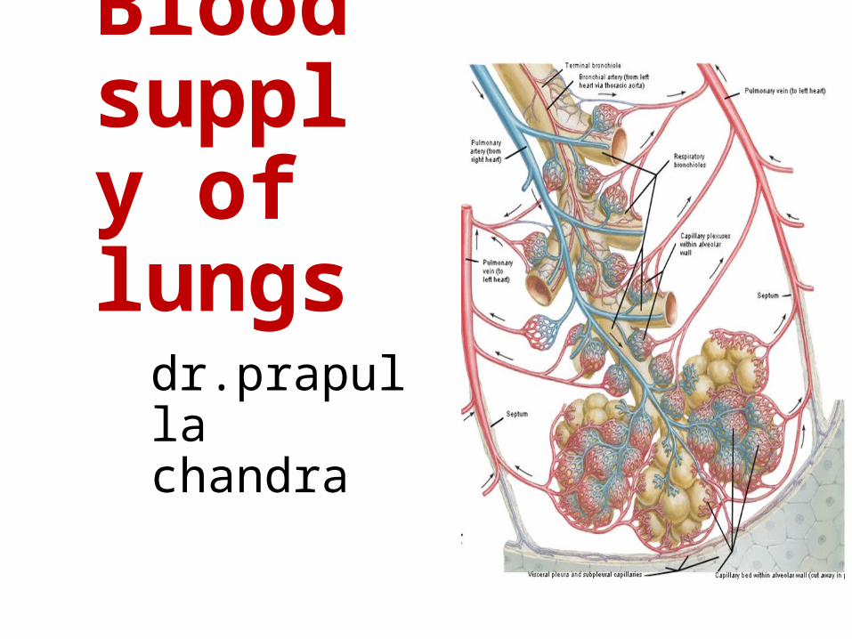
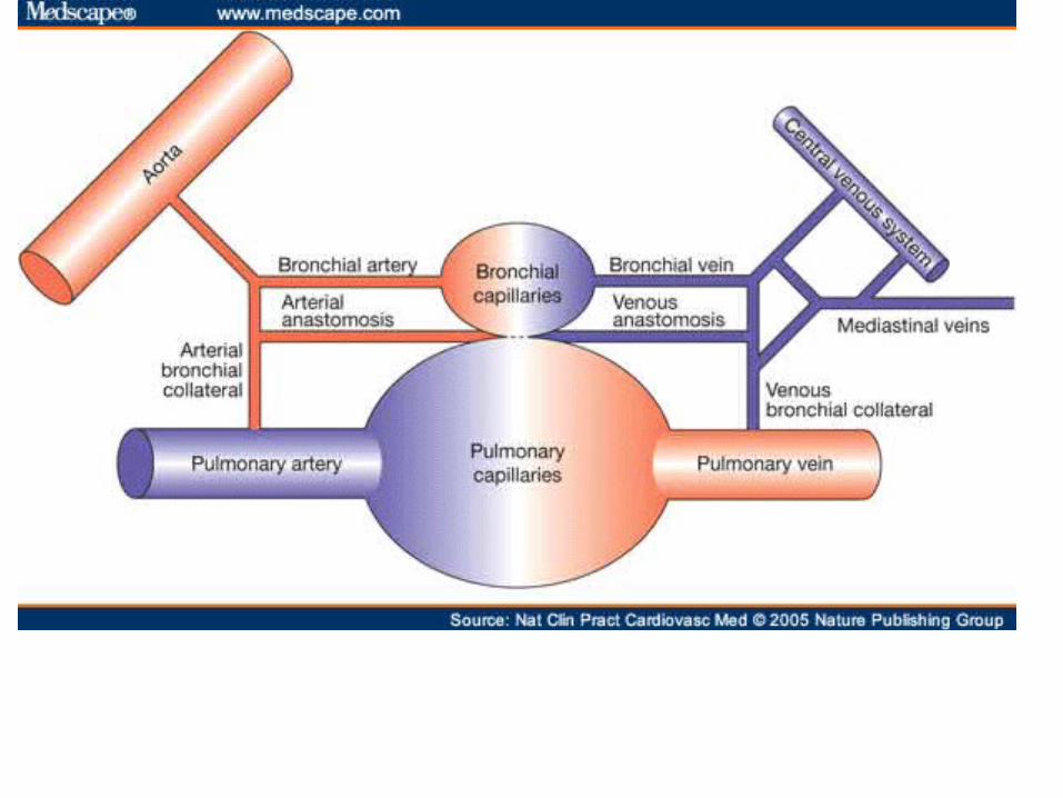
Blood supply of lungs
dr.prapulla chandra
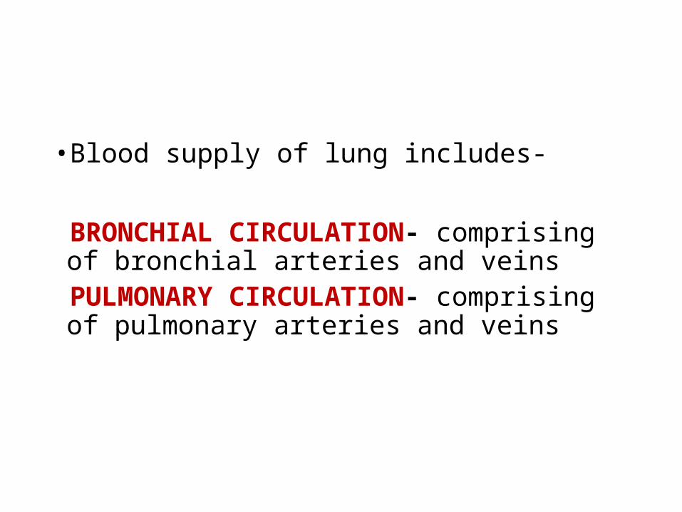
• Blood supply of lung includes-
BRONCHIAL CIRCULATION- comprising of bronchial arteries and veins
PULMONARY CIRCULATION- comprising of pulmonary arteries and veins
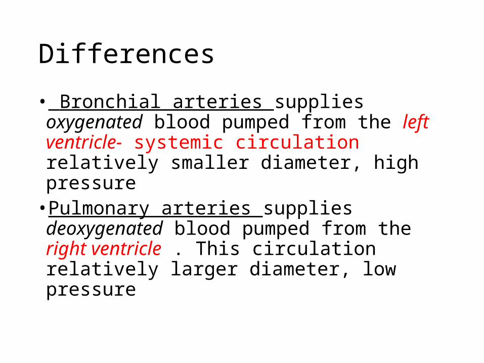
Differences
• Bronchial arteries supplies oxygenated blood pumped from the left ventricle- systemic circulation relatively smaller diameter, high pressure
• Pulmonary arteries supplies deoxygenated blood pumped from the right ventricle . This circulation relatively larger diameter, low pressure
DEVELOPMENT
• By the completion of 16th week intrauterine life, the preacinar structures of bronchi, pulmonary vessels, bronchial arteries are developed
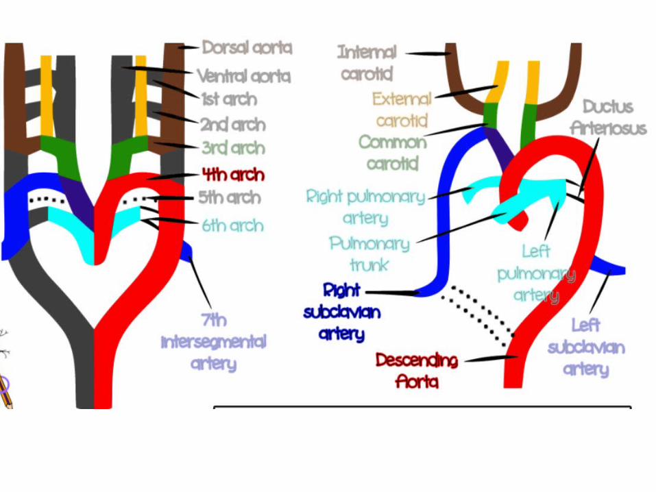
• Pulmonary trunk developed from 6th primitive branchial arch
• Vessels developed from mesenchymal tissue
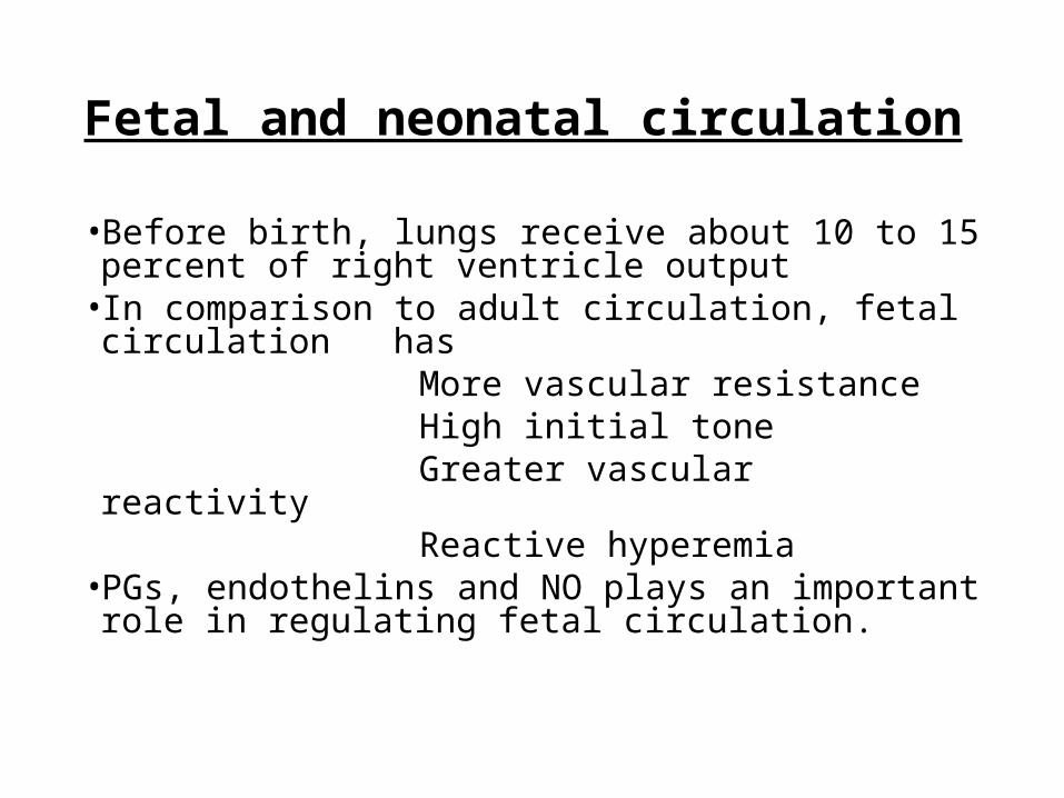
Fetal and neonatal circulation
• Before birth, lungs receive about 10 to 15 percent of right ventricle output
• In comparison to adult circulation, fetal circulation has
More vascular resistance High initial tone Greater vascular reactivity Reactive hyperemia• PGs, endothelins and NO plays an important role in
regulating fetal circulation.
• After first breath there is marked drop in PVR Due to: Increased Po2 Expansion of the lungs
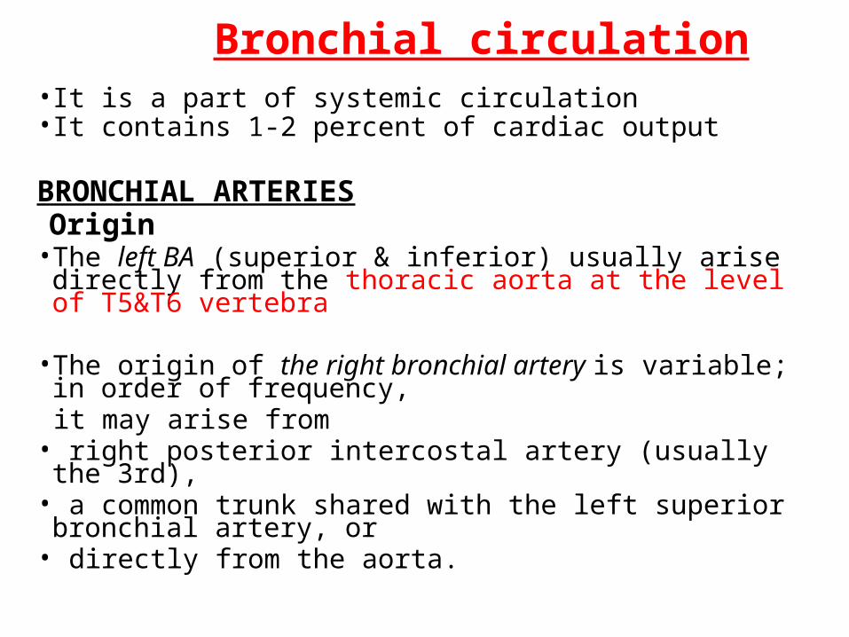
Bronchial circulation• It is a part of systemic circulation• It contains 1-2 percent of cardiac output
BRONCHIAL ARTERIES Origin• The left BA (superior & inferior) usually arise directly from the
thoracic aorta at the level of T5&T6 vertebra • The origin of the right bronchial artery is variable; in order of
frequency, it may arise from• right posterior intercostal artery (usually the 3rd), • a common trunk shared with the left superior bronchial artery, or • directly from the aorta.
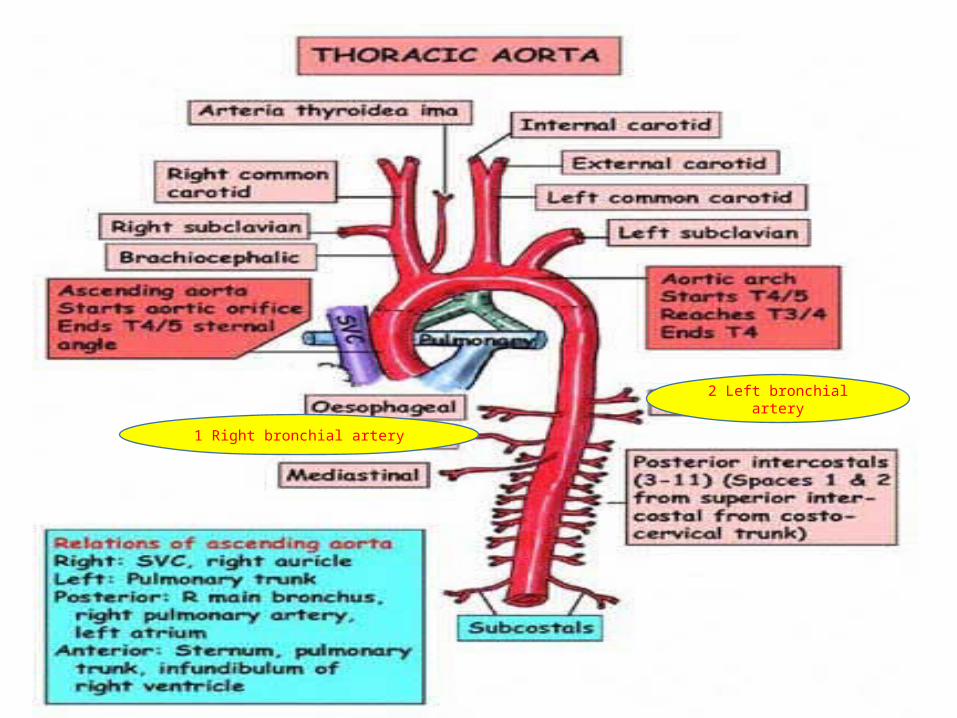
2 Left bronchial artery
1 Right bronchial artery
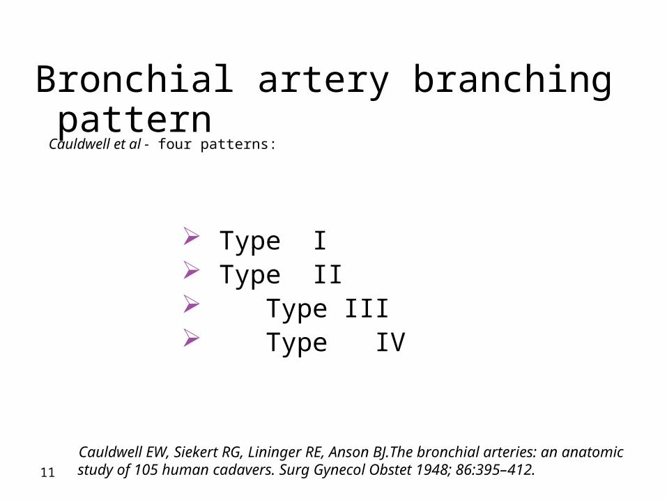
Bronchial artery branching pattern Cauldwell et al - four patterns:
11
Type I Type II Type III Type IV
Cauldwell EW, Siekert RG, Lininger RE, Anson BJ.The bronchial arteries: an anatomic study of 105 human cadavers. Surg Gynecol Obstet 1948; 86:395–412.
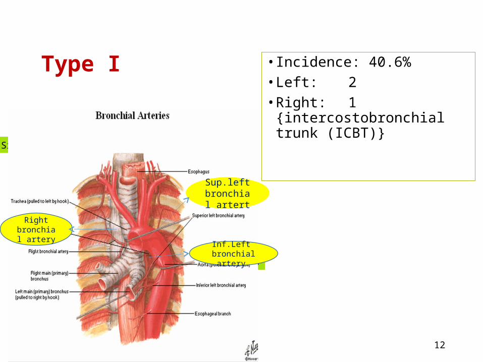
Type I • Incidence: 40.6% • Left: 2• Right: 1 {intercostobronchial
trunk (ICBT)}
12
Single rt.branch
Lt. 2 br.arties
Sup.left bronchial
artert
Inf.Left bronchial
artery.
Right bronchial
artery.
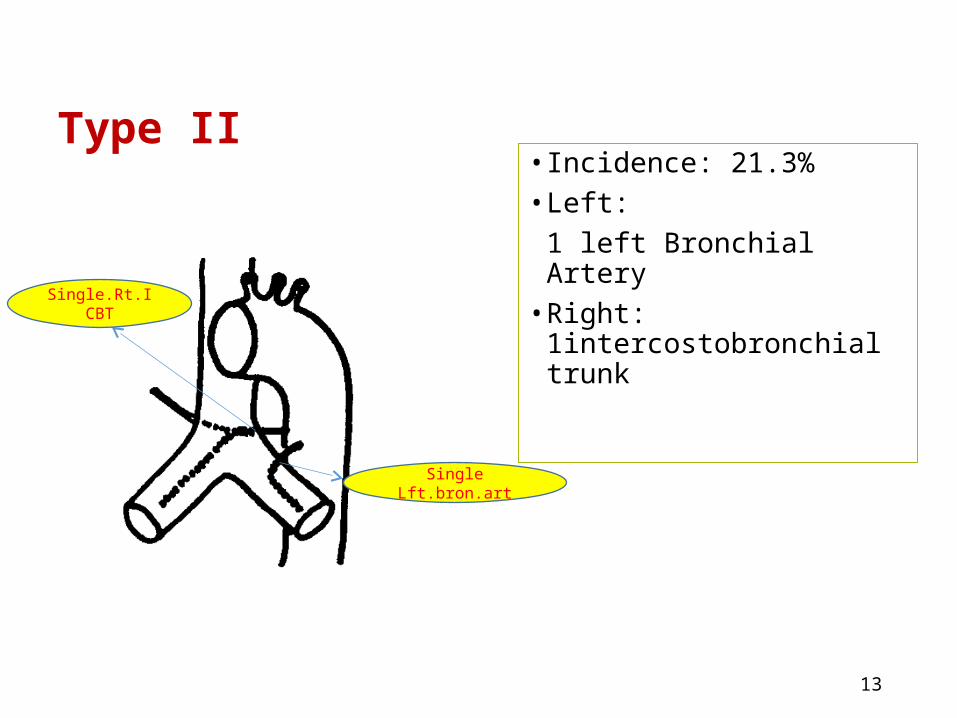
Type II• Incidence: 21.3%• Left:
1 left Bronchial Artery• Right: 1intercostobronchial
trunk
13
Single Lft.bron.art
Single.Rt.ICBT
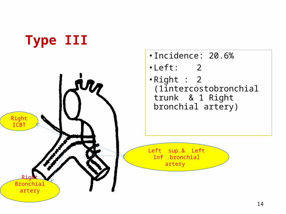
Type III • Incidence: 20.6%• Left: 2• Right : 2
(1intercostobronchial trunk & 1 Right bronchial artery)
14
Left sup.& Left Inf .bronchial artery.
Right ICBT
Right Bronchial artery
.
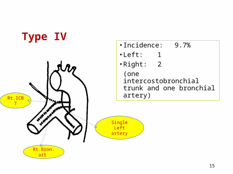
Type IV• Incidence: 9.7% • Left: 1• Right: 2
(one intercostobronchial trunk and one bronchial artery)
15
Single Left artery
Rt.ICBT
Rt.Bron.art.
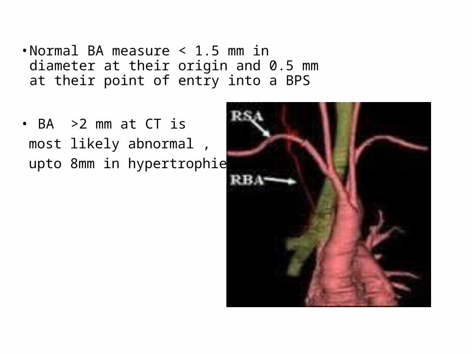
• Normal BA measure < 1.5 mm in diameter at their origin and 0.5 mm at their point of entry into a BPS
• BA >2 mm at CT is most likely abnormal , upto 8mm in hypertrophied
Distribution to lung• The br. A supply blood to the bronchi and connective
tissue of the lungs. They end at the level of the respiratory bronchioles. They anastomose with the branches of the pulmonary arteries, and together, they supply the visceral pleura of the lung
Functions• It has features of nutrient circulation• they supply the lungs with oxygenated blood • It also participates in air conditioning of the inspired
air.
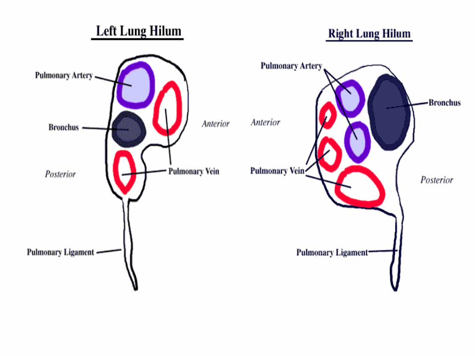
Bronchial veins• There is typically a single bronchial vein at
each hilum, formed from the superficial bronchial veins with deep bronchial veins, draining into the pulmonary veins.
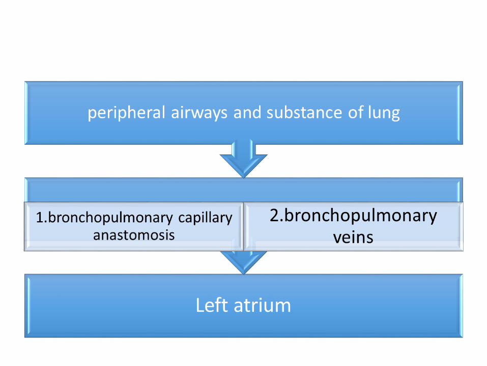
Deep bronchial veins• The deep bronchial veins form from intrabronchial
venous plexuses. These vein drains either directly into the left atrium or into thepulmonary vein. Within the lung there are significant anastmoses with the intrapulmonary pulmonary veins.
• Superficial bronchial veins• The superficial bronchial veins consist of a subpleural
venous network which receive tributaries from extra-pulmonary bronchi, hilar lymph nodes and the vaso vasorum of the hilar pulmonary vessels and unite to form a single bronchial vein at each hilum.
• on the right, they drain into the azygos vein. On the left they drain into the left superior intercostal vein, accessory hemiazygos vein or left brachiocephalic vein. At the hilum they communicate with the pulmonary veins.
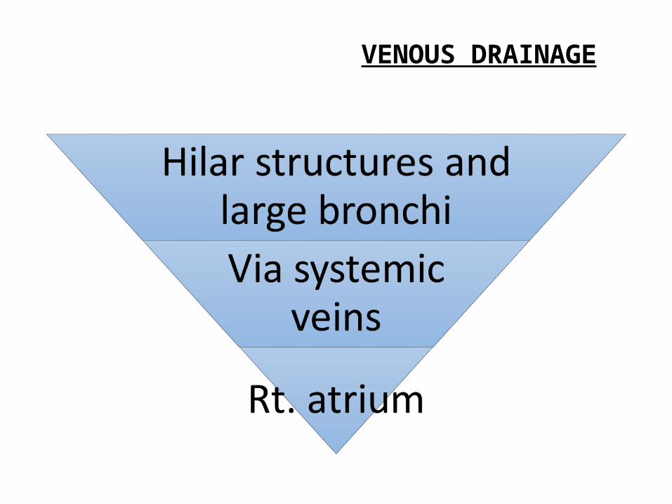
VENOUS DRAINAGE
• The bronchial circulation proliferates beyond the
limits in the following conditions: Pulmonary atresia bronchiectasis lung abscess lung cancer•In chronic MS, Haemoptysis occurs from bronchial
veins
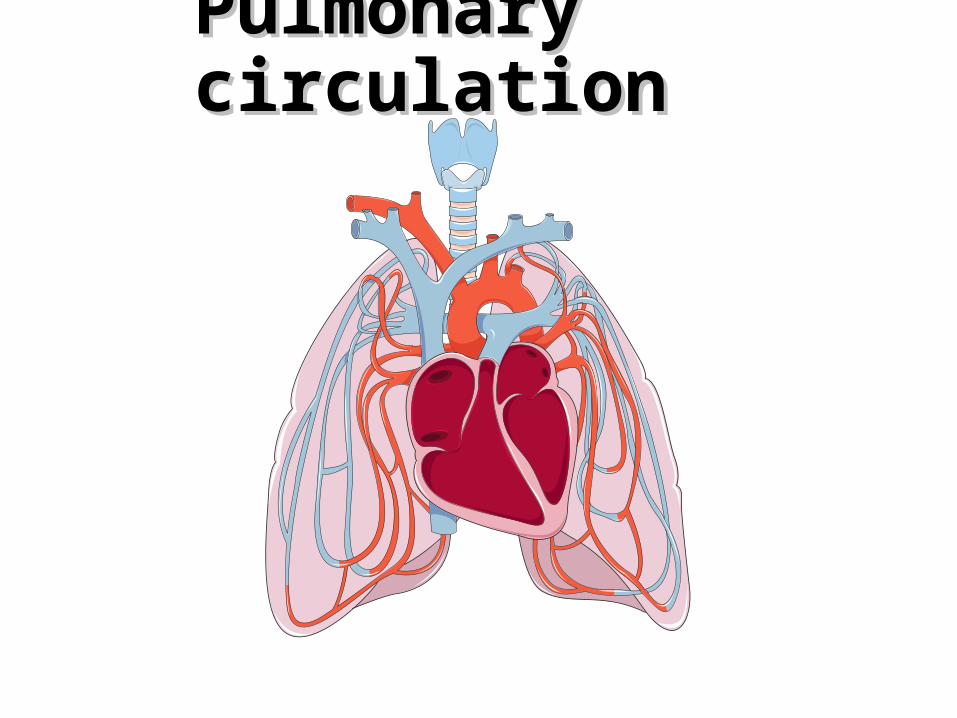
Pulmonary Pulmonary circulationcirculation
PULMONARY CIRCULATION
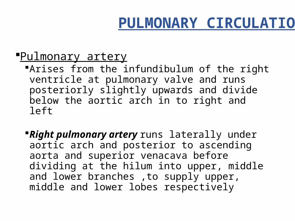
Pulmonary arteryArises from the infundibulum of the right ventricle
at pulmonary valve and runs posteriorly slightly upwards and divide below the aortic arch in to right and left
Right pulmonary artery runs laterally under aortic arch and posterior to ascending aorta and superior venacava before dividing at the hilum into upper, middle and lower branches ,to supply upper, middle and lower lobes respectively
• Left pulmonary artery runs laterally, upwards and
posteriorly,anterior to descending aorta and connected to ligamentum arteriosum.
• It divides into upper and lower branches; they supply upper and lower lobes
• Sub divides in a variable pattern on both sides
• Counting from peripheral small arteries to the main
pulmonary trunk, there are 17 orders of branching on both sides.
• Pulmonary arteries breaking up to pulmonary capillaries that form network around the alveolus.
• Media of the vessels consists of elastic fibrils(5 or 6layers) with smooth muscle fibers, collagen and mucopolysaccharide ground substance.
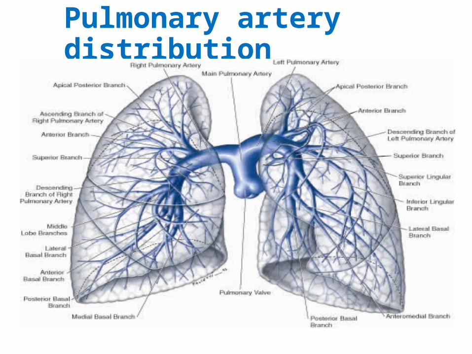
Pulmonary artery distribution
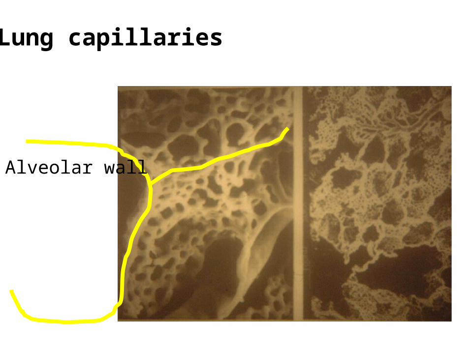
Alveolar wall
Lung capillaries
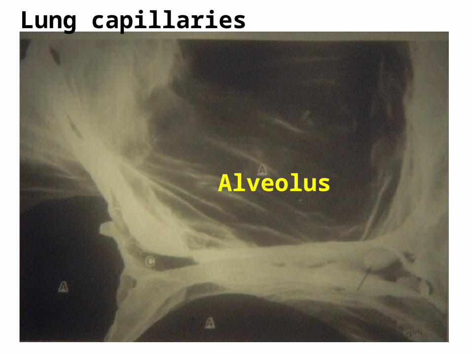
Lung capillaries
Alveolus
Pulmonary veins
• Four in number two on each side They commence in a capillary network upon the
walls of the air sacs, where they are continuous with capillary ramifications of the pulmonary artery and joining together form a single trunk for each lobule
• On right side two in number - because middle lobe
vein joined with upper lobe vein• On left side two in number• These four veins perforate the fibrous layer of the
pericardium and opens separately in to upper and back of the left atrium
• Pulmonary artery—de-oxygenated• Pulmonary veins—only veins which carry oxygenated
blood
Functions of the pulmonary circulation• Respiratory and non-respiratory• Respiratory- gas exchange• Non-respiratory
•Blood filter•Blood reservoir•Metabolic•Nutrient
Gas exchange
The pulmonary artery branches rapidly give rise to nearly 300 million capillaries
Gas exchange between alveolar gases and blood occur within the lung capillaries by simple diffusion.
Oxygen diffuses from the alveolus to pulmonary capillary blood, ,Co2 diffuses in the reverse direction, as determined by their concentration gradient
Blood filter
• Micro vessels are so numerous, that effectively serves as a filter to entrap the foreign materials present in the blood
• This filter mechanism prevent entry of potentially harmful particles in to systemic circulation
• Entrapped materials can be removed by enzymatic process, macrophages or absorption into lymphatic system
Reservoir of blood for left ventricle
• Pulmonary circulation is very compliant and accommodate about 500 ml of blood in an adult
• This can serve as a reservoir for left ventricle particularly during times when output exceeds venous return
• The cardiac output can increase rapidly by drawing upon pulmonary blood volume without depending on instantaneous increase in venous return

Metabolic function of the pulmonary circulation
• Uptake or metabolic conversion of vasoactive substances into circulation done by endothelial cells.
• Conversion of Angiotensin I to angiotensin II
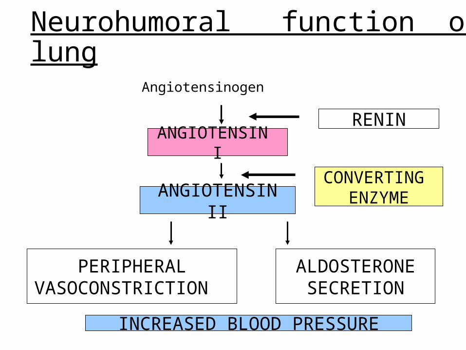
Neurohumoral function of lung
Angiotensinogen
ANGIOTENSIN I
ANGIOTENSIN II
RENIN
CONVERTING ENZYME
PERIPHERAL VASOCONSTRICTION
ALDOSTERONE SECRETION
INCREASED BLOOD PRESSURE
Pulmonary vascular resistance
• Normal pulmonary circulation is a low resistance, high compliant vascular bed
• Calculated by using• R=P.PA-P.LA/QT• Normal value of R for pulmonary circulation is 0.1 mm
hg/L/minIn humans residing at high altitude, will have high PVR,
because muscular media of the small pulmonary arteries and arterioles becomes thicker.
Pulmonary vascular pressures
• Pulmonary pressure drop along the length of pulmonary vascular tree
• Since pulmonary capillary pressure can’t be measured directly, they are generally estimated to be intermediate between the mean pulmonary arterial and pulmonary wedge pressures
• Pulmonary capillary flow recorded by using Body plethysmograph Nitrous oxide methodAverage pul. Art. Pressure is about 10-12 mm Hg(1/8 th
of systemic circulation)During systole 20-30 mm of Hgdiastole 5 to 10mm of HgAgeing associated with slight increase in pulmonary
arterial pressure
LEFT ATRIAL AND PULMONARY WEDGE PRESSURE • In intact, unanaesthetized humans, the mean left
atrial pressure is about 5 to 10 mm Hg.• It is measured by advancing a cardiac catheter
through the right side of the heart and pulmonary arterial tree until it impacts in a small precapillary vessel
Pulmonary blood volume
• It is about 10 percent of total systemic circulation• Measured by using indicator dilution principle• 70 kg human male contains approximately 400-500
ml
• This value is useful for 1.Determination of the mechanical behavior of lung. 2.Provides preload for the left ventricle, acts as a
reservoir3.As a supply of Hb for alveolar-capillary gas exchange4.As a source of water and macromolecules that
engage in alveolar-capillary exchange5.As a potential mechanism for increasing pulmonary
capillary pressures and promoting pulmonary edema.
Pulmonary blood volume Increases in -IV infusion -immersing the body in water -by inflation of an antigravity suit -by negative pressure breathing -systemic vasoconstriction -lying down position
• Decreases -when person stands - After a large venesection -during positive-pressure breathing -valsalva maneuver -systemic vasodilatation
Pulmonary vasomotor
control
•The initial vascular tone of pulmonary circulation is attributed to balance between vasodilation and vasoconstriction caused by various mediators.
•Vasodialtion caused by prostacyclins, nitric oxide
•Constriction by endothelins..
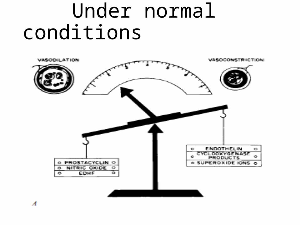
Under normal conditions
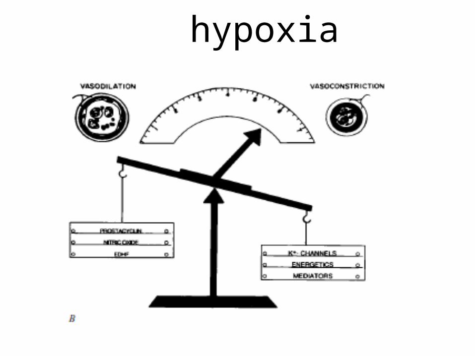
hypoxia
Role of nerves in pulmonary vasomotor control• Sympathetic innervation to pulmonary circulation
includes alpha adrenergic receptors -predominate-(eg:NE)- constrictor beta adrenergic receptors-(eg:isoproterenol)- dilator• Cholinergic activity does not appear to be implicated
Acute hypoxia
• Pressor effect of acute hypoxia on the pulmonary circulation was given by Euler and Liljestrand
• This pressor effect redirects blood flow to better ventilated portion
• Acute hypoxia increases pulmonary arterial pressure, does not affect left atrial pressure, and little increase in cardiac output
• The pressor effect starts within seconds, generally
reaches peak by 3 min, and attenuates gradually as hypoxia continues
• Acidosis augments pressor response• This effect Is predominantly at pre-capillary level
(i.e.: small muscular art. and arterioles)
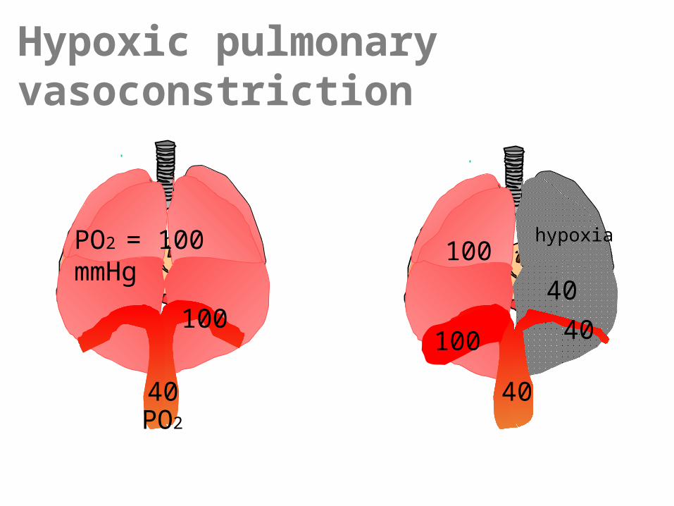
Hypoxic pulmonary vasoconstriction
PO2 = 100 mmHg
100
40
40 100
100
40
40
PO2
hypoxia
Chronic hypoxia• Ex. High altitude• It causes remodeling of small pulmonary arteries and
arterioles leading to high PVR,due to muscular media thickening.
•Acute hypercapnia -Little response is seen if the pH is maintained
at near normal level Acidosis causes pulmonary vasoconstriction, where
as alkalosis causes vasodilatation.
other vasoactive substances
• Vasodilators Ach Bradykinin Isoprenaline• Vasoconstrictors Catecholamine Angiotensin II Histamine Serotonin
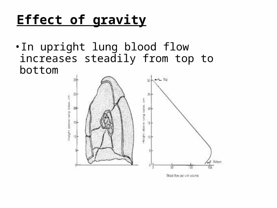
Effect of gravity
• In upright lung blood flow increases steadily from top to bottom
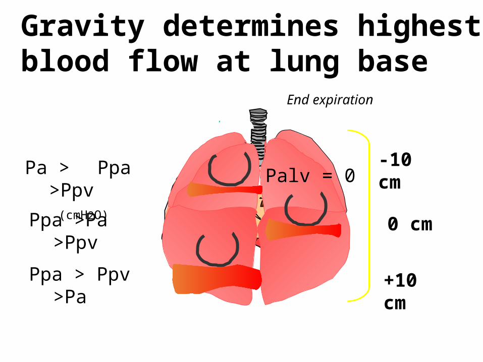
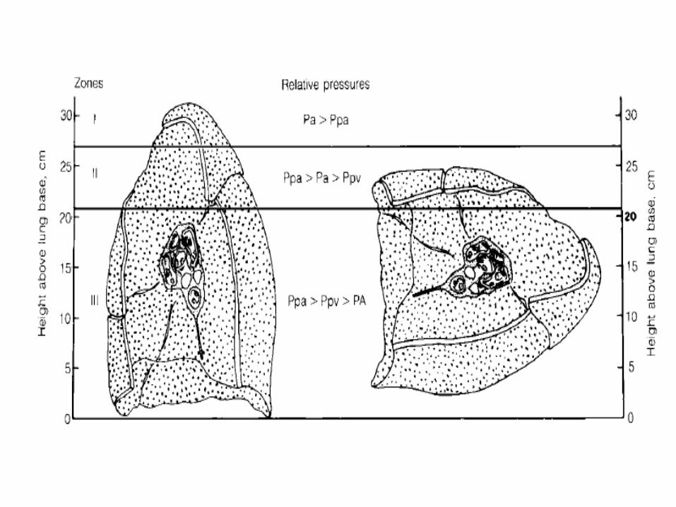
Gravity determines highest blood flow at lung base
0 cm
End expiration
+10 cm
-10 cm
Pa > Ppa >Ppv
(cmH2O)Ppa >Pa >Ppv
Ppa > Ppv >Pa
Palv = 0
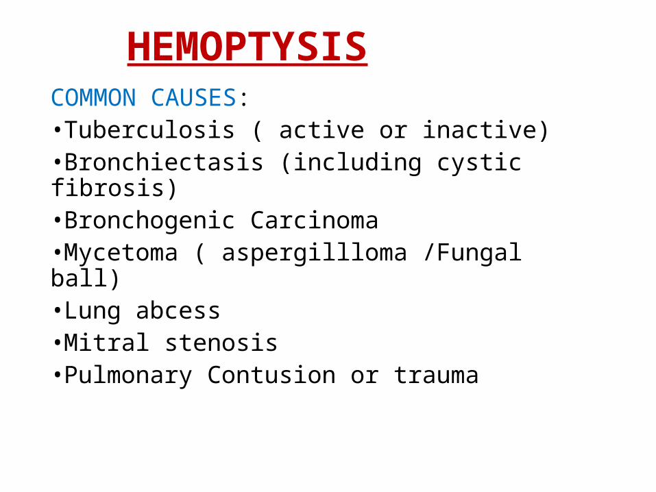
HEMOPTYSISCOMMON CAUSES:•Tuberculosis ( active or inactive)•Bronchiectasis (including cystic fibrosis)•Bronchogenic Carcinoma•Mycetoma ( aspergillloma /Fungal ball)•Lung abcess•Mitral stenosis•Pulmonary Contusion or trauma
Tuberculosis
MECHANISMS•Active tubercular pneumonitis- bronchiolar erosion•Rupture of Rasmussen’s aneurysm (pulmonary. artery)•Healed calcified lymphnode - eroding through bronchial arteries into airway (expectoration of broncholith)
BRONCHIECTASIS
• Pathologically it is destruction of the cartilaginous support of bronchial wall and bronchial dilatation owing to parenchymal retraction from alveolar fibrosis
ANATOMICAL CHANGES:• Bronchial artery hypertrophy• Expansion of peribronchial & sub mucosal
bronchiolar arteriolar plexus• Augmentation of anastomoses with the pulmonary
arterial bed
MYCETOMA
MECHANISMS:• Mechanical trauma of the vascular granulation
tissue by the movement of the fungal ball in the cavity
• Vascular injury from aspergillus associated endotoxin
• Aspergillus related proteolytic activity• Vascular damage from a type 3 hypersensitivity
reaction
Abnormal pulmonary vascular
communications
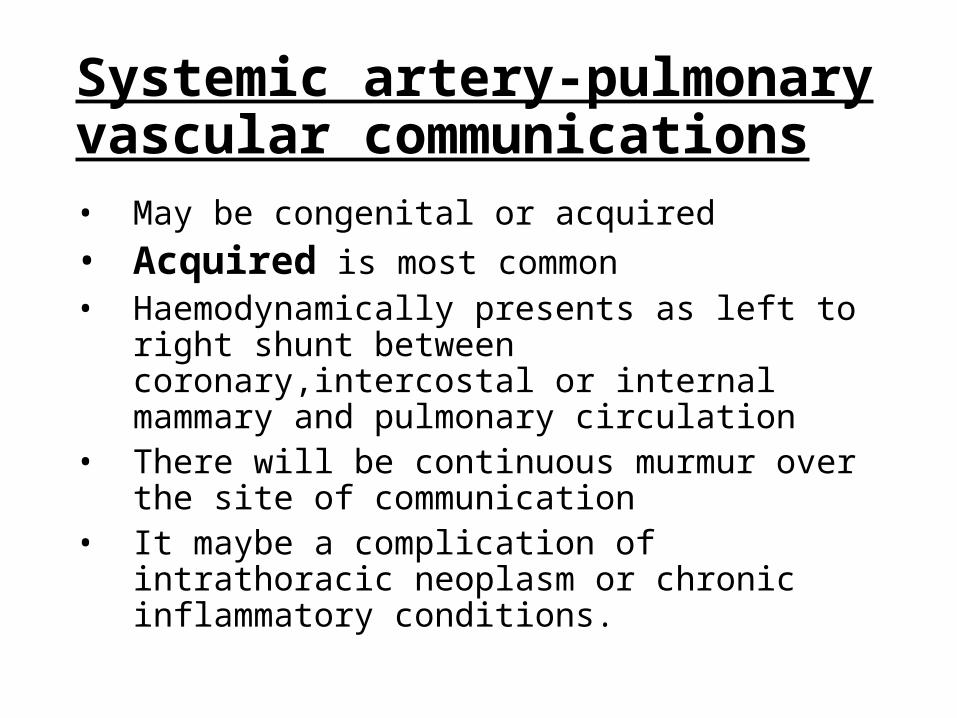
Systemic artery-pulmonary vascular communications• May be congenital or acquired• Acquired is most common• Haemodynamically presents as left to right shunt
between coronary,intercostal or internal mammary and pulmonary circulation
• There will be continuous murmur over the site of communication
• It maybe a complication of intrathoracic neoplasm or chronic inflammatory conditions.
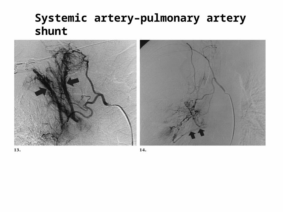
Systemic artery–pulmonary artery shunt
Congenital Pulmonary Arteriovenous Malformation (PAVM)• AKA pulmonary a-v fistulae = pulmonary a-v aneurysm = cavernous angiomas of the lung
• Defined as abnormal vascular communications between pulmonary arteries and veins that lead veno-arterial shunting
There is a close association b/w PAVM and hereditary hemorrhagic telangiectasia (osler weber rendu syndrome),an autosomal dominant condition in 50-60% of cases and
the reverse in 5-15% cases
It has been shown that a gene located on chr 9q3 is strongly associated with the presence of two conditions
• Divided into simple or complex• Simple PAVMs consist of segmental artery joining
thin-walled aneurysmal sac drained by single vein (majority of cases)
• Complex PAVMs involves arterial supply from 2 different segmental arteries (only small percentage)
• Can have multiple lesions(20-30%), • bilateral disease(10%)• More than half found in lower lung fields• Sizes vary typically 1-5 cm and can exceed 10cm
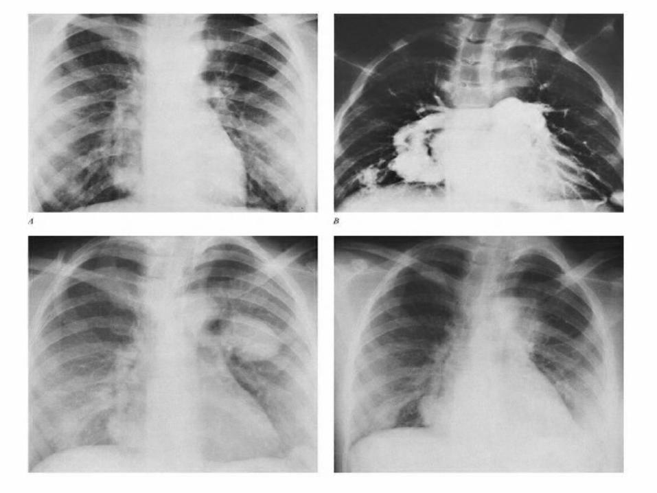
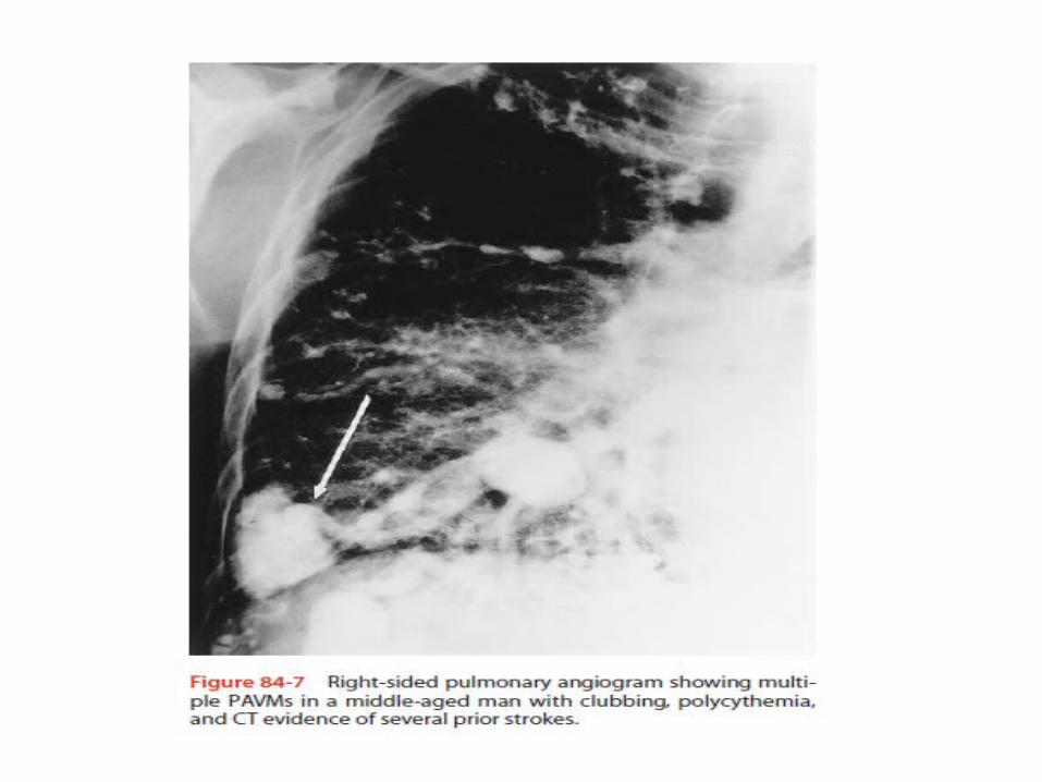
• Most commonly found in lower lobes adjacent to visceral pleura
• Presenting features: dyspnea and epistaxis Cyanosis and clubbing Extra cardiac murmur• Treatment is by excision
ANOMALOUS SYSTEMIC PULMONARY PERFUSION
• Usually from descending thoracic aorta, also from ascending aorta, innominate, subclavian etc
• Asymptomatic but may present with recurrent hemoptysis in some cases
CXR shows normal or increased vascular markings in the vicinity of anomalous vessels
Aortograhy is diagnostic
Treatment consists of surgical ligation, embolisation or even lung resection to control hemoptysis.
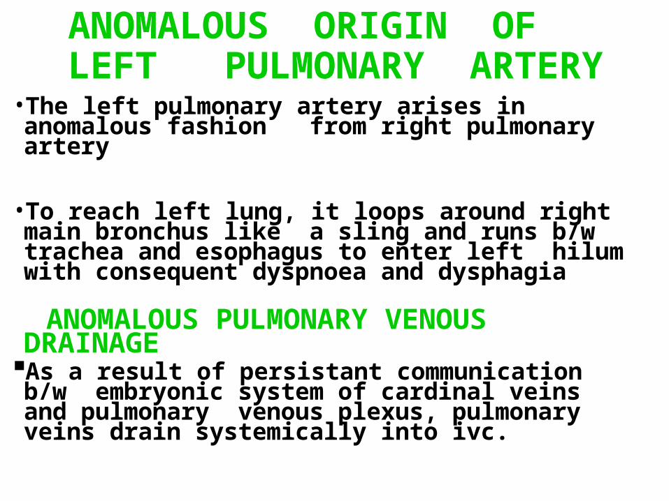
ANOMALOUS ORIGIN OF LEFT PULMONARY ARTERY
• The left pulmonary artery arises in anomalous fashion from right pulmonary artery
• To reach left lung, it loops around right main bronchus like a sling and runs b/w trachea and esophagus to enter left hilum with consequent dyspnoea and dysphagia
ANOMALOUS PULMONARY VENOUS DRAINAGE
As a result of persistant communication b/w embryonic system of cardinal veins and pulmonary venous plexus, pulmonary veins drain systemically into ivc.
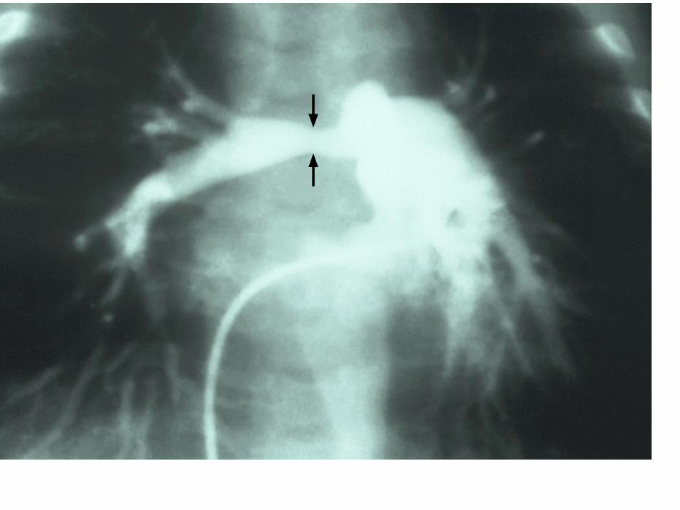
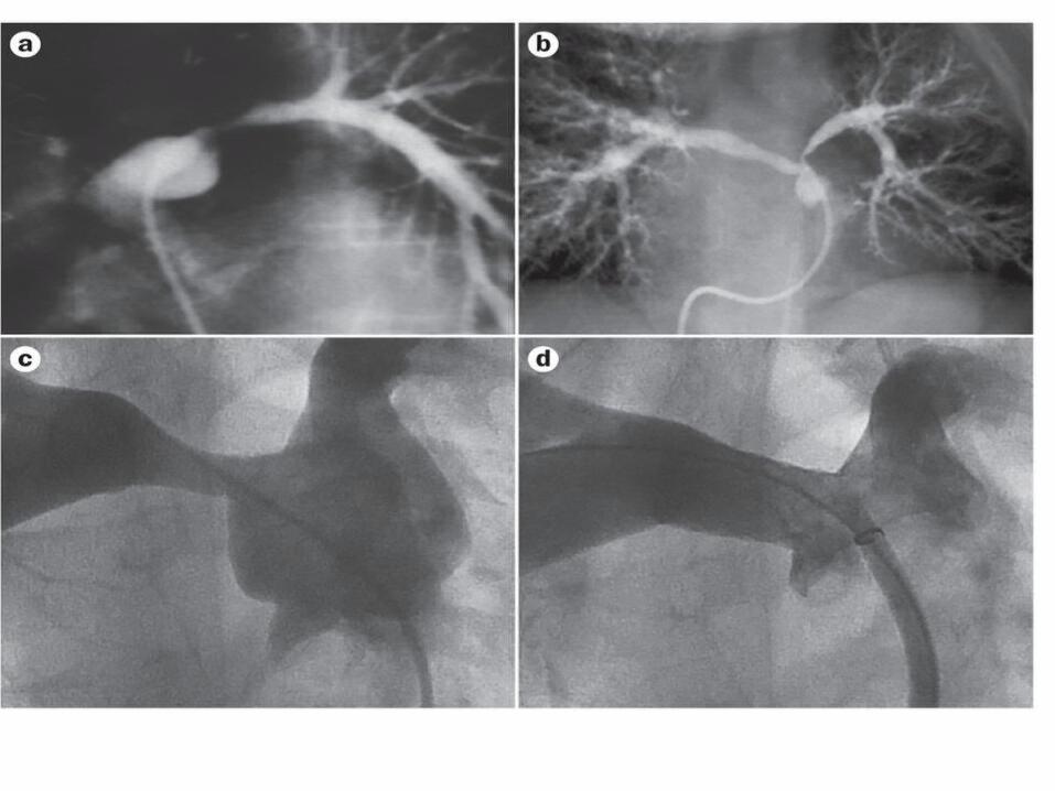
PULMONARY ARTERY STENOSIS
• Charaterised by coarctation of pulmonary arteries with poststenotic dilatation
BAUM and his colleagues divided the patients into two groups
TYPE1: Main pulmonary artery affected; associated with CVS anomalies TYPE2: Peripheral branch involved
CXR may show poststenotic dilatation of affected pulmonary artery branches, oligemia and signs of PAH or cor pulmonale
Diagnosis confirmed by pulmonary angiography
ABSENT UNILATERAL PULMONARY ARTERY
• Failure of development of either right or left side of primitive sixth branchial arch so that no communication exists b/w main pulmonary artery trunk and the lung on affected side, which receives its blood supply systemically most frequently bronchial vessels.
Usually associated with CHD
Associated with pulmonary artery hypertension
Diagnosis by CXR, VP-scan, pulmonary arteriography
Treatment usually not possible, although reconstructive anastomotic surgery and recanalisation possible in some.
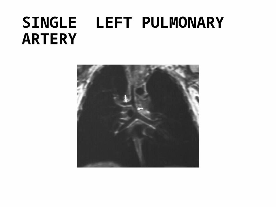
SINGLE LEFT PULMONARY ARTERY
ABSENCE OF MAIN PULMONARY ARTERY
The pulmonary artery trunk may be completely absent as a result of agenesis of primitive sixth branchial arch or of developmental failure of septum dividing truncus arteriosus
L-R shunt exists, the lungs being supplied by bronchial or other systemic aberrant vessels
Poor prognosis, most patients being stillborn or dying in infancy with pulmonary hypertension.
Pulmonary hypertension•Mean pulmonary artery pressure >25mm Hg at rest or >30 mm Hg with exercise
•Anatomical changes Intimal atheromas Medial hypertrophy Remodeling of muscular arteries
Pulmonary thromboembolism
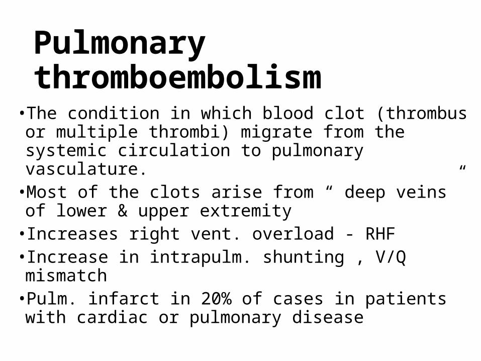
• The condition in which blood clot (thrombus or multiple thrombi) migrate from the systemic circulation to pulmonary vasculature.
• Most of the clots arise from “ deep veins” of lower & upper extremity
• Increases right vent. overload - RHF• Increase in intrapulm. shunting , V/Q mismatch • Pulm. infarct in 20% of cases in patients with cardiac or
pulmonary disease
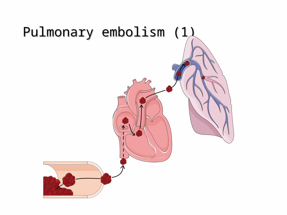
Pulmonary embolism (1)Pulmonary embolism (1)
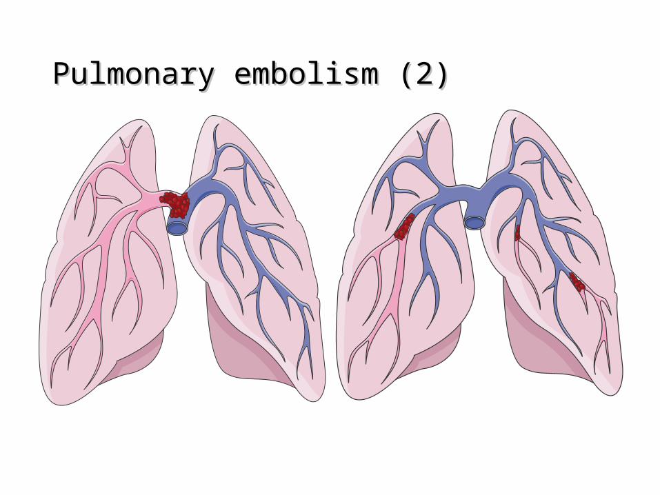
Pulmonary embolism (2)Pulmonary embolism (2)
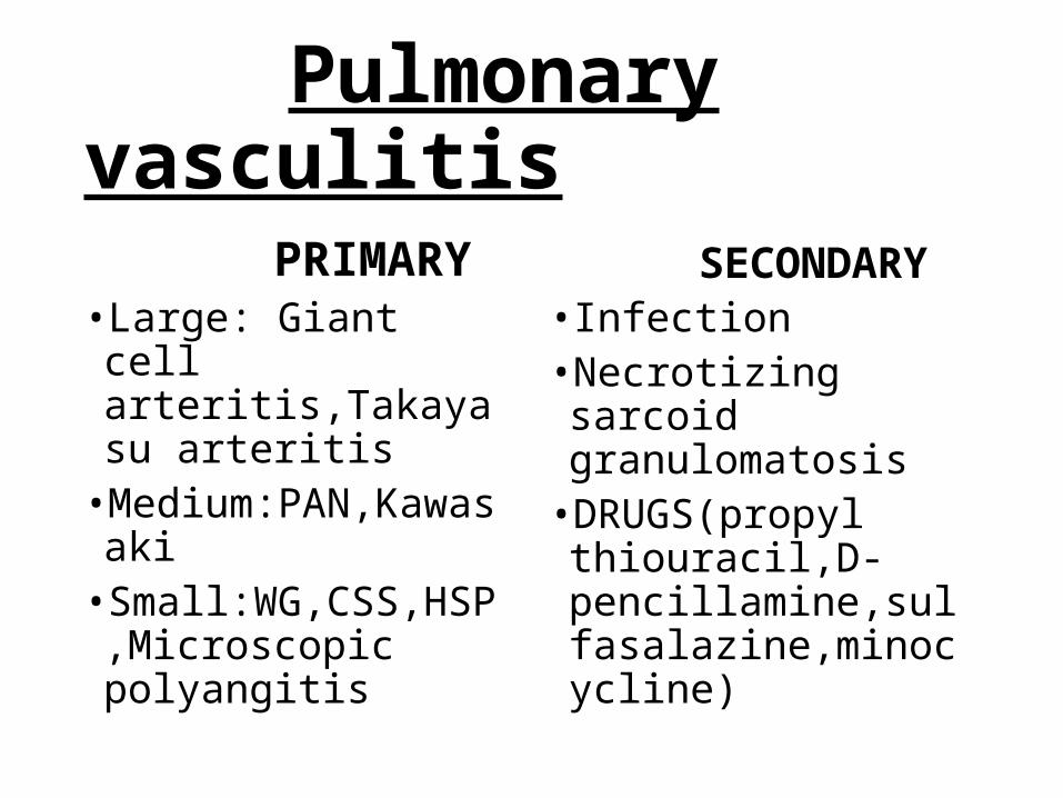
Pulmonary vasculitis PRIMARY• Large: Giant cell
arteritis,Takayasu arteritis
• Medium:PAN,Kawasaki• Small:WG,CSS,HSP,Micr
oscopic polyangitis
SECONDARY
• Infection• Necrotizing sarcoid
granulomatosis• DRUGS(propyl
thiouracil,D-pencillamine,sulfasalazine,minocycline)

THANK YOU
Next seminar on 06-03-2015 SOLITARY PULMONARY NODULE by Dr. deepika