Embed Size (px)
Citation preview
BlOOD CONCENTRATION
METHODS
CHOYTOO Shiksha
Contents
▫ Microfilaria count
▫ Microhaematocrit centrifugation
▫ Triple centrifugation
▫ Buffy coat concentration
▫ Knot concentration
▫ Membrane Filtration
▫ Gradient centrifugation
▫ Malarial Parasite – QBC Method
▫ References
Note:
• The thin and thick blood films for detection of parasites are routinely done
• However, if the parasites are not seen in these blood films, the concentration methods are used

Microfilaria count
• Themicrofilaria is an early stage in the life cycle of certain parasitic nematodes.
• Microfilaria count : with the help of a haemoglobinometer pipette, 29 mm3 of blood is placed in a clean glass, dried as thick film, dehaemoglobinised and stained in the usual manner.
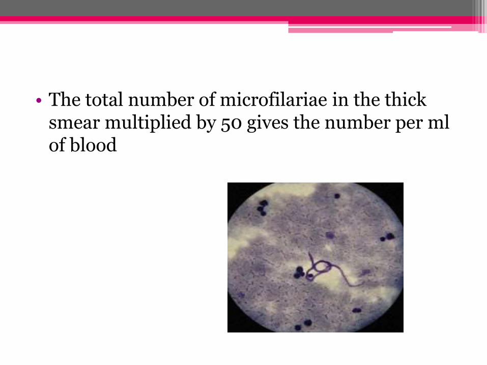
• The total number of microfilariae in the thick smear multiplied by 50 gives the number per ml of blood
Blood Concentration Method
• Concentration methods are used to detect haemoparasites
• Some of the methods are
▫ Microhaematocrit centrifugation
▫ Triple centrifugation
▫ Buffy coat concentration
▫ Knot concentration
▫ Membrane Filtration
▫ Gradient centrifugation
Microhaematocrit Centrifugation
• Blood is collected in a haematocrit tube up to 2/3rd of its volume
• End of the tube is sealed
• Centrifuge at 1500g for 7 minutes
• The RBC-plasma is examine under oil-immersion lens
• Examination of malarial parasite and trypanosomes

Triple Centrifugation
• Brief description:
▫ 9ml of blood is mixed with 1ml of 6%sodium citrate
▫ Centrifuge for 10 min at 100g
▫ The supernatant* is collected and is centrifuge again at 700g for 10 min
▫ Sedimentation is examined under wet film or stained smear
*denoting the liquid lying above a solid residue after crystallization, precipitation, centrifugation, or other process.
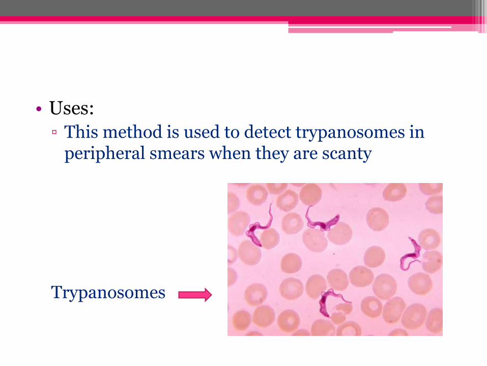
• Uses:
▫ This method is used to detect trypanosomes in peripheral smears when they are scanty
Trypanosomes

Buffy Coat Concentration
• Brief description
▫ 5ml of citrated or oxalated blood is centrifuged in a tube
▫ Buffy coat present between the plasma and packed red cells is collected and stained
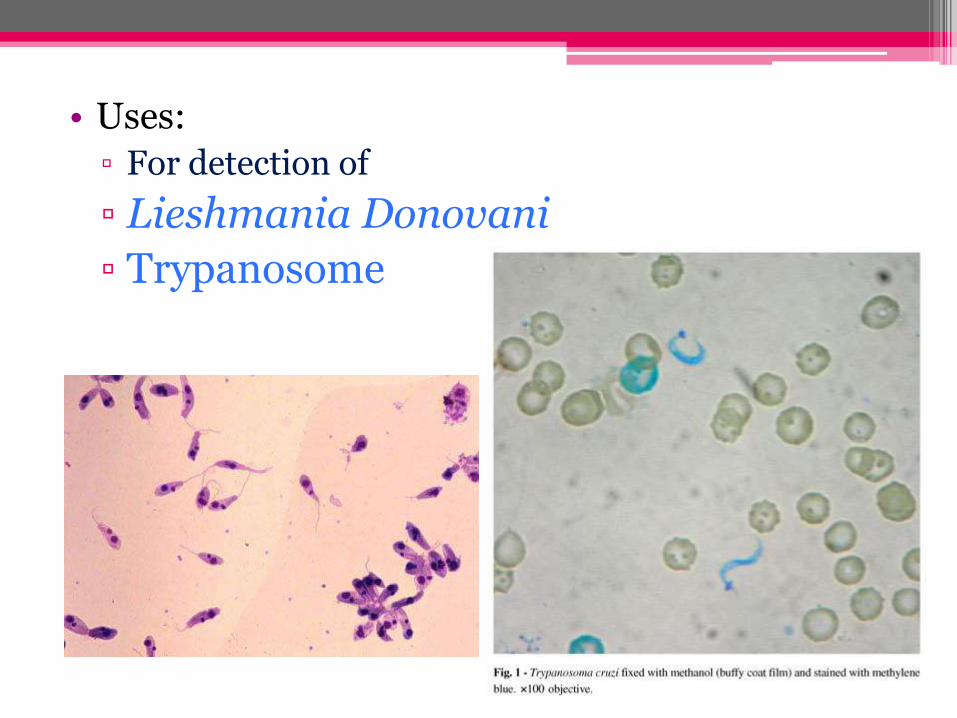
• Uses:
▫ For detection of
▫ Lieshmania Donovani
▫ Trypanosome
Knott Concentration
• Brief Description:
▫ 2ml of blood is thoroughly mixed with 10 ml of 2% solution of formalin
▫ Mixture is allowed to stand for 10 min or longer
▫ Then centrifuged at 200g for 2min

• Use:
▫ It is primarily used to detect microfilariae in blood, especially when a light infection is suspected
▫ Disadvantage of this method is that microfilariaeare killed by the formalin and are therefore not seen as motile organisms.
Membrane filtration
• 1ml of venous blood is drawn into a 10 ml of syringe containing 0.1 ml of a 5% solution of sodium citrate
• In the same syringe, 9ml of 10% solution of Teepolin physiological saline is drawn and shaken gently for 1 min
• Needle is removed and attached to a Swiney filter holder containing a 25 mm membrane filter of 5um porosity placed over a filter paper pad of the same size and moisten with saline
• With gentle and steady pressure the blood is forced through the filter
• The filter is washed three times by passing 10 ml of physiological saline
• Filter is removed and stained for 5 min in hot haematoxilin
• It is then briefly “blued” in running tap water
• It is dried, covered with mounting medium and coverslip, and examined under a microscope
• This technique has been proved highly efficient in demonstrating filarial infections when microfilaremia are of low density
• It has also been successfully used in field surveys.
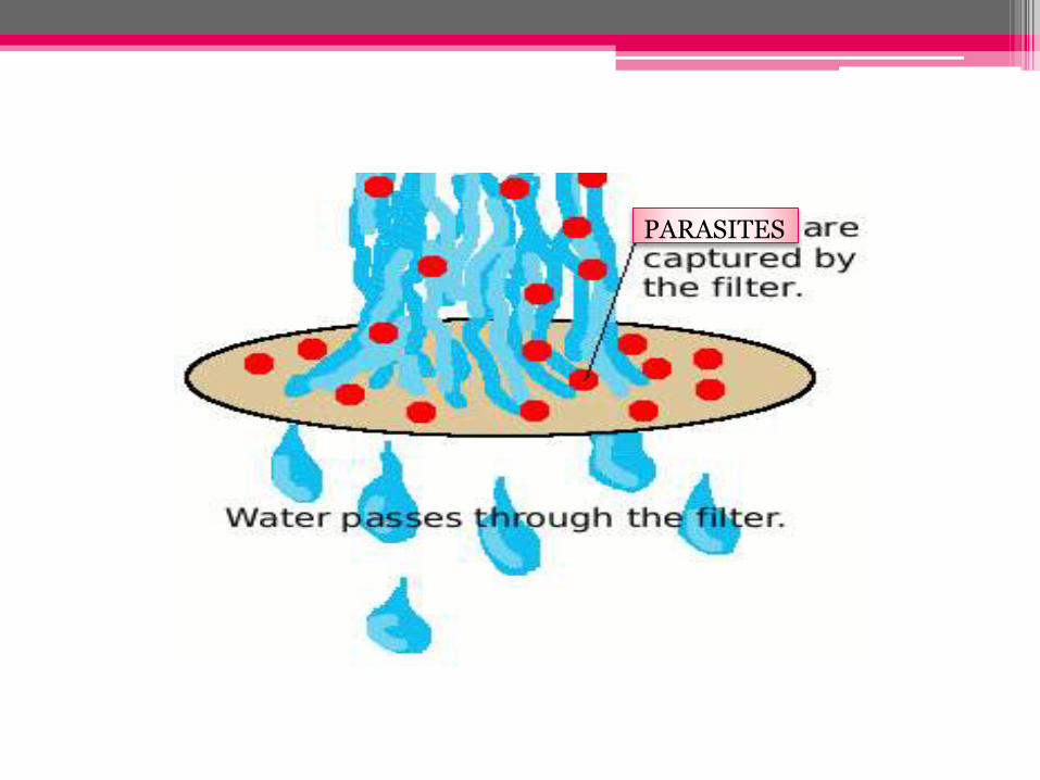
PARASITES

Gradient Centrifugation
• 5ml of Ficoll-Hypaque solution is mixed with an equal volume of heparinised blood.
• This is centrifuged at 150g for 40 min
• Shows three layer:
▫ White cell layer (bottom)
▫ Ficoll-Hypaque layer (middle)
▫ Plasma layer (top)
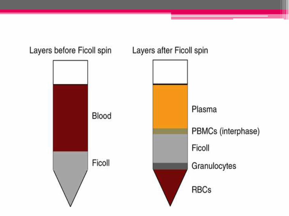

• Uses:
▫ The middle layer is used for detection of microfilariae

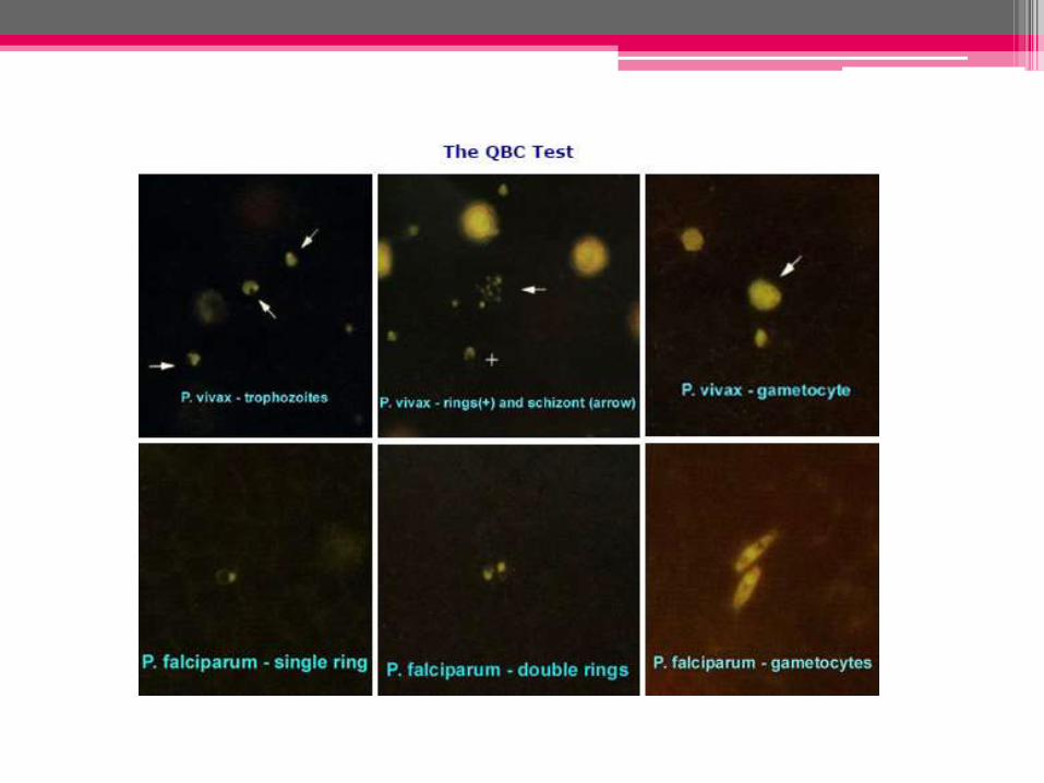
Malarial Parasite – QBC Method
(quantitative buffy coat method)
• new method for identifying the malarial parasite in the peripheral blood
• involves staining of the centrifuged and compressed red cell layer with acridine orange and its examination under UV light source

• QBC test tube (hematocrit tube)- pre-coated internally with acridine orange stain and potassium oxalate
• is filled with 55-56 ml of blood
• centrifuged at 12,000 rpm for 5 minutes
• components of the buffy coat separate according to their densities, forming discrete bands
• The QBC tube is placed on the tube holder and examined using a standard white light microscope equipped with the UV microscope adapter
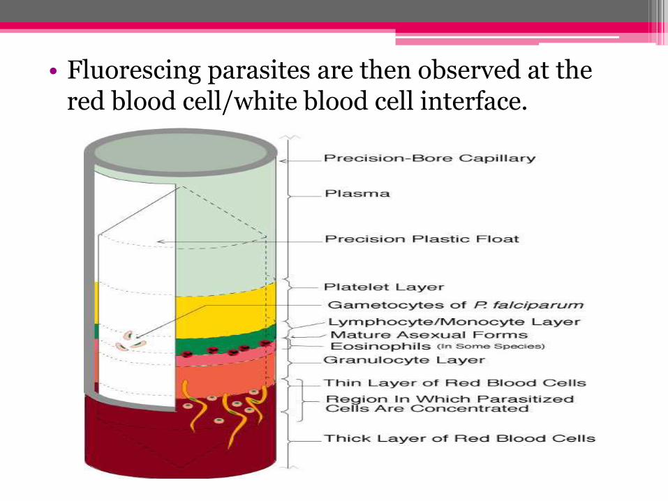
• Fluorescing parasites are then observed at the red blood cell/white blood cell interface.
References

THANK YOU