Embed Size (px)
DESCRIPTION
.
Citation preview
AZOOSPERMIA
BY: MARYAM HOSSEINZADEH
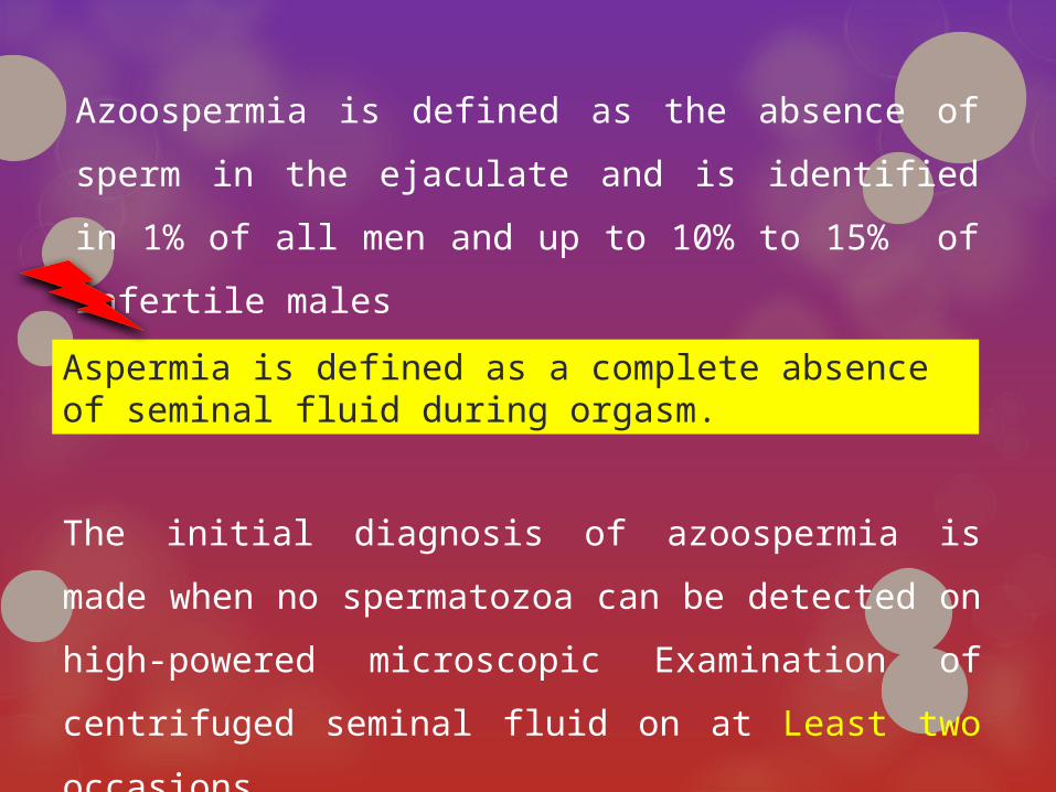
Azoospermia is defined as the absence of sperm in the
ejaculate and is identified in 1% of all men and up to 10%
to 15% of infertile males
Aspermia is defined as a complete absence of seminal fluid during orgasm.
The initial diagnosis of azoospermia is made when no
spermatozoa can be detected on high-powered
microscopic Examination of centrifuged seminal fluid on at
Least two occasions.

The WHO Laboratory Manual for The Examination of Human Semen recommends that the seminal fluid be centrifuged for 15 minutes at a centrifugation speed of, preferably, 3000g or greater.
The complete absence of spermatozoa should be confirmed with repeat testing after a long time, because many external factors
(febrile episodes and some therapies) may cause transient azoospermia.

inability of produced spermatozoa to reach the emitted semen: (excretory or Obstructive Azoospermia)
lack of spermatozoa production in the testes:(Secretory or Non-Obstructive Azoospermia)
Azoospermia may result from
or
NOA
OA
In clinical practice both components are sometimes present in a single patient (mixed genesis azoospermia)
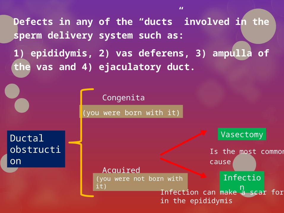
Defects in any of the “ducts” involved in the sperm
delivery system such as:
1) epididymis, 2) vas deferens, 3) ampulla of the vas and 4)
ejaculatory duct.
Ductal obstruction
Congenital
Acquired
(you were born with it)
(you were not born with it)
Vasectomy
Infection
Infection can make a scar form in the epididymis
Is the most common
cause
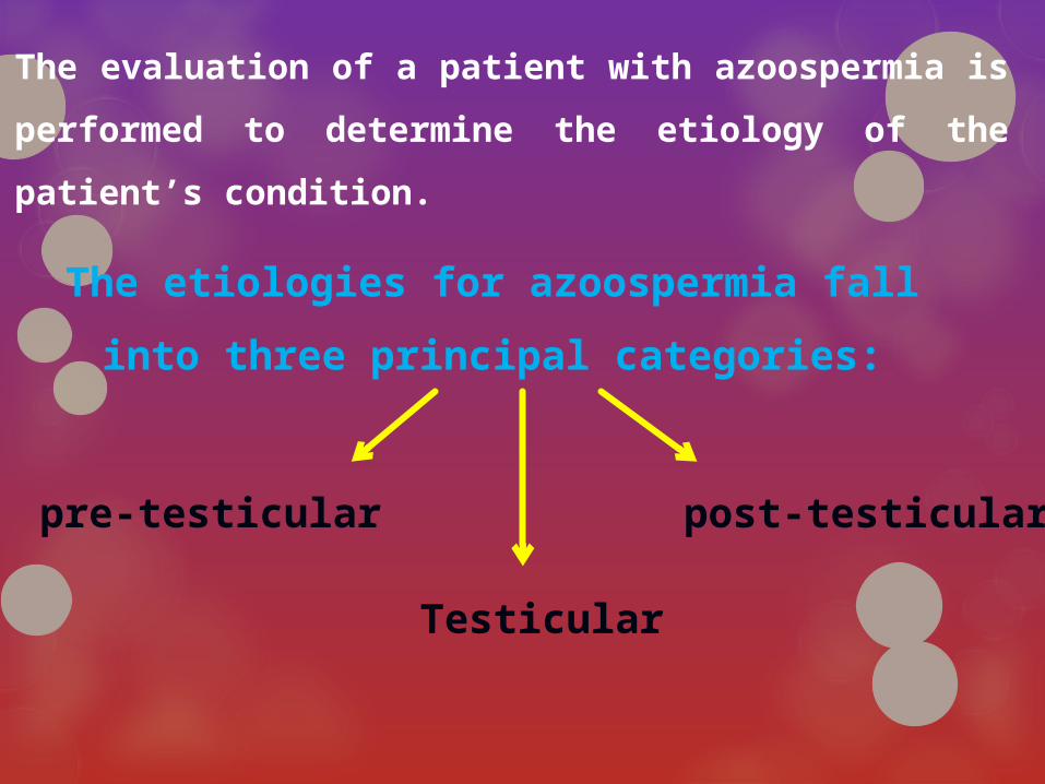
The evaluation of a patient with azoospermia is performed
to determine the etiology of the patient’s condition.
The etiologies for azoospermia fall into three
principal categories:
pre-testicular
Testicular
post-testicular
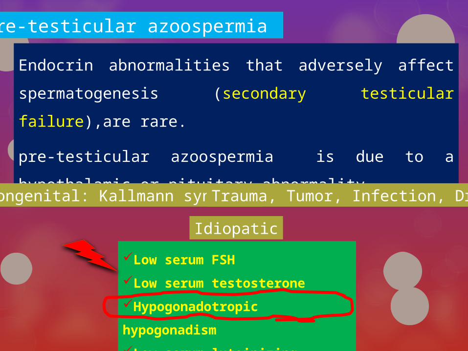
pre-testicular azoospermia
Endocrin abnormalities that adversely affect
spermatogenesis (secondary testicular failure),are
rare.
pre-testicular azoospermia is due to a hypothalamic or
pituitary abnormality.
Low serum FSH
Low serum testosterone
Hypogonadotropic hypogonadism
Low serum luteinizing hormone(LH
congenital: Kallmann syndrome
Idiopatic
Trauma, Tumor, Infection, Drug
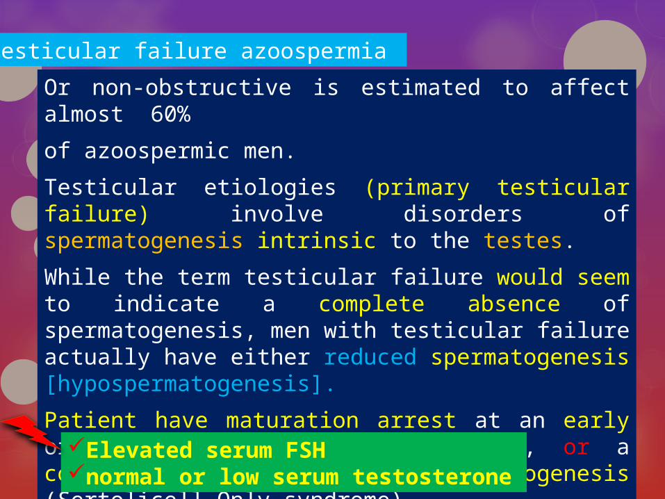
Testicular failure azoospermia
Or non-obstructive is estimated to affect almost 60%
of azoospermic men.
Testicular etiologies (primary testicular failure) involve disorders of spermatogenesis intrinsic to the testes.
While the term testicular failure would seem to indicate a complete absence of spermatogenesis, men with testicular failure actually have either reduced spermatogenesis [hypospermatogenesis].
Patient have maturation arrest at an early or late stage of spermatogenesis, or a complete failure of spermatogenesis (Sertolicell Only syndrome)Elevated serum FSH
normal or low serum testosterone

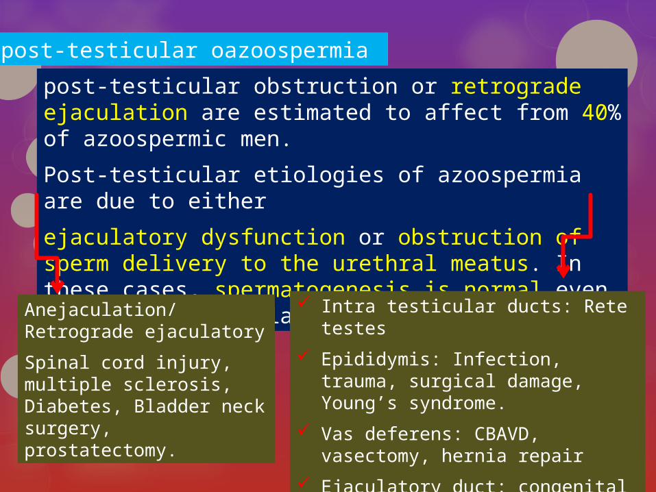
post-testicular oazoospermia
post-testicular obstruction or retrograde ejaculation are estimated to affect from 40% of azoospermic men.
Post-testicular etiologies of azoospermia are due to either
ejaculatory dysfunction or obstruction of sperm delivery to the urethral meatus. In these cases, spermatogenesis is normal even though the semen lacks spermatozoa.
Intra testicular ducts: Rete testes
Epididymis: Infection, trauma, surgical damage, Young’s syndrome.
Vas deferens: CBAVD, vasectomy, hernia repair
Ejaculatory duct: congenital cysts, infection, trauma, urethral surgery
Anejaculation/Retrograde ejaculatory
Spinal cord injury, multiple sclerosis, Diabetes, Bladder neck surgery, prostatectomy.

The pre-testicular and post-testicular
abnormalities that cause azoospermia are
frequently correctable.
Testicular Disorders are generally irreversible,
With the possible exception of impaired
spermatogenesis Associated with varicoceles.

NOA and OA:Results with IVF/ICSI

childhood illnesses: orchitis or cryptorchidism
genital trauma
prior pelvic/inguinal surgery
infections
gonadotoxin exposure: prior radiation therapy/chemotherapy
current medical therapy
familial history of birth defects
mental retardation
reproductive failure
cystic fibrosis
Diagnosismedical history
testis size and consistency (normal
testis volume greater than 19 ml)
consistency of the epididymies
secondary sex characteristics
presence and consistency of the
vasa deferentia
presence of a varicocele
masses upon digital rectal
examination
physical examination
measurement of serum testosterone level (T)
follicle stimulating hormone level (FSH)
Luteinizing hormone (LH)
hormone level measurements
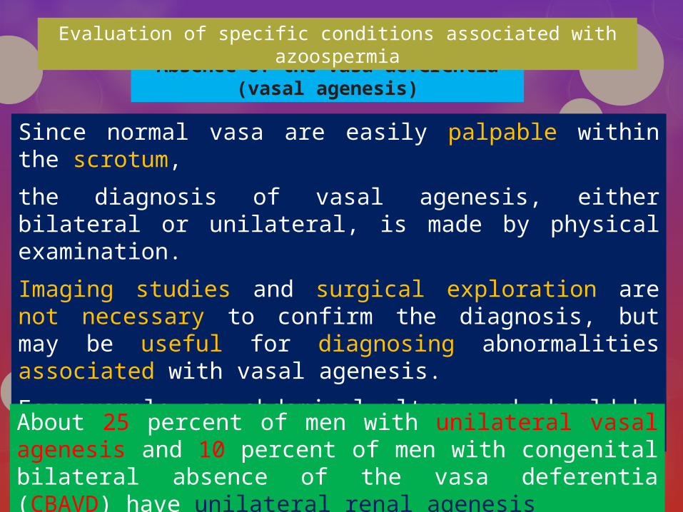
Absence of the vasa deferentia(vasal agenesis)
Since normal vasa are easily palpable within the scrotum,
the diagnosis of vasal agenesis, either bilateral or unilateral, is made by physical examination.
Imaging studies and surgical exploration are not necessary to confirm the diagnosis, but may be useful for diagnosing abnormalities associated with vasal agenesis.
For example, an abdominal ultrasound should be considered to Rule out renal anomalies.
About 25 percent of men with unilateral vasal agenesis and 10 percent of men with congenital bilateral absence of the vasa deferentia (CBAVD) have unilateral renal agenesis
Evaluation of specific conditions associated with azoospermia

Due to the embryological association between the vasa And Seminal
vesicles, most patients with vasal agenesis Also have seminal vesicle
hypoplasia or agenesis. Since the majority of semen is derived from the
seminal vesicles, almost all patients with CBAVD have low semen volume.
There is a strong association between CBAVD and mutations of the cystic
fibrosis transmembrane conductance Regulator (CFTR) gene.
Approximately 70% of men with CBAVD and no clinical evidence of a
cystic fibrosis have an identifiable abnormality of CFTR gene.
Almost all male patients With Clinical cystic fibrosis have CBAVD

Since It can be assumed that a man with CBAVD harbors a genetic
abnormality in the CFTR gene, it is important to test his partner for CFTR
gene abnormalities prior to performing a treatment that utilizes his sperm
because of the (approximately 4%) risk that she may be a carrier. Ideally,
genetic counseling should be offered both before and after genetic testing
of both partners.
At a minimum, genetic testing for CFTR mutations in the female partner
should be offered before proceeding with treatments that utilize the sperm
of a man with CBAVD. If the female partner tests positive for a CFTR
mutation, the male should be tested as well. If the female partner has a
negative test for CFTR mutations, testing of the male partner is optional.
Recommendations:
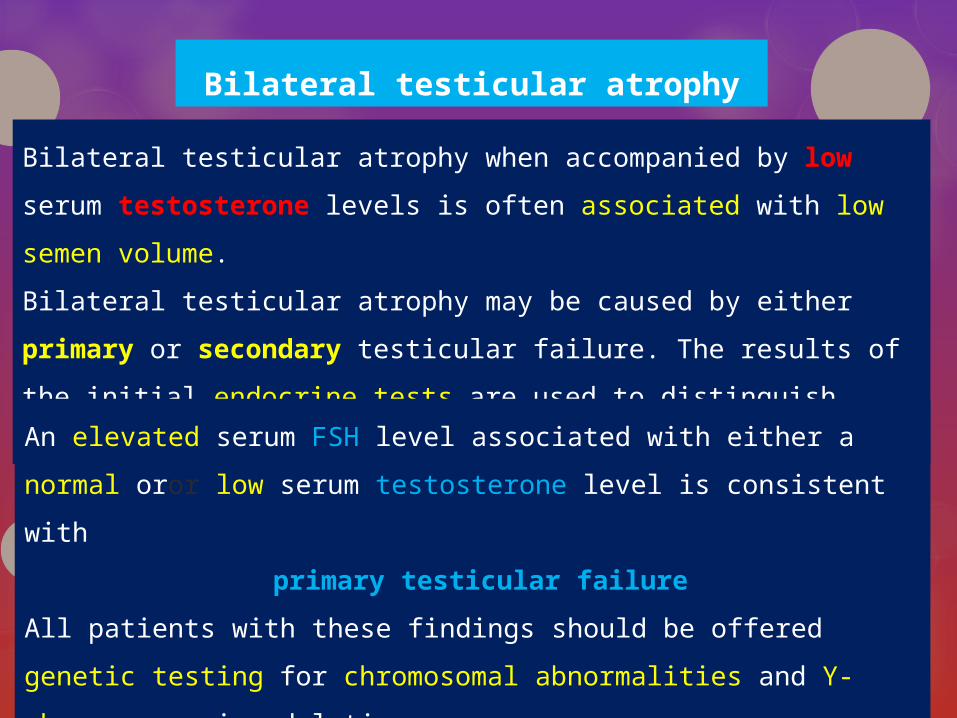
Bilateral testicular atrophy
Bilateral testicular atrophy when accompanied by low serum testosterone
levels is often associated with low semen volume.
Bilateral testicular atrophy may be caused by either primary or secondary
testicular failure. The results of the initial endocrine tests are used to
distinguish between these two possibilities.
An elevated serum FSH level associated with either a normal oror
low serum testosterone level is consistent with
primary testicular failure
All patients with these findings should be offered genetic testing
for chromosomal abnormalities and Y-chromosome microdeletions.
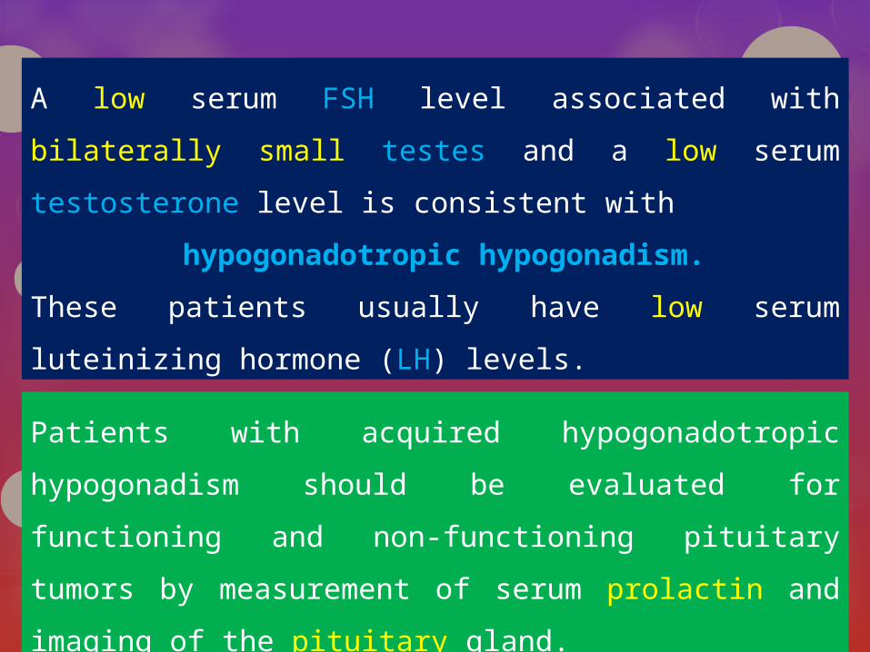
A low serum FSH level associated with bilaterally small testes
and a low serum testosterone level is consistent with
hypogonadotropic hypogonadism.
These patients usually have low serum luteinizing hormone
(LH) levels.
Patients with acquired hypogonadotropic hypogonadism
should be evaluated for functioning and non-functioning
pituitary tumors by measurement of serum prolactin and
imaging of the pituitary gland.
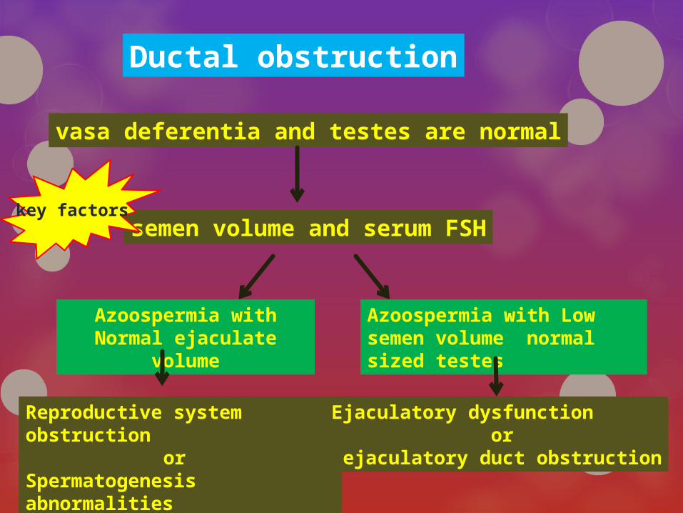
vasa deferentia and testes are normal
semen volume and serum FSH
Azoospermia with Normal ejaculate volume
Reproductive system obstructionor
Spermatogenesis abnormalities
Azoospermia with Low semen volume normal sized testes
Ejaculatory dysfunction or
ejaculatory duct obstruction
Ductal obstruction
key factors

In the azoospermic patient, if the semen volume is
normal (>1ml) and alkaline (pH>7.0), and fructose
poseitive, the seminal vesicles are indeed functional
and emptying through patent ejaculatory ducts.
CBAVD and ejaculatory duct obstruction will not be
diagnostic possibilities (normal testes size).
In these cases, either there exists a blockage to sperm
flow closer to the testes (the vas deferens or the
epididymis) or the testes do not produce sperm
(spermatogenic failure).
Patients with normal ejaculate volume
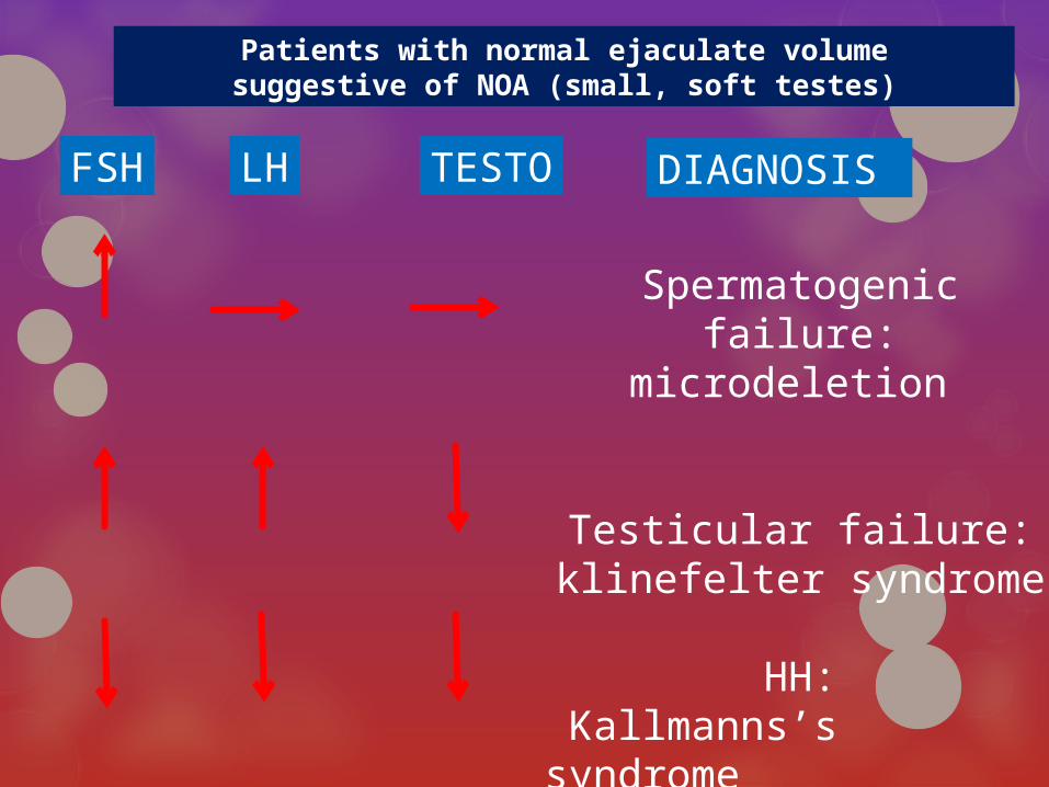
TESTO
Spermatogenic failure: microdeletion
Testicular failure:klinefelter syndrome
HH: Kallmanns’s syndrome
FSH LH DIAGNOSIS
Patients with normal ejaculate volumesuggestive of NOA (small, soft testes)

In order to distinguish between obstructive and nonobstructive
causes of azoospermia, diagnostic testicular biopsy is indicated
for patients with normal testicular size, at least one palpable vas
deferens and a normal serum FSH level.
Recommendations:
The serum FSH of a patient with normal semen volume is a critical factor in determining whether a diagnostic testicular biopsy is needed to establish the presence or absence of normal spermatogenesis.
Marked elevation of serum FSH (greater than two times the upper Limit of normal) is diagnostic of abnormal spermatogenesis.diagnostic testicular biopsy is not necessary in these patients
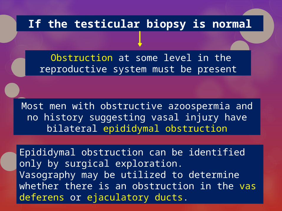
If the testicular biopsy is normal
Obstruction at some level in the reproductive system must be present
Most men with obstructive azoospermia and no history suggesting vasal injury have bilateral epididymal
obstruction
Epididymal obstruction can be identified only by surgical exploration.Vasography may be utilized to determine whether there is an obstruction in the vas deferens or ejaculatory ducts.
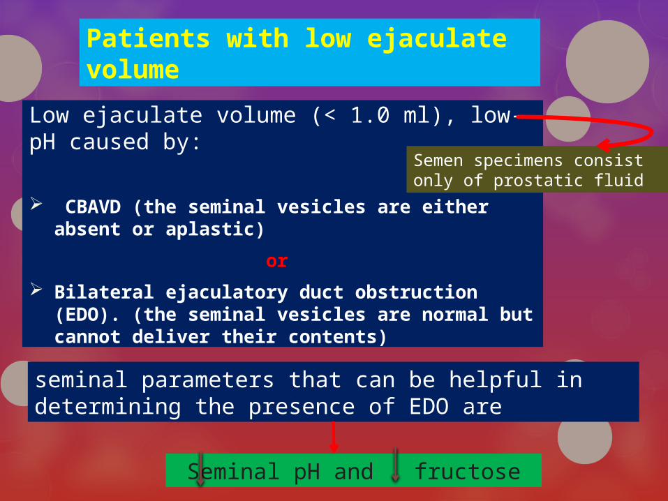
Patients with low ejaculate volume
Low ejaculate volume (< 1.0 ml), low-pH caused by:
CBAVD (the seminal vesicles are either absent or aplastic)
or
Bilateral ejaculatory duct obstruction (EDO). (the seminal vesicles are normal but cannot deliver their contents)
Seminal pH and fructose
seminal parameters that can be helpful in determining the presence of EDO are
Semen specimens consist only of prostatic fluid
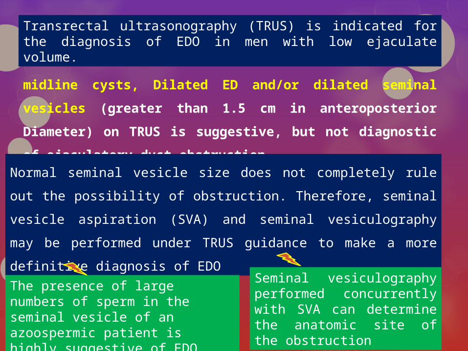
Transrectal ultrasonography (TRUS) is indicated for the diagnosis of EDO in men with low ejaculate volume.
midline cysts, Dilated ED and/or dilated seminal vesicles (greater
than 1.5 cm in anteroposterior Diameter) on TRUS is suggestive,
but not diagnostic of ejaculatory duct obstruction.
Normal seminal vesicle size does not completely rule out the possibility of
obstruction. Therefore, seminal vesicle aspiration (SVA) and seminal
vesiculography may be performed under TRUS guidance to make a more
definitive diagnosis of EDO
The presence of large numbers of sperm in the seminal vesicle of an azoospermic patient is highly suggestive of EDO
Seminal vesiculography performed concurrently with SVA can determine the anatomic site of the obstruction
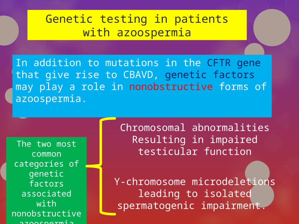
Genetic testing in patients with azoospermia
Chromosomal abnormalities Resulting in impaired testicular
function
Y-chromosome microdeletions leading to isolated spermatogenic
impairment.
In addition to mutations in the CFTR gene that give rise to CBAVD, genetic factors may play a role in nonobstructive forms of azoospermia.
The two most common
categories of genetic factors associated with nonobstructive
azoospermia are
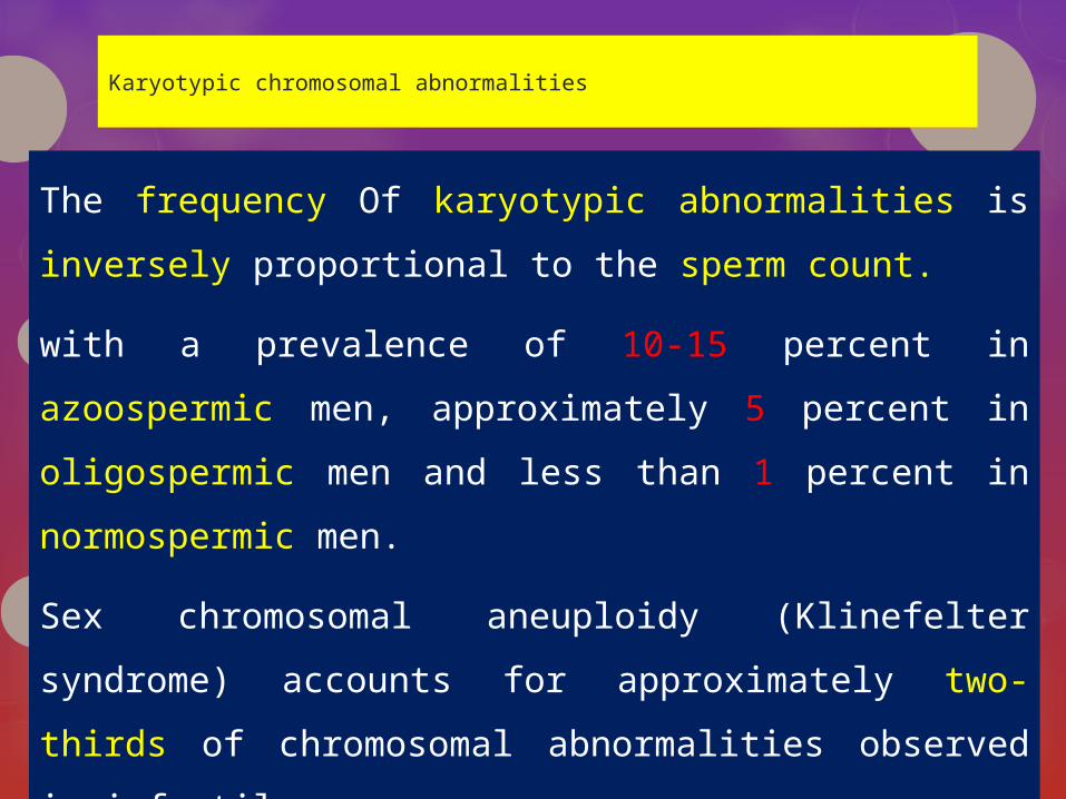
Karyotypic chromosomal abnormalities
The frequency Of karyotypic abnormalities is inversely
proportional to the sperm count.
with a prevalence of 10-15 percent in azoospermic
men, approximately 5 percent in oligospermic men
and less than 1 percent in normospermic men.
Sex chromosomal aneuploidy (Klinefelter syndrome)
accounts for approximately two-thirds of chromosomal
abnormalities observed in infertile men.
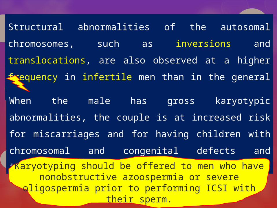
Structural abnormalities of the autosomal
chromosomes, such as inversions and translocations,
are also observed at a higher frequency in infertile men
than in the general population.
When the male has gross karyotypic abnormalities, the
couple is at increased risk for miscarriages and for
having children with chromosomal and congenital
defects and infertility in male offspring
Karyotyping should be offered to men who have nonobstructive azoospermia or severe oligospermia
prior to performing ICSI with their sperm.
Y-chromosome microdeletions
Microdeletions of the Y chromosome may be found in 10-15 percent of men with azoospermia or severe oligospermia .
The human Y chromosome plays an essential role in
the genetic regulation of spermatogenesis.
Most of the genes involved in spermatogenesis have been mapped to the proximal long arm of the Y chromosome (Yq11) and are arranged in azoospermia factor (AZF) region. deletions in this region are specifically related to failure of spermatogenesis.
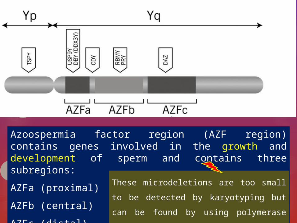
Azoospermia factor region (AZF region) contains genes involved in the growth and development of sperm and contains three subregions:
AZFa (proximal)
AZFb (central)
AZFc (distal)
These microdeletions are too small to be
detected by karyotyping but can be
found by using polymerase chain reaction
(PCR)

It appears That these regions of the Y chromosome contain multiple genes necessary for spermatogenesis.
The specific location of the deletion along the Y chromosome may significantly affect spermatogenesis. If the deleted region of the Y chromosome is in the AZFc region, sperm will be present in the ejaculate in many patients, albeit in severely reduced numbers.
Other patients with AZFc region deletions will be azoospermic but still may have sperm production that is sufficient to allow sperm extraction by testis biopsy.
However, up to 80% of men with AZFc deletions may have retrievable sperm for ICSI.
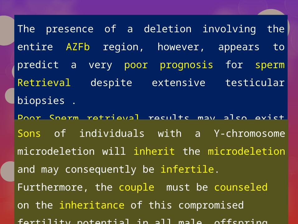
The presence of a deletion involving the entire AZFb region,
however, appears to predict a very poor prognosis for sperm
Retrieval despite extensive testicular biopsies .
Poor Sperm retrieval results may also exist for men with
deletions involving the AZFa region.
Sons of individuals with a Y-chromosome microdeletion will
inherit the microdeletion and may consequently be infertile.
Furthermore, the couple must be counseled on the
inheritance of this compromised fertility potential in all male
offspring
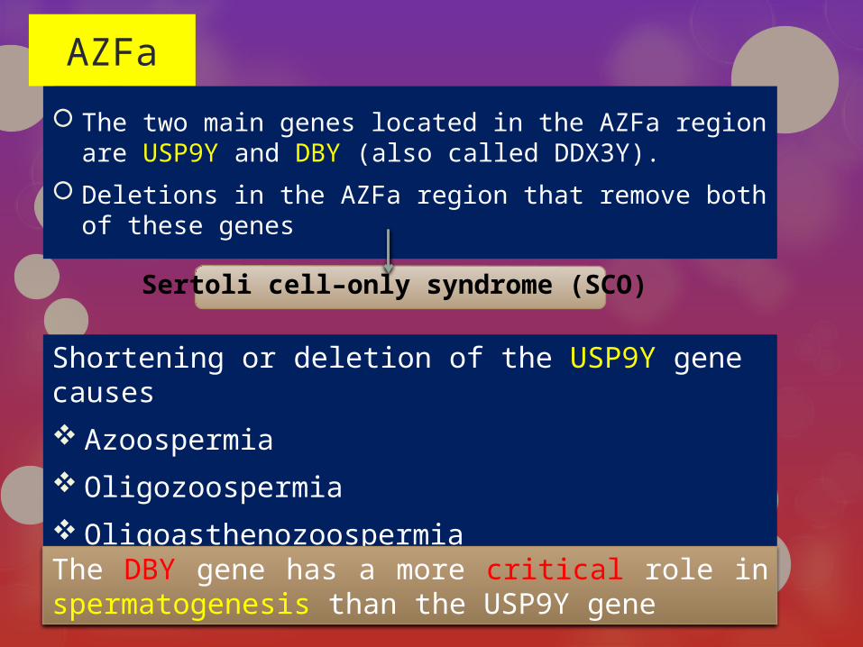
AZFa
The two main genes located in the AZFa region are USP9Y and DBY (also called DDX3Y).
Deletions in the AZFa region that remove both of these genes
Sertoli cell–only syndrome (SCO)
Shortening or deletion of the USP9Y gene causes
Azoospermia
Oligozoospermia
Oligoasthenozoospermia
The DBY gene has a more critical role in spermatogenesis than the USP9Y gene

AZFb
Deletions of the AZFb region cause arrest of spermatogenesis at the primary spermatocyte stage, indicating that the region is essential for fertility
The main genes in the AZFb region is RBMY and a family of PRY genes.
RBMY1 codes for an RNA binding protein , which is a testis-specific splicing factor expressed in the nuclei of spermatogonia, spermatocytes, and round spermatids.
The PRY genes are involved in the regulation of apoptosis,
(an essential process that removes abnormal sperm)
If both the RBMY and PRY genes are removed, spermatogenesis is arrested completely

AZFc
Studies demonstrate that only the AZFa and AZFb regions are needed to initiate spermatogenesis but without the AZFc region, spermatogenesis will not be completely normal
The AZFc region also contains genes involved in spermatogenesis.The DAZ (deleted in Azoospermia) gene which encodes a transcription factor that is usually present in men with Normal fertility, is located in the AZFc region.AZFc deletions cause approximately 12% of non-obstructive azoospermia and 6% of severe oligozoospermia.In many cases, men can still achieve fertilization with the assistance of ART.

Thanks for your attention….


![Azoospermia and embryo morphokinetics: testicular sperm ... · AZF (azoospermia factor) region are clinically important due to their association with failure or disruption of spermatogen-esis[1–3].Deletionsonthe](https://img.pdfslide.us/doc/110x75/5c4596b593f3c34c377ddd20/azoospermia-and-embryo-morphokinetics-testicular-sperm-azf-azoospermia.jpg)