Embed Size (px)
Citation preview

Ascites
Dr Mohammed Hussien
Assistant Lecturer of Hepatology & Gastroentrology
Kafrelsheik University

What is Ascites ?
• Ascites is the presence of excess fluid in the peritoneal cavity.
• It is a common clinical finding with a wide range of causes, but develops most frequently as a part of the decompensation of previously asymptomatic chronic liver disease.
3
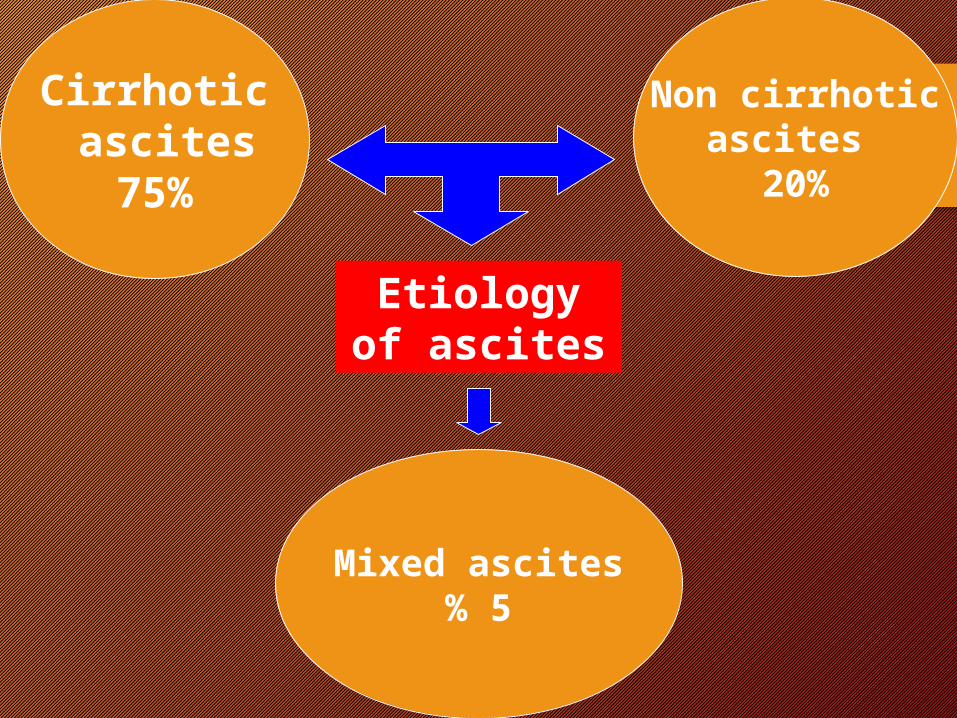
Cirrhotic ascites
75%
Non cirrhotic ascites
20%
Mixed ascites5%
Etiology of ascites
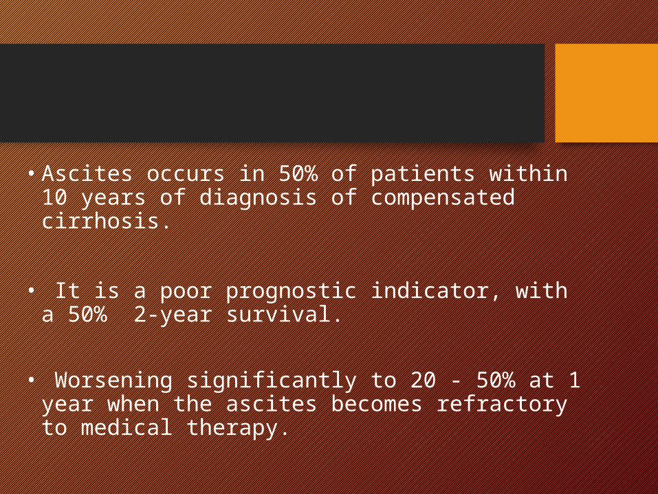
• Ascites occurs in 50% of patients within 10 years of diagnosis of compensated cirrhosis.
• It is a poor prognostic indicator, with a 50% 2-
year survival. • Worsening significantly to 20 - 50% at 1 year
when the ascites becomes refractory to medical therapy.

Mechanism of ascites formation
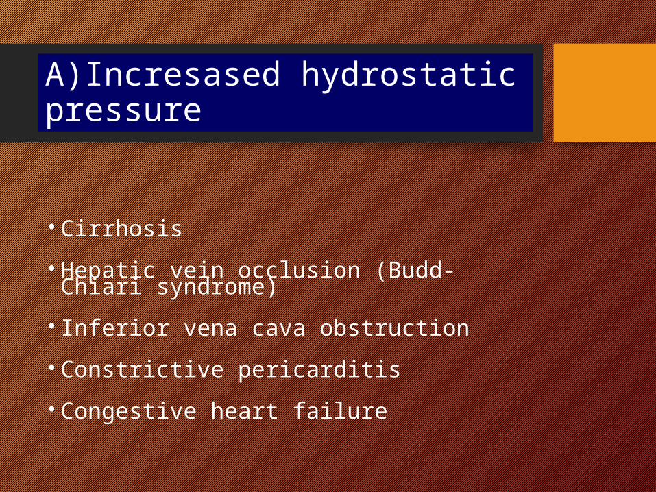
A)Incresased hydrostatic pressure
• Cirrhosis• Hepatic vein occlusion (Budd- Chiari syndrome)• Inferior vena cava obstruction• Constrictive pericarditis• Congestive heart failure
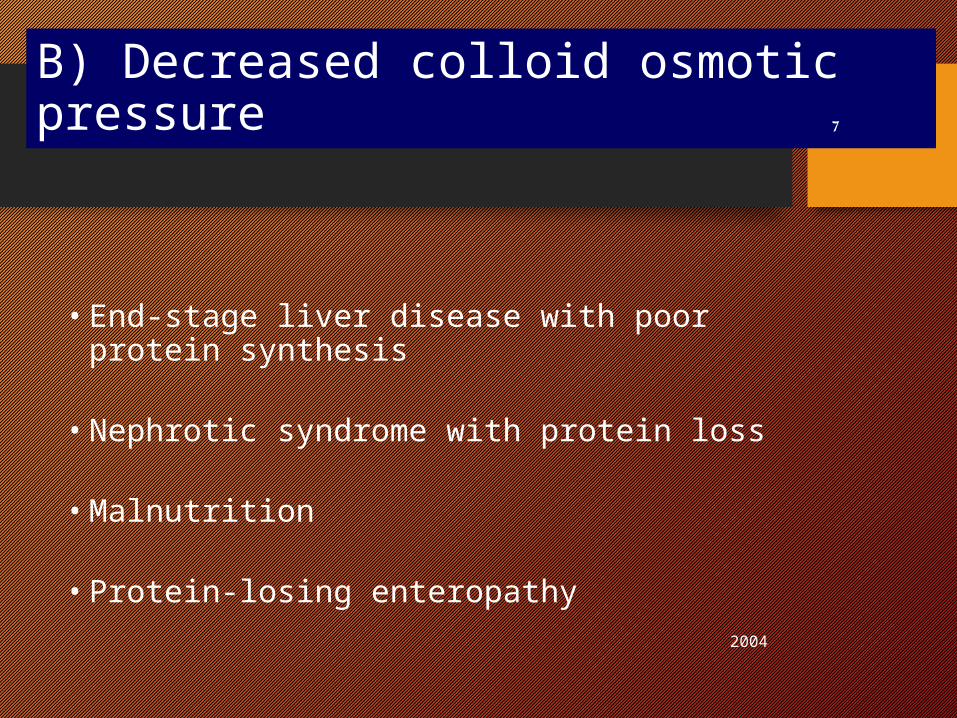
B) Decreased colloid osmotic pressure
• End-stage liver disease with poor protein synthesis
• Nephrotic syndrome with protein loss
• Malnutrition
• Protein-losing enteropathy 2004
7
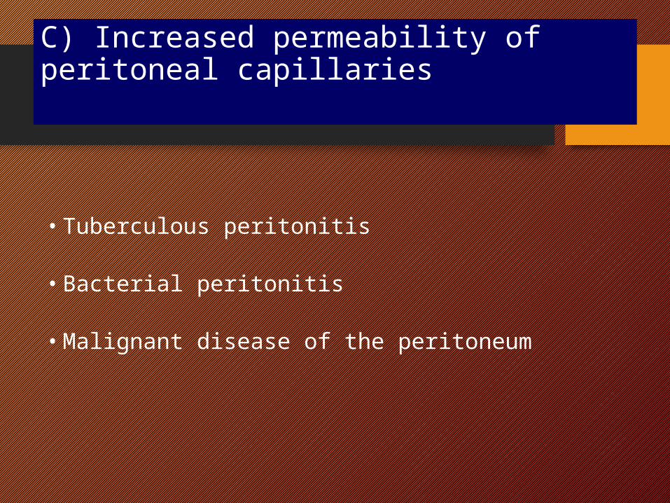
C) Increased permeability of peritoneal capillaries
• Tuberculous peritonitis
• Bacterial peritonitis
• Malignant disease of the peritoneum
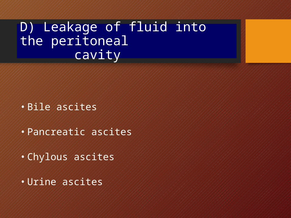
D) Leakage of fluid into the peritoneal cavity
• Bile ascites
• Pancreatic ascites
• Chylous ascites
• Urine ascites
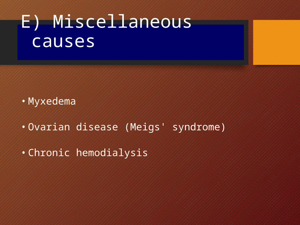
E) Miscellaneous causes
• Myxedema
• Ovarian disease (Meigs' syndrome)
• Chronic hemodialysis
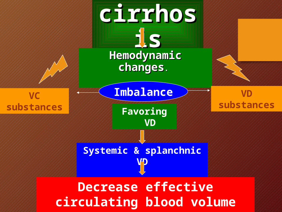
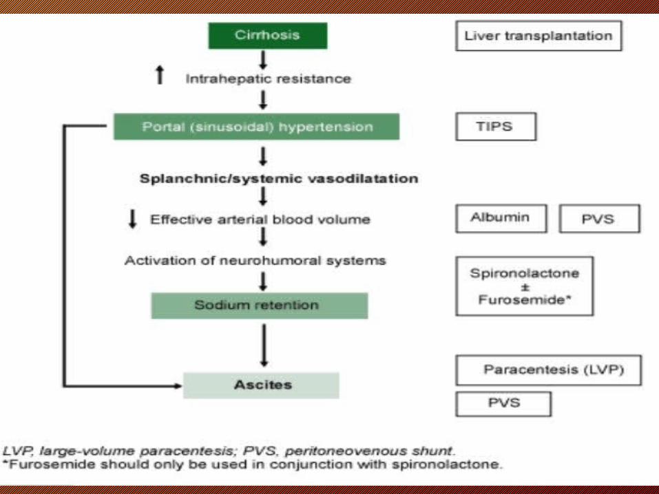
Pathogenesis of ascites in cirrhosis
2004
cirrhosiscirrhosisHemodynamic changesHemodynamic changes. .
VC substances
VD substancesFavoring VD
Systemic & splanchnic VD
Decrease effective circulating blood volume
Imbalance
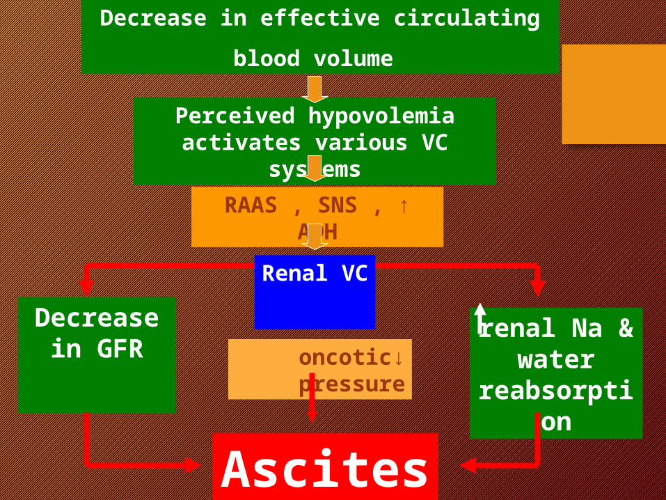
Perceived hypovolemia activates various VC systems
↑RAAS , SNS , ADH
Renal VC
renal Na & water
reabsorption
Decrease in GFR
Ascites
Decrease in effective circulating
blood volume
↓oncotic pressure
2004
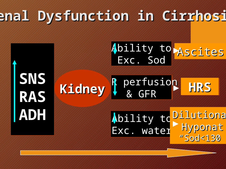
SNSSNSRASRASADHADH
KidneyKidney R perfusionR perfusion& GFR& GFR
Ability toAbility toExc. SodExc. Sod
Ability toAbility toExc. waterExc. water
HRSHRS
AscitesAscites
DilutionalDilutionalHyponatHyponat
““Sod<130Sod<130””
Renal Dysfunction in CirrhosisRenal Dysfunction in Cirrhosis
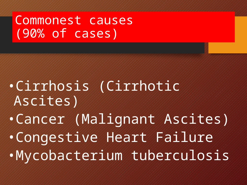
Commonest causes (90% of cases)
•Cirrhosis (Cirrhotic Ascites) •Cancer (Malignant Ascites) •Congestive Heart Failure •Mycobacterium tuberculosis
Clinical Manifestations and Diagnosis
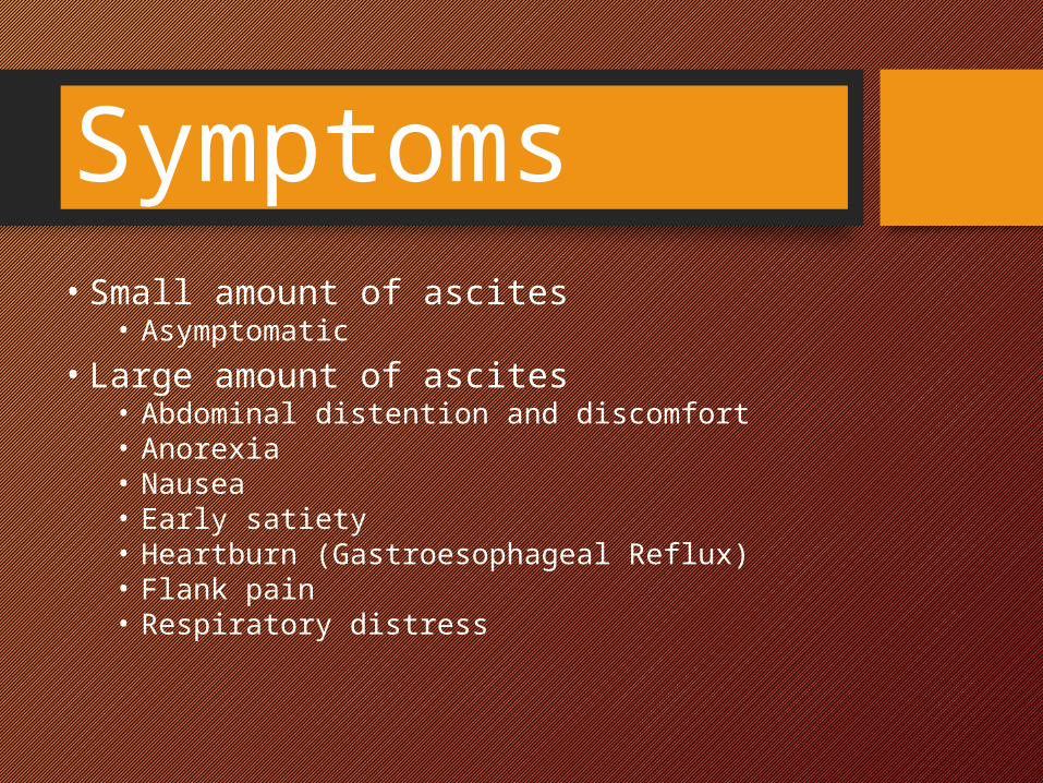
Symptoms• Small amount of ascites
• Asymptomatic • Large amount of ascites
• Abdominal distention and discomfort • Anorexia • Nausea • Early satiety • Heartburn (Gastroesophageal Reflux) • Flank pain • Respiratory distress
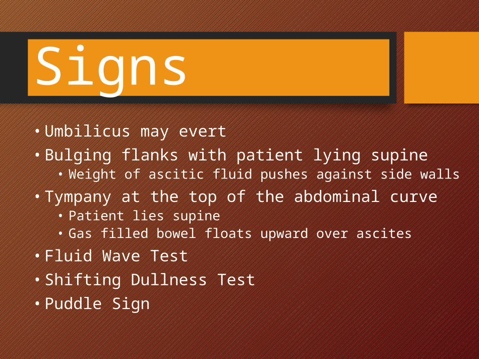
Signs
• Umbilicus may evert • Bulging flanks with patient lying supine
• Weight of ascitic fluid pushes against side walls • Tympany at the top of the abdominal curve
• Patient lies supine • Gas filled bowel floats upward over ascites
• Fluid Wave Test • Shifting Dullness Test • Puddle Sign
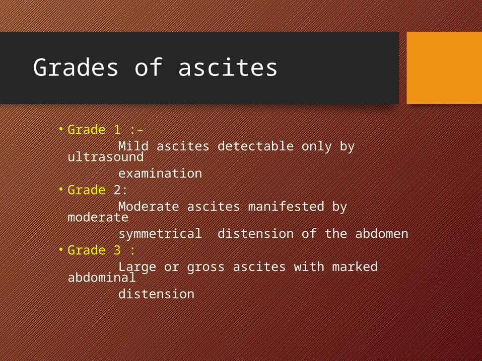
Grades of ascites
• Grade 1 :– Mild ascites detectable only by ultrasound examination• Grade 2: Moderate ascites manifested by moderate symmetrical distension of the abdomen• Grade 3 : Large or gross ascites with marked abdominal distension
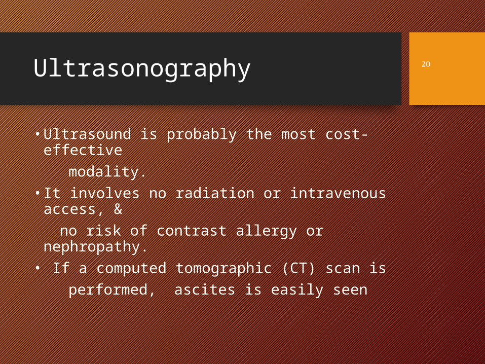
Ultrasonography
• Ultrasound is probably the most cost-effective
modality. • It involves no radiation or intravenous
access, & no risk of contrast allergy or nephropathy. • If a computed tomographic (CT) scan is performed, ascites is easily seen
20
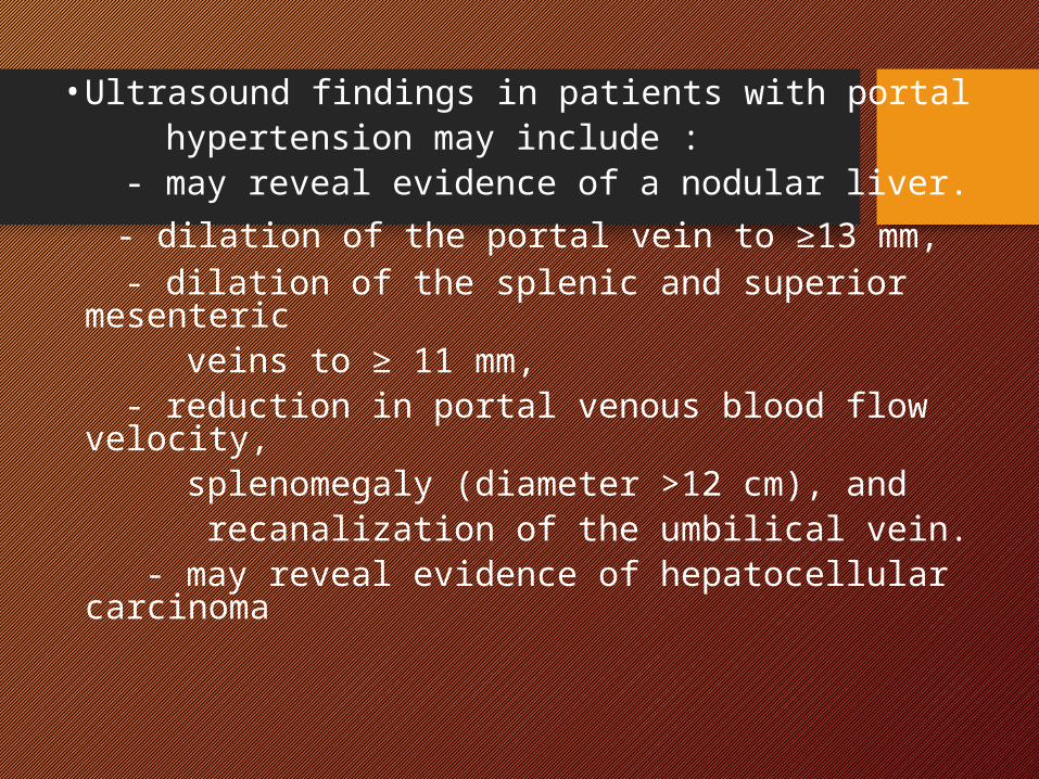
• Ultrasound findings in patients with portal hypertension may include : - may reveal evidence of a nodular liver. - dilation of the portal vein to ≥13 mm, - dilation of the splenic and superior
mesenteric veins to ≥ 11 mm, - reduction in portal venous blood flow
velocity, splenomegaly (diameter >12 cm), and recanalization of the umbilical vein. - may reveal evidence of hepatocellular
carcinoma

Analysis of Ascitic Fluid
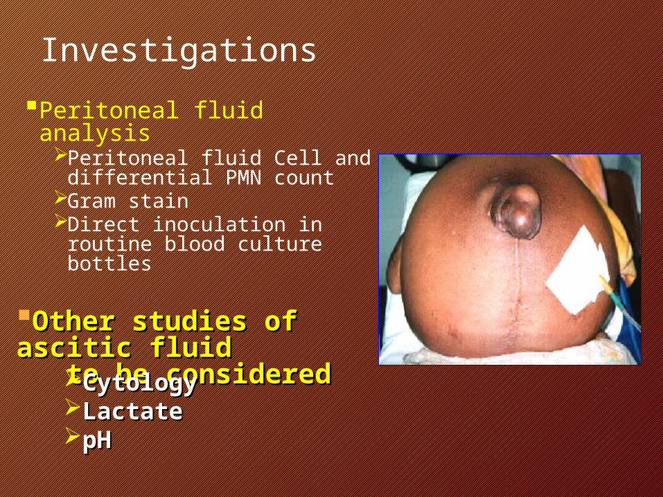
InvestigationsPeritoneal fluid analysis
Peritoneal fluid Cell and differential PMN count
Gram stainDirect inoculation in
routine blood culture bottles
Other studies of Other studies of ascitic fluid ascitic fluid to be consideredto be consideredCytologyCytology
LactateLactatepHpH
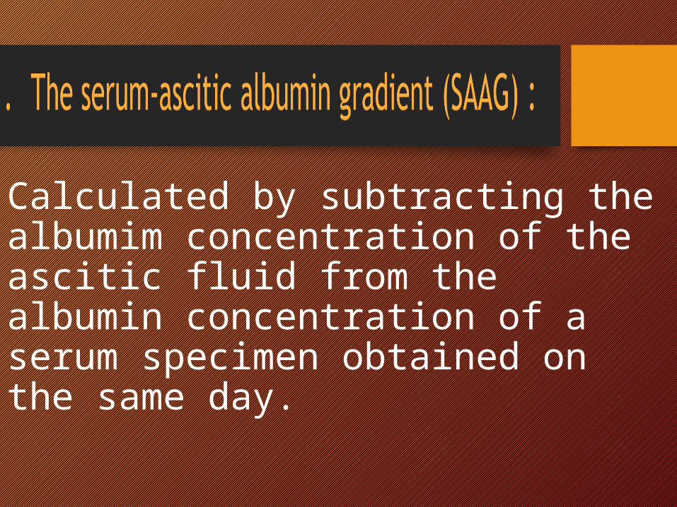
Calculated by subtracting the albumim concentration of the ascitic fluid from the albumin concentration of a serum specimen obtained on the same day.

Serum Ascites Albumin Gradient (SAAG)
SAAG
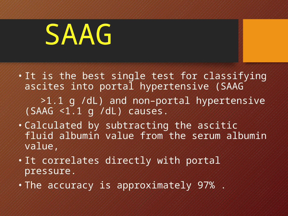
• It is the best single test for classifying ascites into portal hypertensive (SAAG
>1.1 g /dL) and non–portal hypertensive (SAAG <1.1 g /dL) causes.
• Calculated by subtracting the ascitic fluid albumin value from the serum albumin value,
• It correlates directly with portal pressure. • The accuracy is approximately 97% .
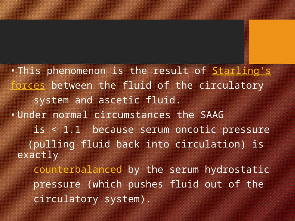
• This phenomenon is the result of Starling'sforces between the fluid of the circulatory system and ascetic fluid. • Under normal circumstances the SAAG is < 1.1 because serum oncotic pressure (pulling fluid back into circulation) is exactly counterbalanced by the serum hydrostatic pressure (which pushes fluid out of the circulatory system).
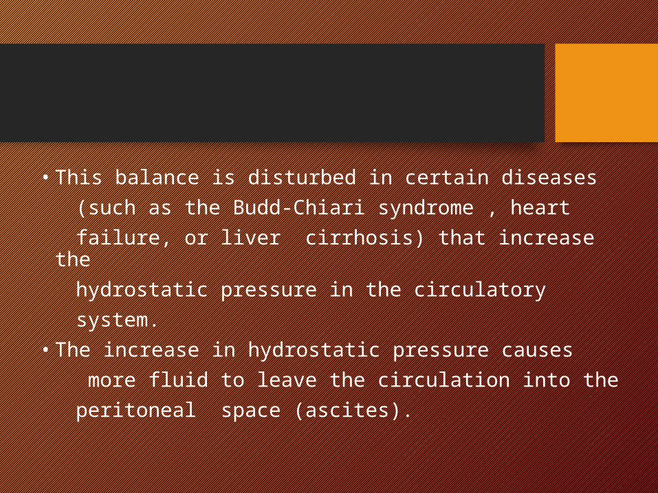
• This balance is disturbed in certain diseases (such as the Budd-Chiari syndrome , heart failure, or liver cirrhosis) that increase the hydrostatic pressure in the circulatory system.• The increase in hydrostatic pressure causes more fluid to leave the circulation into the peritoneal space (ascites).
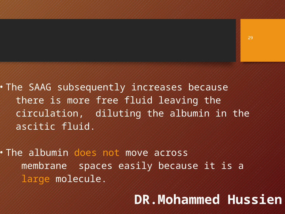
• The SAAG subsequently increases because there is more free fluid leaving the circulation, diluting the albumin in the ascitic fluid.
• The albumin does not move across membrane spaces easily because it is a large molecule.
DR.Mohammed Hussien
29
2004
30
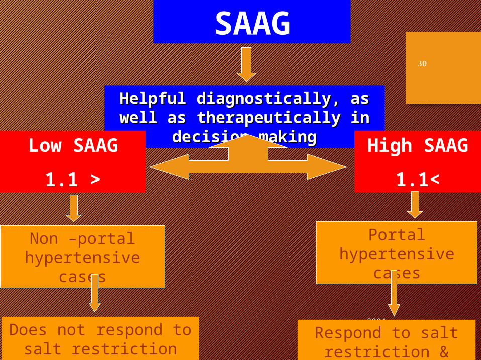
SAAG
Helpful diagnostically, as well as Helpful diagnostically, as well as therapeutically in decision makingtherapeutically in decision making
Low SAAG
<1.1
High SAAG
>1.1
Non –portal hypertensive cases
Portal hypertensive cases
Does not respond to salt restriction nor Diuretics
Respond to salt restriction & Diuretics
2004
31
2004
32
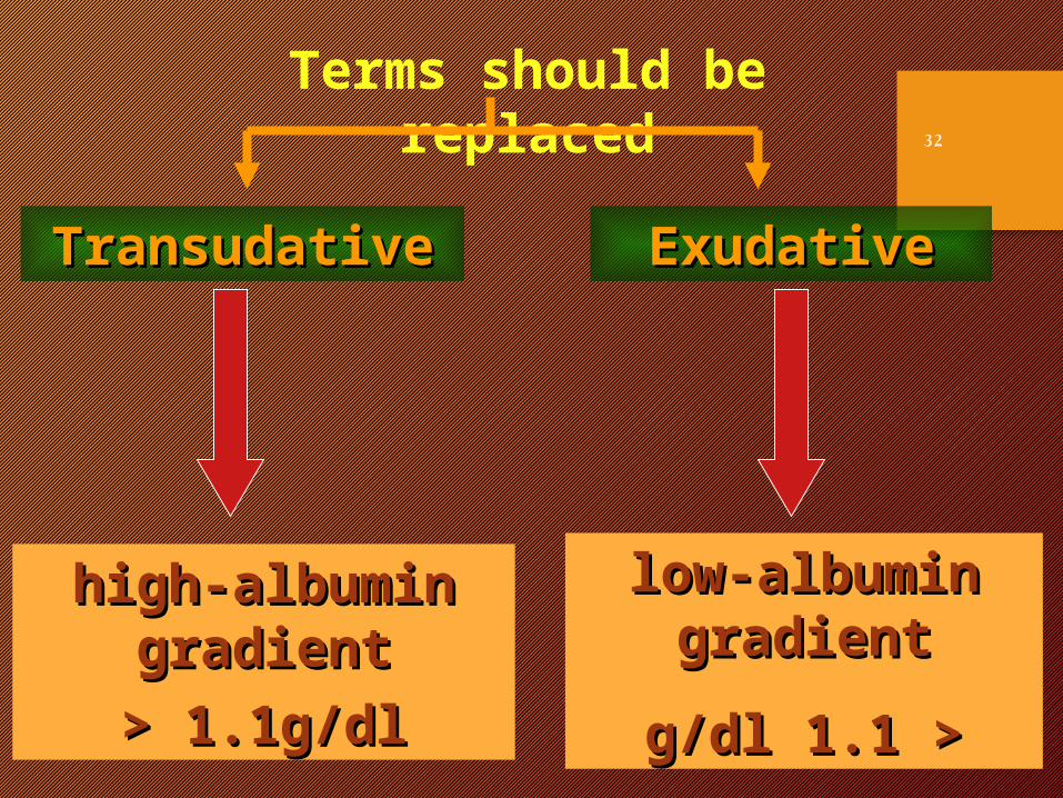
high-albumin high-albumin gradientgradient< 1.1g/dl< 1.1g/dl
low-albumin low-albumin gradientgradient
< <1.11.1 g/dlg/dl
TransudativeTransudative ExudativeExudative
Terms should be replaced
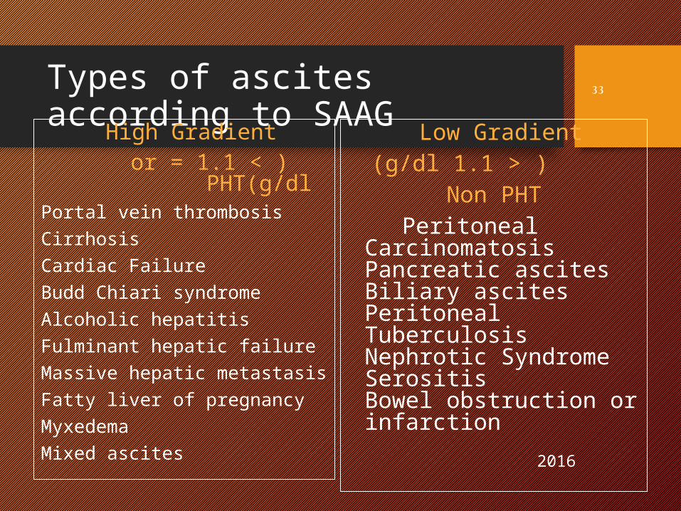
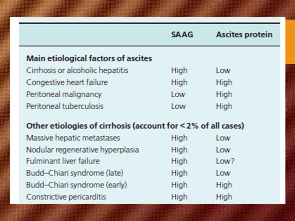
Types of ascites according to SAAGHigh Gradient
> ( or = 1.1 g/dl)PHT Portal vein thrombosisCirrhosisCardiac FailureBudd Chiari syndromeAlcoholic hepatitisFulminant hepatic failureMassive hepatic metastasisFatty liver of pregnancyMyxedemaMixed ascites
Low Gradient < ( 1.1 g/dl)
Non PHT Peritoneal
CarcinomatosisPancreatic ascitesBiliary ascitesPeritoneal TuberculosisNephrotic Syndrome Serositis Bowel obstruction or infarction
2016
33
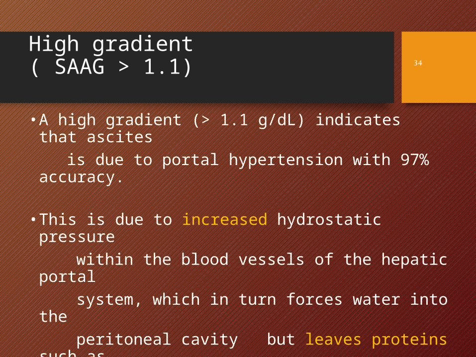
High gradient ( SAAG > 1.1)
• A high gradient (> 1.1 g/dL) indicates that ascites is due to portal hypertension with 97% accuracy.
• This is due to increased hydrostatic pressure within the blood vessels of the hepatic portal system, which in turn forces water into the peritoneal cavity but leaves proteins such as albumin within the vasculature.
34
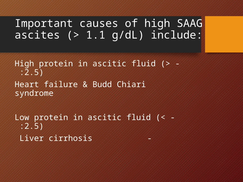
Important causes of high SAAG ascites (> 1.1 g/dL) include:
- High protein in ascitic fluid (> 2.5) : Heart failure & Budd Chiari syndrome
- Low protein in ascitic fluid (< 2.5) :- Liver cirrhosis
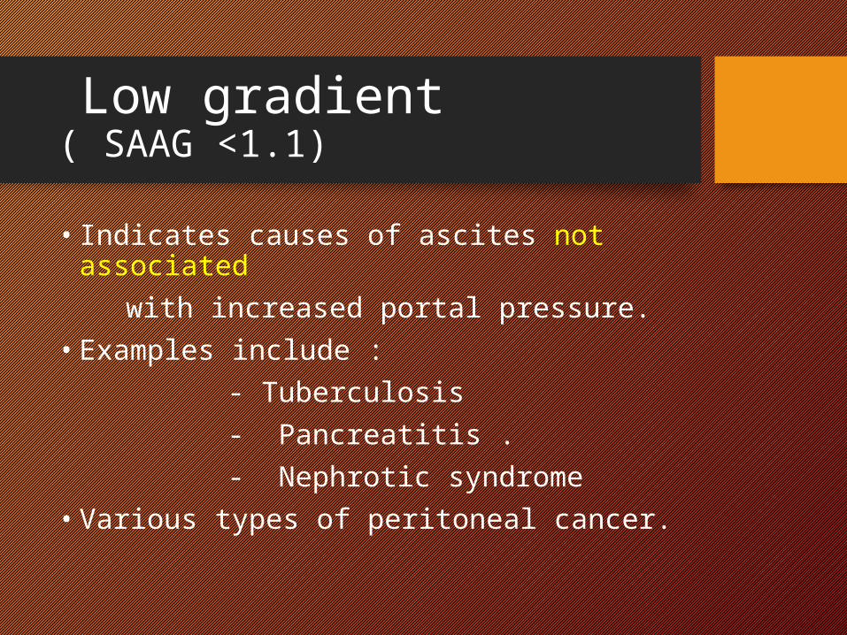
Low gradient( SAAG <1.1)
• Indicates causes of ascites not associated with increased portal pressure. • Examples include : - Tuberculosis - Pancreatitis . - Nephrotic syndrome• Various types of peritoneal cancer.
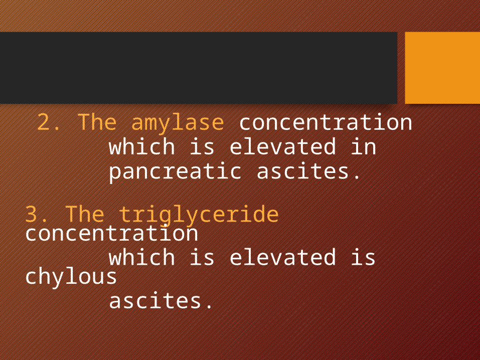
2. The amylase concentration which is elevated in pancreatic ascites. 3. The triglyceride concentration which is elevated is chylous ascites.
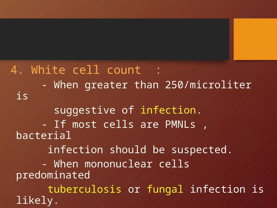
4. White cell count : - When greater than 250/microliter is suggestive of infection. - If most cells are PMNLs , bacterial infection should be suspected. - When mononuclear cells predominated
tuberculosis or fungal infection is likely.
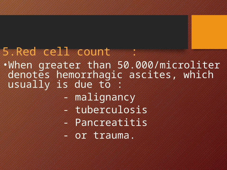
5.Red cell count : • When greater than 50.000/microliter denotes hemorrhagic ascites, which usually is due to :
- malignancy - tuberculosis - Pancreatitis - or trauma.
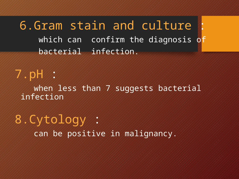
6.Gram stain and culture : which can confirm the diagnosis of bacterial infection.
7.pH : when less than 7 suggests bacterial infection
8.Cytology : can be positive in malignancy.
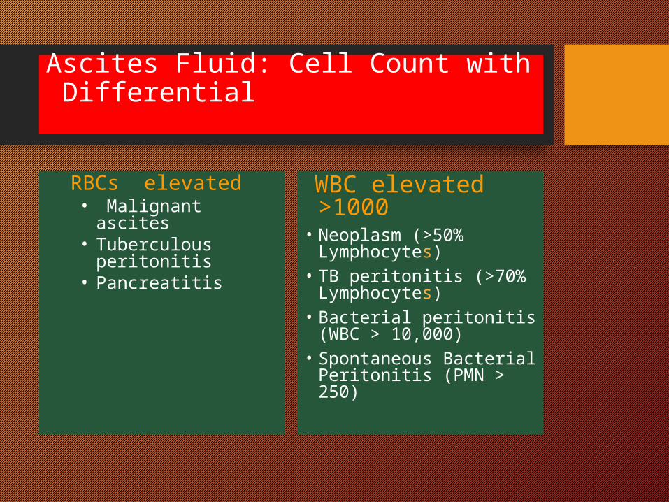
Ascites Fluid: Cell Count with Differential
RBCs elevated • Malignant ascites• Tuberculous
peritonitis • Pancreatitis
WBC elevated >1000
• Neoplasm (>50% Lymphocytes)
• TB peritonitis (>70% Lymphocytes)
• Bacterial peritonitis (WBC > 10,000)
• Spontaneous Bacterial Peritonitis (PMN > 250)

Management of ascites
2016
43
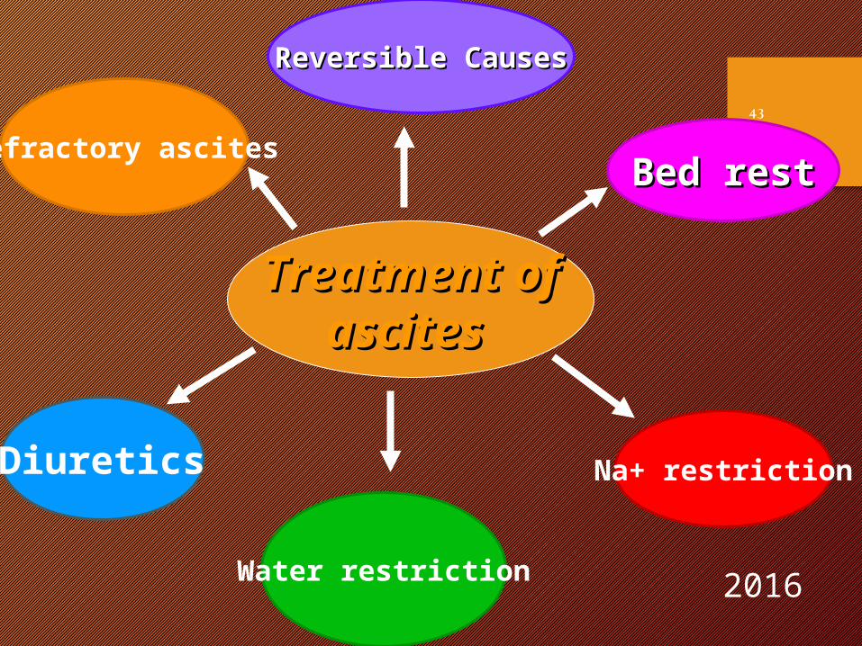
Treatment ofTreatment of ascitesascites
Bed restBed rest
Water restriction
Diuretics
Refractory ascites
Reversible CausesReversible Causes
Na+ restriction
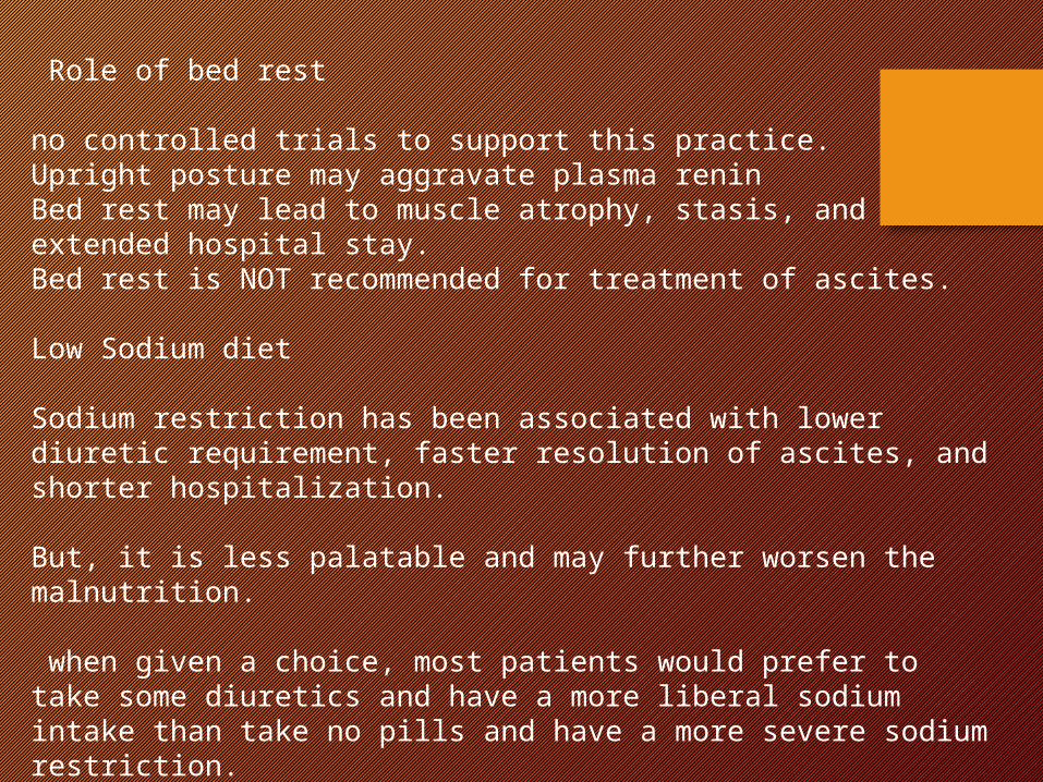
Role of bed rest
no controlled trials to support this practice. Upright posture may aggravate plasma reninBed rest may lead to muscle atrophy, stasis, and extended hospital stay. Bed rest is NOT recommended for treatment of ascites.
Low Sodium diet
Sodium restriction has been associated with lower diuretic requirement, faster resolution of ascites, and shorter hospitalization.
But, it is less palatable and may further worsen the malnutrition.
when given a choice, most patients would prefer to take some diuretics and have a more liberal sodium intake than take no pills and have a more severe sodium restriction.
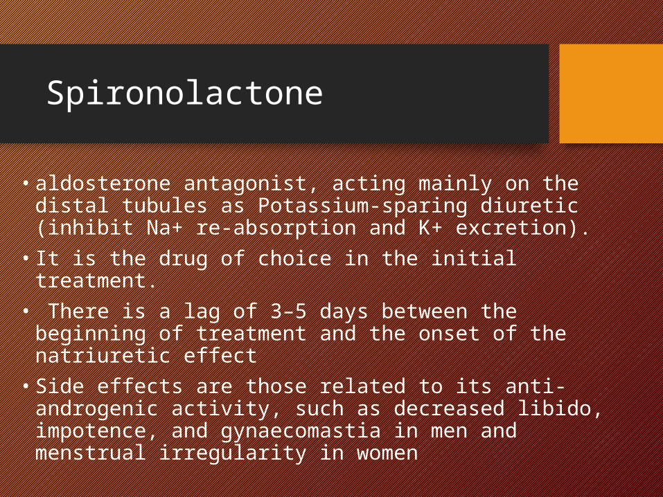
Spironolactone
• aldosterone antagonist, acting mainly on the distal tubules as Potassium-sparing diuretic (inhibit Na+ re-absorption and K+ excretion).
• It is the drug of choice in the initial treatment.• There is a lag of 3–5 days between the beginning of
treatment and the onset of the natriuretic effect• Side effects are those related to its anti-androgenic
activity, such as decreased libido, impotence, and gynaecomastia in men and menstrual irregularity in women
Frusemide
• Frusemide is a loop diuretic that generally used as an adjunct to spironolactone
• it inhibit re-absorption of Na+/K+/2Cl- in the ascending limb of the loop of Henle.
• High doses of frusemide are associated with severe electrolyte disturbance and metabolic alkalosis, and should be used cautiously.
Other diuretics
• Amiloride and triamterene act on the distal tubule. It blocks Na reabsorption and induces diuresis in 80% of patients at doses of 15–30 mg/day. It is less effective compared with spironolactone.
• Bumetanide is similar to frusemide in its action and efficacy
Single or combination therapy• The initial combination treatment shortens the time
to mobilization of moderate to tense ascites and better for inpatient treatment.
• So it is preferred approach in achieving rapid natriuresis and maintaining normokalemia.
• An alternative approach would be to start with Spironolactone, in particular in the outpatient setting, then monitoring the patient for adding loop diuretics after 400mg Spironolactone failure.
Dosage
• The doses of both oral diuretics can be increased simultaneously every 3-5 days (maintaining the 100 mg:40 mg ratio) if weight loss and natriuresis are inadequate.
• This ratio maintains normo-kalemia.• Usual maximum doses are 400 mg/day of
spironolactone and 160 mg/day of furosemide• Over diuresis is associated with intravascular
volume depletion leading to renal impairment, hepatic encephalopathy, and hyponatraemia.
Therapeutic Paracentesis
• Although initially the recommendation was to perform daily 5-L paracentesis until the disappearance of ascites, it was subsequently determined that total paracentesis (i.e., removal of all ascites in a single procedure accompanied by the concomitant infusion of 6–8 g albumin per liter of ascites removed) was as safe as repeated partial paracenteses
LVP
• LVP associated with i.v. plasma expander is effective and associated with a significantly faster resolution and a lower rate of complications than repeated paracentesis with intensive diuretics.
• However, it is a local therapy (does not act on the mechanisms of ascites formation) and ascites recurrence is the rule.
• Additionally, it is more costly and requires more resources than the administration of diuretics.
Use of plasma expanders
• Paracentesis of <5 L of uncomplicated ascites does not require volume expansion
• Plasma volume expander should always be used whenever >5 L of ascites are removed.
Stepwise treatment of ascites
• Sodium restriction (88 mmol /d = 2 g) • Titrate spironolactone (to Na+u / K+u > 1)• If no success add loop diuretic• Fluid restriction only if Na+ < 120 mmol/l • Bed rest is not recommended. • Aim for weight loss < 1/2 kg/d in non-edematous pts ,
but should not exceed 1 kg/day when edema is present.•
Please don’t forget
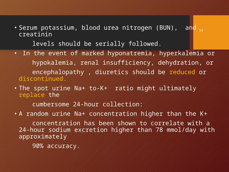
• Serum potassium, blood urea nitrogen (BUN), and creatinin levels should be serially followed.• In the event of marked hyponatremia, hyperkalemia or hypokalemia, renal insufficiency, dehydration, or encephalopathy , diuretics should be reduced or
discontinued.• The spot urine Na+ to-K+ ratio might ultimately replace
the cumbersome 24-hour collection: • A random urine Na+ concentration higher than the K+ concentration has been shown to correlate with a 24-hour
sodium excretion higher than 78 mmol/day with approximately
90% accuracy.
54
2004
55
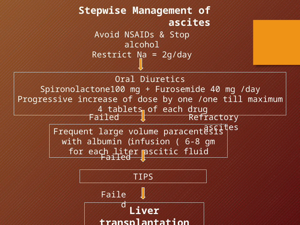
Avoid NSAIDs & Stop alcoholRestrict Na = 2g/day
Oral DiureticsSpironolactone100 mg + Furosemide 40 mg /day
Progressive increase of dose by one /one till maximum 4 tablets of each drug
Frequent large volume paracentesis with albumin (infusion ( 6-8 gm for each
liter ascitic fluid
TIPS
Liver transplantation
Stepwise Management of ascites
Failed Refractory ascites
Failed
Failed
2004
57
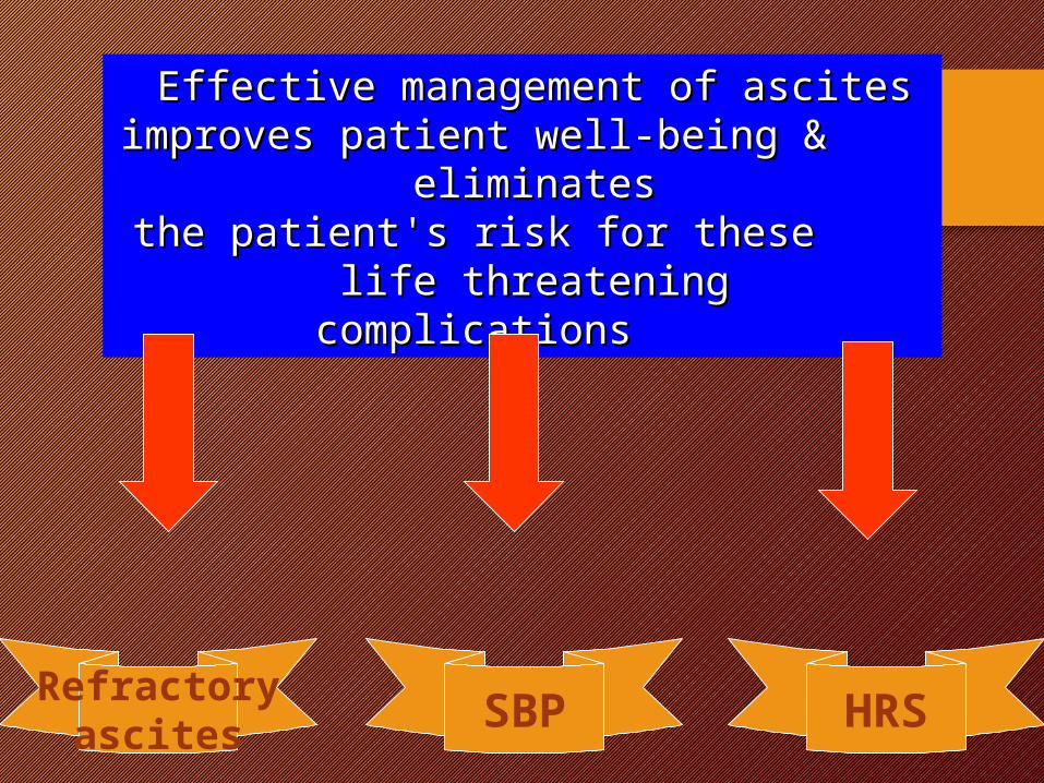
Effective management of ascitesEffective management of ascites improves patient well-being & eliminatesimproves patient well-being & eliminates the patient's risk for these life threateningthe patient's risk for these life threatening
complicationscomplications
HRSSBPRefractoryascites

Refractory ascites
2004
59
2004
60
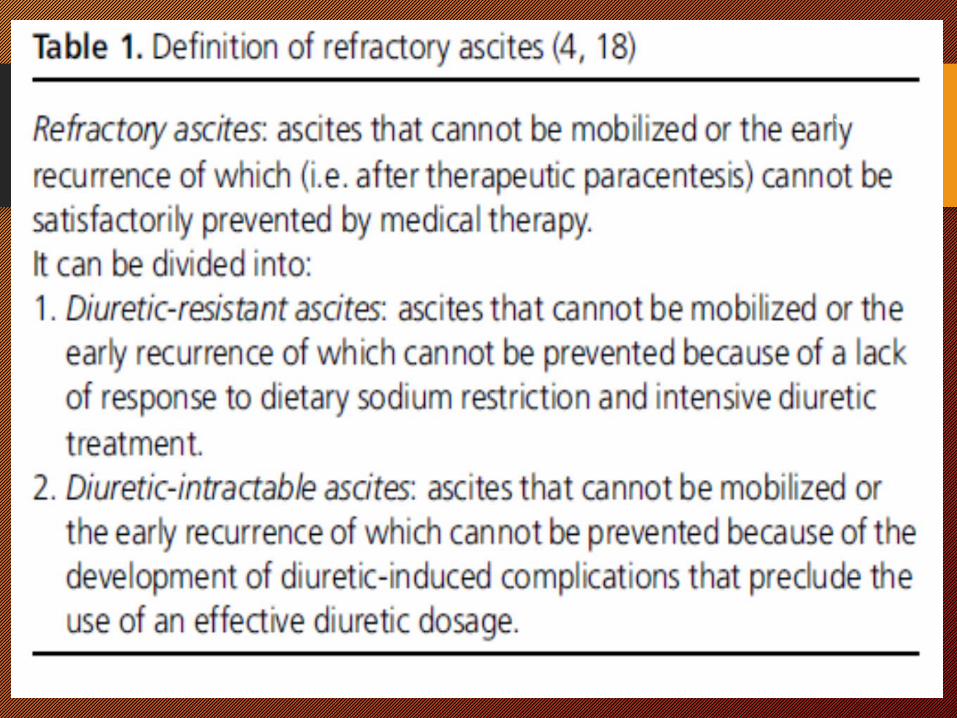
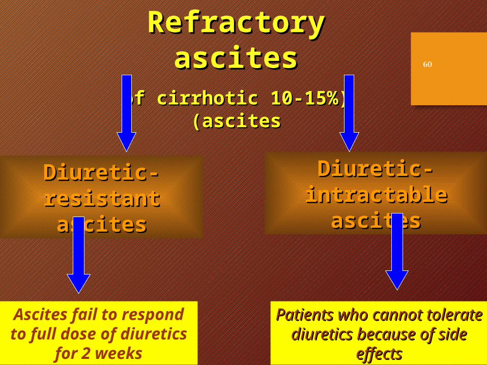
Refractory ascitesRefractory ascites((10-15%10-15% of cirrhotic ascitesof cirrhotic ascites))
Diuretic-resistant Diuretic-resistant ascitesascites
Diuretic- intractableDiuretic- intractable ascitesascites
Ascites fail to respond to full dose of diuretics
for 2 weeks
Patients who cannot tolerate Patients who cannot tolerate diuretics because of side diuretics because of side
effectseffects
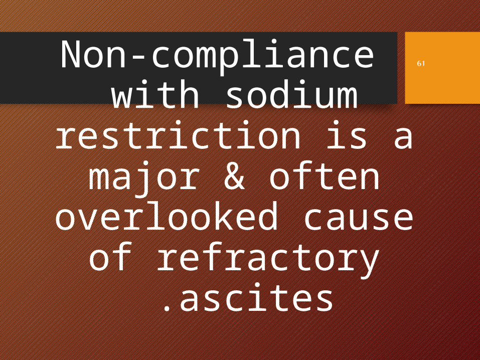
Non-compliance with sodium
restriction is a major & often
overlooked cause of refractory ascites .
61
2004
62
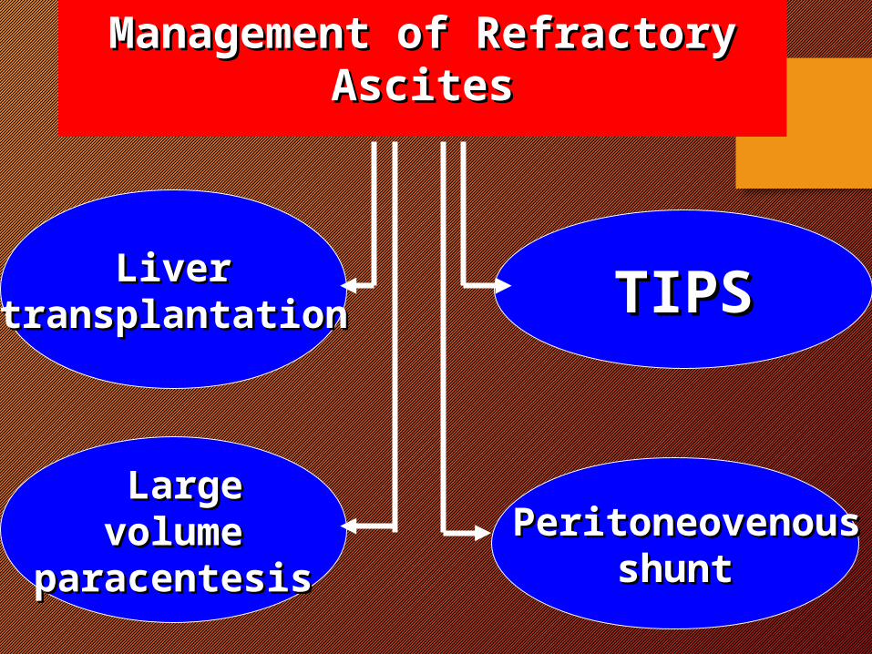
Management of Refractory Management of Refractory AscitesAscites
LiverLivertransplantationtransplantation
LargeLarge volumevolume
paracentesisparacentesisPeritoneovenousPeritoneovenous
shuntshunt
TIPSTIPS
Liver transplantation
• It is the most effective & definitive treatment but ؟؟؟؟؟؟؟
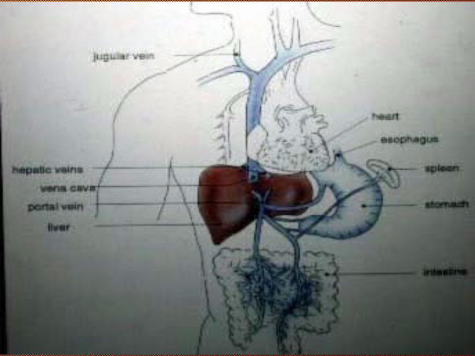
Transjugular intrahepatic Porto systemic shunt (TIPS)
2004
65
2004
66
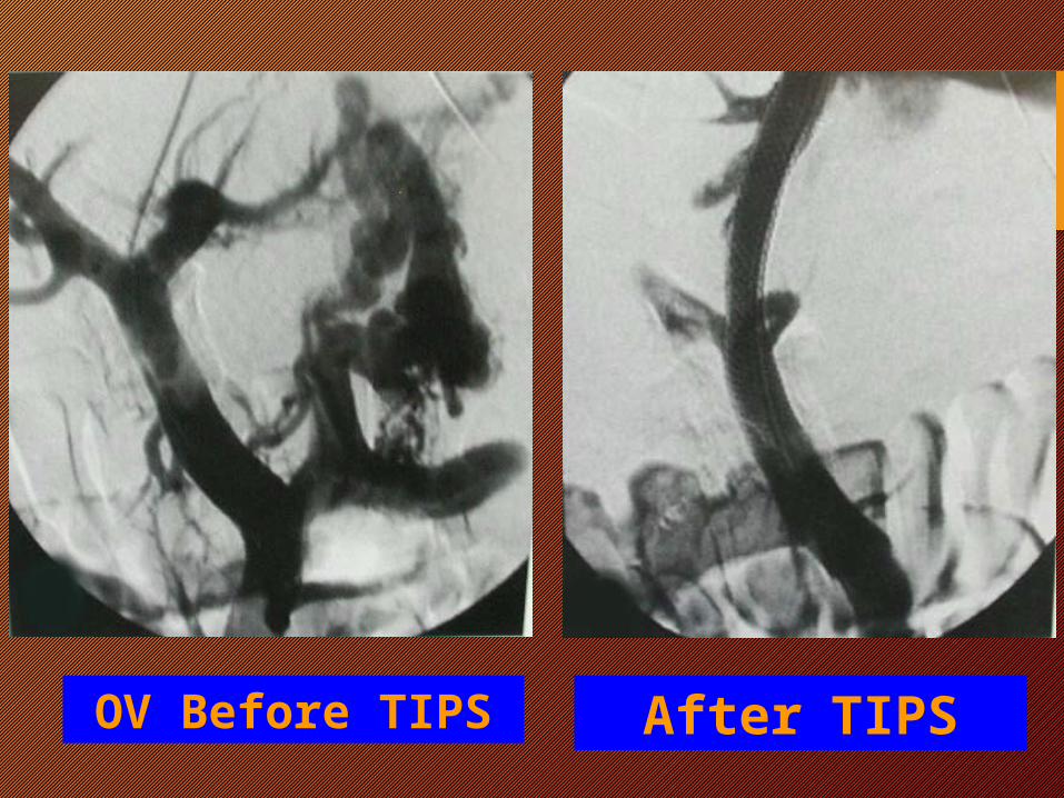
OV Before TIPS After TIPS
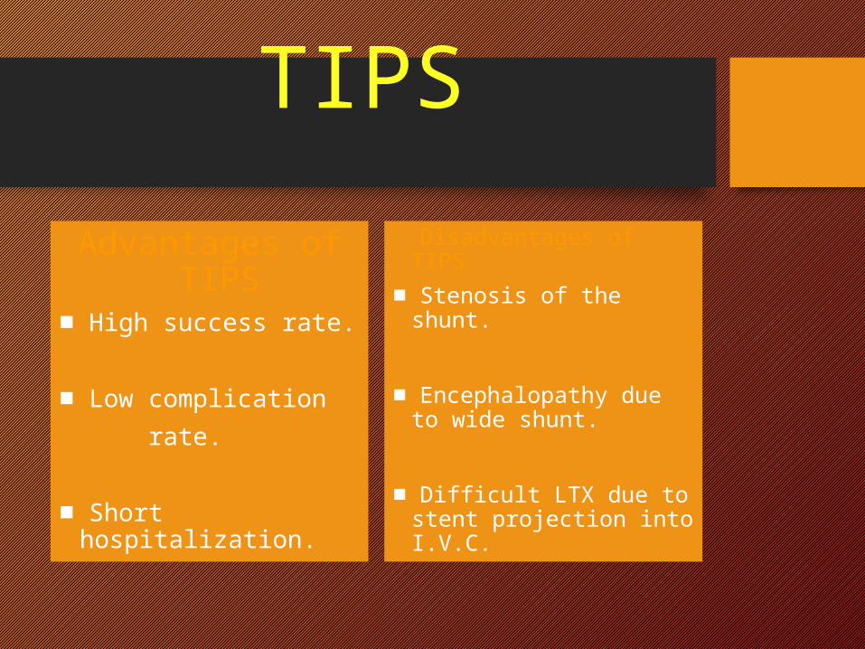
TIPSAdvantages of
TIPS■ High success rate.
■ Low complication rate.
■ Short hospitalization.
Disadvantages of TIPS■ Stenosis of the shunt.
■ Encephalopathy due to wide shunt.
■ Difficult LTX due to stent projection into I.V.C.
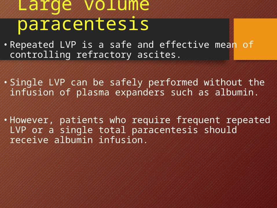
Large volume paracentesis
• Repeated LVP is a safe and effective mean of controlling refractory ascites.
• Single LVP can be safely performed without the infusion
of plasma expanders such as albumin.
• However, patients who require frequent repeated LVP or a single total paracentesis should receive albumin infusion.
2004
69
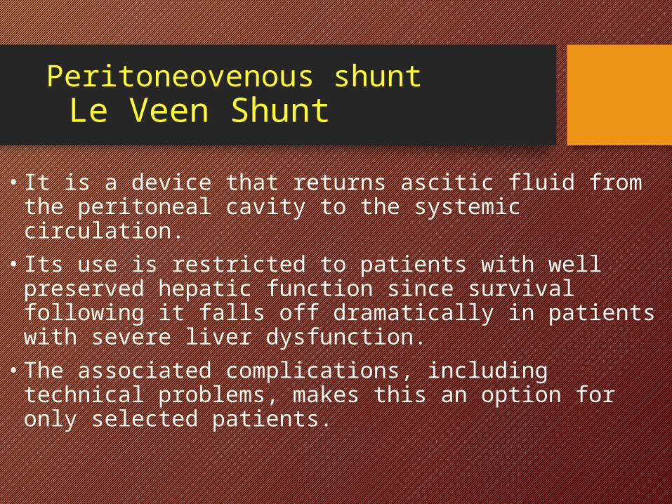
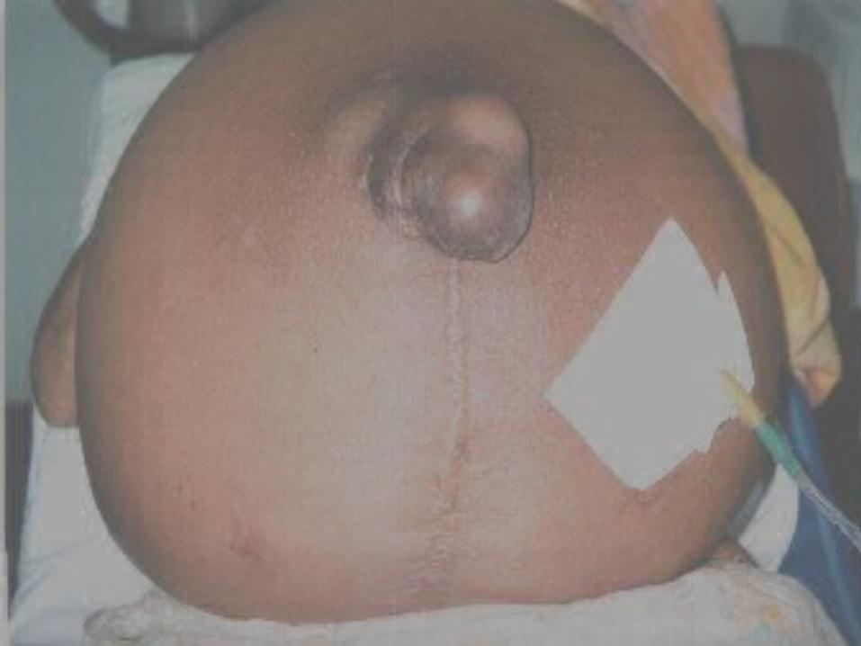
Peritoneovenous shunt Le Veen Shunt
• It is a device that returns ascitic fluid from the peritoneal cavity to the systemic circulation.
• Its use is restricted to patients with well preserved hepatic function since survival following it falls off dramatically in patients with severe liver dysfunction.
• The associated complications, including technical problems, makes this an option for only selected patients.
2004
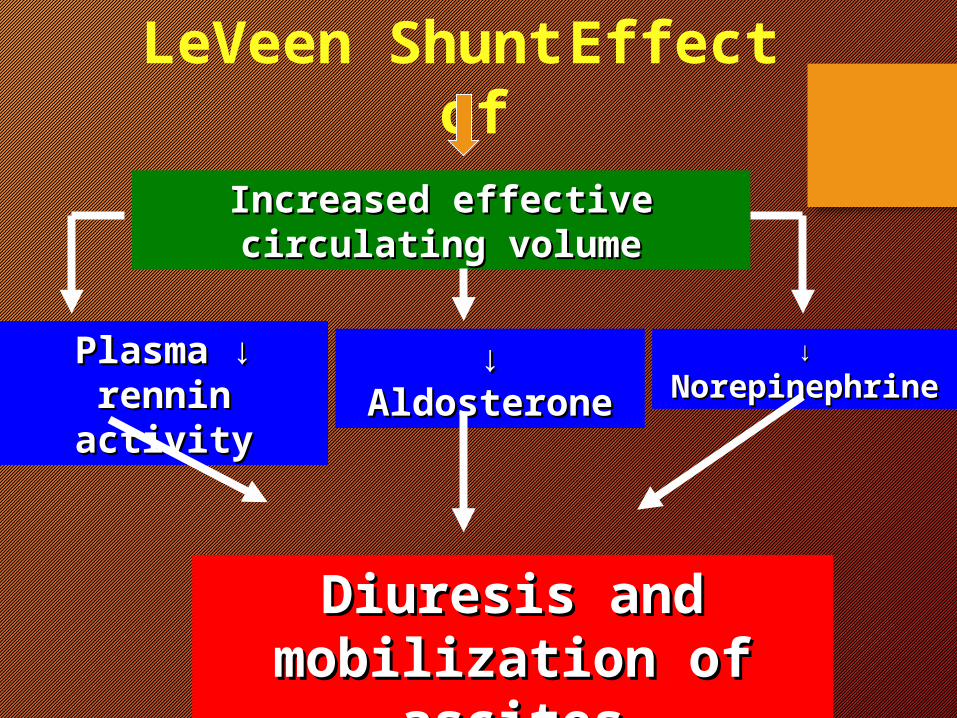
LeVeen Shunt Effect of
Increased effective circulating Increased effective circulating volumevolume
↓ ↓Plasma rennin Plasma rennin activityactivity
↓↓ AldosteroneAldosterone ↓↓ NorepinephrineNorepinephrine
Diuresis and Diuresis and mobilization of ascitesmobilization of ascites

Sponataneous Bacterial Peritonitis
(SBP)
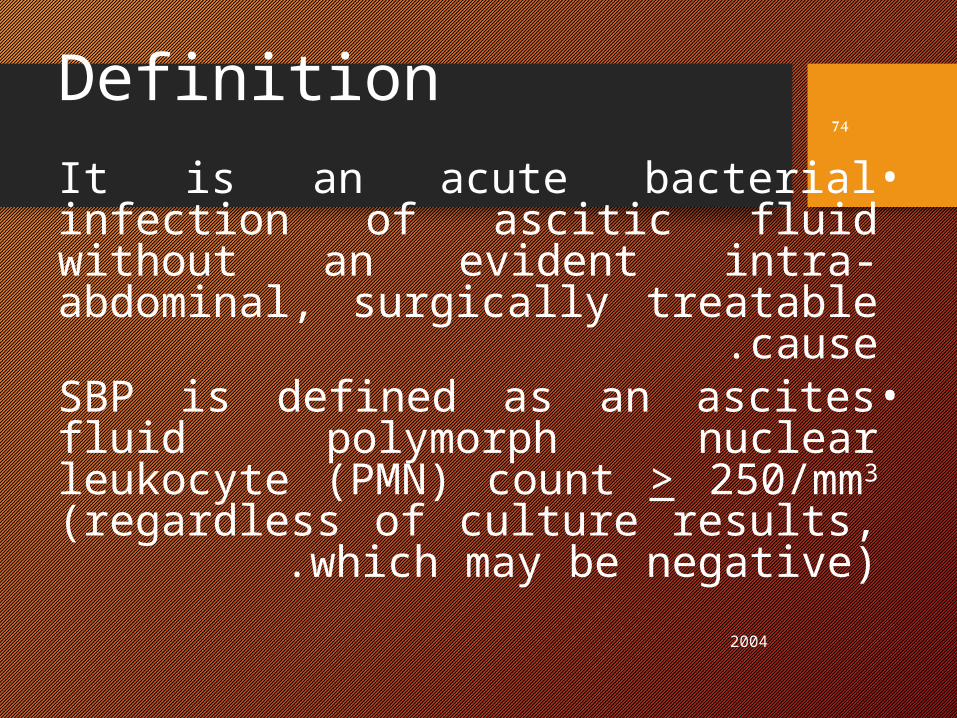
Definition•It is an acute bacterial infection
of ascitic fluid without an evident intra-abdominal, surgically
treatable cause .•SBP is defined as an ascites fluid
polymorph nuclear leukocyte (PMN) count > 250/mm3
(regardless of culture results, which may be negative) .
2004
74
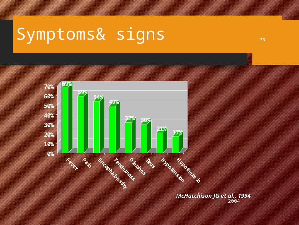
Symptoms& signs
69%59%
54%49%
32% 30%21% 17%
0%10%20%30%40%50%60%70%
FeverPain
Encephalopathy
TendernessDiarrheaIleus
HypotensionHypothermia
2004
75
McHutchison JG et al., 1994McHutchison JG et al., 1994
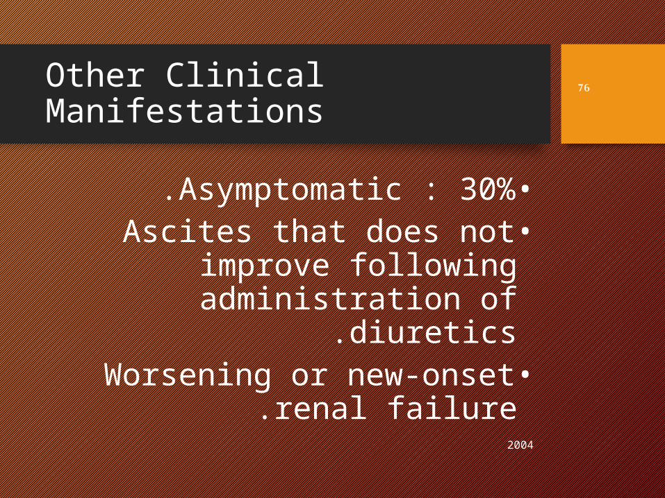
Other Clinical Manifestations
•Asymptomatic : 30%.•Ascites that does not
improve following administration of diuretics.
•Worsening or new-onset renal failure.
2004
76
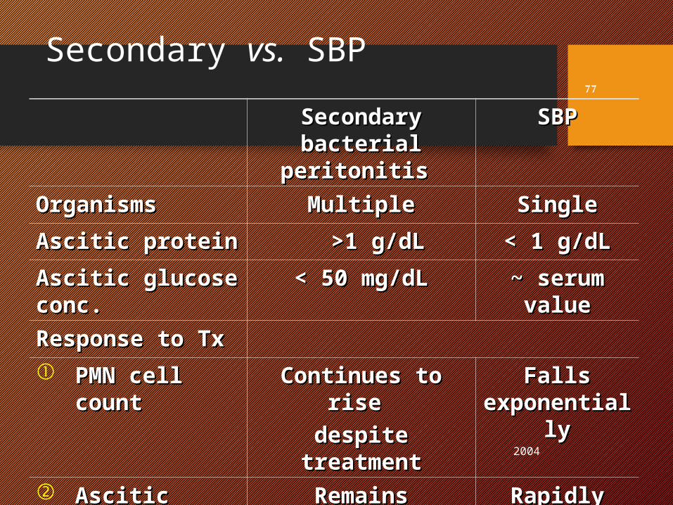
Secondary vs. SBP
Secondary Secondary bacterial bacterial
peritonitis peritonitis
SBPSBP
OrganismsOrganisms MultipleMultiple SingleSingleAscitic protein Ascitic protein >1 g/dL>1 g/dL < 1 g/dL< 1 g/dLAscitic glucose Ascitic glucose conc.conc.
< 50 mg/dL< 50 mg/dL ~ serum ~ serum valuevalue
Response to TxResponse to Tx PMN cell PMN cell
countcountContinues to Continues to
rise rise despite despite
treatmenttreatment
Falls Falls exponentialexponential
lyly
Ascitic Ascitic cultureculture
Remains positiveRemains positive Rapidly Rapidly becomes becomes sterilesterile
2004
77
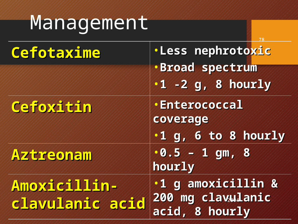
Management Cefotaxime Cefotaxime •Less nephrotoxicLess nephrotoxic
•Broad spectrumBroad spectrum•1 -2 g, 8 hourly1 -2 g, 8 hourly
CefoxitinCefoxitin •Enterococcal Enterococcal coveragecoverage•1 g, 6 to 8 hourly1 g, 6 to 8 hourly
AztreonamAztreonam •0.5 0.5 –– 1 gm, 8 hourly 1 gm, 8 hourly
Amoxicillin-Amoxicillin-clavulanic acid clavulanic acid
•1 g amoxicillin & 1 g amoxicillin & 200 mg clavulanic 200 mg clavulanic acid, 8 hourly acid, 8 hourly 2004
78
Hepatorenal Syndrome
HRS2004
79
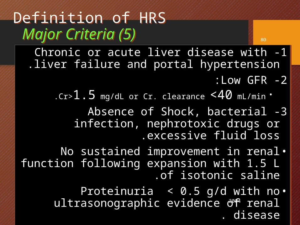
Definition of HRS Major Criteria (5)
1 -Chronic or acute liver disease with liver failure and portal hypertension.
2 -Low GFR :•Cr>1.5 mg/dL or Cr. clearance <40 mL/min.
3 -Absence of Shock, bacterial infection, nephrotoxic drugs or excessive fluid loss.
•No sustained improvement in renal function following expansion with 1.5 L of isotonic
saline.•Proteinuria < 0.5 g/d with no
ultrasonographic evidence of renal disease. 2004
80
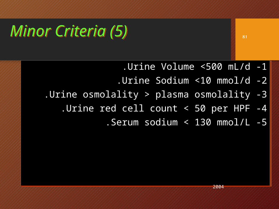
Minor Criteria (5)
1 -Urine Volume <500 mL/d.2 -Urine Sodium <10 mmol/d.3 -Urine osmolality > plasma osmolality.4 -Urine red cell count < 50 per HPF.5 -Serum sodium < 130 mmol/L.
2004
81
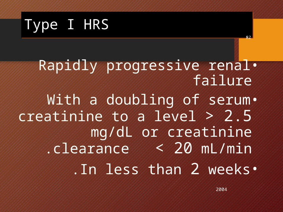
Type I HRS
•Rapidly progressive renal failure•With a doubling of serum
creatinine to a level > 2.5 mg/dL or creatinine clearance
< 20 mL/min.•In less than 2 weeks.
2004
82
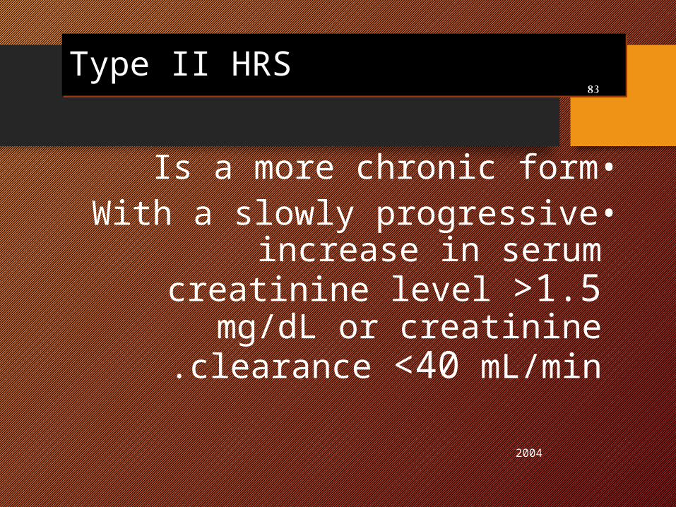
Type II HRS
•Is a more chronic form•With a slowly progressive
increase in serum creatinine level >1.5 mg/dL or creatinine
clearance <40 mL/min.
2004
83
2004
84
Management Management of of
HRSHRS
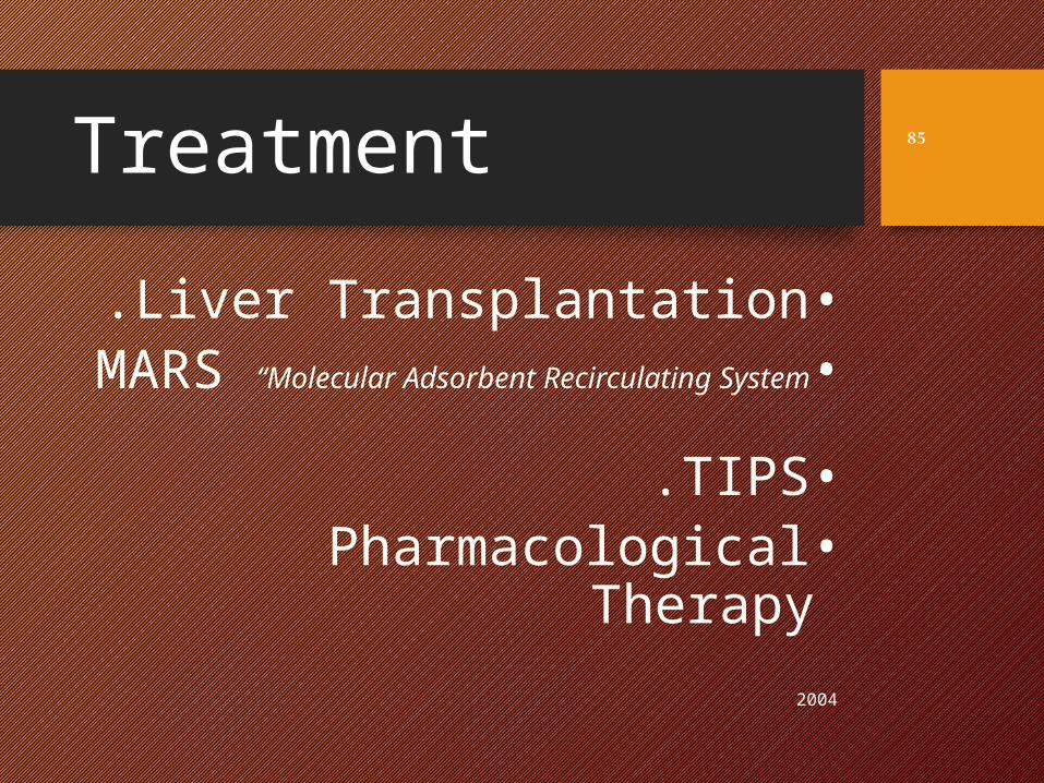
Treatment•Liver Transplantation.•MARS “Molecular Adsorbent
Recirculating System ”
•TIPS.•Pharmacological
Therapy2004
85

2004
86