Embed Size (px)
Citation preview

APOPTOSISDR.AYESHA FARHEEN

Objectives
Definition of Apoptosis
ProcessDifferentiation from Necrosis
PathwaysApplied Aspect
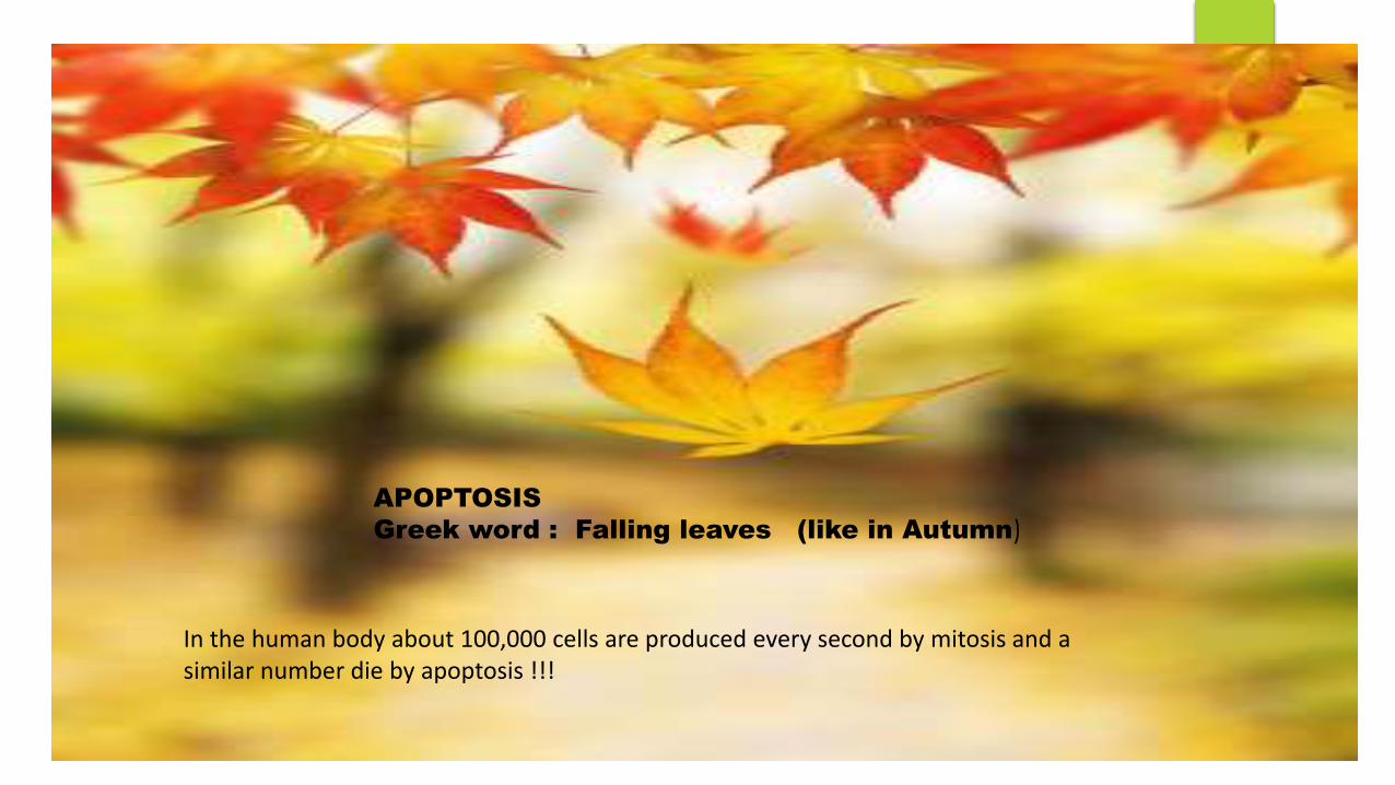
APOPTOSIS
Greek word : Falling leaves (like in Autumn)
In the human body about 100,000 cells are produced every second by mitosis and a similar number die by apoptosis !!!

History
Apoptotic principle was first described by KARL VOGT in 1842
“APOPTOSIS” term was coined by JAMES CORMACK
2002 NOBEL PRIZE IN MEDICINE was awarded to SYDNEY
BRENNER,HORVITZ and JOHN.E.SULSTON for their work in identifying genes
that control Apoptosis.
John E Sulton won Nobel Prize in 2002
for his pioneering research in Apoptosis
PROGRAMMED CELL DEATH
CELL SUICIDE
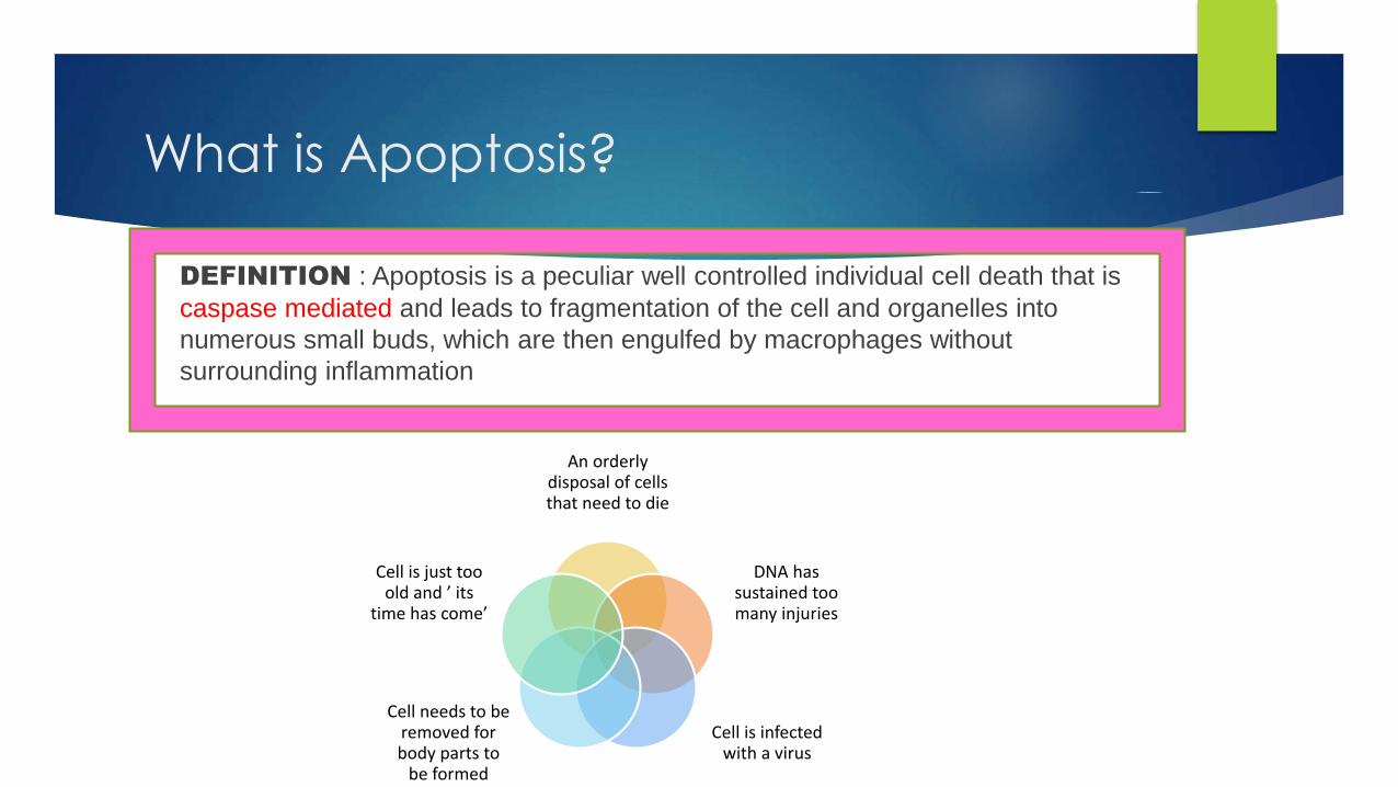
What is Apoptosis?
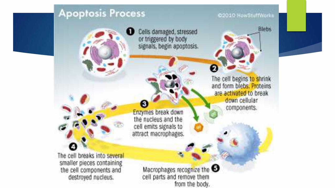
DEFINITION : Apoptosis is a peculiar well controlled individual cell death that is
caspase mediated and leads to fragmentation of the cell and organelles into
numerous small buds, which are then engulfed by macrophages without
surrounding inflammation
An orderly disposal of cells that need to die
DNA has sustained too many injuries
Cell is infected with a virus
Cell needs to be removed for body parts to
be formed
Cell is just too old and ’ its
time has come’
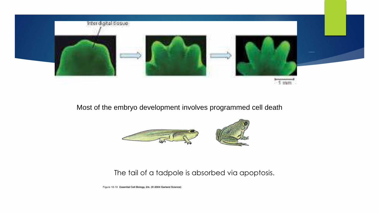
The tail of a tadpole is absorbed via apoptosis.
Most of the embryo development involves programmed cell death

Importance of Apoptosis
1) Crucial for embryonic development
-Errors in Apoptosis can lead to Birth Defects
2) Important for maintaining homeostasis
- Cell death is balanced with mitosis to regulate cell number.
3) Improper regulation contributes to human disease
- Neurodegenerative diseases
Parkinson’s
Alzheimer’s
- Cancer
- Autoimmune diseases e.g. (diabetes type I)
- Viral diseases
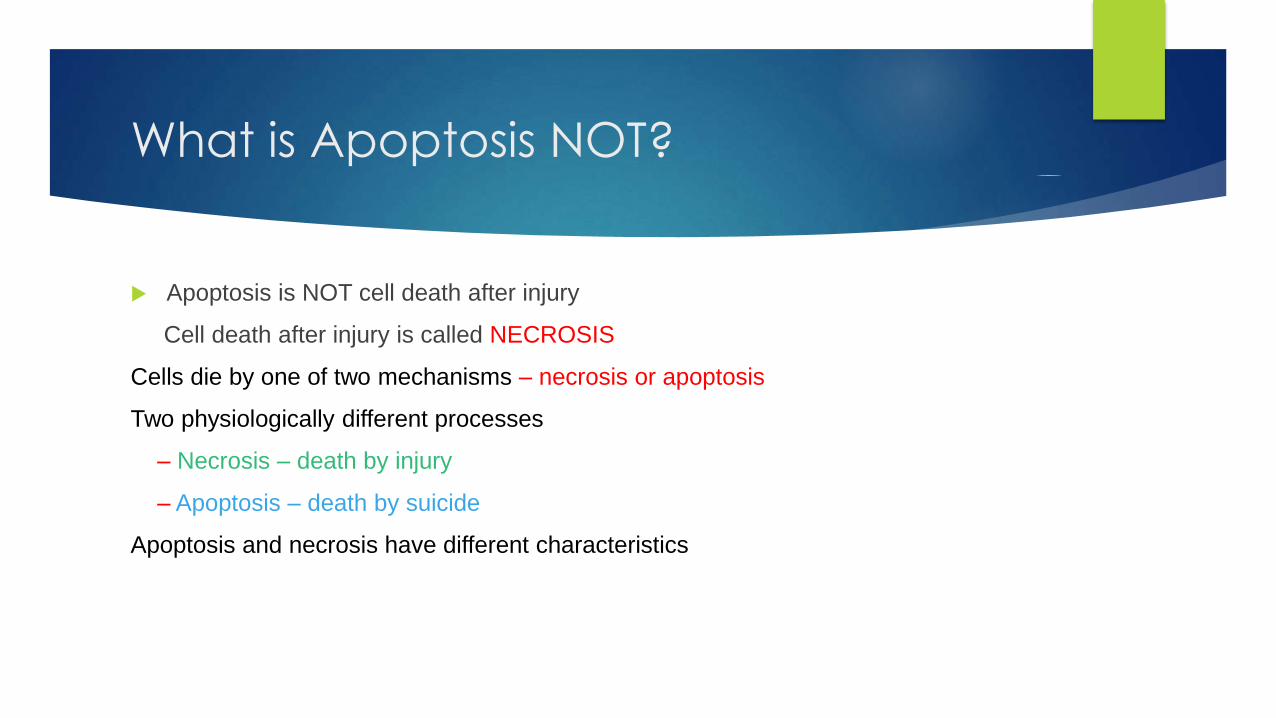
What is Apoptosis NOT?
Apoptosis is NOT cell death after injury
Cell death after injury is called NECROSIS
Cells die by one of two mechanisms – necrosis or apoptosis
Two physiologically different processes
– Necrosis – death by injury
– Apoptosis – death by suicide
Apoptosis and necrosis have different characteristics


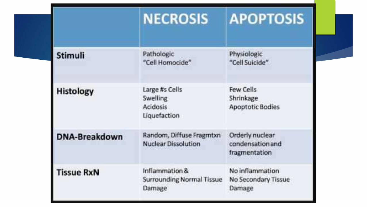
How Apoptosis Differs from Necrosis?
1. Apoptosis is intrinsically controlled, necrosis is not
2. Apoptosis is more rapid (12-24 hours) than necrosis
3. Apoptosis is induced by endogenous or exogenous stimuli, necrosis is always induced by exogenous harms
4. Apoptosis is limited to single or few cells at a time, and occurs among healthy cell population, necrosis is usually more extensive & occurs in tissue exposed to injuries
5. Cell cytoplasm shrinks in apoptosis and swells in necrosis.
6. Nucleosomes of apoptotic cells are 180 bp fragments, contrary to the irregular ones in necrosis
7. Apoptosis has no inflammation, while necrosis leads to liberation of pro-inflammatory mediators
8. Apoptosis has no systemic manifestations contrary to most inflammations
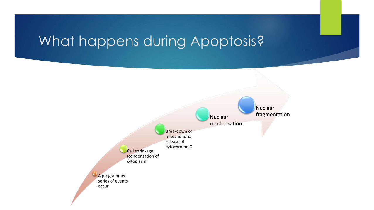
What happens during Apoptosis?
A programmed series of events occur
Cell shrinkage (condensation of cytoplasm)
Breakdown of mitochondria; release of cytochrome C
Nuclear condensation
Nuclear fragmentation

Cellular changes associated with apoptosis

Biochemical features of Apoptosis
• By activation of caspases
• Caspases activate DNAses
Protein Cleavage
• Cleavage into oligonucleosomes
• By Ca2+-and Mg2+-dependent endonucleases
DNA Breakdown
• Phosphatidylserine
• Thrombospondin
Phagocytic Recognition

Mechanisms of Apoptosis

Mechanisms of Apoptosis
Active cysteine residue in the catalytic site
Specificity in cleavage after an Asp residue
Synthesized as inactive zymogens (PROCASPASES)


Digestion of DNA starts after
2 hrs
3&4 hrs after initiation of apoptosis DNA is almost all degraded
DNA is fragmented with restriction endonucleases
Apoptosis induces 180 bp Laddering of DNA
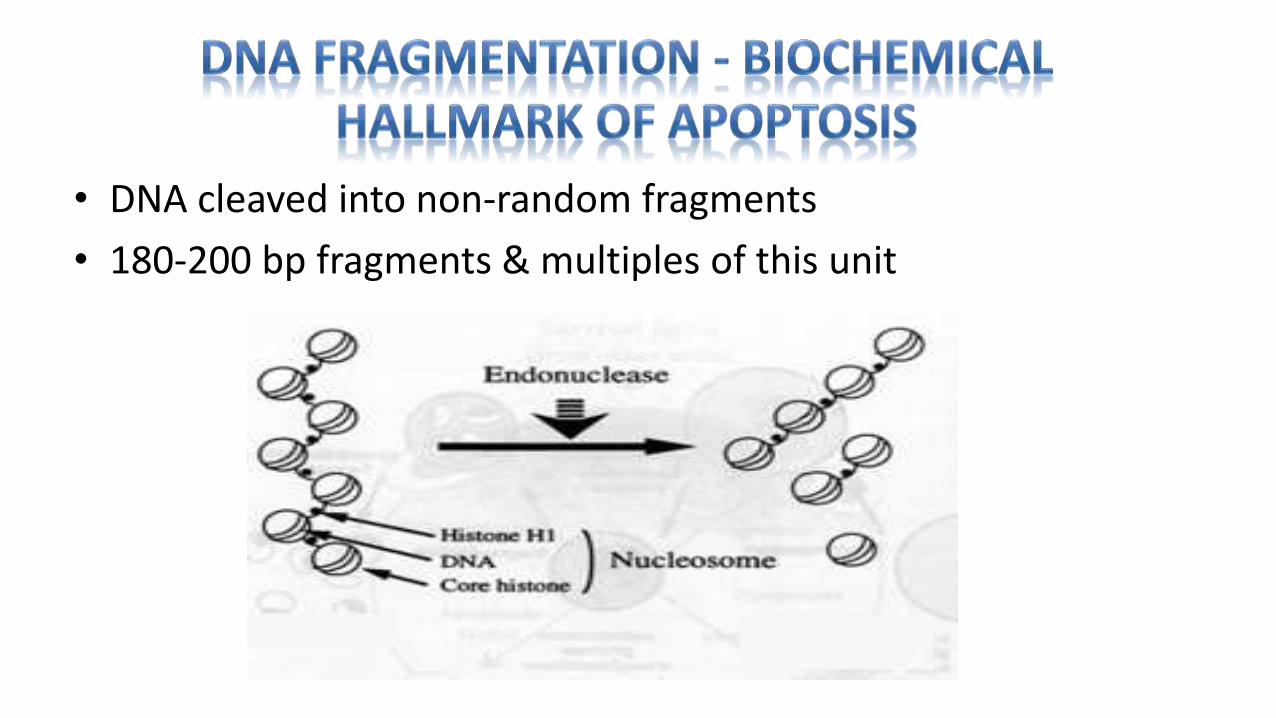
• DNA cleaved into non-random fragments
• 180-200 bp fragments & multiples of this unit

Mechanism
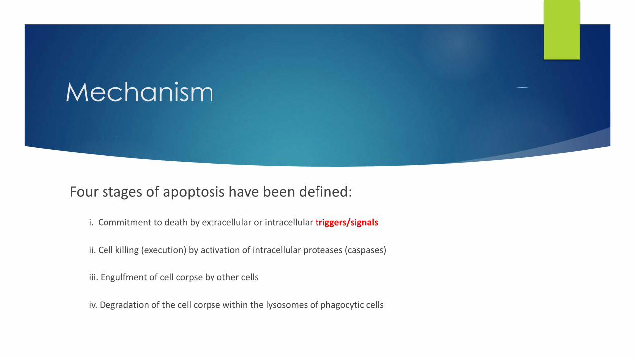
Four stages of apoptosis have been defined:
i. Commitment to death by extracellular or intracellular triggers/signals
ii. Cell killing (execution) by activation of intracellular proteases (caspases)
iii. Engulfment of cell corpse by other cells
iv. Degradation of the cell corpse within the lysosomes of phagocytic cells

DAMAGE Physiological death signals
DEATH SIGNAL
PROAPOPTOTICPROTEINS
ANTIAPOPTOTICPROTEINS
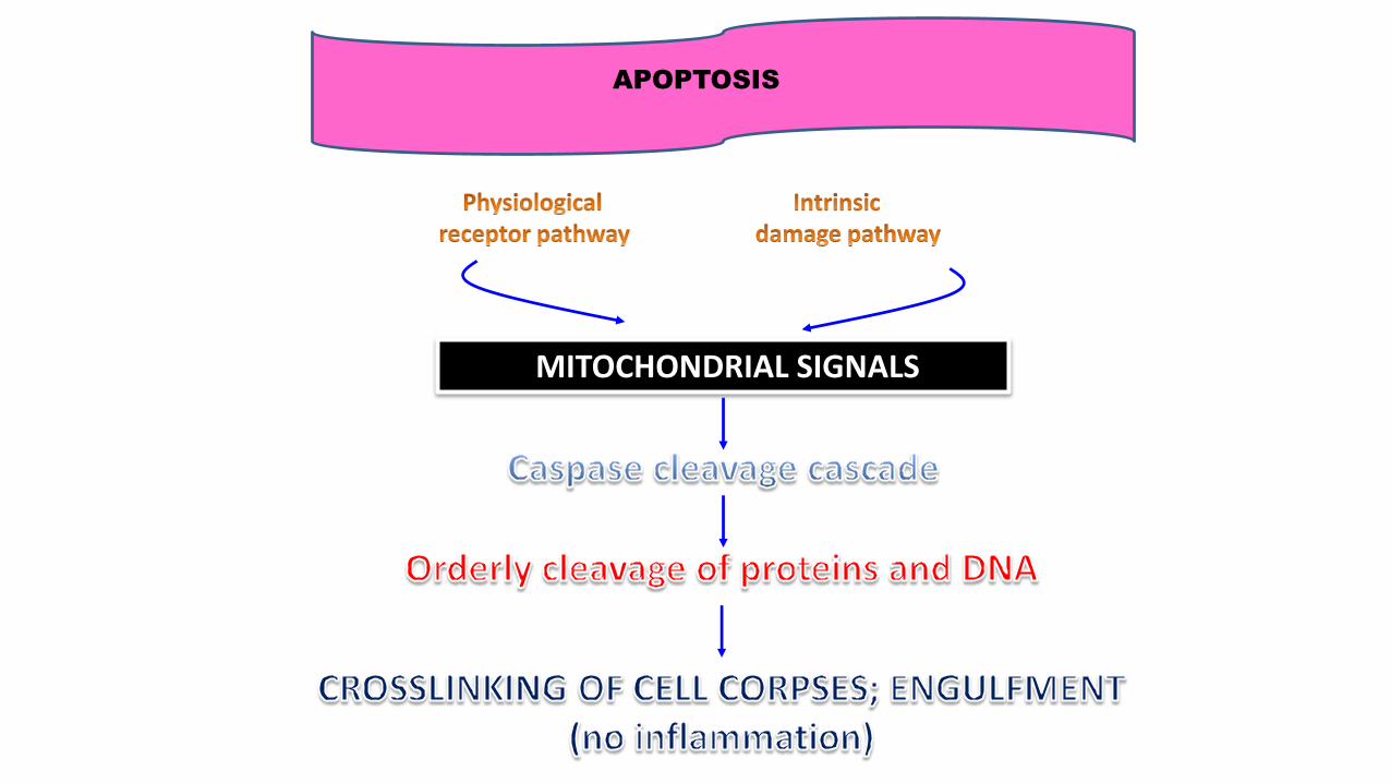
MITOCHONDRIAL SIGNALS
APOPTOSIS
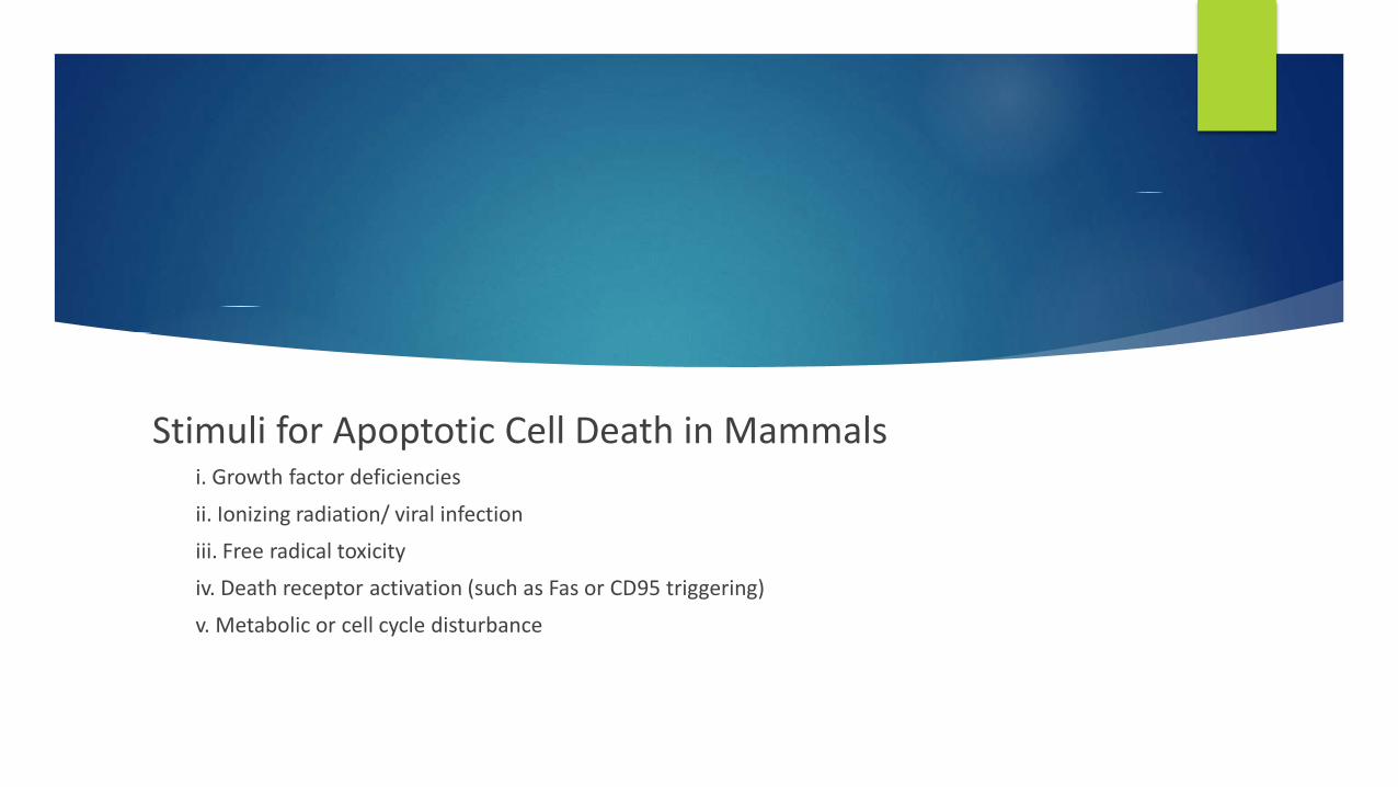
Stimuli for Apoptotic Cell Death in Mammals i. Growth factor deficiencies
ii. Ionizing radiation/ viral infection
iii. Free radical toxicity
iv. Death receptor activation (such as Fas or CD95 triggering)
v. Metabolic or cell cycle disturbance

Stimuli for Apoptotic Cell Death

Death Factors
Definition: Cytokines that activate an apoptosis program by binding to their specific receptor. Typical examples of death factors are:
1. Fas ligand,
2. TNF (tumor necrosis factor) and
3. TRAIL (TNF-related apoptosis-inducing ligand).
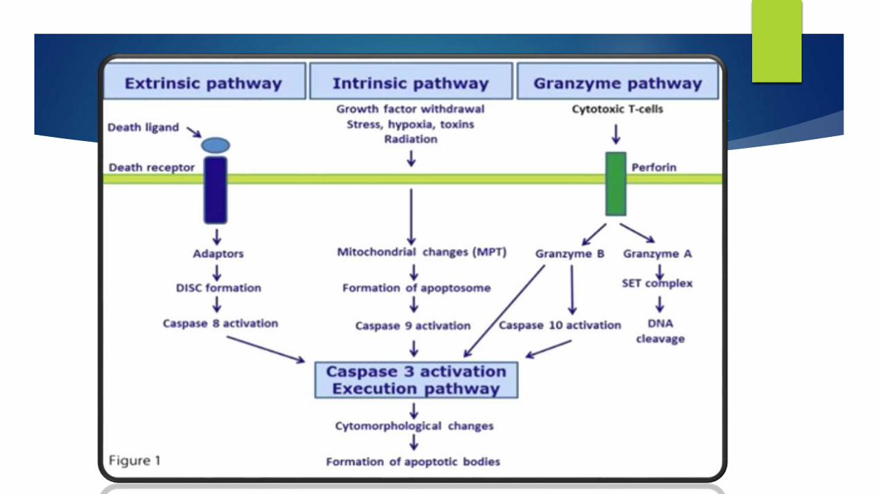
- Apoptosis can also be induced by cytotoxic T-lymphocytes using the enzyme granzyme.

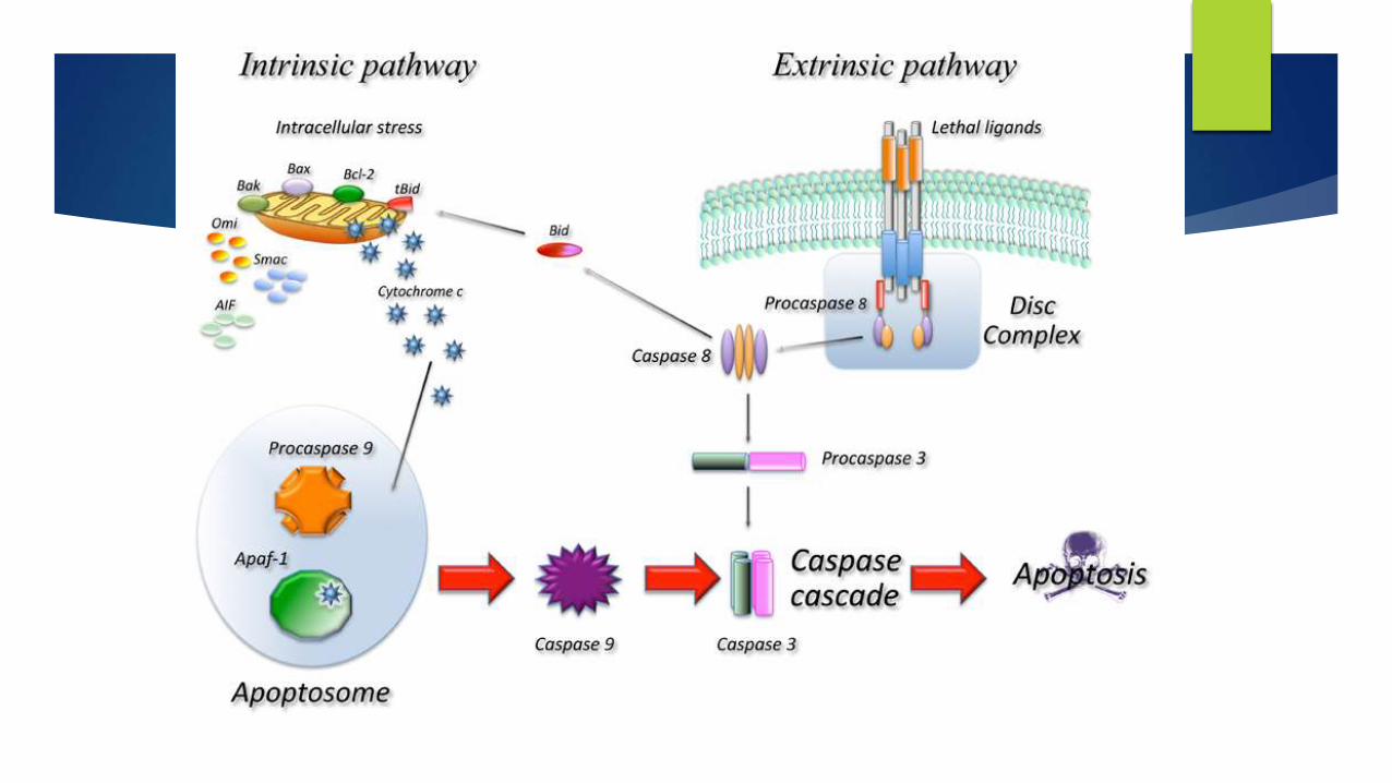
Apoptosis
Extrinsic pathway
Intrinsic pathway
Granzyme pathway
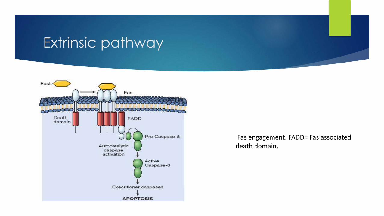
Extrinsic pathway
Fas engagement. FADD= Fas associated death domain.

Extrinsic pathway


Intrinsic Pathway
AIF= Apoptosis inhibitory factor; IAPs= Inhibitors of apoptosis proteins; Apaf-1= apoptosis protease activating factor
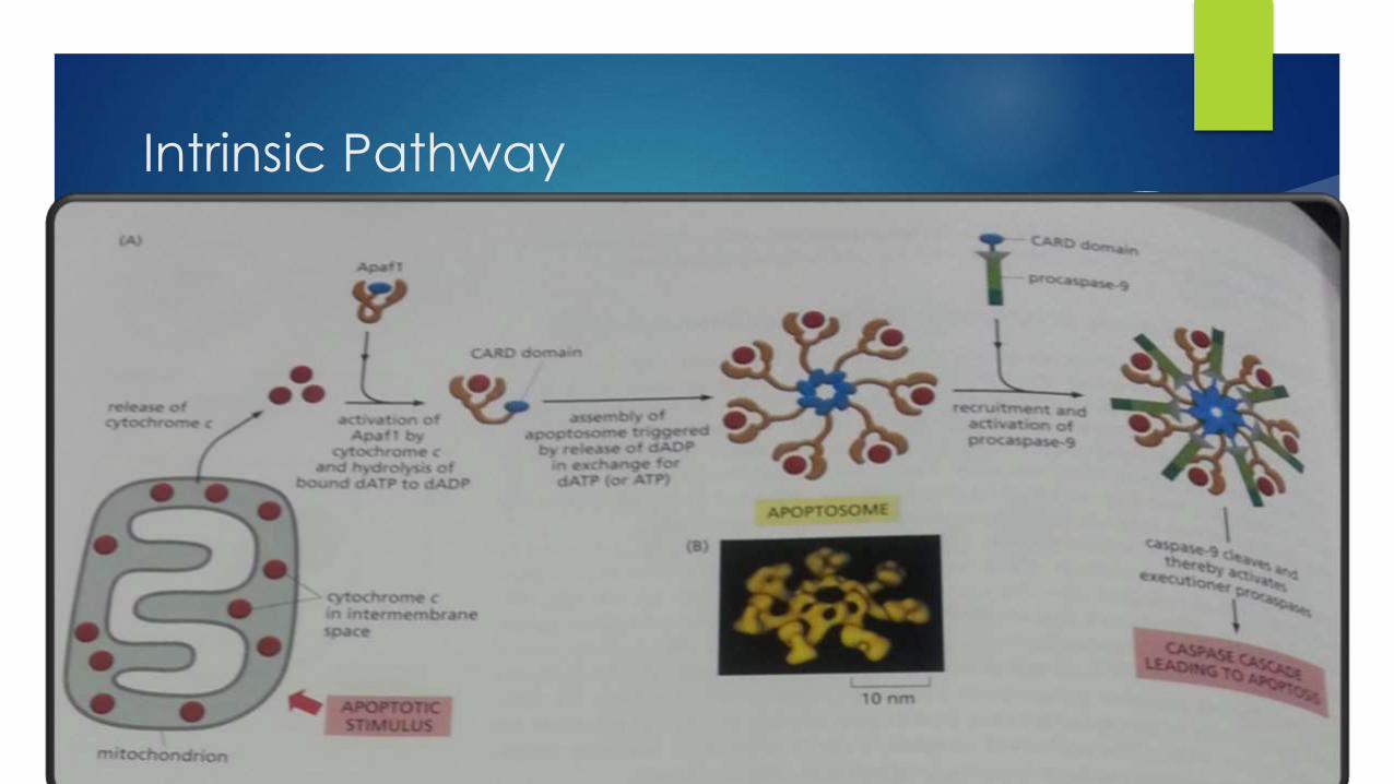
Intrinsic Pathway

Bcl2 was the first apoptosis-related gene ,recognized to play a role tumor genesis
BCL-2 is a human proto-oncogene located on chromosome 18.
Its product is an integral membrane protein (called Bcl-2) located in the membranes of the endoplasmic reticulum , nuclear envelope, and in the outer membrane of mitochondria.
The gene was discovered as the translocated locus in a B-cell leukemia (hence the name). This translocation is also found in some B-cell lymphomas
Apoptosis and Cancer


Apoptosis and Cancer
• Apoptosis does not occur in Cancer
• Cancerous cells trick and skip Apoptosis in number of ways
Inactivation of p53 [shooting the guard]
Produce Bcl-2 or a protein which mimics Bcl-2
Inhibits expression of Apaf-1
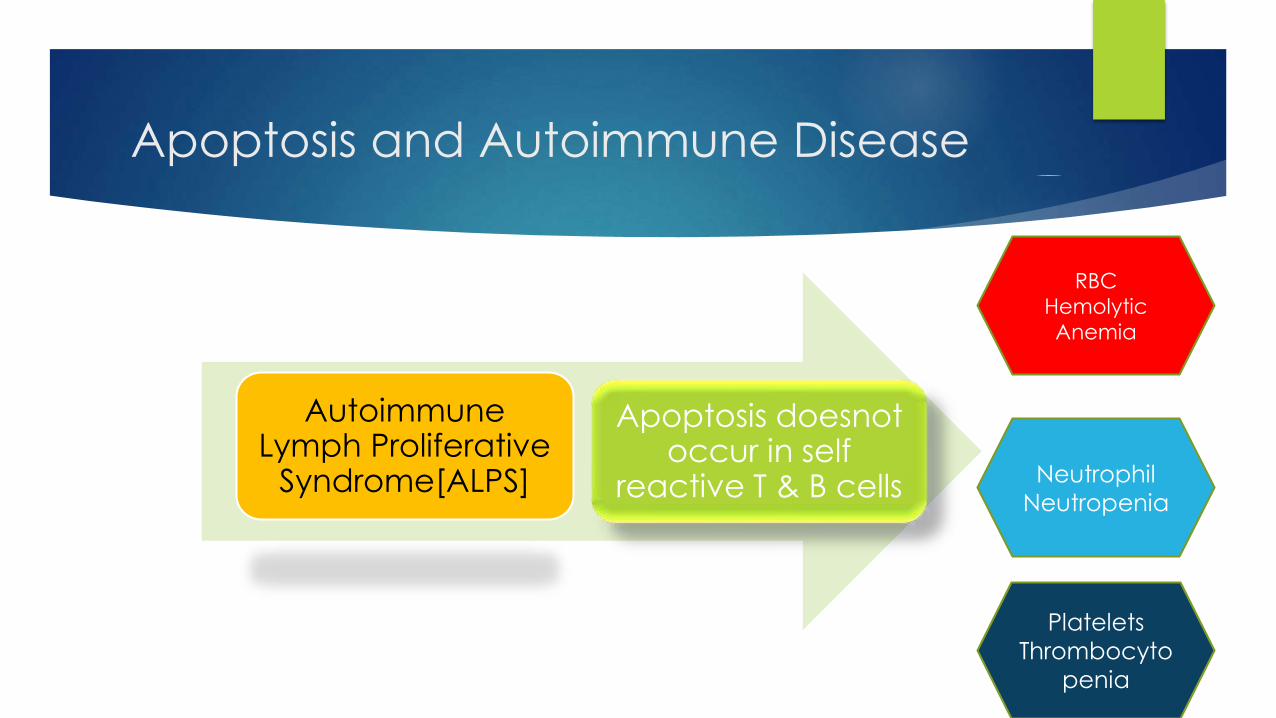
Apoptosis and Autoimmune Disease
Autoimmune Lymph Proliferative
Syndrome[ALPS]
Apoptosis doesnot occur in self
reactive T & B cells
RBC
Hemolytic
Anemia
Neutrophil
Neutropenia
Platelets
Thrombocyto
penia

Apoptosis and HIV
Infected CD4
cell induces
Apoptosis in
surrounding
cells
• Deactivated
Bcl-2
• Decreases
CD4
Glycoprotein
markers on
innocent T
cells ,getting
them killed
Infected CD4 cell
avoids Apoptosis in
itself
Decreases
Phosphatdylserine
marker for itself
allowing longer
survival.
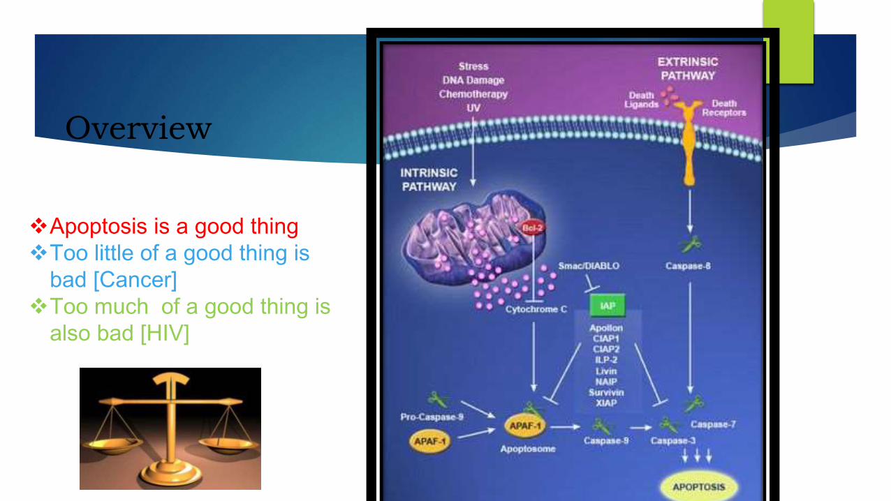
Overview
Apoptosis is a good thing
Too little of a good thing is
bad [Cancer]
Too much of a good thing is
also bad [HIV]

Sources
Textbook of Medical Physiology –Guyton & Hall [12th edition]
Review of Medical Physiology-William F Ganong [24th edition]
Internet Sources :
• www.ncbi.nlm.nih.gov/books/NBK26873
• http://en.wikipedia.org/wiki/Apoptosis
Articles :
Apoptosis- Molecular mechanisms and Pathogenicity
http://link.springer.com/article/10.1023/A:1009616228304#pg2
http://www.excli.de/vol8/Rastogi_08_2009/Rastogi_030809_proof.pdf

”
“Queries?

THANK YOU