Embed Size (px)
Citation preview
EWMA Document:Antimicrobials and Non-healing WoundsEvidence, controversies and suggestions
A EWMA Document

S 2 J O U R N A L O F WO U N D C A R E VO L 2 2 . N O 5 . E W M A D O C U M E N T 2 0 1 3
© EWMA 2013
All rights reserved. No reproduction, transmission or copying of this publication is allowed without written
permission. No part of this publication may be reproduced, stored in a retrieval system, or transmitted in any form or
by any means, mechanical, electronic, photocopying, recording, or otherwise, without the prior written permission
of the European Wound Management Association (EWMA) or in accordance with the relevant copyright legislation.
Although the editor, MA Healthcare Ltd. and EWMA have taken great care to ensure accuracy, neither
MA Healthcare Ltd. nor EWMA will be liable for any errors of omission or inaccuracies in this publication.
Published on behalf of EWMA by MA Healthcare Ltd.
Publisher: Anthony Kerr
Editor: Daniel Shanahan
Designer: Alison Cutler
Published by: MA Healthcare Ltd, St Jude’s Church, Dulwich Road, London, SE24 0PB, UK
Tel: +44 (0)20 7738 5454 Email: [email protected] Web: www.markallengroup.com
F. Gottrup,1 (Editor) MD, DMSci, Professor of Surgery, Chair of Antimicrobial Document author group;
J. Apelqvist,2 (Co-editor) MD, PhD, Senior Consultant, Associate Professor;
T. Bjansholt,3 MSc, PhD, Associate Professor;
R. Cooper,4 BSc, PhD, PGCE, CBIOL, MSB, FRSA, Professor of Microbiology;
Z. Moore,5 PhD, MSc, FFNMRCSI, PG Dip, Dip Management, RGN, Lecturer in Wound Healing & Tissue Repair;
E.J.G. Peters,6 M.D., PhD, Internist- Infectious Diseases Specialist;
S. Probst,7 DClinPrac, RN, Lecturer;
1 Bispebjerg University Hospital, Copenhagen, Denmark;
2 Skåne University Hospital, Malmoe, Sweden;
3 University of Copenhagen and Copenhagen University Hospital, Copenhagen, Denmark;
4 Cardiff Metropolitan University, Cardiff, Wales, UK;
5 Royal College of Surgeons in Ireland, Dublin, Ireland;
6 VU University Medical Center, Amsterdam, the Netherlands;
7 Zurich University of Applied Sciences, Winterthur, Switzerland.
Editorial support and coordination: EWMA Secretatiat
Email [email protected] Web:www.Ewma.org
The document is supported by an unrestricted grant from B. Braun Medical, BSN Medical, ConvaTec, PolyMem, Flen
Pharma, Lohmann & Rauscher, Mölnlycke Health Care, Schülke & Mayr, Smith & Nephew and sorbion.
Eucomed Advanced Wound Care Sector Group provided initial funding for the document.
This article has not undergone double-blind peer review.
This article should be referenced as: Gottrup, F., Apelqvist, J., Bjansholt, T. et al. EWMA Document: Antimicrobials and
Non-healing Wounds—Evidence, Controversies and Suggestions. J Wound Care. 2013; 22 (5 Suppl.): S1–S92.
J O U R N A L O F WO U N D C A R E VO L 2 2 . N O 5 . E W M A D O C U M E N T 2 0 1 3 S 3
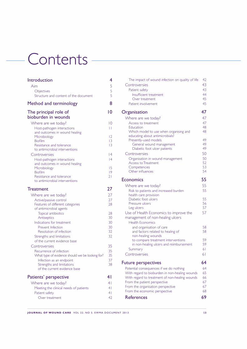
ContentsIntroduction 4
Aim 5Objectives 5 Structure and content of the document 5
Method and terminology 8
The principal role of 10 bioburden in wounds
Where are we today? 10Host-pathogen interactions 11 and outcomes in wound healing Microbiology 12 Bio!lm 13 Resistance and tolerance 13 to antimicrobial interventions
Controversies 14Host-pathogen interactions 14 and outcomes in wound healing Microbiology 15 Bio!lm 19 Resistance and tolerance 21 to antimicrobial interventions
Treatment 27Where are we today? 27
Active/passive control 27 Features of different categories 28 of antimicrobial agents
Topical antibiotics 28 Antiseptics 30
Indications for treatment 30Prevent Infection 30 Resolution of infection 32
Strengths and limitations 32 of the current evidence base
Controversies 35Recurrence of infection 35 What type of evidence should we be looking for? 35
Infection as an endpoint 37 Strengths and limitations 38 of the current evidence base
Patients’ perspective 41Where are we today? 41
Meeting the clinical needs of patients 41Patient safety 41
Over treatment 42
The impact of wound infection on quality of life 42 Controversies 43
Patient safety 43Insuf!cient treatment 44 Over treatment 45
Patient involvement 45
Organisation 47Where are we today? 47
Access to treatment 47 Education 48 Which model to use when organising and 48 educating about antimicrobials? Presently-used models 49
General wound management 49 Diabetic foot ulcer patients 49
Controversies 50Organisation in wound management 50 Access to Treatment 52 Competencies 53 Other in"uences 54
Economics 55Where are we today? 55
Risk to patients and increased burden 55 health care provision Diabetic foot ulcers 55 Pressure ulcers 56 Leg ulcers 57
Use of Health Economics to improve the 57 management of non-healing ulcers
Health Economicsand organisation of care 58and factors related to healing of 58 non-healing woundsto compare treatment interventions 59in non-healing ulcers and reimbursement 59
Summary 61Controversies 61
Future perspectives 64Potential consequences if we do nothing 64With regard to bioburden in non-healing wounds 65With regard to treatment of non-healing wounds 66From the patient perspective 67From the organisation perspective 67From the economic perspective 68
References 69
S 4 J O U R N A L O F WO U N D C A R E VO L 2 2 . N O 5 . E W M A D O C U M E N T 2 0 1 3
Non-healing wounds are a signi!cant problem for health-care systems worldwide. In the industrialised world,
almost 1–1.5% of the population will have a problem wound at any one time. Furthermore, wound management is expensive; in Europe, the average cost per episode is €6650 for leg ulcers and €10 000 for foot ulcers, and wound management accounts for 2–4% of health-care budgets. These !gures are expected to rise along with an increased elderly and diabetic population.1–4
Infection is one of the most frequent complications of non-healing wounds. It can jeopardise the progression towards healing, result in longer treatment times and increase the resource use. In the worst cases, it can result in a major amputation or a life-threatening condition. Wounds are disposed to infection, as the exposure of subcutaneous tissue following a loss of skin integrity provides a moist, warm, and nutrient-rich environment, which is conducive to microbial colonisation and proliferation. Consequently, use of antimicrobial agents is important in wound management.
Inappropriate use of antimicrobials (especially antibiotics) creates an environment for the selection of resistance against the currently available antimicrobial products, with the potential consequence of signi!cantly jeopardising patients’ health status. The development of so called ‘superbugs’ is foreseeable and is the background for increased political involvement.5–7
In 2009, the EU member states adopted council conclusions concerning innovative incentives for effective antibiotics. This is one of the single most powerful, concerted political stances on antibiotic resistance ever. Here it is recognised that the spread of antibiotic resistance is a major threat to public health security worldwide and requires action at all levels. Hence, they call upon the member states to develop and implement strategies to ensure awareness among the public and health professionals of the threat of antibiotic resistance and of the measures available to counter the problem.
This has been followed by several pan-European initiatives, such as the conference ‘Combating Antimicrobial resistance—Time for Joint Action’ in March 2012,7 in which the European Wound Management Organisation (EWMA) participated. The conference conclusions were that there was a substantial gap in the knowledge in this area.
Furthermore, the European Commission has followed this by a report on implementation of the council recommendations on patient safety, in which they conclude that ‘even if many member states have taken a variety of actions, there is still considerable room for improvement’.8,9
Resistance to antibiotics results in a considerable decrease in the possibility of effectively treating infections, and increases the risk of complications and death.10 In the European Union (EU) alone, it is estimated that 2 million patients acquire nosocomial (hospital-acquired) infections each
Introduction

J O U R N A L O F WO U N D C A R E VO L 2 2 . N O 5 . E W M A D O C U M E N T 2 0 1 3 S 5
year,11 of which more than half are drug-resistant.12 Infections based on resistant bacteria are associated with up to two-fold increase in mortality compared with infection with susceptible microbes.13
Coupled with insuf!cient investment in the development of new antibiotic treatments, the issue of drug-resistant bacteria is becoming a pressing public-health concern. In 2007, the European Antimicrobial Resistance Surveillance System (EARSS) reported that Staphylococcus aureus had become resistant to the antibiotic meticillin (MRSA), indicating that beta-lactam antibiotics are not suitable for empiric treatment of wound infections in Europe.14 To date, there is no collection of data for bacterial resistance in wounds.
Despite a tremendous amount of literature covering the effects and use of antimicrobials, and the development of resistance in the wound area, there is a lack of a consistent and reproducible approach to de!ning, evaluating and measuring the appropriate and adequate use of antimicrobials locally/topically in wound management, from a clinical and industry perspective.
This lack of information can best be illustrated by the fact that, despite the extensive use of antimicrobials in wounds, their use remains controversial for wound management. These controversies have never been discussed and evaluated in detail, which is a major reason for wound infection persisting as one of the most serious in$uencing factors for the existence of non-healing wounds.
This document describes these controversies and hopes to raise interest in how to solve these problems for the future use of antimicrobials. For this reason, EWMA established the group, which produced this document.
By discussion and clari!cation, we hope to contribute to a reduction in the burden of care, in an ef!cient and cost-effective way.
StatementThere are a large number of antimicrobial wound care products available, but we need to be better prepared for selecting the right product for the right patient, for the right wound, at the right time. There is confusion among policy makers, patients, clinicians and researchers as to the controversies for the use of antimicrobials in wounds. Most discussions and recommendations do not differentiate between different types of antimicrobials, especially with regard to antibiotics and antiseptics.5
‘This document describes the
controversies surrounding use
of antimicrobials in wound
management, and hopes to raise
interest in how to solve these
problems for the future use
of antimicrobials
’
S 6 J O U R N A L O F WO U N D C A R E VO L 2 2 . N O 5 . E W M A D O C U M E N T 2 0 1 3
Aim The intention of this document is to focus on the responsible design and use of antimicrobial strategies in wounds that fail to progress through an orderly and timely sequence of repair. In this document, these types of wounds are de!ned as ‘non-healing’.15 The focus is not on a speci!c type of non-healing wound, but to provide more general recommendations for these types of wounds.
Animal and cellular models, acute wound (surgical/trauma wounds) and burns are excluded from this document. Systemic infections, debridement as a bioburden control and other types of wound management strategies will not be covered in detail.
The document structure is inspired from the different elements that are normally included in the health technology assessment (HTA) approach.
It is not a traditional position document that discusses different treatment strategies, when to use which product, or an assessment of one product over another.
The overall aim of this document is to highlight current knowledge regarding use of antimicrobials, particularly in non-healing wounds, to discuss what still is controversial and give suggestions for future actions.
ObjectivesThese goals will be achieved by the following:
1 Producing an update of each topic mentioned, including statements on which items have been shown to be based on evidence at the highest level.
2 Uncovering controversies and issues related to use of antimicrobials in wound management; describe possible solutions and the pros and cons of each
3 Summarising the information presented and offer perspectives for further work.
The intentions of the document are to present a platform of viewpoints from which we can build messages for the different stakeholders, including patients, health professionals, policy makers, politicians, industry and hospital administrators.
Structure and content of the documentThe document includes the different aspects of health-care perspectives surrounding the central theme of antimicrobials in wounds. Each chapter begins with an introduction to the current knowledge and status of the speci!c topic; we have called this ‘where are we today.’ This section also covers an assessment of the current literature and what evidence there is for the existing consensus.
The method for the evidence assessment builds upon EWMAs previous work with outcomes15 and
‘The intention of this document
is to focus on the responsible
design and use of antimicrobial
strategies in wounds that fail to
progress through an orderly
and timely sequence of repair
’
J O U R N A L O F WO U N D C A R E VO L 2 2 . N O 5 . E W M A D O C U M E N T 2 0 1 3 S 7
is the foundation for the recommendations made in this document.
The second section of each chapter will address the relevant controversies. Each controversy has its own subtitle, which is stated below the the author group’s statement. Following the statement, the controversy is discussed and a short conclusion is given.
The present document tries to uncover the controversies with regard to the use of antimicrobials in wound care, with a focus on non-healing wounds. Most research with regard to infection and wound healing is related to acute wounds and a minor part is related to non-healing wounds; however, some evidence from acute wounds will be presented when applicable.
The document will focus on local (topical) treatment with antimicrobials, such as antibiotics and antiseptics. Treatment with systemic antibiotics
is not within the scope of the present document, but results may be used in case of lacking evidence for local treatment. The document will consider infection rate as a continuum (for the document’s de!nition of infection please refer to Table 2-1). We will present overall treatment strategies, but not judge whether one treatment is better than another or compare treatment strategies (or products). Therefore, there will be no discussion of practical treatments or descriptions of clinical guidelines; however, the organisational aspects of treatment will be explored. Since the authors are residents of Europe and EWMA is a European association, the document will only take European patients and health-care systems into consideration.
The opinions stated in this document have been reached by a consensus of the authors involved, weighing their professional opinions based on their individual research and that of their peers as well as their own clinical experience.
S 8 J O U R N A L O F WO U N D C A R E VO L 2 2 . N O 5 . E W M A D O C U M E N T 2 0 1 3
Search history and development of the documentEach chapter of the document has been divided between the authors and the editor, and the co-editor has provided feedback in an edited draft. This process has been repeated several times; the group edited the !nal document and all authors agreed on all controversies, statements and discussions. The !nal draft was sent to a review process during which resource persons, EWMA Council members and supporters were asked to comment on the draft in an internal validation process.
Method and terminology
Term De!nitionAntibiotics A chemical substance that either kills or inhibits the growth of a microorganism, such as bacteria, fungi or
protozoa. Antibiotics have three major sources of origin: (i) naturally isolated, (ii) chemically synthesised, or (iii) semi-synthetically derived. They can be classi!ed according to their effect on bacteria—those that kill bacteria are bactericidal, while those that inhibit the growth of bacteria are bacteriostatic. Antibiotics are de!ned according to their mechanism for targeting and identifying microorganisms—broad-spectrum antibiotics are active against a wide range of microorganisms; narrow-spectrum antibiotics target a speci!c group of microorganisms by interfering with a metabolic process speci!c to those particular organisms.6
Antimicrobial agents Any substance with the ability to inhibit a microorganism, which means that the de!nition inludes both antibiotics and antiseptics, irrespective of being in the form of a dressing, solution, gel or drug.
Antimicrobial resistance The ability of a microorganism to survive and even replicate during a course of treatment with a speci!c antibiotic or antiseptic. It can arise from gene acquisition and/or mutation. Failure to resolve an infection with the !rst course of an antibiotic or antiseptic treatment may mean that the infection spreads or becomes more severe.Intrinsic resistance Bacteria have never been shown to be susceptibleAcquired resistance Previously susceptible bacteria have become resistant as a result of adaptation through
genetic changeMultidrug resistance Corresponds to resistance of a bacterium to multiple antibiotics.6
Antimicrobial tolerance The ability of a microorganism to survive and even replicate during a course of treatment with a speci!c antibiotic or antiseptic. Tolerance is distinct from resistance, since resistance is caused by the acquisition of determinants that regulate active mechanisms, which directly diminish the action of the antimicrobial agent and allow cell division and microbial growth, whereas tolerance enables the cells in bio!lms to sustain long-term exposure to the antimicrobial agents without loss of viability or genetic change. Antimicrobial tolerance is not due to a permanent genetic change.16
Table 2-1. De!nitions used in the document
Besides an initial literature search, a speci!c literature search was made with regard to the study design, endpoints and outcomes in comparative/randomised controlled trials (RCTs) on the antimicrobial treatment of wounds. This systematic review was made to supplement an earlier literature search conducted in 2009.
De!nitionsFor the full list of de!nitions used in the document, please refer to Table 2-1.

J O U R N A L O F WO U N D C A R E VO L 2 2 . N O 5 . E W M A D O C U M E N T 2 0 1 3 S 9
Term De!nitionAntiseptic Agents inhibiting the growth and development of microorganisms. An antiseptic is a
non-speci!c chemical possessing antimicrobial properties that can be used on skin, wounds and mucous membranes.17
Bacteria Prokaryotes can be divided into categories, according to several criteria. One means of classifying bacteria uses staining to divide most bacteria into two groups (Gram-positive, Gram-negative), according to the properties of their cell walls.6
Bioburden Bioburden is the population of viable microorganisms on/in a product, or on a surface.17
Bio!lm A coherent cluster of bacterial cells imbedded in a biopolymer matrix, which, compared with planktonic cells, shows increased tolerance to antimicrobials and resists the antimicrobial properties of host defence.16
Colonisation Microbial multiplication in or on the wound without an overt immunological host reaction.16
Contamination Microbial ingress into the wound without growth and division.17
Empirical antibiotic therapy
Antibiotic therapy covering at the most probable or important micro organism with the most probable resistance pattern.17
Endpoints The occurrence of a disease, symptom, sign, or laboratory abnormality that constitutes one of the target outcomes of a clinical trial.18
Host defence The capacity of an organism or a tissue to withstand the effects of a harmful environmental agent.16
Infection Invasion and multiplication of microorganisms in body tissues, evoking an in"ammatory response (systemic and/or local) and causing local signs of in"ammation, tissue destruction, and fever.6 It is perhaps worth noting that de!nitions of wound infection vary,19 but that diagnosis is based on clinical signs and symptoms.16
Outcome Documentation of the effectiveness of health care services and the end results of patient care.15
Recurrence of infection A reoccurrence of the same illness from which an individual has previously recovered.17
Reduction of bioburden Reduction of the size and diversity of a microbial population.17
Resource utilisation The total amount of resources actually consumed, compared against the amount of resources planned for a speci!c process.6
Wound cleansing Removing harmful substances (for example, microorganisms, cell debris and soiling) from the wound, so that the healing process is not delayed/hindered, or to reduce the risk of infection.17
Table 2-1. De!nitions used in the document continued
S 1 0 J O U R N A L O F WO U N D C A R E VO L 2 2 . N O 5 . E W M A D O C U M E N T 2 0 1 3
This chapter will describe the controversies surrounding the signi!cance of bioburden in wounds from a scienti!c point of view:
Host-pathogen interactions and outcomes in wound healing
Q Does infection impair wound healing?
Q Do bacteria impair wound healing in a non-infected, non-healing wound?
Microbiology
Q Is the number of a speci!c bacterium per gramme/cm3 of tissue an adequate indicator of infection in all types of wounds?
Q Should microbial organisms always be eliminated from a wound?
Q Do we know enough to set an indication for topical antimicrobial intervention from a microbiological perspective?
Q Is the type or virulence of bacteria important?
Q What is critical colonisation?
Q Is removal of microorganisms from wounds a suf!cient endpoint for the ef!cacy of the use of antimicrobials in wounds?
The principal role of bioburden in wounds
Bio!lm
Q Does the presence of a bio!lm itself in$uence wound healing?
Q Is the presence of a bio!lm in a wound always undesirable?
Q How can bacteria in bio!lms be removed from wounds?
Resistance and tolerance to antimicrobial interventions
Q Is there any antimicrobial agent that is not expected to select for resistance or tolerance in bacteria in the wound?
Where are we today?Historical backgroundThe formulation of the germ theory of disease by Koch in 1876 established the role of infectious agents in the causation of infection; from this, the relevance of antimicrobial agents in treating and preventing infections became evident. The use of antimicrobial interventions in treating wounds has a long history and even ancient civilisations are known to have devised crude antimicrobial topical wound remedies from local materials, such as wine, vinegar, honey, plant extracts and minerals. With the development of

J O U R N A L O F WO U N D C A R E VO L 2 2 . N O 5 . E W M A D O C U M E N T 2 0 1 3 S 1 1
the chemical industry during the 19th century, antiseptics became available for treating wounds. Surgical procedures were feared as they often resulted in life-threatening infections, known as hospital gangrene, and mortality rates were 70–80%.20 The need for handwashing was !rst recognised by Ignaz Semmelweis and, in the late 19th century, Joseph Lister developed a concept of aseptic surgery in which carbolic acid was used to reduce the microbial contamination of surgical instruments, the operating theatre environment, incision sites and the surroundings.
The systemic use of chemical agents as ‘magic bullets’ to treat infection was pioneered by Paul Ehrlich at the beginning of the 20th century. Later, the discovery of antibiotics (Alexander Fleming) provided a variety of natural and semi-synthetic antimicrobial agents that were able to limit the growth of speci!c infectious agents, by targeting a precise intracellular site or pathway. Clinicians began to rely on antibiotics instead of antiseptics for preventing and treating systemic and localised wound infections, due to their rapid mode of action and effectiveness. Additionally, reports of cytotoxicity obtained from animal models21,22 discouraged use of antiseptics in wound care.
Antibiotics have been used extensively in medicine and agriculture. During the 1950s, antibiotic-resistant bacteria were !rst reported; more recently,
antiseptic-resistant bacteria have been detected. Continual microbial evolution and the spread of resistant strains have led to increased prevalence and emergence of multidrug-resistant strains. This has reduced the ef!cacy of antimicrobial agents in contemporary practice and the dilemma of managing wound infection effectively in the future must be carefully considered. Although a wide range of antimicrobial products are available for treating wounds, few are without limitations (Table 3-1 and Table 3-2).
Host-pathogen interactions and outcomes in wound healingLoss of integrity of the skin provides an opportunity for the ingress of microbial cells, and the presence of microorganisms in wounds is not uncommon. The outcome of complex interactions between the
‘The formulation of the germ
theory of disease by Koch
in 1876 established the role
of infectious agents in the
causation of infection; from this,
the relevance of antimicrobial
agents in treating and preventing
infections became evident
’
S 1 2 J O U R N A L O F WO U N D C A R E VO L 2 2 . N O 5 . E W M A D O C U M E N T 2 0 1 3
human host and wound bioburden is not readily predictable, but three conditions are recognisable:
1 When conditions within a wound do not favour the multiplication of any of the contaminating microbes present, their persistence is short-term and wound healing may not be affected (contamination)17
2 Colonisation occurs when a stable equilibrium is reached by microbes that successfully evade host defences and grow without eliciting a systemic immune responses or overt clinical symptoms.23 There is evidence that colonisation does not impair wound healing in venous leg ulcers24
3 When an imbalance arises because host immunological competence is compromised and/or microbes manifest virulence factors, overt wound infection results and microbial invasion into host tissues leads to cellular damage, immunological responses, and the development of clinical signs and symptoms.25
The factors that determine the outcome of host-pathogen interactions are not completely understood,26,27 and the impact of microbial cells and their products on healing are also not yet fully elucidated. Furthermore, the reasons for the transition of an acute wound to a chronic wound are, at present, only partially explained.
MicrobiologyThe bacterial diversity in non-healing wounds is high.28,29 In investigating the bacterial $ora by conventional culturing, it was observed that chronic venous leg ulcers harbour S. aureus (in 93.5% of the ulcers examined), Enterococcus faecalis (71.7%), Pseudomonas aeruginosa (52.2%), coagulase-negative staphylococci (45.7%), Proteus spp. (41.3%) and anaerobic bacteria (39.1%).30 Another study of chronic venous leg ulcers found the most common bacteria to be S. aureus (65%), Enterococcus (62%)
and Pseudomonas (35%).31 All of the studies characterising the microbial $ora of non-healing wounds agree on the nearly universal presence of S. aureus.31–34 In addition, most studies recovered P. aeruginosa in approximately half of the investigated venous leg ulcers and showed that the deep dermal tissues of all non-healing wounds harbour multiple bacterial species.30,33,35 The organisation and distribution of two bacterial species in the chronic wound bed has been explored in two studies.35,36 Two speci!c peptide nucleic acid (PNA) probes for $uorescent in situ hybridisation (FISH) analysis, one for S. aureus and one for P. aeruginosa, in combination with a universal bacterial probe were used in both studies. The observations revealed that both bacteria might be present in the same wound but at distinct locations, and that very few bacteria of different species were observed in close proximity to each other.31
In diabetic foot wounds, Gram-positive aerobic cocci were found in 59% of cultures (of which 24% were S. aureus), and Gram-negative aerobes were found in 35% of cultures (23% Enterobacteriaceae, of which 29% were Escherichia coli and 28% were Proteus mirabilis). P. aeruginosa was present in 8% of all isolates and anaerobes accounted for fewer than 5% of all isolates.37 Other groups have used molecular techniques, such as 16S sequencing and denaturing gradient gel electrophoresis (DGGE), to elucidate the microbiota of non-healing wounds,23,38–40 and found more diverse microbial communities, including anaerobic bacteria, in many wounds. In diabetic foot ulcers, De Sotto and coworkers37 found that taking deep tissue cultures, as opposed to super!cial wound swabs, led to a substantial reduction in the number of cultured species, and a reduction in the prevalence of multidrug-resistant organisms and the number of organisms considered mere colonisers. Therefore, it can be concluded that there is substantial evidence for the presence of considerable amounts of bacteria in all types of non-healing wounds.

J O U R N A L O F WO U N D C A R E VO L 2 2 . N O 5 . E W M A D O C U M E N T 2 0 1 3 S 1 3
Traditional culturing techniques are normally used to provide qualitative information on the presence of potential pathogens and their antibiotic sensitivities. However, antimicrobial interventions will be chosen on empirical criteria when patients present with spreading wound infections. Rapid molecular characterisation of wound microbial $ora is not routinely available and does not yet provide adequate information on antimicrobial susceptibility.
Bio!lmsUntil 40 years ago, medical scientists thought bacteria to exist solely as free-living organisms and, as such, were studied in laboratory experiments in shaken cultures. This form is now described as the planktonic phenotype. In the late 1970s, it was realised that bacteria may occur in aggregates in nature and in chronic infections.41,42 This aggregating process was later termed the bio!lm growth phenotype.43 The planktonic and bio!lm growth phenotypes are distinct not only because bacteria in bio!lms are sessile, but because they exhibit extreme resistance/tolerance to antibiotics and many other conventional antimicrobial agents, as well as an extreme capacity to evade host defences.33,34,44–46
Bio!lm in woundsBio!lm were !rst associated with healed wounds when they were detected on sutures and staples that had been removed from surgical incision sites.47 Murine models were used to investigate the ability of staphylococci to form bio!lm in acute wounds48–50 and to delay healing.51 The !rst direct evidence of the presence of bio!lm in non-healing wounds was based on the microscopic observation of bacterial aggregates.52–54 The bio!lm growth phenotype protects the bacteria from antibiotics and other antimicrobial agents, such as silver, and host defence mechanisms (such as the immune system). The phenotype has been de!ned as:
‘A coherent cluster of bacterial cells imbedded in a matrix, which are more tolerant to most antimicrobials and the host defence, than planktonic bacterial cells’.55
This suggests that if the bacteria succeed in forming a bio!lm within the wound bed, they will be extremely dif!cult to eradicate, other than by surgical or mechanical wound debridement. Essentially, bio!lm consist of aggregated bacteria in multiple layers. It is not know how many bacterial layers it takes for the aggregate to reach the bio!lm-tolerant phenotype. Most of our knowledge is derived from in vitro studies where tolerant bacteria are dormant and closely resemble the stationary growth of planktonic bacteria. This dormancy is thought to be established by increasing gradients of nutrients and oxygen, as the layers of bacteria increase.56
The matrix of the bio!lm also plays a role. It is not a bullet-proof physical shell surrounding the bacteria; instead, the matrix components chelate and/or neutralise different antimicrobial agents, whereas others freely penetrate. A secondary effect of many bacterial aggregates is the initiation of cell-to-cell signalling, also termed quorum sensing, which initiates virulence factors and increased antimicrobial and host tolerance.
Resistance and tolerance to antimicrobial interventions Resistance to an antimicrobial agent can arise by mutation and/or gene acquisition.
Reduced susceptibility of bio!lm to antimicrobial agents and host defence mechanisms is correlated to the development of bacterial aggregation and is referred to as tolerance. Tolerance is distinct from resistance, since resistance is caused by the acquisition of determinants that regulate active mechanisms, which directly diminish the action of the antimicrobial agent and allow cell division and
S 1 4 J O U R N A L O F WO U N D C A R E VO L 2 2 . N O 5 . E W M A D O C U M E N T 2 0 1 3
microbial growth. Conversely, tolerance enables the cells in bio!lm to sustain long-term exposure to the antimicrobial agents without loss of viability.
Bio!lm disruption and dispersal experiments suggest that tolerance is readily reversible, whereas resistance due to mutational events is not.57 The many cell layers in bio!lm cause metabolic activity gradients that mediate slower growth rate of the inner part of the bio!lm and decrease access to nutrients and oxygen. The matrix of the bio!lm also contributes to tolerance, as some of the matrix components, such as extracellular DNA and alginate, are known to chelate antibiotics.58 Many antibiotics show high levels of antimicrobial activity only on metabolically active bacteria.
ControversiesHost-pathogen interactions and outcomes in wounds
Q Does infection impair wound healing?
StatementWound infection may interrupt the wound healing process.
DiscussionWound healing is normally expected to proceed according to expected timeframes,59 but can be prolonged by various intrinsic and/or extrinsic factors. At present, there is insuf!cient information on the way in which either acute or chronic infection impacts the events of healing.
ConclusionMore research into the effects of microbial cells and their products on the cells and components involved in wound repair is indicated.
(For further discussion, look at the in$uence of bacteria on wound healing below).
Q Do bacteria impair wound healing in a non-infected, non-healing wound?
StatementSome bacteria have the potential to impair wound healing in the absence of infection, but there is insuf!cient evidence from a clinical perspective. However, there are in vitro data that have shown that some bacteria can impair wound healing.
DiscussionEven though no de!nite conclusions can be drawn at the moment, a study by James et al.54 established an elevated presence of microbial aggregates in non-healing wounds compared with acute wounds, using scanning electron microscopy (SEM). In addition, it has been reported that P. aeruginosa-infected wounds appear signi!cantly larger in size than wounds that do not contain P. aeruginosa.60–62
Both cellular and humoral responses take part in the in$ammatory process of non-healing wounds.
‘Some bacteria have the
potential to impair wound
healing in the absence of
infection, but there is insuf!cient
clinical evidence
’
J O U R N A L O F WO U N D C A R E VO L 2 2 . N O 5 . E W M A D O C U M E N T 2 0 1 3 S 1 5
Similar to any other infection, polymorphonuclear leucocytes (PMNs; the majority of white blood cells) are detected in high amounts in non-healing wounds, especially when infected with P. aeruginosa.63 But what role does P. aeruginosa possibly play? It was demonstrated by Jensen et al.64 that P. aeruginosa bio!lms are capable of eliminating human neutrophils by excreted rhamnolipids. Bjarnsholt et al.52 proposed that this elimination also occurs in infected wounds. The consequences are a chronic in$ammatory condition, a continuous in$ux of neutrophils and an ef$ux of intracellular degradation enzymes from dead neutrophils, such as reactive oxygen species (ROS) and matrix metalloproteinases (MMPs). P. aeruginosa also seems to play a role in the success rate of split-thickness skin grafting, substantiating the negative role of bacteria in wound healing.65
In a recent study,66 the bioburden of 52 non-healing, neuropathic, non-ischaemic, diabetic foot ulcers, without clinical evidence of infection, was investigated. It was found that microbial load, diversity and the presence of potential pathogens was grossly underrepresented by swabs processed by conventional bacterial culture compared with those whose DNA was characterised by sequencing bacterial ribosomal genes. Ulcer depth was positively correlated with abundance of anaerobes and negatively correlated with abundance of Staphylococcus. Ulcer duration was positively correlated with bacterial diversity and higher levels of Gram-negative bacteria, but not Staphylococcus. Ulcers in patients with poor glycaemic control had higher levels of Staphylococcus and Streptococcus.
ConclusionIn laboratory studies, it has been shown that some bacteria have the potential to impair wound healing in the absence of infection, but there is insuf!cient clinical evidence to draw de!nitive conclusions. Further studies elucidating the precise role of bacteria are urgently needed.
MicrobiologyQ Is the number of a speci!c bacterium per
gramme/cm3 tissue an adequate indicator of infection in all types of wounds?
StatementWe believe that the de!nition of infection for acute wounds (≥ 105 bacteria/cm3 tissue67) may not be appropriate for non-healing wounds.
DiscussionA relationship between skin graft survival in animal wounds and the presence of bacteria was demonstrated by Liedburg, Reiss and Artz,68 and con!rmed in humans by Krizek, Robson and Kho.67 Krizek et al.67 showed that, on average, 94% of grafts survived when ≤ 105 cfu/g bacteria were present in biopsies and only 19% survived when the count exceeded 105 cfu/g. Quantitative bacteriology was performed on wounds undergoing delayed closure and those with ≤ 105 cfu/g bacteria at closure healed successfully, but those with > 105 cfu/g bacteria did not.69 Similarly, bacterial numbers were shown to in$uence infection70 and the successful closure of pedicled $aps.71
In 1969, a rapid means of estimating bacterial numbers using a stained slide prepared immediately from biopsy material was developed.72 Hence, the 105 cfu/g threshold became the generally accepted de!nition of infection.73,74 However, multiple sampling of seven decubitus ulcers and two postoperative samples showed the limited value of a single tissue sample;75 also, estimating bacterial numbers in tissue collected from burn patients failed to distinguish between colonised and infected patients.76 Therefore, relevance of determining bioburden size in non-healing wounds and the 105 guideline has been challenged.77
Laboratory protocols for the routine processing of wound swabs usually aim to isolate and identify potentially pathogenic organisms. They do not
S 1 6 J O U R N A L O F WO U N D C A R E VO L 2 2 . N O 5 . E W M A D O C U M E N T 2 0 1 3
normally include the quantitative assessment of bacterial cells, whereas those for biopsies may. However, biopsies are not often employed in the diagnosis of infection. In enumerating bacterial numbers, methods are generally designed to estimate the total viable number of aerobic bacteria, even though no single method can provide suitable laboratory conditions to support the cultivation of all aerobic bacteria. Numbers of a speci!c bacterium could be reasonably and accurately estimated, but this would not necessarily re$ect the total viable count of all bacteria. Moreover, compared with a quantitative molecular technique, conventional bacterial counting gave an underestimate on average of 2.34 log and a maximum difference of more than 6 log.66 It is important to note that swabs are used to recover bacteria from the wound surface, whereas biopsies sample deeper tissue. Since varying protocols may have been used in different laboratories, comparison of bacterial numbers in different studies is unwise. Furthermore, methods to detect bio!lm during the routine processing of clinical specimens derived from wounds are not yet available.
Many different bacterial and fungal species have been identi!ed in non-healing wounds. The quantity of each species may vary and whether small amounts of one bacterium might boost one of the major inhabitants of a wound is not known. From microscopic investigations, we know that the bacteria in non-healing wounds are primarily found in small, local and very heterogeneously distributed bio!lm aggregates;78–80 however, some of these small aggregates elicit a massive neutrophil in!ltration and a delay in healing, whereas others do not. This indicates that the number of bacteria per cm3 tissue may not be relevant, while which species are present may.
ConclusionThere is a need to investigate the relationship between microbial population sizes in non-healing wounds and clinical indicators of infection.
Q-i Should microbial organisms always be eliminated from a wound?
StatementThe causal relationship between the presence of microorganisms in a wound and the progress of wound healing is not entirely understood, but we believe that not all microbial organisms must be eliminated from the wound.
Q-ii Do we know enough to agree on an indication for use of topical antimicrobial intervention from a microbiological perspective?
StatementUnlike indications for initiating systemic antibiotic therapy for wound infections, indications for initiating topical antimicrobial agents are less well-de!ned. We believe that it is likely that both indications for systemic and topical antimicrobial agents are equal.
DiscussionThe human body is not germ free, but supports a diverse natural $ora of microbial species without detriment. Some evidence demonstrates that healing in a sterile wound proceeds at slower rates than in non-sterile wounds. Animal models have been used to explore the effects of bacteria on healing rates. Faster healing in wounds that had been inoculated with staphylococci compared with similar wounds protected from environmental contamination by dressings was reported by Carrel in 1921,81 and wounds inoculated with either S. aureus or Bacillus subtilis showed a rapid gain in tensile strength.82
Accelerated healing has also been reported in wounds infected with Gram-negative bacteria where the presence of Proteus or E. coli, or both evoked a greater in$ammatory response and increased wound strength due to increased collagen content.83 Some evidence suggests that this effect was related to inoculum size. Wounds
J O U R N A L O F WO U N D C A R E VO L 2 2 . N O 5 . E W M A D O C U M E N T 2 0 1 3 S 1 7
that received 107 cfu or more E. coli exhibited signs of infection by gross appearance and higher tensile strength, those with 103–106 cfu E. coli had a high tensile strength but variable signs of infection, and those with 102 cfu E. coli were weaker than control wounds and without infection.84
The involvement of different microbial species in delayed healing has been extensively investigated; however, con$icting evidence linking bioburden to healing progress exists. Although S. aureus is commonly isolated from wounds, it has not always been linked to infection.85 P. aeruginosa was associated with enlarged ulcers61 and enlarged pressure sores,86 but was not thought to cause delayed healing. This pathogen produces a range of virulence determinants, of which expression is in$uenced by bacterial numbers via chemical signalling or quorum sensing. For example, rhamnolipids from P. aeruginosa impair neutrophil function and impact healing.52 Incidence of anaerobes and chronic wound infection has been linked,85 and synergistic relationships between anaerobes and coliforms facilitate infections at low population densities.87 Hence, determining the number of speci!c bacteria may be more informative than determining total bacterial numbers in the future.
Longitudinal studies have indicated that the presence of a diverse $ora, rather than any particular species, is linked to recalcitrant wounds.88,89 Since the impact of microbial $ora on wounds does not yet seem to be adequately explained, it is dif!cult to predict how antimicrobial interventions will affect rates of healing. However, it should be cautioned against dismissing the presence of certain combinations of bacteria detected in wounds, such as coliforms and anaerobes, since they can act synergistically to facilitate infection.
A correlation between decreasing bacterial load and the rate of wound healing was demonstrated by Lyman et al. in 1970,45 and the need to reduce
microbial populations to less than 106 cfu/ml wound exudate to abolish delayed healing in pressure ulcers was demonstrated.46
In a recent retrospective cohort study,90 it was demonstrated that individualised topical treatment regimens, including topical antibiotic therapy aimed at speci!c bacterial species identi!ed with molecular diagnostics, resulted in signi!cantly improved healing outcomes compared with either the use of systemic antibiotics indicated by molecular diagnostics or to standard care.
Molecular characterisation of strains of S. aureus isolated from diabetic foot ulcers suggested that strains isolated from uninfected ulcers that healed or had a favourable outcome differed from those derived from infected ulcers.91
ConclusionAt present, the evidence to show that controlling wound bioburden improves healing outcomes is limited. There is a need to determine the effects of each individual species as well as the effects of combinations of species on healing outcomes.
Q Is the type or virulence of bacteria important?
StatementSome bacteria are more aggressive than others in causing infection in a wound.
DiscussionIdenti!cation of serious pathogens, such as beta-haemolytic (Group A and G) Streptococcus, is always of clinical signi!cance in a non-healing wound. However, studies correlating speci!c bacterial species to wound healing indicate that the presence of P. aeruginosa plays an important role in wound healing and the success rate of skin grafting.65 Additionally, it has been reported that P. aeruginosa-infected wounds appear signi!cantly larger in terms of area than wounds that do not contain P. aeruginosa.60–62

S 1 8 J O U R N A L O F WO U N D C A R E VO L 2 2 . N O 5 . E W M A D O C U M E N T 2 0 1 3
The expression of virulence determinants in bacteria is often in$uenced by the numbers of individuals present in the population of a species. This is known as quorum sensing and explains why bacteria present in high numbers may be virulent, but the same organism at low numbers is not. It also indicates that enumerating speci!c bacteria rather than whole communities may be more informative for initiating antimicrobial interventions.
ConclusionGroup A and G beta-haemolytic streptococci are clinically signi!cant in wounds. In some studies and in certain wounds, P. aeruginosa seems to play an important role.
Q What is critical colonisation?
StatementCritical colonisation is a term used to describe wounds that fail to heal due to microbial multiplication, without tissue invasion or an overt host immunological response.
DiscussionThe term critical colonisation was !rst used in 1996 to explain delayed wound healing that was ameliorated by topical antimicrobial treatment.92,93 It was used to modify the conventional model of wound infection (where contamination, colonisation and infection were distinct outcomes), to explain the wide spectrum of conditions between wound sterility and infection. This model later became known as the wound infection continuum, where increasing bioburden was related to clinical circumstances and critical colonisation was intermediate to colonisation and infection.94 Hence critical colonisation might be considered to be synonymous with local infection, or covert infection.
Traditionally, indicators of wound infection were considered to be swelling, erythema, pain,
increased temperature and loss of function. Additional indicators have been identi!ed,95,96 but their importance depends on wound type. Sometimes, critical colonisation is de!ned as ≥ 105 or ≥ 106 organisms per gramme of tissue.97–99 Mnemonic terms have been suggested to evaluate clinical signs and symptoms that distinguish between critical colonisation and infection;100 indicators of critical colonisation were a non-healing wound, increased exudation, red friable tissue, the presence of debris and malodour. Indicators of infection were de!ned as increasing wound size and temperature, ability to probe to bone, new breakdown, oedema, erythema, increased exudation and malodour. In a study to evaluate the ability of these clinical indicators to discriminate between critical colonisation and infection, with respect to bacterial burden according to semi-quantitative swab culture, combining any three signs gave sensitivity and speci!city of 73.3% and 80.5% for critical colonisation, and 90% and 69.4% for infection, respectively.101 While wounds containing debris, friable tissue and exhibiting increased exudate (critically colonised) were found to be !ve times more likely to yield scant or light bacterial growth, those with elevated temperature (infected) were eight times more likely to give moderate or heavy growth. Thus some indicators had greater weight than others.101
In a clinical study, inclusion criteria for patients with chronic venous leg ulcers with signs of critical colonisation stipulated that only one of four clinical signs was required,102 suggesting that different ways of de!ning critical colonisation exist. Recently, the extent of critical colonisation in combat wounds was thought to be associated with in$ammatory response.103 One of the important arguments against using the term critical colonisation and against its importance in wound healing is that evidence does not support using systemic antibiotic therapy for

J O U R N A L O F WO U N D C A R E VO L 2 2 . N O 5 . E W M A D O C U M E N T 2 0 1 3 S 1 9
treating clinically uninfected wounds, either to enhance healing or as prophylaxis against clinically overt infection.34,36 As mentioned earlier, the relationship between high bacterial load and clinical outcome is uncertain.
With this in mind, it does not seem appropriate to use bacterial load, critical colonisation or bioburden as outcomes for studies on topical antimicrobial agents, until further studies clarify how these outcomes should be de!ned.
ConclusionAt present, a consensus on how to de!ne and identify critical colonisation has not been reached. We believe the term is confusing and needs a stricter de!nition before it can be used in clinical practice or as an endpoint in research. Further investigation into the relationship between bioburden, in$ammatory response and clinical outcome is needed. It does not seem appropriate to use bacterial load, critical colonisation or bioburden as outcomes in studies of topical antimicrobial agents.
Q Is removal of microorganisms from wounds a suf!cient endpoint for demonstrating the ef!cacy of the use of a topical antimicrobial agent in wounds?
StatementRemoval of microorganisms is not a suf!cient endpoint for the ef!cacy of a topical antimicrobial agent. It is not a very good surrogate parameter to demonstrate the clinical signi!cant effect of an antimicrobial product.
DiscussionThe ef!cacy of systemic antimicrobial agents, as well as topical antimicrobial agents, has traditionally been evaluated using a combination of in vitro tests, in vivo models and clinical studies. Few clinical studies have monitored wounds for
the eradication of microorganisms. Clinical studies designed to evaluate topical antimicrobial agents often use infection or time to healing as endpoints, rather than the eradication of microbial species from wounds. As mentioned earlier, many different microbial species have been identi!ed in non-healing wounds. The quantity of each species may vary and whether small amounts of one bacterium might boost one of the major inhabitants of a wound is not known. Microscopic investigations showed that the bacteria in non-healing wounds are primarily found in small bio!lm aggregates;78–80 however, while some of these small aggregates elicit a massive neutrophil in!ltration and delay in healing, others do not.65,104 This might indicate that the number of bacteria may be less relevant than which species are present.
ConclusionIf an antimicrobial agent is intended to eradicate a speci!c organism from a wound, then monitoring its persistence during a clinical trial is justi!ed. Otherwise, until the impact of a given species or mixed community on wound healing is understood, monitoring bioburden may not yield meaningful information.
‘Removal of microorganisms
is not a suf!cient endpoint
for the ef!cacy of a topical
antimicrobial agent
’

S 2 0 J O U R N A L O F WO U N D C A R E VO L 2 2 . N O 5 . E W M A D O C U M E N T 2 0 1 3
Bio!lmQ Does the presence of a bio!lm itself in$uence
wound healing?
StatementBio!lm may be present in non-healing wounds, but their in$uence on wound healing in the clinical setting is uncertain. The major issue is the lack of a clinical de!nition.
DiscussionThe !rst direct evidence of bio!lm involvement in non-healing wounds was based on the detection of bacterial aggregates.52–54 These three publications were preceded by a number of reports suggesting the presence of bio!lms in wounds and were followed by articles elaborating on and expanding the observations of bio!lm in non-healing wounds.105,106
In a previous study,80 Kirketerp-Møller et al. collected and examined chronic wound samples obtained from 22 different patients, all clinically suspected to be infected by P. aeruginosa. Using classic culturing methods, S. aureus was detected in the majority of the wounds, whereas P. aeruginosa was observed less frequently. In contrast, using PNA FISH, the authors found that a large fraction of the wounds that harboured P. aeruginosa aggregated as microcolonies imbedded in a bio!lm. These microcolonies were detected inside the wound bed, whereas S. aureus, when present, was detected on the surface of the wounds. This !nding is supported by other observations,53 demonstrating that S. aureus forms microcolonies encased in an extracellular matrix on the surface of the wound bed.
In one study,54 a statistically signi!cant association between the presence of microbial aggregates in non-healing wounds compared with acute wounds was established by SEM. However, not all non-healing wounds contain bio!lms; thus, the presence of bio!lms in non-healing wounds does not by itself account for failure to heal.
ConclusionBio!lm have been demonstrated to be present in non-healing wounds and seem to interact with the wound bed. However, the clinical in$uence of bio!lm on wound healing is not yet fully elucidated. Evidence that bio!lm contribute to chronic in$ammation in a wound exists, but how that in$uences wound healing remains unclear.
Q Is the presence of bio!lm in a wound always undesirable?
StatementThe presence of a bio!lm in a wound does not always lead to treatment failure and/or delayed healing.
DiscussionAlthough wound chronicity was associated with the presence of bio!lm,54 not all non-healing wounds can be assumed to contain bio!lm. The discovery of bio!lm on the intradermal surfaces of closures in healed wounds,47 for example, demonstrates that the presence of bio!lm does not always result in adverse effects in surgical wounds.
ConclusionIt is presently not known whether the effects of bio!lm in any wound always lead to problems. No speci!c indications for treatment of bio!lms have been established for non-healing wounds and may have differing outcomes in differing circumstances. This is an emerging area of research.
Q How can bacteria in bio!lms be removed from wounds?
StatementBacteria in bio!lms will be dif!cult to remove, other than by mechanical or surgical means.
DiscussionIt is well established from in vitro, in vivo and patient

J O U R N A L O F WO U N D C A R E VO L 2 2 . N O 5 . E W M A D O C U M E N T 2 0 1 3 S 2 1
studies that bacteria growing in bio!lms are almost impossible to eradicate with antibiotics.107 On the other hand, bacteria in acute infections that are not in the bio!lm mode of growth are still susceptible to appropriate antibiotics. One approach to managing bio!lm in non-healing wounds has been suggested, whereby physical removal of the bio!lm by sharp debridement is immediately followed by antimicrobial strategies targeted at planktonic bacteria to prevent the re-establishment of the bio!lm.54,108
Treating non-healing wounds containing bio!lm with antibiotics alone is unlikely to lead to bacterial eradication, but could select antibiotic-resistant bacteria. Evasion of immune defence is supported by observations that P. aeruginosa bio!lms are surrounded by neutrophils, but are not penetrated.52,63 This is very similar to what has been observed with in vitro bio!lms overlaid with freshly-isolated human PMNs.56 There seem to be similarities between patients with cystic !brosis (CF) and those with a chronic wound. Both patient groups suffer from defects in the primary line of defence. CF patients experience a build-up of thickened mucus that hampers the mechanical process of clearing bacteria. Non-healing wounds consist primarily of granulation tissue composed of a network of collagen !bres, new capillaries, and extracellular matrix together with PMNs, macrophages, and !broblasts. Embedded in this environment are bio!lm, but these are not eradicated by PMNs. The bio!lm seem to suppress the activity of the cellular defence system, which might explain the lack of wound healing with the presence of bio!lm or vice versa.
Several antimicrobial agents have been shown to inhibit bio!lms in vitro (Table 3-1). In one model,109 iodine was shown to be more effective at disrupting mixed bio!lms of Pseudomonas and Staphylococcus than either antibiotics or silver-containing dressings.
The resistance or tolerance to antibiotics and antiseptics, and the evasion of the host’s immune system would imply that if bacteria succeed in forming a bio!lm in the wound bed, they would be extremely dif!cult to eradicate other than by surgical or mechanical wound debridement. The re-establishment of a bio!lm relies initially on planktonic cells, which may be susceptible to antimicrobial agents; thus, bio!lm removal coupled with methods to prevent new bio!lm formation may offer a future management strategy.
ConclusionBacteria in bio!lm are tolerant to antibiotics, some antiseptics and the host immune defence mechanisms; they seem to be most effectively removed by mechanical or surgical means. The re-establishment of a bio!lm relies initially on planktonic cells, which may be susceptible to antimicrobial agents, so bio!lm removal coupled with methods to prevent new bio!lm formation may offer a future management strategy. Additional innovative anti-bio!lm agents also need to be found.
Resistance and tolerance to antimicrobial interventions Q Is there any antimicrobial agent that is not
expected to select for resistance or tolerance in bacteria in the wound?
StatementEventually, it is likely that resistance will develop against any topical antimicrobial. In experiments, bacteria treated with honey, povidone iodine, octenidine, polyhexanide and chlorhexidine in vitro have not been shown to develop resistance. Resistance against silver has been described; however, its consequences and clinical impact is controversial or not known.
DiscussionThe more frequently an agent is utilised, the greater the opportunity to select for resistant mutants
S 2 2 J O U R N A L O F WO U N D C A R E VO L 2 2 . N O 5 . E W M A D O C U M E N T 2 0 1 3
and for transmission to susceptible individuals. Resistance to an antimicrobial agent can arise by spontaneous mutation, by chemically or physically induced mutation, and by gene acquisition.
Gene transfer between bacterial species is achieved by three distinct processes: transformation, transduction and conjugation. Resistance determinants are transferred between strains on plasmids, transposons and integrons. Possession of a resistance determinant may go undetected until selection pressure is applied. In the presence of an inhibitor, such as an antibiotic or antiseptic, susceptible microbial cells will be inhibited, leaving resistant strains unaffected and able to $ourish without competition.
Antibiotic resistance is well documented.110 Resistance to some topical agents used in wound care has also been reported (Table 3-1 and Table 3-2) and instances of resistance to both antibiotics and antiseptics are known.111 At present, most information is obtained from in vitro data, which is out of the scope of the present document. However, resistance to bacteria can only be tested in vitro.
The interval between the introduction of an antimicrobial agent and the emergence of resistant strains is unpredictable. The likelihood that resistant strains will arise can be estimated in training experiments where cultures are repeatedly subcultured in low concentrations of an inhibitor. To date honey, povidone iodine, octenidine and polyhexaninde (PHMB) failed to select for resistant organisms using this approach (Table 3-3). A caveat to this remark is that these mentioned substances have not been as thoroughly studied as other products, such as chlorhexidine and silver. Resistance against silver has been described; however, its consequences and clinical impact are controversial, or not known. More studies
performed to resistance increase the chance that resistance against the substance will be found.
Bio!lm disruption and dispersal experiments suggest that tolerance is readily reversible, but resistance due to mutational events is not.57 Tolerance is correlated to the aggregation of bacteria. The many cell layers in the aggregates cause metabolic activity gradients. This mediates a slower growth rate of the inner part of the bio!lm and decreases access to nutrients and oxygen. Many antibiotics show only high levels of antimicrobial properties on bacteria with metabolic activity or bacteria that multiply. The matrix of the bio!lms also contributes to tolerance, as some of the matrix components are known to chelate antibiotics such as extracellular DNA and alginate.49
Since chronic infections, by de!nition, last for long periods, the development of genetic and induced resistance also plays a major role in treatment failure. Exposure of microbial cultures to antimicrobial agents increases the selection pressure for resistant variants to grow and multiply.
ConclusionResistance to antimicrobial agents seems to be possible with most antimicrobials, even though bacteria treated with honey, povidone iodine, octenidine and polyhexanide in in vitro experiments thus far did not develop resistance. The more frequently an agent is used, the greater is the opportunity to select for resistant mutants and for transmission to susceptible individuals. We have to recognise that resistance of wound pathogens against the wide range of antimicrobial agents used in wound care is not routinely measured, either due to lack of available technology or resources. There may come a time when this is necessary and suitable methods will have to be introduced.

J O U R N A L O F WO U N D C A R E VO L 2 2 . N O 5 . E W M A D O C U M E N T 2 0 1 3 S 2 3
Clin
ical
us
eA
ntib
iotic
Targ
et s
ite/
mod
e of
act
ion
Resis
tant
bac
teri
a iso
late
d
and
cita
tion
Ant
ibio
!lm
ac
tivity
Loca
l cy
toto
xici
tySy
stem
ic
toxi
c ef
fect
sA
llerg
enic
ity
1948
Bacit
racin
Inte
rfere
s w
ith b
acte
rial
cell-
wal
l syn
thes
isS.
aure
us11
2
Beta
-hae
mol
ytic
stre
ptoc
occi
(2)11
3
N/A
—+
++
+
1948
Maf
enid
eIn
hibi
ts fo
lic a
cid b
iosy
nthe
sisN
/A+
++
++
+
1950
sPo
lymyx
in E
(c
olist
in)
Disr
upts
bac
teria
l cel
l mem
bran
es
by b
indi
ng to
pho
spho
lipid
sP.
aeru
ginos
a114
Acin
etob
acte
r bau
man
nii
Kleb
siella
spp
.
++
++
+
1960
sN
eom
ycin
Inhi
bits
bac
teria
l pro
tein
syn
thes
isS.
aure
us11
5
E. c
oli11
6
P. ae
rugin
osa11
7
N/A
++
++
++
+
1967
Silve
r su
lpha
diaz
ine
Prev
ents
folic
acid
bio
synt
hesis
Gra
m-n
egat
ive b
acilli
118
N/A
++
++
++
1971
Gen
tam
icin
Inte
rrup
ts b
acte
rial p
rote
in s
ynth
esis
by
bin
ding
to 3
0s r
ibos
omal
sub
unit
Gra
m-n
egat
ive b
acilli
119
S. au
reus
118
Hig
h le
vel r
esist
ance
in
ente
roco
cci11
9
++
++
++
1985
Mup
irocin
Inhi
bits
bac
teria
l pro
tein
syn
thes
is an
d RN
A s
ynth
esis
S. au
reus
119
++
—+
1987
Am
phot
erici
nD
isrup
ts c
ell m
embr
anes
Cand
ida
albi
cans
120
N/A
++
++
++
Table 3-1. Active bioburden control: Properties of topical antibiotics utilised in wound care
— Not detected + Weak effects ++ Signi!cant effects +++ Severe effects

S 2 4 J O U R N A L O F WO U N D C A R E VO L 2 2 . N O 5 . E W M A D O C U M E N T 2 0 1 3
Clin
ical
us
eTo
pica
l ant
imic
robi
al
agen
tTa
rget
site
/ m
ode
of a
ctio
nRe
sista
nt
bact
eria
!rs
t iso
late
d
Exam
ples
of
antib
io!l
m a
ctiv
ity
Exam
ples
of
cyto
toxi
city
(in v
itro
test
s)
Exam
ples
of
syst
emic
toxi
city
an
d al
lerg
enic
ityA
ntiq
uity
Silve
rIn
tera
cts
with
thio
l gro
ups
in
mem
bran
e-bo
und
enzy
mes
an
d bi
nds
to D
NA
to c
ause
st
rand
bre
akag
e
E. c
oli12
3
Ente
roba
cter
clo
acae
122
P. ae
rugin
osa12
3
A. b
aum
anni
i124
P. ae
rugin
osa12
5
10 m
ultid
rug
resis
tant
ba
cter
ia12
6
P. ae
rugin
osa
and
S. au
reus
109
Hum
an k
erat
inoc
ytes
127
Mon
olay
ers,
expl
ants
an
d m
urin
e m
odel
128
Hum
an d
iabe
tic
!bro
blas
ts12
9
Mur
ine
!bro
blas
ts13
0
Arg
yria
and
arg
yros
is131
Ant
iqui
tyH
oney
Prev
ents
cel
l div
ision
in
stap
hylo
cocc
i and
disr
upts
ou
ter
mem
bran
es o
f Ps
eudo
mon
as
—P.
aeru
ginos
a, S.
aure
us13
2
MRS
A13
3—
—
1827
Hyp
ochl
orite
(al
so k
now
n as
Ea
u de
Jave
l, EU
SOL,
Dak
in’s
solu
tion
and
blea
ch)
Supe
roxi
disin
g ag
ent—
inhi
bitio
n of
glu
cose
ox
idat
ion
and
DN
A
repl
icatio
n, de
plet
ion
of
aden
ine
nucle
otid
es, p
rote
in
dena
tura
tion
—E.
col
i, S. a
ureu
s134
MRS
A13
5
P. ae
rugin
osa,
S. au
reus
136
P. aer
ugino
sa, S
. aur
eus13
7
Rabb
it ea
r ch
ambe
r21
Hum
an !
brob
last
s22C
orro
sive
to s
kin,
depe
ndin
g on
co
ncen
trat
ion
(HPA
)
1839
Iodi
neO
xida
tion
of th
iol g
roup
s, am
ino
grou
ps, b
indi
ng to
D
NA
and
red
uctio
n of
fatty
ac
ids
in m
embr
anes
——
—Re
nal a
nd th
yroi
d dy
sfunc
tion13
8
1887
Hyd
roge
n pe
roxi
deFo
rms
free
radi
cals,
whi
ch
oxid
ise th
iols
grou
ps in
pr
otei
ns a
nd c
ause
bre
aks
in
DN
A s
tran
ds
—S.
epid
erm
idis13
9
P. ae
rugin
osa,
S. au
reus
136
P. aer
ugino
sa, S
. aur
eus13
7
Hum
an !
brob
last
s22C
ardi
ac a
rres
t due
to
embo
lism
140
1933
Qua
tern
ary
amm
oniu
m
com
poun
ds (
cetr
imid
e,
benz
alko
nium
chl
orid
e)
Disr
uptio
n of
the
bact
eria
l in
ner
mem
bran
eE.
col
i141
Serra
tias
mar
cesc
ens14
2
P. ae
rugin
osa14
3
E. c
oli, S
. aur
eus13
4M
urin
e !b
robl
asts
144
Mur
ine
!bro
blas
ts13
0Po
ssib
le
hype
rsen
titiv
ity14
5
Table 3-2. Active bioburden control: Properties of antiseptic agents used in antimicrobial wound dressing

J O U R N A L O F WO U N D C A R E VO L 2 2 . N O 5 . E W M A D O C U M E N T 2 0 1 3 S 2 5
Clin
ical
us
eTo
pica
l ant
imic
robi
al
agen
tTa
rget
site
/ m
ode
of a
ctio
nRe
sista
nt
bact
eria
!rs
t iso
late
d
Exam
ples
of
antib
io!l
m a
ctiv
ity
Exam
ples
of
cyto
toxi
city
(in v
itro
test
s)
Exam
ples
of
syst
emic
toxi
city
an
d al
lerg
enic
ity
1954
Chl
orhe
xidi
neD
isrup
tion
of th
e ba
cter
ial
inne
r m
embr
ane
and
coag
ulat
ion
of c
ytop
lasm
ic co
mpo
nent
s
Prot
eus
mira
bilis
146
Pseu
dom
onas
sp
.147
S. au
reus
148,
149
E. c
oli,
S. au
reus
134
P. a
erug
inos
a150
P. ae
rugin
osa,
S. au
reus
137
Mur
ine
!bro
blas
ts14
4
Mur
ine
!bro
blas
ts13
0Ri
sk o
f ana
phyla
ctic
reac
tion
to
chlo
rhex
idin
e al
lerg
y151
1956
Povi
done
iodi
neO
xida
tion
of th
iol g
roup
s, bi
ndin
g to
DN
A a
nd
redu
ctio
n of
fatty
acid
s in
mem
bran
es
—P.
aeru
ginos
a, S.
aure
us10
9
S. ep
ider
mid
is139
Hum
an !
brob
last
s22
Mur
ine
!bro
blas
ts13
0Re
nal a
nd th
yroi
d dy
sfunc
tion13
8
Alle
rgic
reac
tions
152
1981
Cad
exom
er io
dine
Oxi
datio
n of
thio
l gro
ups,
bind
ing
to D
NA
and
re
duct
ion
of fa
tty a
cids
in
mem
bran
es
—S.
aure
us15
3H
uman
!br
obla
sts15
4Re
nal a
nd th
yroi
d dy
sfunc
tion13
8
1984
Oct
enid
ine
Disr
uptio
n of
bac
teria
l m
embr
anes
—P.
aeru
ginos
a, S.
aure
us15
5M
urin
e !b
robl
asts
144
Mur
ine
!bro
blas
ts13
0
Chr
onic
veno
us
leg
ulce
rs15
6
—
1994
Polyh
exan
ide
(pol
yhex
amet
hyle
ne
bigu
anid
e [P
HM
B])
Disr
uptio
n of
bac
teria
l m
embr
anes
by
bind
ing
to
phos
phol
ipid
s
—E.
coli, S
. aur
eus13
4
P. aer
ugino
sa15
0M
urin
e !b
robl
asts
144
Mur
ine
!bro
blas
ts13
0H
yper
sens
itivi
ty r
are,
bu
t pos
sible
157
2005
Slow
-rel
ease
hyd
roge
n pe
roxi
de p
rodu
cts
(bas
ed
on g
luco
se o
xida
se a
nd
lact
oper
oxid
ase)
Form
s fre
e ra
dica
ls, w
hich
ox
idise
thio
ls gr
oups
in
prot
eins
and
cau
se b
reak
s in
D
NA
str
ands
—P.
aeru
ginos
a, M
RSA
158
——
Table 3-2. Active bioburden control: Properties of antiseptic agents used in antimicrobial wound dressing continued

S 2 6 J O U R N A L O F WO U N D C A R E VO L 2 2 . N O 5 . E W M A D O C U M E N T 2 0 1 3
Antimicrobial agent Organisms tested No. of passagesChlorhexidine S. aureus159 100
Manuka (Leptospermum) honey S. aureus, P. aeruginosa160
E. coli, P. aeruginosa, S. aureus, MRSA161Not stated28
Octenidine MRSA162
S. aureus159> 13100
Polyhexanide (polyhexamethylene biguanide [PHMB]) S. aureus159 100
Povidone iodineE. coli, Klebsiella aerogenes, P. aeruginosa, Serratia marcescens163
S. aureus159
20
100
Silver S. aureus164 42
Table 3-3. Active bioburden control: Antimicrobial agents demonstrated not to select for resistant mutants (listed alphabetically)

J O U R N A L O F WO U N D C A R E VO L 2 2 . N O 5 . E W M A D O C U M E N T 2 0 1 3 S 2 7
T he purpose of this chapter is to cover the controversies, as they are seen from the perspective of care providers:
Recurrence of infection
Q Do we have clinical data that prove that the use of topical antimicrobial treatment prevents/ resolves infection in wounds and non-healing wounds, and/or decreases/increases wound healing rate?
Q Does the use of topical antimicrobial treatment in wounds reduce the recurrence of infection?
What type of evidence should we be looking for?
Q Should wound dressings and antimicrobial agents be tested only against planktonic bacteria?
Q What endpoints do we need to justify the use of topical and local antimicrobial treatments in non-healing wounds?
Infection as endpoint
Q Can infection be used as an endpoint in wound healing studies?
Strengths and limitations of the current evidence base
Q What are the controversies?
Treatment
Q What are we looking for from these products and are RCTs an adequate way to evaluate them?
Where are we today?Decisions relating to the antimicrobial treatment of wounds are in$uenced by clinical evidence, the availability of appropriate antimicrobial interventions, patient need and practitioner expertise. The choice between systemic or local treatment depends on the perception of signs and symptoms of infection, and previous management regimes. In cases of spreading infection, systemic antibiotics are normally selected on an empirical basis. Otherwise, local wound care strategies are chosen and/or prophylactic measures are initiated.
Expert opinion and personal preferences are factors in selecting treatments, but decisions are primarily informed by available evidence. The quantity of published evidence relating to wound care is substantial but con$icting, and high-level evidence derived from meta-analyses and RCTs is limited. A recent analysis of 149 Cochrane systematic reviews assessed the strength of the evidence presented in 44 reviews and demonstrated that few interventions for local and systematic wound care demonstrated strong conclusions regarding effectiveness.165
Active/passive controlStrategies to manage the bioburden of wounds can be divided into active and passive processes. Those
S 2 8 J O U R N A L O F WO U N D C A R E VO L 2 2 . N O 5 . E W M A D O C U M E N T 2 0 1 3
antimicrobial interventions that inhibit the growth and division of microbial cells associated with wound tissue exert active control, whereas those that facilitate the removal of material from wounds without necessarily inhibiting the microbial $ora can be regarded as passive control.
Active control of bioburden can be achieved by topical antibiotics and antiseptics (Table 3-1 and Table 3-2). Many are employed in the decontamination of wounds colonised by antibiotic-resistant strains. Antiseptics used for skin disinfection or wound cleansing are included in Table 3-2. Inhibitors formulated into antimicrobial agents include cadexomer and povidone iodine, honey, hydrogen peroxide-generating systems, hypochlorite, PHMB, octenidine and silver. Antimicrobial dressings normally act as a barrier either to prevent microbes from gaining access to the wound, or to prevent them from escaping from the wound and contributing to cross-infection. In some dressings, the active antibacterial component migrates into the wound bed, whereas in others it is con!ned to the dressing. Evidence that effective concentrations of the active components are achieved within the wound is limited.
Passive control of bioburden occurs when microbial cells bind to dressings and are removed from the wound environment when the dressing is changed. This can happen with dressings that incorporate antimicrobial components, as well as dressings without active inhibitors. In the latter case, a device may exploit the net negative charge associated with the surface of the microbial cells or hydrophobic/hydrophilic interactions to establish irreversible binding between the bioburden and the dressing. Examples of these bacteria-removing agents are limited at present. Hydration Response Technology or Dialkylcarbamoylchloride (DACC) has been able to bind and inhibit the growth of bacteria and resistance has not been described.166
Features of different categories of antimicrobial agentsThe antimicrobial agents used in wound care can generally be divided in antibiotics, antiseptics and disinfectants. As disinfectants are not used on living tissue, and therefore not applied to humans, we will only discuss antibiotics and antiseptics below. The de!nitions of antibiotics and antiseptics are provided in Table 2-1. While antibiotics are enterally or parenterally administered to patients, and can be transported through the blood or lymphatic system to other parts of the body, antiseptics (and a few antibiotics when applied locally) are con!ned to topical use locally. In this document, systemic application of antibiotics will not be covered.
Ideally, antimicrobial preparations destined for wound care should possess a broad spectrum of antimicrobial activity, be fast acting and stable, without selecting for resistant strains. Furthermore, these agents should not be cytotoxic to host tissue, induce adverse effects, possess mutagencity, be carcinogenic or prolong wound healing, or be expensive. Mutagenic and carcinogenic agents have no place in wound care, but balancing antimicrobial effectiveness against cytotoxicity is dif!cult.
Antimicrobial ef!cacy is evaluated in vitro. Although standardised tests to determine minimum inhibitory concentration (MIC) and minimum bactericidal concentration (MBC) by suspension tests have been used for antiseptic solutions,167 and challenge tests are available for ointments, standardised methods for evaluating wound dressings or bio!lms have not yet been established. However, a biocompatibility index was developed to evaluate antiseptic ef!cacy of planktonic antibacterial activity in relation to cytotoxicity, which divides the concentration at which a 50% solution of murine !broblasts are damaged by the concentration required to achieve a 3-log reduction of test bacterium within 30 minutes at 37°C. The ideal topical antimicrobial agent

J O U R N A L O F WO U N D C A R E VO L 2 2 . N O 5 . E W M A D O C U M E N T 2 0 1 3 S 2 9
would be one that inhibits a wide range of potential pathogens without exhibiting cytotoxicity.130
Topical antibioticsGuidelines for using antibiotics both therapeutically and prophylactically have been developed,168–170 but it is apparent that compliance has been less than satisfactory,171 and the quality of the evidence used to formulate these guidelines may appear weak.172 In a British hospital, a varied choice of treatment regimens was selected for treating wound infections,173 demonstrating the dif!culties in compliance with the guidelines. Furthermore, it is thought that more than 50% of all medicines are inappropriately prescribed, dispensed or sold, and that half of all patients fail to take them correctly.174
Resistance to an antimicrobial agent may be an inherent feature of an organism; otherwise, it can be acquired by mutation or gene acquisition. Since antibiotic-producing organisms are widely distributed in nature, it is not surprising that antibiotic resistance determinants have been identi!ed in DNA extracted from 30 000-year-old samples of permafrost recovered from the Yukon (Canada).175 The use of antimicrobial agents removes sensitive strains and allows resistant strains to increase prevalence. A suitable example is mupirocin. In 100 different countries where mupirocin was available, mupirocin-resistant strains were detectable; however, in Norway, where mupirocin was not licensed, mupirocin-resistant S. aureus has not been detected.176 In Brazil, the incidence of mupirocin-resistant MRSA was found to increase over a 5-year period, but was reduced during the next 5 years when the use of mupirocin was restricted.159,176,177
Genetic analysis of antibiotic resistance determinants suggests widely differing origins for drug-resistant organisms (MDROs), such as MRSA,178 and extended spectrum beta-lactamase-producing organisms (ESBLs).179 Recently, antibiotic-resistant strains with antiseptic-resistance have also been reported,180,181
and the selection of MDROs by biocides, such as antiseptics, has been recognised.182,183
The continued emergence of antibiotic-resistant strains and limited investment by pharmaceutical companies in new antibiotics has curtailed the clinical ef!cacy of antibiotics.184,185 Despite increasing awareness of antibiotic resistance, it has been shown that the possibility of contributing to the problem of antibiotic resistance does not in$uence physicians’ attitudes with regard to prescribing patterns,186 as patient needs are prioritised over broader public-health issues. Although this study investigated the treatment of a hypothetical patient with community-acquired pneumonia, such a con$ict will exist in treating many other infections.
The risk of developing side effects, such as allergy and antibiotic resistance, has in some countries, such as Denmark, resulted in recommendations stating that it is contraindicated to use topical antibiotics for treatment of non-healing wounds.187
‘It is thought that more than
50% of all medicines are
inappropriately prescribed,
dispensed or sold, and that
half of all patients fail to take
them correctly
’
S 3 0 J O U R N A L O F WO U N D C A R E VO L 2 2 . N O 5 . E W M A D O C U M E N T 2 0 1 3
AntisepticsAntiseptics are used extensively in health care on human tissue, while disinfectants are restricted for the decontamination of environmental surfaces and medical equipment. However, their bene!ts have not been unchallenged. Concerns about their effects on wound tissue were raised in 1915,188 and have continued until present. Over the years, cytotoxicity tests have relied on either animal models or the culture of keratinocytes, !broblasts, lymphocytes, and neutrophils in vitro. Two notable preclinical studies discouraged the use of antiseptics in wound care.21,22 Cytotoxicity has been reported for some of the agents used topically in wounds (Table 3-1 and Table 3-2). Another limitation for some antiseptics and antibiotics is the sensitisation of patients (Table 3-1 and Table 3-2). Sensitisation or allergic reactions could be found with every ingredient and can lead to anaphylactic reactions in extreme cases.189,190
The emergence of microbes with reduced susceptibility to antiseptics was !rst recognised in the 1950s,191 and is a continuing problem.149,192,193 While the microbial adaptations that confer antibiotic resistance are well characterised,194 they are less well understood for antiseptics and generally depend on either restricting access of agents into the cell or actively pumping them out.193,195–197 The prevalence of organisms with cross-resistance to antibiotics and antiseptics is currently low; however, in order to minimise the risk of prevalence, it is important to monitor the use of antiseptics in the health-care environment.193,198,199
Indications for treatmentPrevent InfectionGuidelines on diabetic foot infection rececently published by the International Working Group on the Diabetic Foot (IWGDF) and the Infectious Diseases Society of America (IDSA) discuss how and when to treat diabetic foot infections.200–204 The limited available evidence does not support
use of systemic antibiotics for treating clinically uninfected wounds in the diabetic foot, to either enhance healing or prevent clinical infection.36,205
Currently, there is little evidence to support the beliefs of some wound specialists that diabetic foot wounds that lack clinical signs of infection may be ‘subclinically’ infected. In such subclinical infections, wounds contain a high bioburden of bacteria (usually de!ned as ≥ 105 organisms per gramme of tissue) that would result in non-healing wounds34,35 (see Chapter 3). In some cases, when it is dif!cult to decide whether a chronic wound is clinically infected (such as in case of ischaemia), it may be appropriate to seek secondary signs of infection, such as abnormal colouration, malodour, friable granulation tissue, undermining of the wound edges, unexpected wound pain or tenderness, or failure to show healing progress despite proper treatment.206 In these unusual cases, a brief, culture-directed course of systemic antibiotic therapy may be appropriate. However, in the strictest sense antibiotic treatment of such wounds should be called treatment of acute infection, not prophylactic treatment or prevention of infection. Additionally, in a systematic review, most patients were on systemic antibiotics.204
In another systematic review of wound-care management in diabetic foot wound healing, the use of aminoglycoside-loaded beads as a topical antibiotic on the wound at the time of forefoot amputation was described.205 In a non-randomised cohort study, the treatment seemed to have a weak but signi!cant effect on the need for later surgical revision. However, little can be drawn from this study, as the apparent effect could have resulted from confounding in$uences.207
To date, there have been several studies of antiseptics, dressing products and wound care management. The above-mentioned systematic review on the use of these products in diabetic foot ulcers was published in early 2012.208 In it, a large,

J O U R N A L O F WO U N D C A R E VO L 2 2 . N O 5 . E W M A D O C U M E N T 2 0 1 3 S 3 1
good-quality, observer-blinded RCT was identi!ed, which reported no differences between three products with or without topical antiseptic effects in terms of healing by 24 weeks, as well as between a variety of secondary outcome measures, including the incidence of secondary infection.209 Another large, non-blinded RCT reported no differences between an alginate- and a silver-impregnated dressing in the incidence and velocity of healing, with no signi!cant differences in occurrences of infection between the groups.210 The results of these large, well-designed trials contradicted the results of a small, earlier study that suggested some bene!t of the silver dressing. In a Cochrane database systemic review regarding topical silver for preventing wound infection, it was concluded that there is insuf!cient evidence to establish whether silver-containing dressings or topical agents promote wound healing or prevent wound infection.211
However, a small study on the use of oak bark extract compared with silver sulphadiazine for 6 weeks showed a signi!cant bene!t in terms of healing for oak bark extract. Although, the effect on bacteria in the wound and the quality of the study were dif!cult to assess due to missing details.212
Only one controlled clinical study was performed to assess the effects of honey on diabetic foot ulcers.213 This study, a small, non-blinded study of poor design, reported no differences in healing time between the use of honey and of povidone iodine; antimicrobial features of honey were not speci!cally assessed in this study.213
In summary, there is little evidence to support the use of antibiotic or antiseptic topical treatments to prevent wound infection, particularly in diabetic foot ulcers. In addition, there was little evidence to support the choice of any one dressing or wound application in preference to any other in attempts to promote healing of chronic ulcers of the foot in diabetic patients in this systematic review.208
Another systematic review of wound-care management included antimicrobial agents used for non-healing wounds.214 Thirty studies were evaluated, of which nine concerned the use of systemic antibiotics and 21 topical agents. No evidence to support systemic antibiotics in venous leg ulcers, mixed aetiology wounds, pressure ulcers, pilonidal sinuses or diabetic foot ulcers was found. Con$icting evidence for silver-based products in venous leg ulcers was reported, none of the topical agents examined were effective in preventing infection in pressure sores and the evidence for other topical agents was equivocal. This has been con!rmed by Cochrane database systemic reviews.211,215,216
In an RCT comparing manuka honey with hydrogel, manuka honey was shown to eradicate MRSA from 70% of chronic venous leg ulcers at 4 weeks compared with 16% in those treated with hydrogel.217 The potential to prevent infection was thought to be increased by removing MRSA.
The clinical evidence to support the use of topical antimicrobial interventions to prevent infection in pressure leg ulcers is also sparse. One systematic review concerning topical silver211 identi!ed 26 RCTs (2066 patients) in which silver-containing dressings and topical agents containing silver, compared with non-silver-containing comparators, were evaluated in uninfected wounds. The authors concluded that there was insuf!cient evidence to demonstrate that either silver-containing dressings or topical agents prevented wound infection or enhanced wound healing. Some weak evidence suggested sustained silver-releasing dressings showed a tendency to reduce the risk of infection in chronic pressure ulcers was reported, but sample sizes were too small for either statistical analysis or formulating conclusions.218
The use of honey- and silver-coated bandages improved the outcomes of malignant wounds.219
S 3 2 J O U R N A L O F WO U N D C A R E VO L 2 2 . N O 5 . E W M A D O C U M E N T 2 0 1 3
No differences were found between the two regimens, and both types of dressings are recommended for use by patients with malignant wounds containing tumour debris and necrosis.219
Resolution of infectionThere are a limited number of comparative studies with resolution of infection as an endpoint, predominantly in the diabetic foot. (Please see Table 4-1 for more details, please refer to Appendix 1 for further overview). In the previously mentioned systematic review, 33 studies were identi!ed with controlled studies of (systemic) diabetic foot infections;203 one publication on the use of a topical antibacterial peptide in combination with oral antibiotics in mildly infected diabetic foot ulcers showed comparable outcomes with fewer side effects.220 Two small, single-centre RCTs compared topical treatments of superoxidised water with other topical antiseptics in diabetic foot ulcers. Odour reduction, cellulitis and extent of granulation tissue
were signi!cantly better in the group of patients treated with superoxidised water than in controls treated with another topical disinfectant.221 There was an 81% reduction in periwound cellulitis in the intervention group versus 44% reduction in the controls. In patients with post-surgically infected diabetic foot wounds, patients treated with superoxidised water seemed to do better than those treated with iodine, although details of interventions and outcomes were suboptimal and were not in chronic ulcers but in surgical wounds.222 In another study of topical disinfectants, iodophor application signi!cantly reduced the amount of bacteria in a wound compared with either acrinol or a control group. No outcomes were reported on wound healing, infection occurrence or resolution.223
In 2007, a Cochrane review of the clinical evidence for the ef!cacy of silver in treating contaminated and infected wounds identi!ed three RCTs (877 patients).224 No improvement in healing was observed and insuf!cient evidence to support the use of silver-containing dressings or topical agents in treating contaminated and infected wounds was found. Another systematic review of literature included RCTs and non-randomised studies, identifying 14 pertinent studies (1285 patients).225 Here, some evidence that silver-releasing dressings had positive effects on infected wounds was found, but the need for further well-designed studies was emphasised. A PHMB-containing dressing was recently shown to reduce bacterial bioburden in infected wounds at 4 weeks compared with the control group treated with a foam comparator.226
Strengths and limitations of the current evidence baseThe cornerstone of evidence-based practice is the integration of high-quality research evidence into clinical decision making. This evidence is used in combination with clinical judgement and experience to plan the most appropriate patient treatment.227 Poorly-conducted research will only
‘There is little evidence to
support the use of antibiotic
or antiseptic topical treatments
to prevent wound infection,
particularly in diabetic foot ulcers
’

J O U R N A L O F WO U N D C A R E VO L 2 2 . N O 5 . E W M A D O C U M E N T 2 0 1 3 S 3 3
yield poor results, which have no place within the clinical arena.228,229 Laboratory tests—unless for microbial resistance—can only strive to simulate clinical conditions and may not be con!rmed by clinical performance. Hence, clinical evidence has greater importance than in vitro data. Nevertheless the in vitro data are a part of the scienti!c puzzle to understand the disease and to develop strategies for their therapy.
The chosen outcomes should be clinically relevant and, where possible, measured in an objective fashion. If objectivity is not possible, some control over a subjective assessment is desirable. Blinding assessors to the treatment allocation, for instance, is a powerful tool for reducing measurement bias. Intervention studies of cutaneous non-healing wounds rely heavily on observational data and use outcomes with varying degrees of reproducibility that usually focus on the condition of the wound.
The development of tests and techniques to improve tissue sampling and analysis, imaging technology, and scienti!c progress in cellular and molecular biology has enabled the development of more ‘objective’ wound outcome parameters (surrogate outcome parameters) that relate to both the wound condition and the treatment intervention being assessed (for example, exudation rate, pain, granulation rate, resolution of necrosis or infection).
However, tests that use physiological changes and molecular biology to assess wound healing are still not widely used in the clinical setting.
The challenge, especially with regard to non-healing wounds, is that subjective endpoints are dif!cult to achieve and maintain. If the only gold standard were total wound closure, no therapy would ever be considered ef!cacious. Conversely, if a non-speci!c endpoint is chosen, any positive !ndings may not translate into a clear clinical bene!t at the bedside.
Therefore, the primary outcome measure selected for any wound study should be appropriate to the intended purpose of the intervention. For this reason, it is important that the study protocol clearly de!nes the primary intention of wound treatment/intervention and provide a rationale for the outcome measures selected to assess this aim.
To assess how outcome parameters with regard to antimicrobial treatment and wounds are used, de!ned and evaluated, a literature search on chronic/problem wounds/ulcers was performed, with the objective of examining and registering their use of endpoints, the quality of their endpoint de!nitions and the robustness of their methodologies from perspective of the EWMA Patient Outcomes Group (POG) document. The search criteria were limited further and included comparative studies and RCTs published from 2003 to September 2009. The primary objective of the analysis was to identify outcome parameters used as primary and secondary endpoints, and to examine how these were de!ned.
The search was then completed with an additional search for studies published 2009–2011. Additional articles were also identi!ed from Cochrane and systematic reviews published 2008–2012 with regard to RCTs of wounds treated with antimicrobials or with an aim to prevent infection in wounds with a focus on non-healing ulcers. After evaluation of abstracts, these articles were selected for analysis.
All articles were reviewed with the primary objective of examining which outcomes were used as the primary or secondary endpoint(s) of the study.
The analysis identi!ed 66 studies (24 in leg ulcers, 18 in diabetic foot ulcers, four in pressure ulcers, four in burns, and the remaining in mixed ulcers and other wounds), of which !ve included systemic antibiotic treatment and four focused on prevention of infection as endpoints.

S 3 4 J O U R N A L O F WO U N D C A R E VO L 2 2 . N O 5 . E W M A D O C U M E N T 2 0 1 3
The remaining studies were RCTs with topical antimicrobial agents (n=47) with a total of 89 presented endpoints. In 17 of these studies, a primary endpoint was prede!ned.
The endpoints were divided into categories and number of studies. As shown in Table 4-1, the most commonly-used endpoints were changes in wound condition, reduction rate and wound closure. A substantial number of endpoints were either not prede!ned or insuf!ciently de!ned. Seventeen studies had either ‘resolution of infection’ (n=11) or ‘prevention of infection’ (n=6) as the given endpoint, without giving further operational de!nitions of infection. In studies, involving antimicrobial agents,
in which the endpoint could be considered as prede!ned, only four of these were studies involving infection, or resolution or control of infection.
A major problem with regard to the clinical evaluation of the use of antimicrobials in the treatment of wounds is the lack of consensus on the classi!cation of infection, the de!nition of a wound with an infection and the resolution of infection. The most frequent de!nition with regard to resolution of infection in studies was ‘at the discretion of the physician.’
Different classi!cation systems have been suggested for clinical infections, primarily relating to acute
Endpoints Total no. of
studies
Leg ulcers
Diabetic foot
ulcers
Malignant fungating wounds
Pressure ulcers
Burns Mixed ulcers
Other
Rate of reduction 15 5 2 1 1 — 4 2
Signs of infection 15 2 8 — 2 — 3 —
Healing time 11 4 4 — 1 2 — —
Biomarkers and bacteriology 9 3 1 — — 1 4 —
Dressing performance 4 3 — — — 1 — —
Wound closure 4 3 1 — — — — —
Symptoms, signs 3 2 1 — — — — —
Change in wound condition 2 1 — — — — 1 —
Costs and resources used 2 1 1 — — — — —
Table 4-1. Endpoints in comparative clinical studies of antimicrobial agents in non-healing wounds (for more details, please refer to Appendix 1)
J O U R N A L O F WO U N D C A R E VO L 2 2 . N O 5 . E W M A D O C U M E N T 2 0 1 3 S 3 5
skin infection, acute surgical infection and chronic diabetic foot infections. Until recently, there was no widely accepted method for classifying the severity of infection; however, two classi!cations have now been designed to assess the severity of diabetic foot ulcer infections. They were developed by the IWGDF and the IDSA, and have been evaluated and suggested to be useful tools for grading foot infections and predicting clinical outcomes.
There is much controversy concerning how infection should be measured—should it be by examination of clinical signs, by microbiology, by laboratory parameters indicating in$ammation, or by a combination of these parameters? Infection in wound management can be evaluated in different ways, focusing on the possibility of prevention, its resolution and/or the time to resolution. Some composite measures have been suggested to overcome the variability that occurs when different clinicians are involved. In the present analysis, infection (resolution of infection or infection episodes) was an endpoint in 19% (n=17) of the endpoints in the comparative studies. Five of these studies were in subjects with acute super!cial skin infections treated with systemic antibiotics, four studies were in subjects with burns and a substantial number were performed on patients with so-called mixed ulcers. It must be recognised that most of the available data on infection relate to acute skin infections; the use of systemic antibiotics and outcomes are frequently not prede!ned. A major conclusion is that there are a limited number of comparative studies with regard to antimicrobials in non-healing wounds and that these studies frequently lack adequately prede!ned or evaluated endpoints, also with regard to infection.
The limitations of adequately prede!ned endpoints in these studies are a major barrier for evaluating the importance of various strategies, such as antimicrobials. The most important endpoints should be prevention of infection, resolution of
infection, wound healing, wound healing time or time for resolution of an infection. To be able to properly evaluate the value of antimicrobials in wounds, we need a new set of tools and endpoints for these studies, which are clearly illustrated by the enclosed evaluation of endpoints in the presented studies. Better infection measurement could have signi!cant impact on study participants in terms of being exposed to more invasive procedures and wounding, such as biopsies. The bene!ts/risks need to be carefully weighted.
ControversiesRecurrence of infectionQ Do we have clinical data that prove that the use
of topical antimicrobial treatments prevents reinfection in non-healing wounds?
StatementThere are no clinical data to support that the use of topical antibiotic or antiseptic treatments can prevent recurrence of infection.
DiscussionTo our knowledge, there are no clinical data to support that the use of antiseptic treatments can prevent recurrence of infection. The few studies on the prevention of recurrence that have been performed investigated systemic antibiotics. Possible endpoints that can be used in studies of prevention of recurrence are identical to the ones used for prevention.
ConclusionThere are no clinical data to support that the use of topical antibiotic or antiseptic treatments can prevent recurrence of infection.
What type of evidence should we be looking for?Q Should wound dressings and antimicrobial
agents be tested only against planktonic bacteria?

S 3 6 J O U R N A L O F WO U N D C A R E VO L 2 2 . N O 5 . E W M A D O C U M E N T 2 0 1 3
StatementWe think that if bio!lms impact wound healing, antimicrobial treatments should be tested against bio!lms.
DiscussionIt could be argued that the reason so many dressings and antimicrobial agents fail to eradicate bacteria from non-healing wounds and other chronic infections is that they were designed only for planktonic bacteria.
With the knowledge that bacteria may be present in bio!lm in non-healing wounds, dressings and antimicrobial agents should be tested for their ef!cacy against bio!lm using appropriate test models. Most important is to culture the bacteria in the bio!lm. Several in vitro model systems have been developed during the last decades, both for high-throughput screening and in-depth investigations. For high-throughput screening, static microtitre plate assays,230 or the Calgary Bio!lm Device,231 in which 96 (or more) pegs !t into microtitre plates, are the most common. These assays can be used to test for biomass accumulation by staining the biomass using crystal violet. Crystal violet staining on the other hand does not discriminate between live and dead bacteria. To test whether the bacteria are being killed in these assays, the bacteria must be cultivated to determine the number of viable cells.
For more in-depth investigations, a continuous $ow-cell system,232 colony bio!lms,233 drip $ow reactors,234 or the rotating disk reactors235 can be used. Regrettably, these models are only used in experimental laboratories. No methods for susceptibility testing of bio!lms are currently available for clinical microbiology. Few antibiotics are ef!cient in killing bacteria in bio!lms, making susceptibility testing not a valid option at the moment. However, in the future it will be important as new drugs are developed.
For all the methods, it must be emphasised that the bacteria need to adapt to the bio!lm phenotype. For aerobic bacteria, approximately 12 hours are required for a young semi-tolerant bio!lm to develop, but 24–36 hours are needed for a fully mature and tolerant bio!lm to develop
As for any drugs, further testing in appropriate animal bio!lm models are needed.236
ConclusionIt is logical to test for antimicrobial effects on cells in a bio!lm, as well as cells in the planktonic phase. Several methods exist to test for susceptibility of bio!lm phenotypic bacteria. However, few antibiotics and disinfectants ef!ciently kill bacteria in mature bio!lms at present and bio!lm susceptibility testing is not yet available for clinical purposes.
Q What endpoints do we need to justify the use of topical and local antimicrobial treatments in non-healing wounds?
StatementWe think that, to justify the use of topical and local antimicrobial treatments in non-healing wounds, the endpoints should primarily be either prevention of infection or resolution of infection. The use of increased healing rates or shorter healing times as primary endpoints is also valid, but the study must then be adequately designed so the correlation between the antimicrobial intervention and outcome can be validated.
DiscussionTo justify the use of topical antimicrobial treatments in non-healing wounds, the endpoints should primarily be either prevention of infection or resolution of infection. As infection should be de!ned clinically and the number of bacteria in wounds has no clear relation with infection (Chapter 3), the use of bacterial quanti!cation (such as ‘reduction of bioburden’) or sterility to
J O U R N A L O F WO U N D C A R E VO L 2 2 . N O 5 . E W M A D O C U M E N T 2 0 1 3 S 3 7
de!ne resolution of infection is not desirable. The use of increased healing rate or shorter healing time as a primary endpoint is also valid, but the study must then be adequately designed so the correlation between the antimicrobial intervention and outcome can be validated.
Many writers discuss what is termed the hierarchy (or pyramid) of evidence.237 Systematic reviews and meta-analyses are at the top of the hierarchy because pooling of good-quality studies, using similar methodologies, on similar cohorts of patients, gives greater weight of evidence either for or against an intervention, compared with the interpretation of the outcomes of one study alone. However, where few studies pertaining to a particular aspect of clinical care exist, RCTs with de!nitive results are next on the pyramid,237 followed by RCTs with non-de!nitive results, cohort studies and case reports.237 In order to place trial evidence on the correct rung of the hierarchy ladder, it must be appraised for the relative merits of results achieved. Fundamentally, individuals conducting critical appraisal ask whether the study !ndings can be believed.228
Level 1A evidence is preferred, but if not available, we will use other evidence levels.
ConclusionTo justify the use of topical antimicrobial treatments in non-healing wounds, the endpoints should primarily be either prevention or clinical resolution of infection. Use of increased healing rates or shorter healing times as a primary endpoint is also valid, but the study must then be adequately designed so the correlation between the antimicrobial intervention and outcome can be validated.
Infection as endpointQ Can resolution of infection be used as an
endpoint in wound healing studies?
StatementWe think that wound infection is a valid endpoint in a wound healing study and that clinical parameters should be used for the de!nition of wound infection.
DiscussionClinical infection of a wound leads to non-healing wounds, increased treatment times, higher expenses, increased suffering, and risk of severe complications. For this reason, infection is a clinically important factor for healing and could be a valuable endpoint in an RCT. As mentioned, the commonly used endpoints of wound closure, healing rate, epithelialisation, quality of life, and wound environment are all to some extent dependent on the presence of infection.
The critical point is how infection should be evaluated. Should clinical signs, bacterial load or laboratory parameters (for example, leukocytosis, C-reactive protein [CRP] or erythrocyte sedimentation rate) de!ne presence of infection?
There have been few published papers on infection as an endpoint. Resolution of infection has been used as an endpoint in comparative studies at the
‘There are no clinical data
to support that the use of
topical antibiotic or antiseptic
treatments can prevent
recurrence of infection
’
S 3 8 J O U R N A L O F WO U N D C A R E VO L 2 2 . N O 5 . E W M A D O C U M E N T 2 0 1 3
discretion of the physician and sometimes supported by a scoring system. Those studies where infection has been used as an endpoint may not have de!ned it adequately (if at all) and it has frequently been de!ned as ‘at the discretion of the physician.’
A few studies have used a scoring system. Since infection is a clinical diagnosis, it would make sense to use a clinical scoring system to de!ne infection. Several scoring systems have been used in the past. Examples of classi!cations are the Meggit-Wagner, PEDIS and IDSA, SAD/SAD and SINBAD, and UT systems. All were originally diabetic foot ulcer classi!cations and therefore include typical diabetic foot ulcer outcome indicators, such as neuropathy and arterial disease. Other schemes were speci!cally developed as wound scores. Examples of these are the USC,238 the DUSS and MAID, and the DFI.239–242 The IDSA and the IWGDF classi!cation system might be most suitable to describe infection and can also be used to guide therapy. The Meggit-Wagner and SINBAD classi!cations are not useful to describe infection, as they provide a dichotomous description of infection without further de!nitions of infection. The UT classi!cation uses a dichotomous description for infection, but infection is better de!ned in stages and there is evidence that the system adequately predicts outcome. The PEDIS, IDSA, and S(AD)/SAD provide a semi-quantitative, four-point scale to describe infection and may better predict outcomes of diabetic foot infections. The Ulcer Severity Index is complex and there are no data available on the predictive qualities for infection. The DUSS and DFI are less complex and provide wounds scores that have been successfully tested in large clinical trials. There is no evidence that one classi!cation or wound score is better than another.
Decisions on a local or systemic treatment, or a combination of these treatments, must follow the diagnosis of infection. In clinical trials, an externally blinded evaluation of the wound is preferable to eliminate investigator bias.
ConclusionWound infection is a valid primary, but most often secondary, endpoint. It should be recognised by clinical signs and may be supported by laboratory parameters. Decisions on a local or systemic treatment, or a combination of these, must follow the diagnosis of infection. In clinical trials, an externally blinded evaluation of the wound is preferable to eliminate investigator bias.
Strengths and limitations of the current evidence baseQ What are the controversies with regard to the
methodology of studies providing evidence for topical antimicrobial treatment?
StatementThere is a lack of agreement among clinicians regarding the conduct of research in wound management. The generation of a strong evidence base is fraught with methodological challenges.
DiscussionThere is much debate within the published literature and media alike pertaining to the use of antimicrobial agents in wound management. At the essence of these arguments are issues of ef!cacy, ef!ciency and value for money.243 In other words, do the products do what they are supposed to do and, in doing so, are they safe and cost effective? Practicing clinicians are continuously challenged to provide high-quality care with limited resources. However, the ability to manage increasing demands on the health service is greatly in$uenced by the available resources.244 It is unlikely that there will ever be suf!cient revenue to meet all health-care challenges; therefore, prevention of unnecessary health-related complications is more important than ever.245 Inherent in this aspiration is the need for clinicians to adopt the concept of evidence-based practice into daily care delivery.246
The generation of new evidence in the wound healing and tissue repair !eld is fraught with

J O U R N A L O F WO U N D C A R E VO L 2 2 . N O 5 . E W M A D O C U M E N T 2 0 1 3 S 3 9
challenges. RCTs are considered the gold standard for conducting clinical trials and are one of the most powerful tools in research today.247 The argument prevails that the way in which evidence is generated in wound care remains challenging because of dif!culties in achieving all of the quality markers of the RCT.248 As a result of issues such as inadequate sample sizes, non-blinded outcome assessment, inadequate follow up and lack of clear descriptions of interventions, wound-care research often falls short of expectations.249 Therefore, Gottrup248 argues that the foundation of the problem lies in the lack of agreement regarding the conduct of research in wound management. Furthermore, Gottrup248 argues that the time has arrived for the development of consensus on what parameters/outcomes are the most important to explore in order to have acceptable evidence.
The increasing prevalence of chronic, non-healing wounds, combined with the fears regarding antibiotic resistance,29 has meant that clinicians are continuously seeking alternate methods of treating these wounds.243,250 However, in doing this, there is the uncertainty regarding the evidence base to support or refute use of antimicrobial agents for the management of infection and bacterial burden.215,224,251–253 For clinicians, this makes funding and subsequent availability of the different treatment options, challenging.254 The Cochrane Collaboration is explicit in the type of evidence eligible for inclusion in their reviews of interventions.255 RCTs are the main studies, although controlled clinical trials and cluster trials are commonly included.255 As such, the Cochrane reviewers do not propose that they are summarising all of the evidence available, rather are focussing on a particular type of evidence.255
The choice of the type of evidence to include relates to the desire to reduce the margin for bias, thereby increasing the believability of the results.255 As discussed previously, a major limitation is the lack of evidence of ef!cacy, as there are limited
trials available.256 It is important to highlight that lack of evidence of ef!cacy is not the same as evidence of inef!cacy and those who interpret the !ndings as such, are very much misguided.
ConclusionPractitioners are challenged by the lack of clear evidence to support the use of many topical antimicrobial products used in clinical practice. Lack of evidence of ef!cacy is not the same as evidence of inef!cacy, and often the foundation of the problem lies in the lack of agreement regarding the conduct of research in wound management. The time has arrived for the development of consensus on what parameters are the most important to explore, in order to have an acceptable evidence base for practice.
Q What are we looking for from these products and are RCTs an adequate way to evaluate?
StatementWe believe that, for certain approval processes, an RCT is the appropriate way to compare between products. However, because clinicians need to know how the products will work on their cohort of patients, other types of study designs may also be relevant. Due to the healthy selection bias in all RCTs, there is an additional need for larger cohort or data collection studies to understand how a product acts or work in an unselected population. Therefore, there is an urgent need for larger cohort studies from which natural outcomes, as well as criteria for future endpoint parameters, could be de!ned and evaluated.
DiscussionIt is in recognising the limitations of the evidence base that Jadad and Haynes257 highlighted the importance of considering the wider context of evidence-based practice. They argue that much of the advances in health care knowledge of the past decades has not arisen due to intervention studies

S 4 0 J O U R N A L O F WO U N D C A R E VO L 2 2 . N O 5 . E W M A D O C U M E N T 2 0 1 3
with large outcomes, but rather has arisen from the accumulation of many smaller scale studies.257 This is very much in keeping with the arguments of EWMA, where they have always stressed the important role of controlled trials, cohort studies and case reviews in contributing to our understanding of how interventions impact on clinical outcomes.15
Clearly, for the practicing clinicians, all of this information is relevant as it re$ects more accurately the cohort of patients they encounter on a daily basis.258 The external validity of the studies therefore becomes increasingly important.259 Thus, Gottrup et al.15 argue that the essential issue is to develop a consistent and reproducible approach to de!ne, evaluate and measure appropriate and adequate outcomes in RCTs, as well as other clinical studies, such as cohort studies, comparison studies of treatment regimens with registry data and real-life studies. Furthermore, the recommendation is that the particular properties (such as substance, total content of substance release kinetics etc, and how that matters for the wound bioburden) of a wound dressing and its reasons for use should guide the outcome measure of choice for evaluation purposes, as well as the development and certi!cation/reimbursement process.15 It is clear from these recommendations that this is the direction needed for the further development of our understanding of the role of antimicrobial agents in wound management.
‘The time has arrived for the
development of consensus on
what parameters are the most
important to explore, in order
to have an acceptable evidence
base for practice
’ConclusionIn generating an evidence base pertaining to antimicrobial products, it is important to consider both the internal and external validity of the study design. The essential issue is to develop a consistent and reproducible approach to de!ne, evaluate and measure appropriate and adequate outcomes, which are clinically relevant. It is in this way that the drive for an evidence-based practice can be enhanced.

J O U R N A L O F WO U N D C A R E VO L 2 2 . N O 5 . E W M A D O C U M E N T 2 0 1 3 S 4 1
This chapter will cover the controversies as they are seen from the patients’ perspective. Below are listed the
controversies discussed in this chapter.
Meeting the clinical needs of patients
Q Does the lack of appropriate attention to the clinical needs of the patient lead to an increased risk of bioburden?
Patient safety as it applies to wounds
Q Is the link between inappropriate management of individuals with wounds and patient safety clearly appreciated?
Q How do we secure patient safety?
Patient involvement
Q How is the patient integrated in the treatment?
Where are we today?Meeting the clinical needs of patientsThe UN Committee on Economic, Social, and Cultural Rights argues that the right to health contains four elements: availability, accessibility, acceptability, and quality.260 For individuals with non-healing wounds, the right to health means that
Patients’ perspective
they should expect to have access to treatments that are timely, appropriate, patient centred and of the highest quality. The EU report on the rising threat of antibiotic resistance stresses that, in order to maintain ef!ciency, they should only be used when strictly necessary; thus, in wound management, the availability of alternate therapies is seen as being increasingly important.7,261 Thus, in dealing with wounds with a problematic bio-burden, accurate and on-going assessment is central in ensuring that the clinical needs of the patient are identi!ed, and appropriate interventions are employed. Furthermore, the planning of care should be cognisant of the ethical and cultural principles of care and, as such, including the patient, where possible, in all decision making is central to success.
Patient safetyThe concept of patient safety as it applies to woundsOver the past years, changes in the traditional role of the heath professional, increased patient empowerment, greater demand for safety in the delivery of high-quality health care and an increased awareness of the incidence of adverse clinical events, have stimulated a growing interest in patient safety.262 Therefore, the concept of patient safety has become a key issue in the provision of health care today.263 At its essence, patient safety aims to ensure prevention of errors and adverse effects to patients associated with health care.264 Further, WHO265 argues that

S 4 2 J O U R N A L O F WO U N D C A R E VO L 2 2 . N O 5 . E W M A D O C U M E N T 2 0 1 3
challenges to achieving safe patient care do not necessarily relate to individual practitioners, but rather are associated with failing processes and weak systems. Thus, they emphasise the important role of education and training, integrated standards of care, communication and team work in achieving a robust patient safety culture within health-care services.265
The increasing prevalence and incidence of nosocomial wounds is closely linked with quality of care and, as such, these rising !gures reduce society’s con!dence in the health service’s ability to deliver care that is timely, appropriate and effective.266 The OECD Health Care Quality Indicators (HCQI) Project267 includes hospital-acquired pressure ulcers and surgical site infection rates as a key quality measures for international benchmarking of medical care at the health-system level. From a health care delivery perspective, 25–50% of acute hospital beds are occupied by patients with a wound, with up to 60% of these representing non-healing wounds (infected surgical wounds, pressure ulcers, leg/foot ulcers).4 It is argued that surgical site infections (SSIs) account for 17% of all nosocomial infections.268 Furthermore, European !gures suggest that the mean length of extended hospital stay attributable to SSIs is 9.8 days, at an average cost per day of €325.269
Over treatmentWith the emergence of antibiotic-resistant strains of bacteria, the need for topical antimicrobial agents that effectively manage wound infection becomes increasingly more clinically relevant. In keeping with the patient safety agenda, use of antimicrobial products should be underpinned by a clear understanding of how these products work, including their relative indications and contraindications. In the absence of such an understanding, the safety of the patient may be compromised. Systematic patient and wound assessment are central to providing the information needed to plan effective management strategies; however, therein is the challenge. While
the recognition of overt wound infection is often relatively easy, some wounds do not necessarily display very distinctive characteristics, making assessment challenging. This in itself poses a problem for the practicing clinician in balancing the desire to make the right choice of topical wound treatment with the risk of unnecessary use of an antimicrobial product. Fletcher270 argues that clinicians may overuse antimicrobials in an attempt to manage bioburden; however, in doing so, they may not actually be clear whether the wound had a problematic bioburden or not. Other authors have also suggested that clinicians should currently use topical antiseptics only selectively for a short duration, since there is little information on systemic absorption of antiseptic agents, evidence of clinical ef!cacy is meagre and we need information on development on resistance.243
The impact of wound infection on quality of lifeIt is accepted that wound infection causes pain,271 odour272–274 and production of exudate.275 These wound-related symptoms have a big impact on patients and families. For most, the wound becomes the centre of their lives. They must adjust and dispense their activities of daily living to the needs of the wound. Due to wound infection, some patients report a lack of movement and an increased dependence.
The effect of pain on lifestyle is devastating and, as it is a complex phenomenon, has a serious impact on the quality of life of patients.276 In the literature it is widely understood that wound infection causes pain. Furthermore, it is recognised that there is an association between pain and stress. This stress may intrude with healing. Through a Delphi study of 21 wound experts, Cutting and colleagues investigated whether there was a causal relationship between wound infection and the onset of, or a change in, the nature of pain.277 The authors claim that patients with a wound infection generally experience more pain than those with non-infected wounds.277
J O U R N A L O F WO U N D C A R E VO L 2 2 . N O 5 . E W M A D O C U M E N T 2 0 1 3 S 4 3
Wound exudate due to an infection is another symptom that has an impact on the quality of life of patients and their families. It is reported that patients express that they are distressed about the leaking from the wound and that they are concerned that this might be obvious to others, especially if the exudate is extruding through the clothes.275 Most patients express concerns that there is uncertainty whether the dressing is applied correctly, due to the constant leaking.
This means that there is a stressful demand of frequent clothes washing, which could lead to patients cutting themselves off socially. Management of a leaking wound necessitates frequent dressing changes, and there is an increasing risk of maceration and malodour that may not be eliminated in an effective way.272,278,279
Wound odour is identi!ed in most research as one of the symptoms that causes the most distress to patients, families and health professionals. Wound infection related odour is one of the most dif!cult symptoms to treat.208 It has been recorded in the literature that odour is a very distressing factor in wound management, as most patients with a wound experience mental anguish.281 It is a subjective issue that depends on many variables, such as the patient’s ability to perceive odour.274 The problem with wound odour is that it is dif!cult to hide, as the management possibilities are limited.275
Gethin et al.217 demonstrated in their study that antimicrobials were not the most frequently-used dressings in managing malodour in wounds. However, the results demonstrate that professionals ranked antimicrobials highest in terms of levels of ef!cacy for odour management. The results demonstrate that in clinical practice, there is an interesting disparity between what is used and what is considered effective. One reason for this might be that there is little literature that addresses patient safety and antimicrobials. The
available literature is mostly of qualitative nature and deals with the experiences of patients or the perspectives of clinicians.
In conclusion, evidence in this area is not strong and more research is needed to support clinicians’ decision making when and how to use antimicrobials in the context of patient safety.
ControversiesPatient safetyThe concept of patient safety as it applies to woundsQ Is the link between inappropriate management
of individuals with wounds and patient safety clearly appreciated?
StatementOften, the relationship between wound infection and patient safety is not clearly appreciated; however, from an EU perspective, the effective prevention and management of infected wounds is closely linked to patient safety.
DiscussionThe increasing prevalence and incidence of health care-acquired wounds are closely linked with quality of care and, as such, these rising
‘In clinical practice, there is an
disparity between what is used
and what is considered effective
’

S 4 4 J O U R N A L O F WO U N D C A R E VO L 2 2 . N O 5 . E W M A D O C U M E N T 2 0 1 3
!gures reduce society’s con!dence in the health service’s ability to deliver care that is timely, appropriate and effective.266 These infections are associated with substantial morbidity, mortality and excessive health-care costs.282 In response, the OECD HCQI Project267 includes hospital-acquired infection and, more speci!cally, SSI rates as key quality measures for the international benchmarking of medical care at the health-system level. Thus, the effective prevention and management of infected wounds are closely linked to quality of care, with the rational use of antibiotics and focused use of antimicrobial agents having an important capacity to positively in$uence clinical outcomes.
Avoidance of unnecessary side effects of treatments employed, such as anaphylaxis or cytotoxicity, is also a central concern.283 Overall, the lack of focused attention on the judicious use of antimicrobial treatments is accelerating the emergence of drug-resistant organisms, primarily through the improper use of antimicrobials, all of which have a signi!cant impact on the potential for delivery of safe, effective patient care.261
ConclusionPrevention and management of infected wounds is closely linked to quality of care and patient safety. Focused use of antimicrobial agents is an important consideration in the drive for enhanced clinical outcomes.
Insuf!cient treatmentQ Does insuf!cient application of agreed-upon
standards of care for infection in non-healing wounds impact patient outcomes?
StatementLack of adherence to agreed standards of care for the prevention and management of infection impacts negatively on clinical outcomes and the achievement of patient safety initiatives.
DiscussionThe decision to use a topical antimicrobial agent should be based on the clinical needs of the patient.284 It is here that the concepts of health and social gain importance, as fundamentally all clinical decision making has an effect on the individual, the health service and, in the long term, society as a whole.282 However, it is important to note that failure to address the speci!c symptoms experienced by the individual with an infected wound can cause them to become non-concordant with treatment strategies, thereby worsening clinical outcomes and increasing the risk of further complications associated with infection.286
It is evident from the literature that the incidence of infection in both surgical wounds and wounds in general is closely linked to quality of care and patient safety.287 More worrisome, however, is the impact of SSI on the individual. Indeed, those with SSI display signi!cantly lower scores on the Medical Outcomes Study 12-Item Short-Form Health Survey (SF-12) post-surgery (p=0.004).268 They also have far greater opportunity costs in terms of requirements for outpatient visits, emergency room visits, readmissions and home-care services than their matched counterparts.268 Although this data relates to acute wounds, and the current document is focussing mainily on non-healing wounds, it is important to mention SSI because any infected wound could potentially become a chronic wound, if the infection is not managed appropriately.
Therefore, central to the achievement of standards that potentiate clinical outcomes matched with patient safety initiatives is the correct assessment and management of wounds and their associated problems.288 Inherent in this goal are the appropriate use of antimicrobial products and the judicious use of antibiotic therapy.283 Insuf!cient treatment of wound infection compromises the health and well-being of the individual, increasing morbidity and

J O U R N A L O F WO U N D C A R E VO L 2 2 . N O 5 . E W M A D O C U M E N T 2 0 1 3 S 4 5
mortality. Furthermore, poor treatment strategies can compromise the effective use of the increasingly limited number of existing antimicrobials.261
ConclusionAccurate and on-going assessment of infected wounds is the key to identifying the correct treatment pathway. Failure to provide appropriate care pathways for those with infected wounds compounds the burden on the individual and society as a whole.
Over treatmentQ Is the risk of over treatment and its potential
contribution to the development of resistance clearly appreciated?
StatementOveruse of antimicrobials has a negative impact on health and social gain, and on the availability of effective treatments in the future.
DiscussionAt the essence of choosing an antimicrobial product is the knowledge that the patient can bene!t from such a treatment plan; if the decision to use antimicrobials is based on guesswork rather than on objective criteria, the balance between effectiveness and ef!ciency can never be achieved.289 Not only is this clinically unhelpful, it also contributes to increasing the economic burden of wound care, which, in the long term, has an impact on product availability.290 Indeed, today more than ever before, a !ne balance between revenue and expenditure must be achieved.291
Despite the increasing awareness of the importance of judicious use of antibiotic therapy, Gurgen292 identi!ed that in one primary care setting, 57% of all patients with wounds received antibiotics and 13% received more than one course of treatment. Worryingly, Gurgen argues that such interventions do not appear to be related to the wound
presentation and thus concluded that there is an overuse of antibiotic therapy within certain clinical settings. Such !ndings are not unique; indeed, the Committee on the Environment, Public Health and Food Safety clearly articulated how inappropriate prescription of antimicrobial agents by physicians is a major source of overuse and this, in turn, contributes to the rising prevalence of resistance.261 Also of importance is the pressure placed by patients on physicians to prescribe antimicrobials, particularly antibiotics. This is a confounding factor that also must be addressed.261 Thus, the importance of education for both patients and clinicians alike on the appropriate use of antimicrobials is seen as being fundamental in combating the overuse of these therapies. Such strategies are clearly of importance to drive home the link between overuse and the risk of resistance, which is a real, increasing public-health threat.93,261
ConclusionInappropriate prescription of antimicrobials (particularly antibiotics) by physicians is a major source of overuse, which contributes to the rising prevalence of resistance. Education of both patients and health professionals is essential in driving forward the agenda for change where appropriate use of antimicrobial agents is the key to successful outcomes.
Patient involvementQ Are patients considered equal partners in
planning wound care interventions?
StatementThe patient stays are at the centre of all clinical decision making. This is two-sided; it is best for the patient and relies on knowledge.
DiscussionPatient needs in chronic wound care often continue over months, years or even a lifetime. Therefore, planning wound care requires

S 4 6 J O U R N A L O F WO U N D C A R E VO L 2 2 . N O 5 . E W M A D O C U M E N T 2 0 1 3
empowering patients and their families by involving them in decision making and ensuring that they are happy with the care they receive. Probst and colleagues275 demonstrated how patients and their families receive little support and practical information from health professionals. Other literature demonstrated that health professionals need to include patients and their families in their care by providing information and advising them on how to manage a wound, where to source dressings and how to choose the appropriate dressing, and how to cope with wound-related symptoms.275
In some countries, antibiotics are bought over the counter, which puts the patient in control of their own treatment. Some patients can persuade the physician to prescribe antibiotics; therefore, if physicians are handing out more antibiotics, it shows how power and authority has drifted away from the physician. Once the patient would simply ask the doctor’s advice and then follow it
obediently, now the consultation might be seen as a roughly equal exchange. This means that the physician is no longer the only ‘expert’ in the consulting room, since the patient may very well come armed with detailed, even if half-digested, information gleaned from the internet.
The patients usually believe that antibiotics are needed if impaired or sick, even if they have a viral illness. Empowerment, in the form of involvement and education of patients and their families as partners in the care process, eases, among other things, proactive health care-seeking behaviours.293 Empowerment means different things to different people. It assumes that health professionals treat patients and their families as equals, listen to their concerns, and invite and encourage them to be involved in decision-making processes, according to their own capabilities. In addition, patients and their families should show con!dence in their ability to take co-responsibility for their daily management. It also demands that health professionals ensure access for patients to ongoing education and self-management support from all relevant disciplines.294 This can be done through demonstrating the purpose of using antibiotics or antimicrobials.
ConclusionIf a reduction in use of antibiotics/antimicrobials is to be achieved, it demands the involvement of patients and their families. Patients and their families must be empowered. This can be achieved through a multidisciplinary wound care team. Nurses and physicians need the skills to empower patients, as well as to plan suf!cient time to assess the situation of the patient.‘Overuse of antimicrobials
has a negative impact on
health and social gain, and
on the availability of effective
treatments in the future
’
J O U R N A L O F WO U N D C A R E VO L 2 2 . N O 5 . E W M A D O C U M E N T 2 0 1 3 S 4 7
Organisation
This chapter will cover the controversies as they are seen from the point of view of health administrators.
General organisation in wound management
Q Does organisation have any in$uence on the treatment of patients with non-healing wounds?
Q Does organisation of the use of antimicrobials have any in$uence on the development of antimicrobial resistance in wound management?
Access to treatment
Q Do patients have equal access to treatment (such as infection treatment)?
Competencies
Q Should wound care of infection be provided by all staff, or only by those trained in the assessment and management of individuals with infected wounds?
Q Does education have any in$uence?
Other in"uences
Q Should the use of antimicrobial agents in non-therapeutic situations be monitored?
Q Would it help to monitor agriculture production and the consumption of antimicrobial products in the primary and secondary sectors?
Specialised antimicrobial treatment is an important part of the present health care. A need for a formal education and organisation is of pivotal importance. The organised wound area should be an integrated and accepted part of the health-care system. In this section, suggestions for models will be described and evaluated.2,295–297
Where are we today?Organisation The ideal concept seems to be a wound-healing centre consisting of multidisciplinary, well-educated personnel working full time with wound problems and able to care for patients with all types of wound problems throughout the entire course of treatment.2,295
The employees of the centre should be recruited from relevant specialties and form a multidisciplinary team of staff.
In primary care, these teams should organise the plans for treatment in the primary sector and local hospitals, and should coordinate teaching and education of local health professionals.298 The team should also be the central referral organisation for wound patients in the local region and, in the case of healing problems, it should also serve as a referral to specialised wound healing centres.
Access to treatment Fortinsky et al.299 identi!ed that the odds of being hospitalised as being much higher for a home-care patient with a wound compared with
S 4 8 J O U R N A L O F WO U N D C A R E VO L 2 2 . N O 5 . E W M A D O C U M E N T 2 0 1 3
one without. This suggests that the appropriate management of infected wounds has a central role to play in patient-safety initiatives, as infection contributes to increasing morbidity and mortality, and decreases overall health and social gain.269 Management of non-healing, infected wounds requires a multidisciplinary team approach.269 For example, in the diabetic foot, infection has devastating consequences; therefore, rapid diagnosis and initiation of appropriate local and systemic therapies are essential to avoid loss of limb and threats to life.300
Education A number of Cochrane reviews have explored the impact of different educational strategies on clinical outcomes and concluded that inter-professional education, printed education materials and educational meetings can all positively affect the process and patient outcomes.301–303 The evidence suggests that it is valuable to invest in educational strategies, focusing on mixed approaches with inter-professional attendance because these interventions have a positive impact on clinical outcomes. It is also known that the care delivered to patients with wounds is in$uenced by the knowledge and experience of the individual clinician; therefore, education and training are fundamental to ensuring enhanced clinical outcomes.304
Which model to use when organising and educating about antimicrobials?The !rst question to answer is—what is the role of the microbiology laboratory in guiding antibiotic treatment in wound management?28
1 Microbiological data are important in con!rming that the chosen regimen is appropriate
2 The microbiologist can play an important role in advising on whether to treat a wound and, if so, on the antibiotic treatment choice
3 Most clinicians prescribe broad-spectrum antibiotic agents before reviewing a microbiology report and, in many cases, the treatment may be inappropriate or may not be necessary; this can have a serious impact on hospital budgets. Furthermore, broad-spectrum antibiotics can adversely affect the normal gastrointestinal micro$ora, potentially predisposing patients to Clostridium dif"cile colitis and selecting for resistance in some bacterial strains (e.g., vancomycin-resistant Enterococcus)305
4 The role of the microbiology laboratory is to determine the clinically-signi!cant isolates, perform antimicrobial susceptibility testing and provide subsequent guidance on the most appropriate treatment306
5 Use of microbiologists will facilitate successful wound management and assist in the control of antibiotic usage, thus stemming the spread of antibiotic-resistant bacteria.
The second question, therefore, is which organisational and educational model is best-suited for wound treatment, particularly relating to prophylaxis and treatment of wound patients with antimicrobials, and how the microbiologist can be optimally placed in this organisational/educational model to provide the best-possible continuous dialog between the microbiology department and the wound care practitioner.
To achieve these goals, it is essential to ensure that:
1 Only wounds that are likely to bene!t from a microbiological investigation are sampled (wounds with clinical signs of infection or those that are failing to heal because of infection)
2 The microbiologist has an understanding of the clinical presentation of the wound

J O U R N A L O F WO U N D C A R E VO L 2 2 . N O 5 . E W M A D O C U M E N T 2 0 1 3 S 4 9
3 The microbiologist has an understanding of the method of wound sampling, and the microbiologist is aware of the requirements of the practitioner and the urgency of the results
4 The practitioner understands the rationale for the advice given by the microbiologist (for example, that mixed anaerobic-aerobic culture may not merely indicate a ‘dirty’ wound but may emphasise the signi!cance of microbial synergy).28 By adopting the microbiological approach to the multidisciplinary organisational/educational model, signi!cant cost and time savings may be achieved, resulting in prompt and appropriate treatment for the patient.
Presently-used modelsGeneral wound managementWhen focusing on organisational and educational models for antimicrobial use in the wound area, very few functional clinical models have been described. However, this model has been used in a multidisciplinary/multi-professional organisation, and is an integrated and accepted part of the health care system.2,295,297,307 In these centres, a special model for collaboration has been developed between the centre staff and the microbiology department of the hospital.2,307 In addition to the close teamwork in diagnosing bacterial infections and treating particularly infected wounds, a weekly visit with a senior microbiologist (consultant/professor) has been established in the wound centre. During this visit, all patients being treated with antimicrobials, particularly antibiotics, are discussed and a strict treatment plan for each patient is created. The microbiologist also participates in clinical rounds to better understand what the wounds look like when swabbed. From the microbiologist’s point of view, this provides a better background for evaluating, discussing and recommending the use of antimicrobials in the treatment of wounds.
Is there any prior evidence for similar organisational types in general, or related to collaborations with microbiologist? Not at the highest level, but the general multidisciplinary centre structures has been shown to provide more continuity and standardisation over the treatment course, resulting in 83% satisfactory treatment courses, 80% satisfactory wound diagnosis, and 90% and 73% satisfactory conservative and surgical treatments, respectively.308 Furthermore, multidisciplinary approaches to wound care in both the primary health-care sector and in hospitals have demonstrated a reduction in home visits and the range of products used.309,310 By standardising treatment plans, the healing of certain non-healing wounds is improved.307,311
One of the fundamental parts of the organisational model described here is a standardised education programme for all involved personal.312 Education is one of the fundamentals of such an organisation, and the goal for the future should be to achieve a general consensus on the minimal education programme needed.313
‘The !rst question to answer
is—what is the role of the
microbiology laboratory in
guiding antibiotic treatment in
wound management?
’
S 5 0 J O U R N A L O F WO U N D C A R E VO L 2 2 . N O 5 . E W M A D O C U M E N T 2 0 1 3
Diabetic foot ulcer patientsDiabetic foot ulcer patients with an infection may begin as a minor problem but often progress if not managed appropriately.93,200 Depending on where the patient presents for care, primary-care providers, emergency-department clinicians, internists or hospitalists are often primarily responsible for initially managing a diabetic foot infection. Initial management includes deciding when and with whom to consult for issues beyond the scope of practice or comfort level of the primary clinician. Providing optimal patient care usually requires involving clinicians from a variety of specialties, which may include endocrinology, dermatology, podiatry, general surgery, vascular surgery, orthopaedic surgery, plastic surgery, wound care, and sometimes psychology or social work.
Specialists in infectious diseases or clinical microbiology often make a valuable contribution, particularly when the diabetic foot infection is severe, complex, previously treated or caused by antibiotic-resistant pathogens. In light of the wide variety of causative organisms and the absence of widely accepted, evidence-based antibiotic treatment algorithms, such consultation would be especially valuable for clinicians who are relatively unfamiliar with complex antibiotic therapy.
Care provided by a well-coordinated, multidisciplinary team has been repeatedly shown to improve outcomes.298,307,314–319 Two retrospective studies have shown decreased amputation rates following the establishment of multidisciplinary teams for the treatment of diabetic foot infections.320,321 A prospective observational study also found reduced rates of recurrent foot ulceration by using a multidisciplinary team approach.322
A variant on the multidisciplinary team approach is the diabetic foot care rapid-response team, which can be comprised of an ad hoc group of clinicians, who have mastered at least some of
the essential skills for managing diabetic foot infections.323 Moderate and severe diabetic foot infections frequently require surgical procedures. Severe infections may be pose an immediate life- or limb-threatening risk, and require urgent surgical consultation. The surgeon’s area of specialty training is less important than his or her experience and interest in diabetic foot infection and knowledge of the anatomy of the foot. Following surgery, the wound must be properly dressed and protected.
Clinically, the advantages of introducing an organisational model in wound management seem clear-cut, but evidence at the highest level in the Cochrane system has not yet been produced. Nevertheless, the multidisciplinary model mentioned offers a unique opportunity for recruiting a suf!cient number of patients for clinical and basic research and providing evidence for the materials and procedures used for treatment of infected wounds.
ControversiesOrganisation in wound management Q Does organisation have any in$uence in the
treatment of patients with non-healing wounds?
StatementThe management of non-healing wounds with complications such as infection cannot be considered isolated from the whole patient. Therefore, a multidisciplinary approach is required to enhance clinical outcomes.
DiscussionLack of organisation is demonstrated by a study of the primary health care sector in the central part of Copenhagen.324 A number of general problems were documented for patients with all types of non-healing wounds. Of all patients with wound problems, only 51% had a signi!cant

J O U R N A L O F WO U N D C A R E VO L 2 2 . N O 5 . E W M A D O C U M E N T 2 0 1 3 S 5 1
diagnostic examination; 40% of patients with expected venous leg ulcers were not treated with compression; 34% of patients with foot ulcers were not investigated for diabetes mellitus, and only 50% of patients with a pressure ulcer had of$oading treatment. A lack of organisation seems to be the primary problem and care delivery by individuals, rather than by a team, is not always in the best interest of the patient.325
A team approach with collaboration between all health professionals is required to facilitate quality holistic care326 and increase the chance of success, particularly when the talent and creativity of all employees are recognised.327 Establishment of multidisciplinary teams has been shown to be bene!cial for treatment of patients suffering from complicated wound conditions, including infection. The main objectives for such organisation are to improve prophylaxis and treatment of patients with all types of wound problems. This has essentially been achieved during the establishment of a multidisciplinary/multi-professional organisation in the primary and secondary health-care sectors.2,295,297,307 This system consists of hospital centres and smaller units within the primary health-care sector.
Organisation systems such as this have resulted in a number of improvements. The referral policy has been simpli!ed and centralised. Treatment plans, including diagnostics, treatment and prevention, have been optimised. Different types of educational services, basic and clinical research, and prevention programs have been established. Collaboration models for the relationship between the hospital and community sectors must also be organised.
ConclusionThe clinical outcomes of non-healing wounds with complications such as infection will improve in several ways if the treatment strategy is organised using a multidisciplinary team function.
Q Does organisation of the use of antimicrobials have any in$uence on the development of bacterial resistance in wound management?
StatementIn spite of high-level evidence in the Cochrane system regarding strategies to guide appropriate antibiotic usage, evidence for this has not yet been established in organisational models.
DiscussionTo our knowledge, this type of organisation has only been described in a multidisciplinary/multi-professional centre model.2,307 Here, clinical evidence shows that time for obtaining a microbiological diagnosis, beginning of treatment and controlling the length of antimicrobial treatment can be decreased using this model. Doing this, treatment outcomes were improved and the risk for development of bacterial resistance could be decreased.
Strategies to guide appropriate antibiotic selection in order to reduce the development of antimicrobial resistance have been addressed by national and international organisations. Resistance typically varies regionally, and even between local administrative zones, creating a need to establish both national and local organisation systems.6 ABS (antibiotic stewardship) programmes can encompass a number of different interventions, some of which include education and guidelines; formularies and restricted prescribing; review and feedback for providers; information technology to assist in decisions; and antibiotic cycling.328–330 Proper ABS results in the selection of an appropriate drug, optimisation of the dose and duration, and minimisation of toxicity and conditions for the selection of resistant pathogens.330
Policies that guide appropriate antibiotic use are most commonly based on interventions creating non-!nancial incentives and are generally
S 5 2 J O U R N A L O F WO U N D C A R E VO L 2 2 . N O 5 . E W M A D O C U M E N T 2 0 1 3
categorised as persuasive (facilitating change in prescribing behaviour) or restrictive (forced change) initiatives.6 These initiatives can be subdivided into the following categories:
1 Simple persuasive interventions, such as the use of low-cost interventions (audit, feedback, printed educational material and educational outreach visits by academic detailers). Also, educational outreach visits by academic detailers (university or non-commercial-based educational outreach) and the use of best-practice or consensus-driven guidelines can be a successful intervention to improve antibiotic prescribing331
2 Simple restrictive interventions have demonstrated a more statistically signi!cant reduction in inappropriate antibiotic prescribing332
3 Complex, multifaceted interventions appear to be the most effective mechanism for addressing
antibiotic resistance and inappropriate antibiotic use.333,334 The literature documenting the cost-effectiveness of such interventions is small, but growing.
ConclusionUltimately, there is no evidence on the highest level in the Cochrane system to address this controversy. However, there is some evidence that patient outcomes, health-care organisation and society will improve when wound management is organised, both for wound management in general, and more speci!cally related to use of antimicrobials. Different models are available and both teams focusing on a single wound type and larger specialised wound-healing centres covering different types of wounds and treatment modalities have been shown to improve outcomes. However, a multidisciplinary/multi-professional centre model appears to be the optimal treatment approach in wound management, but the cost-effectiveness of this approach has not yet been determined.
Access to TreatmentQ Do patients have equal access to treatment
of infection?
StatementPatients do not always have equal access to treatment; yet access to appropriate wound-management services is intrinsically linked to the potential for good clinical outcomes
DiscussionFor individuals with infected wounds, it remains important that their clinical needs are met in an appropriate and timely manner. One of the most important lessons we have learned over the lifetime of the EWMA is the distinct difference between the pathophysiological processes in healing and non-healing wounds. The key message is that the lack of attention to the clinical manifestations of the infected wound seriously hampers the
‘Access to treatment is a
function of the availability of
appropriate interventions, the
knowledge and skill of clinical
staff, and !nancial issues
’

J O U R N A L O F WO U N D C A R E VO L 2 2 . N O 5 . E W M A D O C U M E N T 2 0 1 3 S 5 3
ability to make a correct diagnosis and plan for subsequent treatment.312 Clearly, diagnosis should be made at the earliest opportunity, as failure to do so will place the individual at risk of systemic infection and even death.300 Therefore, it is evident that access to appropriate treatment for infected wounds is intrinsically linked to the potential for good clinical outcomes. Interestingly, a study by McCluskey and McCarthy335 noted that, of a sample of 150 nurses in the acute care setting, the majority felt that they were only moderately competent in wound assessment. This suggests that there is some confusion in practice and, as such, patients with infected wounds may not always have their clinical needs met in an appropriate manner.
ConclusionAccess to treatment is a function of the availability of appropriate interventions, the knowledge and skill of clinical staff, and !nancial issues, among others. It is important that those with infected wounds have their clinical needs met in a timely manner. Failure to do so will negatively impact the ability to achieve good clinical outcomes.
CompetenciesQ Should wound care of infection be provided
by all staff, or by those trained in the assessment and management of individuals with infected wounds?
StatementIndividuals with infected wounds should only be cared for by those trained and competent in the provision of wound-management services.
DiscussionHealth professionals are accountable for the provision of safe, evidence based, clinical care to individuals with infected wounds. Competence, the ability to practice safely and effectively, is central to ensuring the safety of those cared for by health professionals.336 This concept is in keeping
with patient safety initiatives,263 and the drive for accountability and quality in health-service delivery.245 Furthermore, it is clearly aligned with the WHO patient safety programme.264
The impact of wound infection on the individual is profound, increasing the risk of signi!cant morbidity and mortality.269 Wound infection prolongs hospital stay, increases health-care costs and impacts negatively on health and social gain.269 Early recognition of infection and rapid intervention with appropriate treatment is essential to enhance clinical outcomes.300 In order to achieve this, competence in wound management is essential.335 Continuing professional development is a lifelong process, ultimately enabling health professionals to develop and maintain the knowledge, skills, attitudes and competence needed to practice appropriate wound management.336 Indeed, the importance of knowledge in facilitating effective clinical decision-making in wound management is well alluded to.337–341
ConclusionFor those with infected wounds, timely provision of appropriate care is closely linked with a patient safety agenda. Thus, it is important that those caring for individuals with infected wounds are competent to do so.
Q Does education have any in$uence at all?
StatementEducation is important to the development of competence in the management of wounds; however, the ability to put into practice what one has been taught is also important.
DiscussionA national cross-sectional investigation showed that almost all general practitioners (98%) believed that wound healing signi!cantly affects their patients; whereas, few (16%) understood basic
S 5 4 J O U R N A L O F WO U N D C A R E VO L 2 2 . N O 5 . E W M A D O C U M E N T 2 0 1 3
wound healing physiology.342 It has also been recommended that it is time to integrate knowledge about wound healing, tissue repair, wound care, long-term scarring and rehabilitation.343
Perceived control, the belief that one can directly in$uence outcomes (such as wound infection), is an important variable in the prediction of behavioural intention of an individual.344 Perceived control is in$uenced by factors such as knowledge, skill, time, opportunity, autonomy and resources—all of which warrant consideration in planning services.344 Individual characteristics of each clinician in$uence their ability to problem solve when dealing with individuals with infected wounds. Of these characteristics, the content of the education received is a central determinant of effective decision making. Therefore, the quality of knowledge gained is a key consideration in ensuring that clinicians are delivering care that is appropriate for those with infected wounds.337
Internal and external in$uences over behaviour also have wide-reaching implications for wound management. Of importance is opportunity, which in this instance is taken to mean the working environment in which the clinician is practising, and which in$uences the clinician’s decision-making. In reality, there are many organisational and environmental factors in the clinical setting that impact one’s ability to practice in a particular manner.345
ConclusionTo achieve a reduction in infected wounds it is not simply a matter of providing education and training, but rather it is also important to provide the necessary resources to ensure that there is ample opportunity to practice what has
been learned. The organisational culture where care is provided plays a key role and, as such, management needs to understand this and foster an environment in which best practice in wound management becomes a reality in the clinical setting. Knowledge comes over time and requires a feedback loop of metrics.
Other in"uencesQ Should the use of antimicrobial agents in non-
therapeutic situations be monitored?
StatementYes.
DiscussionAntimicrobial agents are used in many non-therapeutic situations, particularly to maintain hygienic conditions in hospitals, clinics, schools, nurseries, care homes, toilets, leisure centres, of!ces, kitchens, restaurants, hotels, food processing plants, abattoirs and farms. Appropriate use of antimicrobial agents is needed to reduce and prevent the spread of resistance. Use should be restricted to essential circumstances and follow best-practice guidelines, as inappropriate use promotes the emergence and spread of resistant strains. Injudicious use must be controlled, but the extent of the problem is largely unknown. Surveillance systems to monitor antimicrobial resistance in medical and veterinary practice exist in Europe, but they are not comprehensive. More research into where and how antimicrobial strains evolve and spread is needed.
ConclusionWider antimicrobial surveillance schemes would provide more information on the origin and spread of antimicrobial resistance.

J O U R N A L O F WO U N D C A R E VO L 2 2 . N O 5 . E W M A D O C U M E N T 2 0 1 3 S 5 5
Economics
B elow are listed the controversies discussed in this chapter:
Q What are the economic consequences of not making a diagnosis in due time?
Q What is the cost effectiveness of antiseptic versus antibiotic treatment (not just prices of products, but also societal costs)?
Q Is it cheaper to amputate limbs of an individual with an infected wound than to treat (conservatively) with antibiotics?
Q Do restrictions on the use of products due to their price have consequences, and what are these consequences?
Where are we today?Risk to patients and increased burden on health-care provision Non-healing wounds are associated with long recovery duration, with or without delayed healing, and a high incidence of complications, often resulting in a considerable !nancial burden both from a societal perspective and from the perspective of the health-care providers.4,346,347 Chronic leg ulcers affect approximately 1% of the adult population in developed countries.348,349 It is generally accepted that, where appropriate research-based treatment protocols are in place, about 50% of ulcers will heal within 4 months, 20% will heal within 4 months to 1 year, 20% do
not heal within 2 years, and approximately 8% fail to heal, even after 5 years.349,350 In many countries and in various health-care systems, these data are dif!cult to obtain for several reasons:
1 Lack of adequate population-based data2 Patients who are treated by many disciplines and
at different levels of care (inpatient/outpatient, primary care, home care, or patients/relatives)
3 Patients who are not followed to a speci!c endpoint4 Differences in resources used or available5 Different treatment strategies6 The in$uence of different reimbursement systems7 The economic cost/price for the product or
procedure used.4,346,347
The economic cost of non-healing ulcers are a staggering 2–4% of the health-care budget, but still with a substantial underestimation due to lack of adequate data from many countries and an increasing elderly and diabetic population.4 At the moment there is limited information regarding the cost of wound infection in non-healing wounds, as resources spent are either focused on the total cost of treating individuals with various wounds, or on costs for speci!c interventions or length of stay in hospital. Corresponding challenges are related to patients with other kinds of wounds, such as acute wounds, post-surgical wounds, hospital-acquired infections and wounds of other aetiologies. There is an urgent need for evaluation of strategies and treatments for this patient group to reduce the burden of care, not only with regard to clinical outcome, but also in an ef!cient and cost-effective way.

S 5 6 J O U R N A L O F WO U N D C A R E VO L 2 2 . N O 5 . E W M A D O C U M E N T 2 0 1 3
Diabetic foot ulcersThe International Diabetes Federation347 estimates that the number of people living with diabetes is expected to rise from 366 million in 2011 to 552 million by 2030. Furthermore, almost 183 million people with diabetes are unaware that they have the condition.347 People with diabetes are 50 times more likely to develop a foot ulcer than their non-diabetic counterparts;351 the prevalence of foot ulceration in diabetic patients ranges 3–10%.351 Every 20 seconds, a lower limb is lost as a consequence of diabetes. Globally, approximately 4 million people develop a diabetic foot ulcer, each year.346,347 Up to 85% of diabetes-related amputations are preceded by a foot ulcer.346,347 Furthermore, diabetes is the leading cause of non-traumatic limb amputation and re-amputation in the world.352,353 Up to 25% of the estimated 20 million people with diabetes in the USA will develop a diabetic foot ulcer during their lifetime.354,355 Roughly 50% of diabetic foot ulcers become infected and approximately 20% of these will undergo a lower-extremity amputation (LEA).356 Foot ulcers also cause a loss of mobility for the individual patient, thereby decreasing social functioning.357
The indicative annual cost for EU has been estimated at €4-6 billion; however, from a diabetic foot ulcer perspective, the costs associated with infection management are intrinsically linked to the severity of the disease, the incidence of infection and peripheral arterial disease.314 As such, estimates for Europe are placed as high as €10 billion, annually.314 The direct cost for healing without amputation is estimated at €2157–7169 compared with healing with an amputation, which is estimated to be €14 409–58 700 in various studies (without correction for changes in currency rate, in$ation).346,358 Diabetes consumes 12–20% of health-care resources, of which 20–40% are related to diabetic foot morbidities.204,346,347 These consequences are especially challenging because the prevalence of diabetes is expected to increase to more than 7% of the adult population by 2025.347
The estimated direct cost of treating a diabetic foot ulcer in the USA is up to US$20 000 and a major limb amputation costs approximately US$70 000.346,359,360 Recent estimates suggest that diabetic foot ulcers and amputations alone cost the USA health-care system approximately US$30 billion annually.359,360 In most health-care systems, lower extremity complications account for 20–40% of the total cost of diabetes.346
Pressure ulcersPressure ulcers are a largely preventable problem, yet despite the advances in technology, preventive aids and increased !nancial expenditure, they remain a common and debilitating concern.361 Internationally, prevalence rates range 8.8–53.2%,361–363 and annual incidence rates vary 7–71.6%.364–367 The presence of a pressure ulcer has, for some time, been considered an indicator of the quality of care,368 and incidence !gures reduce society’s con!dence in the health service’s ability to deliver care that is timely, appropriate and effective.266
The proportion of the total health-care budget spent on pressure-ulcer care is about 1% in the Netherlands369 and up to 4% in the United Kingdom (UK).370 However, cost-speci!c !gures for non-healing pressure ulcers are hard to obtain, as most reports do not provide grading. A multiplicity of factors in$uence the total cost of care for pressure ulcers,4,370–372 and reliable data related speci!cally to the costs of non-healing pressure ulcers are limited.4 A study by Bennett370 estimated the cost of healing a category IV pressure ulcer to be about 10 times that of healing a category I ulcer. They also estimated that, in 2000, the cost to heal a category IV non-healing (in this case infected) pressure ulcer was £9670 versus £7750 for a category IV ulcer that healed without complication within the expected time frame.
From a health-care delivery perspective, 25–50% of acute hospital beds are occupied by patients with a wound, with up to 60% of these representing non-

J O U R N A L O F WO U N D C A R E VO L 2 2 . N O 5 . E W M A D O C U M E N T 2 0 1 3 S 5 7
healing wounds (infected surgical wounds, pressure ulcers, leg/foot ulcers).4 In the UK, costs for pressure-ulcer management have been estimated at 4% of the annual health care budget,370 with nurse or health-care assistant time accounting for up to 90% of the overall costs.373 Furthermore, having a pressure ulcer increases length of stay by a median of 4.31 days,374 and is associated with higher mean unadjusted hospital costs (US$37 288 versus US$13 924; p=0.0001)375 and increased risk of mortality (relative risk [RR]=1.92; 95% con!dence interval [CI]=1.52; 2.43).376
Leg ulcers In Europe, the direct cost of treating a leg ulcer varies between € 2500 and €10 800 (averaging €6650), indicating an annual cost in the EU of €6.5 billion for venous ulcers only.4,248,371,377
In 1991, the cost of leg ulcer treatment in the USA was estimated to be between US$775 million and US$1 billion.378
In the UK, the total cost of treating venous leg ulcers for 2005/2006 was estimated to be £168–198 million.379 The factors positively correlated with increasing cost were duration of active therapy, ulcer size and the presence of at least one comorbidity.377,380 However, the epidemiological data suggest an increasing presentation of ulcers that are not of pure venous origin, but are a result of various degrees of arterial disease and other confounding factors. To date, there are limited data available on the natural outcome, resource utilisation, and cost of arterial and mixed leg ulcers.15
Studies have explored the prevalence of chronic venous disease, suggesting that it to be 0.18–1.9%. In 2008, the adult population in the EU was 414 million, with 84 million of those over 65 years. The prevalence in the adult population is 0.12–0.32%, meaning that 490 000–1.3 million adults in the EU have leg ulcers. The prevalence of
leg ulcers increased in the older population (103 in every 10 000 aged ≥ 70 years),381 with an incidence of venous leg ulcers in the population over the age of 65 of 1.16%, meaning that 980 000 people in the EU develop leg ulcers each year. Herber et al.382 identi!ed that the presence of a leg ulcer not only affected the individual from a physical perspective, but also from both a social and psychological perspective. In a cost-of-illness study from Hamburg (Germany), the annual total cost for lower leg ulcer summed up to a mean of €9060/patient/year (€8288 direct, €772 indirect costs). Exploratory predictor analyses suggest that early, inter-professional disease management could lower treatment costs.383
The use of health economics to improve the management of non-healing ulcersDuring recent years, positive examples have illustrated the possibility to reduce both resource
‘Pressure ulcers are a largely
preventable problem, yet
despite the advances in
technology, preventive aids and
increased !nancial expenditure,
they remain a common and
debilitating concern
’

S 5 8 J O U R N A L O F WO U N D C A R E VO L 2 2 . N O 5 . E W M A D O C U M E N T 2 0 1 3
utilisation and costs with simultaneously important improvements in health-related quality of life (HRQoL) for affected patients. Successful projects are often associated with a broader perspective, including not only the costs of dressings and other material, but also costs of staff, frequency of dressing changes, total time to healing and quality of life.358
Health economics and organisation of careIt is less common to study and evaluate organisation of wound care or management systems, but these studies can provide important and useful information to improve outcomes. It is also important to be aware of the costs associated with non-optimal management of ulcers.
The most important factor disclosed in most health economic studies, particularly in the !eld of diabetic foot infections, is the organisation of care and the lack of coordination between various disciplines and levels of care.354–356,384–392 Studies of the economic cost of diabetic foot ulcers, in which patients were followed until healing was achieved, irrespective of the level of care, were a breakthrough for the recognition of the diabetic foot and the need for coordination of knowledge and disciplines to avoid amputation and heal ulcers.346 These !ndings have been con!rmed in various health-care systems globally, indicating the danger with regard to fragmented care and too many caregivers treating too few patients to get experience and, therefore, not recognising high-risk patients in time.354–356,384–392 Management and prevention of diabetic foot infections, according to guideline-based care, are cost-effective and even cost saving, compared with so called ‘standard care’.356,388,389,391 For example, optimal foot care as described by IWGDF204 for diabetic ulcers alone, is cost-effective if at least a 25–40% reduction in the incidence of ulcers or amputation is achieved.356,388,389,391
In the USA, it is estimated that if the above measures were adopted, they could prevent
48–73% of diabetic foot ulcers and LEAs, saving the health-care system up to US$21.8 billion annually. The conclusions from these studies are that the management of diabetic foot infections according to present guidelines would result in improved survival and a reduction in the number of diabetic foot complications.200
Additionally, it is essential to follow resource utilisation until a !nal end point (healing) to achieve a recognition of the total resources and cost.346 Many health-economic studies of non-healing ulcers have focused on reducing hospital stay and treatment at hospital-based specialist clinics. However, a substantial number of resources are used in outpatient facilities in primary care and home care. When analysed by care setting, home health-care accounted for the largest proportion (48%) of the total cost for treatment of venous leg ulcers in the USA. A study in the UK calculated that, in 2000, the mean annual cost per patient for treatment at a leg ulcer clinic was €1205 and €2135 for treatment by community nurses.379 The !nding that home health care accounts for a signi!cant proportion of the total medical costs, suggests that promoting high-quality care in outpatient clinics is likely to improve cost ef!ciency. This is illustrated by a Swedish study in primary care in which a system for early diagnosis of lower-leg ulcers and introducing a strategy to reduce the frequency of dressing changes resulted in a substantial reduction in resources used and economic cost.
All of these studies indicate the importance of organisation in wound care and coordination of treatment strategies to achieve an optimal care, with regard to both outcomes and cost.
Health economics and factors related to healing of non-healing woundsWhen evaluating wound infection, it is essential to consider the consequences of a wound infection as an integrated part of the total management and

J O U R N A L O F WO U N D C A R E VO L 2 2 . N O 5 . E W M A D O C U M E N T 2 0 1 3 S 5 9
resource utilisation, particularly with regard to the treatment of an individual with an infection, through resolution and healing of the infection being achieved. Frequently, the cost to treat the infection has been related to cost for antibiotics and hospital stay. At present, there are few high-quality studies regarding wound management and health economics, and there is confusion regarding how these studies should be performed, particularly with regard to endpoints and resource utilisations.248
In patients with non-healing diabetic foot ulcers, especially those with deep foot infections, primary healing costs on average €15 416 compared with €27 966 for healing with amputation. The dominating factors related to the high cost have been identi!ed as the number of surgical procedures, length of in hospital stay and time to healing.387 In a prospective study following diabetic patients with foot ulcers until healing, with or without amputation,346,384,385 the highest costs were associated with inpatient care and topical treatment of wounds (including staff, transportation and materials). The costs for systemic antibiotics, outpatient clinic visits and orthopaedic appliances were low in relation to the total costs of patients, both with and without amputation.385 In the same study, the total cost for healing a foot ulcer was strongly correlated to the severity of the lesion and comorbidities.385
A number of reports have suggested the cost-effectiveness of different new technologies and dressings for the treatment of non-healing wounds. Although many of these products are more expensive than standard-of-care treatment, their use may be cost-effective if they result in faster healing or reduce the resources used.384 However, it is important to be aware that a treatment could be cost-effective in one group of patients, or for one type of wound, but not in another. An intervention could also be cost-effective when used in one setting, or country but not in another.314,393 394
When assessing use of resources, it is important not to focus on individual items, such as dressings or procedures, but to adopt a broader view of total resource use.314,394 Few studies in wound care provide a full cost-effectiveness analysis. Comparisons of results from various health-economic studies are further complicated by differences in study design. This includes whether the study is prospective or retrospective, the patient inclusion criteria, the type of wound, the health-care setting studied (primary care or secondary care), treatment practices, period of investigation, the reimbursement system and the countries included.15 Most studies focus on clinical outcomes only and include analysis of the estimated direct medical costs for wound treatment, but not indirect costs relating to the loss of productivity, individual costs for patients and families, and reduced quality of life.
Health economics to compare treatment interventionsMany of the design parameters of a study are dependent on the perspective of the analysis (on the perspective of the relevant decision-maker). In wound care, decision makers include clinicians, hospitals or other health-care provider organisations and third-party payers, and the perspective of any analysis determines which costs and outcomes are relevant. Ideally, the prices used to value resources would re$ect their opportunity cost—their value in their best alternative use. In practice, opportunity costs are usually approximated by market prices. When cost is used as an outcome parameter in wound management, it is essential to measure all the quantities of resources used and then add the value of those resources, according to a prede!ned protocol. It is recommended to show resource use and costs separately. Reporting resources separately also allows testing whether differences between programme costs are sensitive to changes in unit prices.
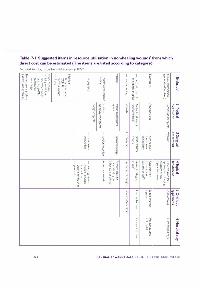
S 6 0 J O U R N A L O F WO U N D C A R E VO L 2 2 . N O 5 . E W M A D O C U M E N T 2 0 1 3
1 Evaluation2 M
edical treatm
ent3 Surgical treatm
ent4 Topical treatm
ent5 O
rthotic appliances
6 Hospital stay
Clinical exam
ination (generalised/localised)
Cardiovascular agents
Vascular:
— percutaneous translum
inal angioplastry
— reconstructive surgery
Time required for
applying and changing dressings or any topical treatm
ent.
Shoes/insolesH
ospital bed days
Laboratory:
— m
etabolic control of diabetes m
ellitus
— haem
orheology
Anticoagulants
Resources for transportation (patient or staff)
Special orthotic appliances
Resources used in hospital
Antibacterial agents
(oral/parenteral)A
vailable category of staff
Total contact castC
ategory of clinic
SteroidsO
rthopaedic:
— incision/drainage
— revision/resection
— m
inor/major
amputation
Frequency of changesProsthesis/w
heelchair
Vascular:
— noninvasive vascular testing
— angiography
Imm
unosuppressive agents
Primary dressing
materials, drugs or
other type of device
Insulin—hypoglycaem
ic agentsA
ccessory material:
— cleansing agents
— !xation (e.g., tape to adapt the dressing to the skin)
— gloves, etc.
Analgesic agents
Infection:—
X-ray, bone scan,
CT, M
RI—
bacterial culture—
biopsy
Socioeconomic:
— living conditions
— A
ttempted D
aily Learning (A
DL)
— com
pliance, know
ledge
Biomechanical (w
alking pattern, foot dynam
ics)
Table 7-1. Suggested items in resource utilisation in non-healing wounds* from which direct cost can be estimated (The items are listed according to category)*Adapted from Ragnarson-Tennvall & Apelqvist (1997)385
J O U R N A L O F WO U N D C A R E VO L 2 2 . N O 5 . E W M A D O C U M E N T 2 0 1 3 S 6 1
Health economics in non-healing ulcers and reimbursement In a study comparing resource use associated with diabetic foot infections in three European foot centres in different countries, substantial differences were identi!ed in inpatient stay, use of antibiotics and vascular surgery.390 The authors concluded that these differences could largely be explained by variations in access to inpatient and outpatient facilities, the patient selection bias, patients’ characteristics, reimbursement systems and health-care systems, and these results were con!rmed in the EURODIALE study.314,394
In a comparison of diabetes-related foot lesions in patients in the Netherlands and California,395 the duration of hospital stay was substantially longer in the Netherlands, but the incidence of lower extremity major amputation was higher in the USA. This has important implications in the drive to cut costs through early discharge. The authors suggested that these differences might be explained by differences in access to health care, health-care !nancing and reimbursement systems. Although hospitalisation is obviously more expensive than home care, the long-term cost effectiveness of these options must be examined. For some patients, wound care strategies (such as of$oading) can be successfully implemented in an inpatient setting, thereby avoiding expensive adverse events, such as amputation. Ultimately, this may be less expensive overall than a prolonged period of home care in which these expensive adverse events are more likely to occur. Reimbursement in some countries favours amputation because of shorter hospital stays and reduced length of time healing.314,394,395
Summary Non-healing wounds often result in a considerable !nancial burden, which is associated with a long healing time and a high incidence of complications. When evaluating the consequences of a wound infection, it is essential to view the consequences
as an integrated part of the total management and resource utilisation of an individual with a non-healing wound. While it is important to identify interventions and strategies early to avoid complications and facilitate healing, these often have cost implications. Clinicians need to be able to present robust economic arguments and strong outcome data to fund holders. A major problem in the analysis of the cost of disease states is that comparisons of cost analyses are compounded by variations in care protocols and the different economic statuses of different countries (such as variations in rates of pay to health professionals and in reimbursements). Substantial efforts will be required to identify a series of standardised criteria for cost analyses that can be used to further identify the most economically effective ways to treat non-healing wounds.
ControversiesQ What are the economic consequences of not
making the correct diagnosis in due time?
StatementThe consequence of not making the correct diagnosis and corresponding treatment strategy will be a delay in adequate treatment and intervention, a delay in healing and, ultimately, increased cost.
DiscussionThe most important factors related to high resource utilisation in treating non-healing ulcers is the need for surgery, in-hospital stays, and wound healing time (duration of ulcer). The consequence of inadequate diagnosis will be a delay in adequate treatment and a subsequent delay in healing, ultimately leading to increased cost. It has to be recognised that health-economic data are essential to describe resources spent with regard to any condition, but especially non-healing ulcers. Treatment of patients with this condition frequently involves many disciplines and incurs large costs. A
S 6 2 J O U R N A L O F WO U N D C A R E VO L 2 2 . N O 5 . E W M A D O C U M E N T 2 0 1 3
description of how these resources could be spent more effectively, both from the patient and the health-care perspective, is essential when evaluating the consequences of a wound infection. Cure of wound infections should be seen as an integrated part of the total management and resource utilisation of an individual with non-healing wound.
ConclusionThe most important factors related to the high resource utilisation in treating non-healing ulcers is the need for surgery, length of inpatient stay and wound healing time (duration of ulcer). Therefore, the consequence of inadequate diagnosis will be a delay in adequate treatment and a subsequent delay in healing, ultimately leading to an increased overall cost.
Q What is the cost effectiveness of antiseptic versus antibiotic treatment (not just prices of products, but also societal costs)?
StatementTo our knowledge there are no studies available differentiating the cost effectiveness of antiseptic compared with local antibiotic treatment.
DiscussionAs part of wound healing, both antiseptics and local antibiotics are used to treat wound infection. When investigating the different outcomes between antiseptics and antibiotic treatments, the outcomes need to be considered as an integrated part of total management and resource utilisation. The total resource utilisation not only involves the direct costs to cure the infection, but also the cost incurred until healing is achieved. Also, the societal costs of healing should be taken into consideration, not just the price of a speci!c item.
There are very limited data comparing cost effectiveness among various treatment strategies in non-healing ulcers. Most studies evaluating
economic cost, or resources used, have been based on clinical trials, which limits their external validity. Frequently, the cost to cure the infection has been related to the cost of antibiotics and/or in-hospital stay. At present, there are few high-quality studies examining wound management and health economics, and to our knowledge there are no studies regarding the difference in cost effectiveness of antiseptic versus local antibiotic treatments.
ConclusionThere are very limited data comparing cost effectiveness between various treatment strategies in non-healing ulcers. When analysing resources spent in treating complex ulcers, it is important to consider all resources spent to achieve healing, not just the price (or cost) of one speci!c item. To our knowledge, there are no studies available analysing the cost effectiveness of antiseptic compared with local antibiotic treatment.
Q Is it cheaper to amputate limbs of an individual with an infected wound than to treat (conservatively) with antibiotics?
‘Most studies evaluating
economic cost, or resources
used, have been based on
clinical trials, which limits their
external validity
’
J O U R N A L O F WO U N D C A R E VO L 2 2 . N O 5 . E W M A D O C U M E N T 2 0 1 3 S 6 3
StatementThe direct cost associated with performing an amputation in diabetic patients with infected wounds (Table 7-1) is higher than when treating without performing an amputation, if diabetic patients are followed to healing.
DiscussionIt is essential to consider the total resources spent to heal a patient with a non-healing infected ulcer. In some countries, the elevated cost for conservative treatment with antibiotics and in-hospital stay may lead to an early amputation. This is a common assumption in countries with health-care systems in which antibiotics have a higher reimbursement price and patients are not followed until healing is achieved.300,346,384,395,396 However, in the few studies that have analysed the total direct cost to achieve healing of an infected foot ulcer, the price for antibiotics comprised 15% of the total cost. Lower-leg amputation is frequently related to high resource utilisation, due to resources spent following amputation. It is essential to analyse and understand that the costs are different due to the different perspective. It is important to evaluate the cost from the societal perspective and not only from the perspective of the hospital. In patients with diabetes and deep foot infection, the total direct cost was twice as high in patients treated with an amputation compared with those treated conservatively. The most important cost-driving factors were wound duration and the number of surgical procedures. The price of antibiotics cannot be used as the only determinant to evaluate treatment cost.387
ConclusionThe limited data available on patients with infected diabetic foot ulcers suggest that the direct costs are higher for healing with an amputation than without.
Q Do recommendations to restrict the use of products due to their price per item have consequences, and what are these consequences?
StatementThe price of a single item in treating individuals with non-healing ulcers should never be the key factor for decision making.
DiscussionIt is very important to recognise the perspective of each of the relevant decision-makers. In wound care, decision makers include clinicians, hospitals or other health-care provider organisations, and third-party payers. For example, from a hospital-management perspective, the cost of intravenous antibiotics or revascularisation could be considered high, particularly because it might prolong the length of the in-hospital stay. However, from a societal perspective, the use of antibiotics and revascularisation in this case is only a fraction of the total cost spent to achieve complete wound healing.
The price of a single item in treating individuals with non-healing ulcers should, therefore, never be the key factor for decision making. Each intervention must be evaluated in light of the total resources spent to achieve a speci!c goal, such as wound healing or resolution of an infection. When cost is used as an outcome parameter in wound management, it is essential to measure all resources used and then add the value of those resources, according to a prede!ned protocol, to a speci!c endpoint (outcome). It is recommended to show resource use and cost separately, as the prices of product/drug/device are set differently in various countries or regions.
ConclusionThe price of a single item in treating individuals with non-healing ulcers should never be the key factor for decision making. Each intervention should be evaluated from the perspective of the total resources spent to achieve a speci!c goal, such as wound healing or resolution of an infection.
S 6 4 J O U R N A L O F WO U N D C A R E VO L 2 2 . N O 5 . E W M A D O C U M E N T 2 0 1 3
Potential consequences if we do nothingJudicious use of all antimicrobial agents is becoming an urgent necessity, in order to retain effective treatments for infection and avoid a return to the conditions that existed before the antibiotic era. Overuse of antibiotics has provided a selection pressure that has allowed antibiotic-resistant strains to emerge and increase in prevalence (see Chapters 3 and 4). Antibiotic resistance increases patient morbidity, extends hospital stay, and increases treatment costs and mortality rates.397,398 These outcomes have a social and economic impact;397 incorrect use of antibiotics wastes time and resources, erodes patient con!dence and reduces staff morale. Many surgical procedures and cancer therapies rely on antibiotics to prevent and/or treat ensuing infections, and these treatments will become impossible without effective antibiotics.398 Moreover, the potential threat of failure to treat wound infections will have an immediate impact in con$ict areas or episodes of natural disasters.
Factors that contribute to the misuse of antibiotics are diverse, the WHO has identi!ed the key issues as diagnostic uncertainty, lack of skills and knowledge, fear of litigation, and failure to properly utilise clinical guidelines.398 The unrestricted use of antibiotics in many non-European countries promotes antibiotic abuse. Additionally, national pharmaceutical policies may be absent; therefore, coordinated opportunities to improve surveillance, regulation and education are lost. The unethical promotion of antimicrobial interventions by
Future perspectives
commercial organisations additionally contributes to misuse. Health professionals with heavy workloads may !nd that time constraints lead to limited opportunities to update knowledge and a reliance on incomplete diagnoses.
Several programmes have been initiated to address the problems emanating from antibiotic resistance, yet few tangible effects have been realised (Table 8-1).
As mentioned in the introduction, infection is one of the most frequently occurring complications of non-healing wounds. There is a concern regarding the use of antimicrobials in the society and, as a consequence, antimicrobial treatment strategies in non-healing wounds have been challenged. The consequences of these controversies have an impact with regard to both overuse or underuse of antimicrobials in wound management.
Therefore, it is essential that management strategies are targeted effectively, to ensure timely and ef!cient wound-management services. Indeed, adopting a systematic approach to patient and wound assessment will lead to early detection of infection and other complications, and the initiation of appropriate treatment plans.399 However, importantly, the process of wound management involves not only the application of an appropriate dressing, drug or device, but also consideration of broader factors that may impede the wound healing process.400
Therefore, as wounds remain a signi!cant health-care problem, effective prevention and

J O U R N A L O F WO U N D C A R E VO L 2 2 . N O 5 . E W M A D O C U M E N T 2 0 1 3 S 6 5
Year Country/Origin Organisation Report1998 UK Select Committee on Science and
Technology of the House of LordsResistance to antibiotics and other antimicrobial agents
2001 Switzerland World Health Organization (WHO) Global strategy for containment of antimicrobial resistance
2004 USA Infectious Diseases Society of America (IDSA)
Bad bugs, no drugs
2009 USA IDSA Bad bugs, no drugs: no ESCAPE!
2009 Sweden European Centre for Disease Prevention and Control
The bacterial challenge: time to react
2011 Belgium European Commission Communication from the European Commission to the European Parliament and the Council: Action Plan against the rising threats from antimicrobial resistance
2011 USA/EU Transatlantic taskforce on antibiotic resistance (TATFAR)
Recommendations for future collaborations between the US and EU
2012 Switzerland WHO The evolving threat of antimicrobial resistance
Table 8-1. The debate and initiatives to control antimicrobial resistance
management strategies should be core components of the strategic planning of health-care services.288
With regard to the bioburden in non-healing wounds, there are three major issues1 The microbiological de!nition of a wound
infection:
Many different bacterial and fungal species have been identi!ed in non-healing wounds. The quantity of each species may vary, and it is not know whether small amounts of one bacterium might boost one of the major inhabitants of a wound. This suggests the number of bacteria/cm3 tissue may not be relevant, but rather which species are present may. We need research that focus on these issues, since most of the information available today are obtained from acute wounds, animal or other experimental models.
There is a need to investigate the relationship between microbial population sizes in non-healing wounds and clinical indicators of infection.
In the future, a stricter de!nition of the terms ‘problematic bacterial load’ or ‘critical colonisation’ will be needed before they can be used in clinical practice or as endpoints in research.
2 Antimicrobial resistance:
There remains a great deal of uncertainty about resistance to topical antimicrobials. The bacterial resistance described in the literature is primarily in relation to the use of antibiotics. Clearly, further systematic reviews of evidence may be warranted and there is a need to monitor indicators for emergence of resistance to antimicrobials in practice settings.
Due to increasing antibiotic resistance, there is an urgent need for adjuvant or alternative treatments, better controls on the use of antimicrobials in human and veterinarian medicine, and consistent restrictions and guidelines in all European countries. Use of excipients may in the future improve the outcome results of antimicrobial treatment.

S 6 6 J O U R N A L O F WO U N D C A R E VO L 2 2 . N O 5 . E W M A D O C U M E N T 2 0 1 3
3 Presence and importance of bio!lms:
From a clinical perspective, we lack a clear understanding of the presence, importance, and proper intervention for bio!lm in non-healing wounds.
With regard to treatment of non-healing wounds, there are several major issues1 Lack of awareness of antibiotic resistance and of
the value of antimicrobial treatment in$uences physicians’ attitudes to prescribing patterns:
Physicians need guidance and education with regard to a structured management of antimicrobial treatment for non-healing wounds. They need to understand that, in order to be effective, antimicrobial treatment should be targeted both to the right wound and to the right patient. There is no proof that routine administration of antibiotics is effective for prophylaxis or treatment in non-infected non-healing wounds.
Due to the increasing resistance towards antibiotics and the need for an effective antimicrobial strategy for non-healing wounds, there is an urgent need for the use of an antimicrobial treatment regime that does not include antibiotics. There needs to be greater clarity about when and where to use each treatment modality.
2 An abundance of products are available, but there is no consensus regarding the value of topical antimicrobials in non-healing wounds.
Most research is conducted by industry rather than government agencies. It is not surprising that the available evidence is mostly brand-speci!c. Companies have little incentive to conduct broad-based research. In combination with a lack of willingness of governments to fund the necessary clinical research, this has created a gap in the present evidence with regard to outcomes and
results of interventions in the general population. A more widespread description covering all aspects of the health care is desirable, and the main problems, such as the availability of evidence, controversies or myths, should be discussed.
3 There is a need for cohort studies, comparative studies, or RCTs with regard to antimicrobial treatments in non-healing wounds with a design and end-points that focus on resolution or prevention of infection:
The majority of comparative studies with regard to the use of antimicrobial agents in wound treatment has been focused on either acute wounds or non-infected wounds. A need for further, well-designed studies has been emphasised; however, the limitations of prede!ned adequate endpoints in studies are a major barrier for evaluating the importance of various treatment strategies, such as antimicrobials. The most important endpoints with regards to antimicrobial treatment should be the prevention of infection, resolution of infection,
‘Due to the increasing resistance
towards antibiotics, there is an
urgent need for the use of an
antimicrobial treatment regime
that does not include antibiotics
’

J O U R N A L O F WO U N D C A R E VO L 2 2 . N O 5 . E W M A D O C U M E N T 2 0 1 3 S 6 7
wound healing, time to wound healing or time for resolution of an infection. Any recommendation needs to acknowledge that, while RCT evidence is ideally required to support proof of ef!cacy, other non-RCT methods may also be useful in determining the impact of antimicrobials in practice settings.
Wound infection is a valid endpoint in a wound healing study and clinical parameters should be used for the de!nition of wound infection. To properly evaluate the value of antimicrobial agents in wound treatment, we need a new set of tools and endpoints for these studies. The commonly-used endpoints of wound closure, healing rate, epithelialisation, quality of life and wound environment are all, to some extent, dependent on the presence of infection. Resolution of infection has been used as an endpoint in some comparative studies, either at the discretion of the physician and sometimes supported by clinical signs and bacterial load, or laboratory parameters. Since infection is a clinical diagnosis, it would make sense to use a clinical scoring system to de!ne infection, as well as resolution of infection.
From the patient perspective—a holistic approach is mandatory• Physicians and caregivers are unaware of the
importance of patients’ and their families’ attitudes towards management.
In the management of a patient, attitude and expectations of treatment have to be considered, especially in health-care systems where management of wounds is relying on relatives and family as a resource. Ultimately, we have to treat not only a wound, but an individual with a wound, also considering the patients’ social environment. Furthermore, patient safety strategies consider ignoring health-care needs or failing to provide adequate health care for appropriate wound management as a form of neglect. Patient safety groups place the onus
of adequate wound management !rmly in the hands of care providers. Therefore, it is of utmost importance that emphasis is placed on a systematic assessment of the patient, the wound and the environment in which care is provided. This will enhance the likelihood that adverse changes in the patient’s condition are readily recognised and appropriate treatment plans are initiated. For this reason, the patient and the family should to be an integral part of the future management of non-healing wounds.
From the organisation perspective, this is the major issue• Individuals with infected wounds should only be
cared for by those trained and competent in the provision of wound management services.
In most health-care systems, policy makers and caregivers are frequently unaware that in most patients with a non-healing wound, with or without infection, the condition is related to comorbidity and concurrent disease, necessitating a multifactorial treatment, in which antimicrobial treatment is a part. More than a decade ago, it was identi!ed that limited availability of adequately-trained personnel and diagnostic equipment compounds the suffering of patients. Furthermore, it increases the costs to an already over-stretched health budget. Especially regarding diabetic services, it has been concluded that structured multi-professional interventions, such as interdisciplinary collaboration and professional and patient education, result in improved patient outcomes and service delivery. To achieve this, all the members of the multidisciplinary team must work together, as no single profession has all the required skills.
The multidisciplinary model offers a unique opportunity for recruiting a suf!cient number of patients for clinical and basic research, thereby producing evidence for the materials and procedures used for treatment of infected wounds.
S 6 8 J O U R N A L O F WO U N D C A R E VO L 2 2 . N O 5 . E W M A D O C U M E N T 2 0 1 3
For this reason, the organisational perspective need to be elaborated and further developed.
From the economic perspective, this is the major issue• Treatment with antimicrobial agents for
non-healing wounds is frequently described in terms of the price of various devices or drugs.
Non-healing wounds often result in a considerable !nancial burden, which is associated with the length of time to heal and the high incidence of complications. The price of a single item in treating individuals with non-healing ulcers should never be the key factor for decision making; each intervention has to be evaluated from the perspective of the total resources spent to achieve a speci!c goal, such as wound healing or resolution of an infection.
While it is important to identify interventions and strategies early to avoid complications and facilitate healing, these often have cost implications.
‘Treatment with antimicrobial
agents for non-healing
wounds is frequently described
in terms of the price of various
devices or drugs
’For the future we need standardised criteria for cost analyses, which can be used to further identify the most economically effective ways to treat non-healing wounds. Q
J O U R N A L O F WO U N D C A R E VO L 2 2 . N O 5 . E W M A D O C U M E N T 2 0 1 3 S 6 9
References
1 Dale, J.J., Callam, M.J., Ruckley, C.V. et al. Chronic ulcers of the leg: a study of prevalence in a Scottish community. Health Bull (Edinb). 1983; 41: 310–314.
2 Gottrup, F. A specialized wound-healing center concepts: importance of a multidisciplinary department structure and surgical treatment facilities in the treatment of chronic wounds. Am J Surg. 2004; 187: 38–43.
3 Hjort, A., Gottrup, F. Cost of wound treatment to increase signi!cantly in Denmark over the next decade. J Wound Care. 2010; 19: 173–184.
4 Posnett, J., Gottrup, F., Lundgren, H., Saal, G. The resource impact of wounds on health-care providers in Europe. J Wound Care. 2009; 18: 154–161.
5 European Commission—Scienti!c Committee on Emerging and Newly Identi!ed Health Risks (SCENIHR). Assessment of the Antibiotic Resistance Effects of Biocides, 2009.
6 Mossialos, E., Morel, C.M., Edwards, S. et al. Policies and incentives for promoting innovation in antibiotic research. World Health Organization—on behalf of the European Observatory on Health Systems and Policies, 2010.
7 Danish Presidency of the Council of the European Union 2012. Combating Antimicrobial Resistance —Time for Joint Action, 2012.
8 European Commission. Report from the Commission to the Council on the basis of Member States’ reports on the implementation of the Council Recommendation (2009/C 151/01) on patient safety, including the prevention and control of healthcare associated infections. European Commission, 2012.
9 European Union. Council conclusions of 1 December 2009 on innovative incentives for effective antibiotics (2009/C 302/05). European Union, 2009.
10 ReAct—Action on Antibiotic Resistance. Cure with Care: Understanding Antibiotic Resistance. Uppsala University, 2007.
11 European Academies Science Advisory Council (EASAC). The Royal Society Tackling Antibiotic Resistance in Europe. EASAC, 2007.
12 Vicente, M. The fallacies of hope: will we discover new antibiotics to combat pathogenic bacteria in time. FEMS Microbiol Rev. 2006; 30: 841–852.
13 Cosgrove, S., Carmeli, S. The impact of antimicrobial resistance on health and economic outcomes; Clin Infect Dis. 2003; 36: 1433–1437.
14 European Antimicrobial Resistance Surveillance System (EARSS). EARSS Annual Report 2006. EARSS, 2007.
15 Gottrup, F., Apelqvist, J., Price, P. Outcomes in controlled and comparative studies on non-healing wounds: recommendations to improve the quality of evidence in wound management. J Wound Care. 2010; 19: 239–268.
16 Burmolle, M., Thomsen, T.R., Fazli, M. et al. Bio!lms in chronic infections—a matter of opportunity - monospecies bio!lms in multispecies infections. FEMS Immunol Med Microbiol. 2010; 59: 324–336.
17 Lee, B.Y. The Wound Management Manual. McGraw-Hill, 2005.
18 Polit, D.F., Beck, C.T. Nursing research: Generating and assessing evidence for nursing practice (9th edn). Lippincott Williams & Wilkins 2012.
19 Leaper, D.J. De!ning infection. J Wound Care. 1998; 7: 373.
20 Altemeier, W. Sepsis in surgery (Presidential address). Arch Surg. 1982; 117: 107–112.
21 Brennan, S., Leaper, D. The effect of antiseptics on the healing wound: a study using the rabbit ear chamber. Br J Surg. 1985; 72: 780–782.
22 Lineaweaver, W., Howard, R., Soucy, D. et al. Topical antimicrobial toxicity. Arch Surg. 1985; 120: 267–270.
23 Mertz, P., Ovington, L. Wound healing microbiology. Dermatol Clin.1993; 11: 739–747.
24 Hansson, C., Hoborn, J., Möller, A., Swanbeck, G. The microbial "ora in venous leg ulcers without clinical signs of infection. Repeated culture using a validated standardised microbiological technique; Acta Dermatol Venereol. 1995; 75, 24–30.
25 Robson, M. Infection in the surgical patient: an imbalance in the normal equilibrium. Clin Plast Surg. 1979; 6: 493–503.
26 Heinzelmann, M., Scott, M., Lam, T. Factors predisposing to bacterial invasion and infection. Am J Surg. 2002; 183: 179–190.
27 Cooper, R. EWMA Position Document: Understanding Wound Infection. EWMA, 2005.
28 Bowler, P.G., Duerden, B.I., Armstrong, D.G. Wound microbiology and associated approaches to wound management. Clin Microbiol Rev. 2001; 14: 244–269.
29 Howell-Jones, R.S., Wilson, M.J., Hill, K.E. et al. A review of the microbiology, antibiotic usage and resistance in chronic skin wounds. J Antimicrob Chemother. 2005; 55: 143–149.
30 Gilchrist, B., Reed, C. The bacteriology of chronic venous ulcers treated with occlusive hydrocolloid dressings. Br J Dermatol. 1989; 121: 337–344.
31 Bendy, R.H., Jnr, Nuccio, P.A., Wolfe, E. et al. Relationship of quantitative wound bacterial counts to healing of decubiti: effect of topical gentamicin. Antimicrob Agents Chemother (Bethesda). 1964; 10, 147–155.
32 Pruitt, B.A. Jnr. The diagnosis and treatment of infection in the burn patient. Burns Incl Therm Inj. 1984; 11: 2, 79–91.
33 Robson, M.C., Lea, C.E., Dalton, J.B., Heggers, J.P. Quantitative bacteriology and delayed wound closure. Surg Forum. 1968; 19: 501–502.
34 Edmonds, M., Foster, A. The use of antibiotics in the diabetic foot. Am J Surg. 2004; 187: 5A (Suppl.), 25S–28S.
35 Gardner, S.E., Frantz, R.A. Wound bioburden and infection-related complications in diabetic foot ulcers. Biol Res Nurs. 2008; 10: 44–53.
36 Chantelau, E., Tanudjaja, T., Altenhofer, F. et al. Antibiotic treatment for uncomplicated neuropathic forefoot ulcers in diabetes: a controlled trial. Diabetes Med. 1996; 13: 156–159.
37 Sotto, A., Richard, J.L., Combescure, C. et al. Bene!cial effects of implementing guidelines on microbiology and costs of infected diabetic foot ulcers. Diabetologia. 2010; 53: 2249–2255.
38 Baxter, C., Mertz, P.M. Local factors that affect wound healing. Nurs RSA. 1992; 7: 2, 16–23.
39 Gilchrist, B. Treating bacterial wound infection. Nurs Times. 1994; 90: 50, 55–58.
S 7 0 J O U R N A L O F WO U N D C A R E VO L 2 2 . N O 5 . E W M A D O C U M E N T 2 0 1 3
40 Hutchinson, J.J., McGuckin, M. Occlusive dressings: a microbiologic and clinical review. Am J Infect Control. 1990; 18: 257–268.
41 Geesey, G.G., Richardson, W.T., Yeomans, H.G. et al. Microscopic examination of natural sessile bacterial populations from an alpine stream. Can J Microbiol. 1977; 23: 1733–1736.
42 Høiby, N. Pseudomonas aeruginosa infection in cystic !brosis. Diagnostic and prognostic signi!cance of Pseudomonas aeruginosa precipitins determined by means of crossed immunoelectrophoresis. Acta Pathol Microbiol Scand Suppl. 1977; 262: 1–96.
43 McCoy, W.F., Bryers, J.D., Robbins, J., Costerton, J.W. Observations of fouling bio!lm formation. Can J Microbiol. 1981; 27: 910–917.
44 Elek, S.D. Experimental staphylococcal infections in the skin of man. Ann NY Acad Sci. 1956; 65: 3, 85–90.
45 Lyman, I.R., Tenery, J.H., Basson, R.P. Correlation between decrease in bacterial load and rate of wound healing. Surg Gynecol Obstet. 1970; 130: 616–621.
46 Bendy, R.H. Jnr, Nuccio, P.A., Wolfe, E. et al. Relationship of quantitative wound bacterial counts to healing of decubiti: effect of topical gentamicin. Antimicrobial Agents Chemother (Bethesda). 1964; 10: 147–155.
47 Gristina, A.G., Price, J.L., Hobgood, C.D. et al. Bacterial colonization of percutaneous sutures; Surgery; 1985; 98:1, 12-19.
48 Akiyama, H., Huh, W.K., Yamasaki, O. et al. Confocal laser scanning microscopic observation of glycocalyx production by Staphylococcus aureus in mouse skin: does S. aureus generally produce a bio!lm on damaged skin? Br J Dermatol. 2002; 147: 879–885.
49 Akiyama, H., Torigoe, R., Arata, J. Interaction of Staphylococcus aureus cells and silk threads in vitro and in mouse skin. J Dermatol Sci. 1993; 6: 247–257.
50 Akiyama, H., Kanzaki, H., Abe, Y. et al. Staphylococcus aureus infection on experimental croton oil-in"amed skin in mice. J Dermatol Sci. 1994; 8: 1, 1–10.
51 Schierle, C.F., De la Garza, M., Mustoe, T.A., Galiano, R.D. Staphylococcal bio!lms impair wound healing by delaying reepithelialization in a murine cutaneous wound model. Wound Repair Regen. 2009; 17: 354–359.
52 Bjarnsholt, T., Kirketerp-Moller, K., Jensen, P.O. et al. Why chronic wounds will not heal: a novel hypothesis. Wound Repair Regen. 2008; 16: 2–10.
53 Davis, S.C., Ricotti, C., Cazzaniga, A. et al. Microscopic and physiologic evidence for bio!lm-associated wound colonization in vivo. Wound Repair Regen. 2008; 16: 23–29.
54 James, G.A., Swogger, E., Wolcott, R. et al. Bio!lms in chronic wounds. Wound Repair Regen. 2008; 16: 37–44.
55 Burmolle, M., Thomsen, T.R., Fazli, M. et al. Bio!lms in chronic infections—a matter of opportunity—monospecies bio!lms in multispecies infections. FEMS Immunol Med Microbiol. 2010; 59: 324–336.
56 Bjarnsholt, T., Jensen, P.O., Burmolle, M. et al. Pseudomonas aeruginosa tolerance to tobramycin, hydrogen peroxide and polymorphonuclear leukocytes is quorum-sensing dependent. Microbiology. 2005; 151: (Pt 2), 373–383.
57 Alhede, M., Kragh, K.N., Qvortrup, K. et al. Phenotypes of non-attached Pseudomonas aeruginosa aggregates resemble surface attached bio!lm. PLoS.One. 2011; 6: 11, e27943.
58 Alipour, M., Suntres, Z.E., Omri, A. Importance of DNase and alginate lyase for enhancing free and liposome encapsulated aminoglycoside activity against Pseudomonas aeruginosa. J Antimicrob Chemother. 2009; 64: 317–325.
59 Overview and general considerations. In: Clark, R.A.F. (ed). The Molecular and Cellular Biology of Wound Repair (2nd edn). Plenum Press, 1996.
60 Gjodsbol, K., Christensen, J.J., Karlsmark, T. et al. Multiple bacterial species reside in chronic wounds: a longitudinal study. Int Wound J. 2006; 3: 225–231.
61 Madsen, S.M., Westh, H., Danielsen, L., Rosdahl, V.T. Bacterial colonization and healing of venous leg ulcers. APMIS. 1996; 104: 895–899.
62 Halbert, A.R., Stacey, M.C., Rohr, J.B., Jopp-McKay, A. The effect of bacterial colonization on venous ulcer healing. Australas J Dermatol. 1992; 33: 2, 75–80.
63 Fazli, M., Bjarnsholt, T., Kirketerp-Moller, K. et al. Quantitative analysis of the cellular in"ammatory response against bio!lm bacteria in chronic wounds. Wound Repair Regen. 2011; 19: 387–391.
64 Jensen, P.O., Bjarnsholt, T., Phipps, R. et al. Rapid necrotic killing of polymorphonuclear leukocytes is caused by quorum-sensing-controlled production of rhamnolipid by Pseudomonas aeruginosa. Microbiology. 2007; 153: (Pt 5), 1329–1338.
65 Hogsberg, T., Bjarnsholt, T., Thomsen, J.S., Kirketerp-Moller, K. Success rate of split-thickness skin grafting of chronic venous leg ulcers depends on the presence of Pseudomonas aeruginosa: a retrospective study. PLoS.One. 2011; 6: 5, e20492.
66 Gardner, S.E., Hillis, S.L., Heilmann, K. et al. The neuropathic diabetic foot ulcer microbiome is associated with clinical factors. Diabetes. 2013; 62: 923–930.
67 Krizek, T., Robson, M., Kho, E. Bacterial growth and skin graft survival. Surg Forum. 1967; 18: 518–519.
68 Liedburg, N.C.F., Reiss, E., Artz, C.P. The effects of bacteria on the take of split-thickness skin grafts in rabbits. Ann Surg. 1955; 142: 92–96.
69 Robson, M.C., Lea, C.E., Dalton, J.B., Heggars, J.P. Quantitative bacteriology and delayed wound closure. Surg Forum. 1968; 19: 501–502.
70 Robson, M.C., Heggars, J.P. Bacterial quanti!cation of open wounds. Mil Med. 1969; 134: 19–24.
71 Murphy, R.C., Robson, M.C., Heggars, J.P., Kadowaki, M. The effect of microbial contamination on musculocutaneous and random "aps. J Surg Res. 1968; 41, 75–80.
72 Heggars, J., Robson, M., Doran, E. The quantitative assessment of bacterial contamination of open wounds by a slide technique. Trans R Soc Trop Med Hyg. 1969; 63: 532–534.
73 Bornside, G., Bornside, B. Comparison between moist swab and tissue biopsy methods for quanti!cation of bacteria in experimental incisional wounds. J Trauma. 1979; 19: 103–105.
74 Pruitt, B. The diagnosis and treatment of infection in the burned patient. Burns. 1984; 11: 79–81.
75 Schneider, M., Vildozola, C.W., Brooks, S. Quantutative assessment of bacterial invasion of chronic ulcers. Am J Surg. 1983; 145: 260–262.
76 Pruitt, B.A. Jnr, McManus, A.T., Kim, S.H., Goodwin, C.W. Burn wound infections: current status. World J Surg. 1998; 22: 135–145.
77 Bowler, P.G. The 10(5) bacterial growth guideline: reassessing its clinical relevance in wound healing. Ostomy Wound Manage. 2003; 49: 1, 44–53.
78 Thomsen, T., Aasholm, M., Rudkjøbing, V. et al. The bacteriology of chronic venous leg ulcer examined by culture-independent molecular methods. Wound Repair Regen. 2010; 18: 38–49.
79 Fazli, M., Bjarnsholt, T., Kirketerp-Møller, K. et al. Non-random distribution of Pseudomonas aeruginosa and Staphylococcus aureus in chronic wounds. J Clin Microbiol. 2009; 47: 4084–4089.
80 Kirketerp-Møller, K., Madsen, K., Jensen, P. et al. The distribution, organization and ecology of bacteria in chronic wounds. J Clin Microbiol. 2008; 46: 2717–2722.
81 Carrel, A. Cicatrization of wounds: XII. Factors initiating regeneration. J Exp Med. 1921; 34: 425–434.
82 Botsford, T. The tensile strength of sutured skin wounds during healing. Surg Gynecol Obstet. 1941; 72: 690–697.

J O U R N A L O F WO U N D C A R E VO L 2 2 . N O 5 . E W M A D O C U M E N T 2 0 1 3 S 7 1
83 Tenorio, A., Jindrak, K., Weiner, M. et al. Accelerated healing in infected wounds. Surg Gynecol Obstet. 1976; 142, 537–543.
84 Raju, D., Jindrak, K., Weiner, M., Enquist, I. A study of the critical bacterial inoculum to cause a stimulus to wound healing. Surg Gynecol Obstet. 1977; 144: 347–350.
85 Bowler, P.G., Davies, B.J. The microbiology of infected and noninfected leg ulcers. Int J Dermatol. 1999; 38: 573–578.
86 Daltrey, D.C., Rhodes, B., Chattwood, J.G. Investigation into the microbial "ora of healing and non-healing decubitus ulcers. J Clin Pathol. 1981; 34: 701–705.
87 Rotstein, O.D., Pruett, T.L., Simmons, R.L. Mechanisms of microbial synergy in polymicrobial surgical infections. Rev Infect Dis. 1985; 7: 151–170.
88 Trengove, N.J., Stacey, M.C., McGechie, D.F., Mata, S. Qualitative bacteriology and leg ulcer healing. J Wound Care. 1996; 5: 277–280.
89 Moore, K., Hall, V., Paull, A. et al. Surface bacteriology of venous leg ulcers and healing outcome. J Clin Pathol. 2010; 63: 830–834.
90 Dowd, S.E., Wolcott, R.D., Kennedy, J. et al. Molecular diagnostics and personalised medicine in wound care: assessment of outcomes. J Wound Care. 2011; 20: 232–234.
91 Sotto, A., Richard, J.L., Messad, N. et al. Distinguishing colonization from infection with Staphylococcus aureus in diabetic foot ulcers with miniaturized oligonucleotide arrays: a French multicenter study. Diabetes Care. 2012; 35: 617–623.
92 White, R.J., Cutting, K.F. Critical colonization—the concept under scrutiny. Ostomy Wound Manage. 2006; 52: 11, 50–56.
93 Dissemond, J., Assadian, O., Gerber, V. et al. Classi!cation of wounds at risk and their antimicrobial treatment with polihexanide: a practice-oriented expert recommendation. Skin Pharmacol Physiol. 2011; 24: 245–255.
94 Kingsley, A. A proactive approach to wound infection. Nurs Stand. 2001; 15: 30, 50–58.
95 Gardner, S.E., Frantz, R.A., Troia, C. et al. A tool to assess clinical signs and symptoms of localized infection in chronic wounds: development and reliability. Ostomy Wound Manage. 2001; 47: 1, 40–47.
96 Cutting, K., Harding, K. Criteria for identifying wound infection. J Wound Care. 1994; 3: 198–201.
97 Edmonds, M., Foster, A. The use of antibiotics in the diabetic foot. Am J Surg. 2004; 187, 25–28.
98 Robson, M. Wound infection: a failure of wound healing caused by an imbalance of bacteria. Surg Clin North Am. 1997; 77, 637–650.
99 Gardner, S.E., Frantz, R.A. Wound bioburden and infection-related complications in diabetic foot ulcers. Biol Res Nurs. 2008; 10, 44–53.
100 Sibbald, R.G., Woo, K., Ayello, E.A. Increased bacterial burden and infection: the story of NERDS and STONES. Adv Skin Wound Care. 2006; 19: 447–461.
101 Woo, K., Sibbald, R. A cross-sectional study of using NERDS and STONES to assess bacterial burden; Ostomy Wound Manage. 2009; 55: 8, 40–48.
102 Jørgensen, B., Bech-Thomsen, N., Grenow, B., Gottrup, F. Effect of a new silver dressings on chronic venous leg ulcers with signs of critical colonisation. J Wound care. 2006; 15: 97–100.
103 Brown, T.S., Hawksworth, J.S., Sheppard, F.R. et al. Effect of a new silver dressings on chronic venous leg ulcers. Surg Infect (Larchmt). 2011; 12: 351–357.
104 Fazli, M., Bjarnsholt, T., Kirketerp-Moller, K. et al. Quantitative analysis of the cellular in"ammatory response against bio!lm bacteria in chronic wounds; Wound Repair Regen. 2011; 19: 387–391.
105 Percival, S.L., Bowler, P.G. Bio!lms and their potential role in wound healing. Wounds. 2004; 16: 7.
106 Mertz, P.M. Cutaneous bio!lms: friend or foe? Wounds. 2003; 15: 5.
107 Stewart, P.S., Costerton, J.W. Antibiotic resistance of bacteria in bio!lms. Lancet. 2001; 358: 9276, 135–138.
108 Wolcott, R.D., Rumbaugh, K.P., James, G. et al. Bio!lm maturity studies indicate sharp debridement opens a time-dependent therapeutic window. J Wound Care. 2010; 19: 320–328.
109 Hill, K.E., Malic, S., McKee, R. et al. An in vitro model of chronic wound bio!lms to test wound dressings and assess antimicrobial susceptibilities. J Antimicrob Chemother. 2010; 65: 1195–1206.
110 Grayson, L.M., Kucers, A., Crowe, S. et al. Kucers’ The Use of Antibiotics Sixth Edition: A Clinical Review of Antibacterial, Antifungal and Antiviral Drugs (6th edn). Edward Arnold, 2010.
111 Suller, M.T., Russell, A.D. Antibiotic and biocide resistance in methicillin-resistant Staphylococcus aureus and vancomycin-resistant enterococcus. J.Hosp.Infect. 1999; 43: 281–291.
112 Stone, J.L. Induced resistance to bacitracin in cultures of Staphylococcus aureus. J Infect Dis. 1949; 85: 91–96.
113 Gezon, H.M., Fasan, D.M. Antigenic and enzyme systems in beat haemolytioc streptococci resistant to penicillin, streptomycin, bacitracin and aureomycin. J Clin Invest. 1949; 28: 886–890.
114 Lockwood, W.R., Lawson, L.A. Studies on the susceptibility of 150 consecutive clinical isolates of Pseudomonas aeruginosa to tobramycin, gentamicin, colistin, carbenicillin, and !ve other antimicrobials. Antimicrob Agents Chemother. 1973; 4: 281–284.
115 Brown, R.L., Evans, J.B. Comparative physiology of antibiotic-resistant strains of Staphylococcus aureus. J Bacteriol. 1963; 85, 1409–1412.
116 Apirion, D., Schlessinger, D. Coresistance to neomycin and kanamycin by mutations in an Escherichia coli locus that affects ribosomes. J Bacteriol. 1968; 96: 768–776.
117 Doi, O., Ogura, M., Tanaka, N., Umezawa, H. Inactivation of kanamycin, neomycin, and streptomycin by enzymes obtained in cells of Pseudomonas aeruginoa. Appl Microbiol. 1968; 16: 1276–1281.
118 Porthouse, A., Brown, D.F., Smith, R.G., Rogers, T. Gentamicin resistance in Staphylococcus aureus. Lancet. 1976; 1: 7949, 20–21.
119 Horodniceanu, T., Bougueleret, L., El-Solh, N. et al. High-level, plasmid-borne resistance to gentamicin in Streptococcus faecalis subspecies zymogenes. Antimicrob Agents Chemother. 1979; 16: 686–689.
120 Dick, J.D., Merz, W.G., Saral, R. Incidence of polyene-resistant yeasts recovered from clinical specimens. Antimicrob Agents Chemother. 1980; 18: 158–163.
121 Jelenko, C. 3rd. Silver nitrate resistant E. coli: report of case. Ann Surg. 1969; 170: 296–299.
122 Annear, D.I., Mee, B.J., Bailey, M. Instability and linkage of silver resistance, lactose fermentation, and colony structure in Enterobacter cloacae from burn wounds. J Clin Pathol. 1976; 29: 441–443.
123 Bridges, K., Kidson, A., Lowbury, E.J., Wilkins, M.D. Gentamicin- and silver-resistant Pseudomonas in a burns unit. BMJ. 1979; 1: 6161, 446–449.
124 Deshpande, L.M., Chopade, B.A. Plasmid mediated silver resistance in Acinetobacter baumannii. Biometals. 1994; 7: 49–56.
125 Bjarnsholt, T., Kirketerp-Moller, K., Kristiansen, S. et al. Silver against Pseudomonas aeruginosa bio!lms. APMIS. 2007; 115: 921–928.
126 Bowler, P.G., Welsby, S., Towers, V. et al. Multidrug-resistant organisms, wounds and topical antimicrobial protection. Int Wound J. 2012; 9: 387–396.
127 Lam, P.K., Chan, E.S., Ho, W.S., Liew, C.T. In vitro cytotoxicity testing of a nanocrystalline silver dressing (Acticoat) on cultured keratinocytes. Br J Biomed Sci. 2004; 61: 125–127.
S 7 2 J O U R N A L O F WO U N D C A R E VO L 2 2 . N O 5 . E W M A D O C U M E N T 2 0 1 3
128 Burd, A., Kwok, C.H., Hung, S.C. et al. A comparative study of the cytotoxicity of silver-based dressings in monolayer cell, tissue explant, and animal models. Wound Repair Regen. 2007; 15: 94–104.
129 Zou, S.B., Yoon, W.Y., Han, S.K. et al. Cytotoxicity of silver dressings on diabetic !broblasts. Int Wound J. 2012; doi: 10.1111/j.1742-481X.2012.00977.x.
130 Muller, G., Kramer, A. Biocompatibility index of antiseptic agents by parallel assessment of antimicrobial activity and cellular cytotoxicity. J.Antimicrob.Chemother. 2008; 61: 1281–1287.
131 Lansdown, A.B. A pharmacological and toxicological pro!le of silver as an antimicrobial agent in medical devices. Adv Pharmacol Sci. 2010; 2010, 910686.
132 Alandejani, T., Marsan, J., Ferris, W. et al. Effectiveness of honey on Staphylococcus aureus and Pseudomonas aeruginosa bio!lms. Otolaryngol Head Neck Surg 2009; 141: 114–118.
133 Merckoll, P., Jonassen, T.O., Vad, M.E. et al. Bacteria, bio!lm and honey: a study of the effects of honey on ‘planktonic’ and bio!lm-embedded chronic wound bacteria. Scand J Infect Dis. 2009; 41: 341–347.
134 Ueda, S., Kuwabara, Y. Susceptibility of bio!lm Escherichia coli, Salmonella enteritidis and Staphylococcus aureus to detergents and sanitizers Biocontrol Sci. 2007; 12: 149–153.
135 Lee, D., Howlett, J., Pratten, J. et al. Susceptibility of MRSA bio!lms to denture-cleansing agents; FEMS Microbiol Lett. 2009; 291: 241–246.
136 Tote, K., Horemans, T., Vanden Berghe, D. et al. Inhibitory effect of biocides on the viable masses and matrices of Staphylococcus aureus and Pseudomonas aeruginosa bio!lms. Appl Environ Microbiol. 2010; 76: 3135–3142.
137 Silva, R.C., Carver, R.A., Ojano-Dirain, C.P., Antonelli, P.J. Ef!cacy of disinfecting solutions in removing bio!lms from polyvinyl chloride tracheostomy tubes. Laryngoscope. 2013; 123: 259–263.
138 Cooper, R.A. Iodine revisited. Int Wound J. 2007; 4: 124–137.
139 Presterl, E., Suchomel, M., Eder, M. et al. Effects of alcohols, povidone-iodine and hydrogen peroxide on bio!lms of Staphylococcus epidermidis. J Antimicrob Chemother. 2007; 60: 417–420.
140 Morikawa, H., Mima, H., Fujita, H., Mishima, S. Oxygen embolism due to hydrogen peroxide irrigation during cervical spinal surgery. Can J Anaesth. 1995; 42: 231–233.
141 Chaplin, C.E. Observations on quaternary ammonium disinfectants. Can J Botany. 1951; 29: 373–382.
142 Chaplin, C.E. Bacterial resistance to quaternary ammonium disinfectants. J Bacteriol. 1952; 63: 453–458.
143 Russell, A.D., Mills, A.P. Comparative sensitivity and resistance of some strains of Pseudomonas aeruginosa and Pseudomonas stutzeri to antibacterial agents. J Clin Pathol. 1974; 27: 463–466.
144 Muller, G., Kramer, A. Comparative study of in vitro cytotoxicity of povidone-iodine in solution, in ointment or in a liposomal formulation (Repithel) and selected antiseptics. Dermatology. 2006; 212: (Suppl. 1), 91–93.
145 Uter, W., Lessmann, H., Geier, J., Schnuch, A. Is the irritant benzalkonium chloride a contact allergen? A contribution to the ongoing debate from a clinical perspective. Contact Dermatitis. 2008; 58: 359–363.
146 Gillespie, W.A., Lennon, G.G., Linton, K.B., Phippen, G.A. Prevention of urinary infection by means of closed drainage into a sterile plastic bag. BMJ. 1967; 3, 90–92.
147 Davies, A., Roberts, W. The cell wall of a chlorhexidine-resistant Pseudomonas. Biochem J. 1969; 112: 1, 15P.
148 Kaatz, G.W., McAleese, F., Seo, S.M. Multidrug resistance in Staphylococcus aureus due to overexpression of a novel multidrug and toxin extrusion (MATE) transport protein. Antimicrob Agents Chemother. 2005; 49: 1857–1864.
149 Lepainteur, M., Royer, G., Bourrel, A.S. et al. Prevalence of resistance to antiseptics and mupirocin among invasive coagulase-negative staphylococci from very preterm neonates in NICU: the creeping threat? J Hosp Infect. 2013; 83: 333–336.
150 Hubner, N.O., Matthes, R., Koban, I. et al. Ef!cacy of chlorhexidine, polihexanide and tissue-tolerable plasma against Pseudomonas aeruginosa bio!lms grown on polystyrene and silicone materials. Skin Pharmacol Physiol. 2010; 23: (Suppl.), 28–34.
151 MHRA; Medical Device Alert, 2012; Available from: http://bit.ly/SA7IOJ [Accessed May 2013].
152 Steen, M. Review of the use of povidone-iodine (PVP-I) in the treatment of burns. Postgrad Med J. 1993; 69: (Suppl. 3), S84–92.
153 Akiyama, H., Oono, T., Saito, M., Iwatsuki, K. Assessment of cadexomer iodine against Staphylococcus aureus bio!lm in vivo and in vitro using confocal laser scanning microscopy. J Dermatol. 2004; 31: 529–534.
154 Zhou, L.H., Nahm, W.K., Badiavas, E. et al. Slow release iodine preparation and wound healing: in vitro effects consistent with lack of in vivo toxicity in human chronic wounds. Br J Dermatol. 2002; 146: 365–374.
155 Zumtobel, M., Assadian, O., Leonhard, M. et al. The antimicrobial effect of Octenidine-dihydrochloride coated polymer tracheotomy tubes on Staphylococcus aureus and Pseudomonas aeruginosa colonisation. BMC Microbiol. 2009; 9, 150.
156 Vanscheidt, W., Harding, K., Teot, L., Siebert, J. Effectiveness and tissue compatibility of a 12-week treatment of chronic venous leg ulcers with an octenidine based antiseptic—a randomized, double-blind controlled study. Int Wound J. 2012; 9: 316–323.
157 Kautz, O., Schumann, H., Degerbeck, F. et al. Severe anaphylaxis to the antiseptic polyhexanide. Allergy. 2010; 65: 1068–1070.
158 Cooper, R. Inhibition of bio!lms by glucose oxidase, lactoperoxidase, guaiacol (GLG)- the active antibacterial component in an enzyme alginogel. Int Wound J. In press.
159 Wiegand, C., Abel, M., Ruth, P., Hipler, U.C. Analysis of the adaptation capacity of Staphylococcus aureus to commonly used antiseptics by microplate laser nephelometry. Skin Pharmacol Physiol. 2012; 25: 288–297.
160 Blair, S.E., Cokcetin, N.N., Harry, E.J., Carter, D.A. The unusual antibacterial activity of medical-grade Leptospermum honey: antibacterial spectrum, resistance and transcriptome analysis. Eur J Clin Microbiol Infect Dis. 2009; 28: 1199–1208.
161 Cooper, R.A., Jenkins, L., Henriques, A.F. et al. Absence of bacterial resistance to medical-grade manuka honey. Eur J Clin Microbiol Infect Dis. 2010; 29: 1237–1241.
162 Al-Doori, Z., Goroncy-Bermes, P., Gemmell, C.G., Morrison, D. Low-level exposure of MRSA to octenidine dihydrochloride does not select for resistance. J Antimicrob Chemother. 2007; 59: 1280–1281.
163 Houang, E.T., Gilmore, O.J., Reid, C., Shaw, E.J. Absence of bacterial resistance to povidone iodine. J Clin Pathol. 1976; 29: 752–755.
164 Randall, C.P., Oyama, L.B., Bostock, J.M. et al. The silver cation (Ag+): antistaphylococcal activity, mode of action and resistance studies. J Antimicrob Chemother. 2013; 68: 131–138.
165 Brolmann, F.E., Ubbink, D.T., Nelson, E.A. et al. Evidence-based decisions for local and systemic wound care. Br J Surg. 2012; 99: 1172–1183.
166 Strohal, R., Apelqvist, J., Dissemond, J. et al. The EWMA document: debridement; J Wound Care; 2013; 22: 1 (Suppl.), S1–S52.
167 Koburger, T., Hubner, N.O., Braun, M. et al. Standardized comparison of antiseptic ef!cacy of triclosan, PVP-iodine, octenidine dihydrochloride, polyhexanide and chlorhexidine digluconate. J Antimicrob Chemother. 2010; 65: 1712–1719.
J O U R N A L O F WO U N D C A R E VO L 2 2 . N O 5 . E W M A D O C U M E N T 2 0 1 3 S 7 3
168 Gemmell, C.G., Edwards, D.I., Fraise, A.P. et al. Guidelines for the prophylaxis and treatment of methicillin-resistant Staphylococcus aureus (MRSA) infections in the UK. J Antimicrob Chemother. 2006; 57: 589–608.
169 Eron, L.J., Lipsky, B.A., Low, D.E. et al. Managing skin and soft tissue infections: expert panel recommendations on key decision points. J Antimicrob Chemother. 2003; 52 (Suppl. 1), i3–17.
170 Enzler, M.J., Berbari, E., Osmon, D.R. Antimicrobial prophylaxis in adults. Mayo Clin Proc. 2011; 86: 686–701.
171 Diana, M., Hubner, M., Eisenring, M.C. et al. Measures to prevent surgical site infections: what surgeons (should) do. World J Surg. 2011; 35: 280–288.
172 Lee, D.H., Vielemeyer, O. Analysis of overall level of evidence behind Infectious Diseases Society of America practice guidelines. Arch Intern Med. 2011; 171: 18–22.
173 Marwick, C., Broomhall, J., McCowan, C. et al. Severity assessment of skin and soft tissue infections: cohort study of management and outcomes for hospitalized patients. J Antimicrob Chemother. 2011; 66: 387–397.
174 Medicines: rational use of medicines. Fact sheet number 338. WHO, 2010.
175 Wright, G.D., Poinar, H. Antibiotic resistance is ancient: implications for drug discovery. Trends Microbiol. 2012; 20: 157–159.
176 Afset, J.E., Maeland, J.A. Susceptibility of skin and soft-tissue isolates of Staphylococcus aureus and Streptococcus pyogenes to topical antibiotics: indications of clonal spread of fusidic acid-resistant Staphylococcus aureus. Scand J Infect Dis. 2003; 35: 84–89.
177 Vivoni, A.M., Santos, K.R., de-Oliveira, M.P. et al. Mupirocin for controlling methicillin-resistant Staphylococcus aureus: lessons from a decade of use at a university hospital. Infect Control Hosp Epidemiol. 2005; 26: 662–667.
178 Enright, M.C., Robinson, D.A., Randle, G. et al. The evolutionary
history of methicillin-resistant Staphylococcus aureus (MRSA). Proc Natl Acad Sci U S A. 2002; 99: 7687–7692.
179 Massova, I., Mobashery, S. Kinship and diversi!cation of bacterial penicillin-binding proteins and beta-lactamases. Antimicrob Agents Chemother. 1998; 42: 1–17.
180 Suller, M.T., Russell, A.D. Triclosan and antibiotic resistance in Staphylococcus aureus. J Antimicrob Chemother. 2000; 46: 11–18.
181 Narui, K., Takano, M., Noguchi, N., Sasatsu, M. Susceptibilities of methicillin-resistant Staphylococcus aureus isolates to seven biocides. Biol Pharm Bull. 2007; 30: 585–587.
182 Russell, A.D. Introduction of biocides into clinical practice and the impact on antibiotic-resistant bacteria. J Appl Microbiol. 2002; 92 (Suppl.), 121S–135S.
183 Poole, K. Ef"ux-mediated antimicrobial resistance. J Antimicrob Chemother. 2005; 56: 20–51.
184 Hunter, P.A., Dawson, S., French, G.L. et al. Antimicrobial-resistant pathogens in animals and man: prescribing, practices and policies. J Antimicrob Chemother. 2010; 65: (Suppl. 1), i3–17.
185 Payne, D.J., Gwynn, M.N., Holmes, D.J., Pompliano, D.L. Drugs for bad bugs: confronting the challenges of antibacterial discovery. Nat Rev Drug Discov. 2007; 6: 1, 29–40.
186 Metlay, J.P. Tensions in antibiotic prescribing. LDI Issue Brief. 2002; 7: 7, 1–4.
187 Kolmos, H. Bacteria and wound infections. In: Gottrup. F., Karlsmark,T. (eds). Wounds, Background, Diagnosis and Treatment (2nd edn). Munksgaard, 2008.
188 Bond, C.J. Remarks on the application of strong antiseptics to infected and non-infected wounds. Br Med J. 1915; 1: 2827, 405–406.
189 Ohtoshi, T., Yamauchi, N., Tadokoro, K. et al. IgE antibody-mediated shock reaction caused by topical application of chlorhexidine. Clin Allergy. 1986; 16: 155–161.
190 Okano, M., Nomura, M., Hata, S. et al. Anaphylactic symptoms due
to chlorhexidine gluconate. Arch Dermatol. 1989; 125: 50–52.
191 Lowbury, E.J. Contamination of cetrimide and other "uids with Pseudomonas pyocyanea. Br J Ind Med. 1951; 8: 1, 22–25.
192 McDonnell, G., Russell, A.D. Antiseptics and disinfectants: activity, action, and resistance. Clin Microbiol Rev. 1999; 12: 147–179.
193 Maillard, J.Y. Antimicrobial biocides in the healthcare environment: ef!cacy, usage, policies, and perceived problems. Ther Clin Risk Manag. 2005; 1: 307–320.
194 Nikaido, H. Multiple antibiotic resistance and ef"ux. Curr Opin Microbiol. 1998; 1: 516–523.
195 Lambert, R.J., Joynson, J., Forbes, B. The relationships and susceptibilities of some industrial, laboratory and clinical isolates of Pseudomonas aeruginosa to some antibiotics and biocides. J Appl Microbiol. 2001; 91: 972–984.
196 Fraise, A.P. Susceptibility of antibiotic-resistant cocci to biocides. J Appl Microbiol. 2002; 92: (Suppl.), 158S–162S.
197 Levy, S.B. Active ef"ux, a common mechanism for biocide and antibiotic resistance. Symp Ser Soc Appl Microbiol. 2002: 31: (Suppl.), 65S–71S.
198 Meyer, B., Cookson, B. Does microbial resistance or adaptation to biocides create a hazard in infection prevention and control? J Hosp Infect. 2010; 76: 200–205.
199 Pagès, J.M., Maillard, J.Y., Davin-Regli, A., Springthorpe, S. Microbicides—the double-edged sword: environmental toxicity and emerging resistance. In: Fraise, A.P., Maillard, J-Y., Sattar, A. (eds). Russell, Hugo and Ayliffe’s Principles and Practice of Disinfection, Preservation and Sterilization (5th edn). Wiley-Blackwell, 2013.
200 Lipsky, B.A., Berendt, A.R., Cornia, P.B. et al. 2012 Infectious Diseases Society of America clinical practice guideline for the diagnosis and treatment of diabetic foot infections. Clin Infect Dis. 2012; 54: 12, e132–173.
201 Lipsky, B.A., Peters, E.J., Berendt, A.R. et al. Speci!c guidelines for the treatment of diabetic foot infections 2011. Diabetes Metab Res Rev. 2012; 28: (Suppl. 1), 234–235.
202 Lipsky, B.A., Peters, E.J., Senneville, E. et al. Expert opinion on the management of infections in the diabetic foot. Diabetes Metab Res Rev. 2012; 28: (Suppl. 1), 163–178.
203 Peters, E.J., Lipsky, B.A., Berendt, A.R. et al. A systematic review of the effectiveness of interventions in the management of infection in the diabetic foot. Diabetes Metab Res Rev. 2012; 28: (Suppl. 1), 142–162.
204 J, A., K, B., WH, v.H. et al. International Consensus on the Diabetic Foot and Practical Guidelines on the Management and the Prevention of the Diabetic Foot.). International Working Group on the Diabetic Foot, 2011.
205 Hirschl, M., Hirschl, A.M. Bacterial "ora in mal perforant and antimicrobial treatment with ceftriaxone. Chemotherapy. 1992; 38: 275–280.
206 Gardner, S.E., Hillis, S.L., Frantz, R.A. Clinical signs of infection in diabetic foot ulcers with high microbial load. Biol Res Nurs. 2009; 11: 119–128.
207 Krause, F.G., deVries, G., Meakin, C. et al. Outcome of transmetatarsal amputations in diabetics using antibiotic beads. Foot Ankle Int. 2009; 30: 486–493.
208 Game, F.L., Hinchliffe, R.J., Apelqvist, J. et al. A systematic review of interventions to enhance the healing of chronic ulcers of the foot in diabetes. Diabetes Metab Res Rev. 2012; 28: (Suppl. 1), 119–141.
209 Jeffcoate, W.J., Price, P.E., Phillips, C.J. et al. Randomised controlled trial of the use of three dressing preparations in the management of chronic ulceration of the foot in diabetes. Health Technol Assess. 2009; 13: 54, 1–86, iii–iv.
210 Jude, E.B., Apelqvist, J., Spraul, M. et al. Prospective randomized controlled study of Hydro!ber dressing containing ionic silver or calcium alginate dressings in non-ischaemic diabetic foot ulcers. Diabet Med. 2007; 24: 280–288.

S 7 4 J O U R N A L O F WO U N D C A R E VO L 2 2 . N O 5 . E W M A D O C U M E N T 2 0 1 3
211 Storm-Versloot, M.N., Vos, C.G., Ubbink, D.T., Vermeulen, H. Topical silver for preventing wound infection. Cochrane Database Syst Rev. 2010; 3: CD006478.
212 Jacobs, A.M., Tomczak, R. Evaluation of Bensal HP for the treatment of diabetic foot ulcers. Adv Skin Wound Care. 2008; 21: 10, 461–465.
213 Shukrimi, A., Sulaiman, A.R., Halim, A.Y., Azril, A. A comparative study between honey and povidone iodine as dressing solution for Wagner type II diabetic foot ulcers. Med J Malaysia. 2008; 63: 44–46.
214 O’Meara, S., Cullum, N., Majid, M., Sheldon, T. Systematic reviews of wound care management: (3) antimicrobial agents for chronic wounds, (4) diabetic foot ulceration. Health Technol Assess. 2000; 4: 21, 1–237.
215 O’Meara, S., Al-Kurdi, D., Ologun, Y., Ovington, L. Antibiotics and antiseptics for venous leg ulcers Cochrane Database Syst Rev. 2010; 1: CD003557.
216 Drosou, A., Falabella, A., Kirsner, R.S. Antiseptics on wounds: an area of controversy. Wounds. 2003; 15: 5, 149–166.
217 Gethin, G., Cowman, S. Bacteriological changes in sloughy venous leg ulcers treated with manuka honey or hydrogel: an RCT. J Wound Care. 2008; 17: 241–247.
218 Health Quality Ontario. Management of chronic pressure ulcers: an evidence-based analysis. Ont Health Technol Assess Ser. 2009; 9: 3, 1–203.
219 Lund-Nielsen, B., Adamsen, L., Kolmos, H.J. et al. The effect of honey-coated bandages compared with silver-coated bandages on treatment of malignant wounds—a randomized study. Wound Repair Regen. 2011; 19: 664–670.
220 Lipsky, B.A., Holroyd, K.J., Zasloff, M. Topical versus systemic antimicrobial therapy for treating mildly infected diabetic foot ulcers: a randomized, controlled, double-blinded, multicenter trial of pexiganan cream. Clin Infect Dis. 2008; 47: 1537–1545.
221 Martinez-De Jesus, F.R., Ramos-De la Medina, A., Remes-Troche, J.M. et al. Ef!cacy and safety of neutral pH superoxidised solution in severe diabetic foot infections. Int Wound J. 2007; 4: 353–362.
222 Piaggesi, A., Goretti, C., Mazzurco, S. et al. A randomized controlled trial to examine the ef!cacy and safety of a new super-oxidized solution for the management of wide postsurgical lesions of the diabetic foot. Int J Low Extrem Wounds. 2010; 9: 10–15.
223 Chen, W., Xu, K., Zhang, H. et al. A comparative study on effect of bacterial load in diabetic foot ulcers dealing with iodophor and rivanol respectively [in Chinese]. Zhongguo Xiu Fu Chong Jian Wai Ke Za Zhi. 2008; 22: 567–570.
224 Vermeulen, H., van Hattem, J., Storm-Versloot, M.N. et al. Topical silver for treating infected wounds. Cochrane Database Syst Rev. 2007; 1: CD005486.
225 Lo, S.F., Hayter, M., Chang, C.J. et al. A systematic review of silver-releasing dressings in the management of infected chronic wounds. J Clin Nurs. 2008; 17: 1973–1985.
226 Sibbald, R.G., Coutts, P., Woo, K.Y. Reduction of bacterial burden and pain in chronic wounds using a new polyhexamethylene biguanide antimicrobial foam dressing-clinical trial results. Adv Skin Wound Care. 2011; 24: 78–84.
227 Sackett, D.L., Rosenberg, W.M.C., Gray, J.A.M. et al. Evidence-based medicine: what it is and what it isn’t. BMJ. 1996; 312, 71–72.
228 Higgins, J.P.T., Altman, D.G. Assessing risk of bias in included studies. In: Higgins, J.P.T., Green, S. (eds). Cochrane Handbook for Systematic Reviews of Interventions. Wiley, 2008.
229 Black, R. Beginning the design process. Doing Quantitative Research in the Social Sciences. Sage, 1999.
230 O’Toole, G.A., Kolter, R. Initiation of bio!lm formation in Pseudomonas !uorescens WCS365 proceeds via multiple, convergent signalling pathways: a genetic analysis. Mol Microbiol. 1998; 28: 449–461.
231 Ceri, H., Olson, M.E., Stremick, C. et al. The Calgary Bio!lm Device: new technology for rapid determination of antibiotic susceptibilities of bacterial bio!lms. J Clin Microbiol. 1999; 37: 1771–1776.
232 Christensen, B.B., Sternberg, C., Andersen, J.B. et al. Molecular tools for study of bio!lm physiology. Methods Enzymol. 1999; 310: 20–42.
233 Anderl, J.N., Franklin, M.J., Stewart, P.S. Role of antibiotic penetration limitation in Klebsiella pneumoniae bio!lm resistance to ampicillin and cipro"oxacin. Antimicrob Agents Chemother. 2000; 44: 1818–1824.
234 Goeres, D.M., Hamilton, M.A., Beck, N.A. et al. A method for growing a bio!lm under low shear at the air-liquid interface using the drip "ow bio!lm reactor. Nat Protoc. 2009; 4: 783–788.
235 Zelver, N., Hamilton, M., Pitts, B. et al. Measuring antimicrobial effects on bio!lm bacteria: from laboratory to !eld. Methods Enzymol. 1999; 310, 608–628.
236 Rumbaugh, K.P., Carty, N.L. In vivo model of bio!lm infections. In: Bjarnsholt, T.M., Moser, C.E., Jensen, P.Ø., Høiby, N. (eds). Bio!lm Infections. Springer, 2010.
237 Guyatt, G., Sackett, D., Sinclair, J. et al. Users’ guides to the medical literature 9: a method for grading health-care recommendations. JAMA. 1995; 274: 1800–1804.
238 Knighton, D.R., Ciresi, K.F., Fiegel, V.D. et al. Classi!cation and treatment of chronic nonhealing wounds. Successful treatment with autologous platelet-derived wound healing factors (PDWHF). Ann Surg. 1986; 204: 322–330.
239 Lipsky, B.A., Armstrong, D.G., Citron, D.M. et al. Ertapenem versus piperacillin/tazobactam for diabetic foot infections (SIDESTEP): prospective, randomised, controlled, double-blinded, multicentre trial. Lancet. 2005; 366: 9498, 1695–1703.
240 Lipsky, B.A., Itani, K., Norden, C., Linezolid Diabetic Foot Infections Study, G. Treating foot infections in diabetic patients: a randomized, multicenter, open-label trial of linezolid versus ampicillin-sulbactam/amoxicillin-clavulanate. Clin Infect Dis. 2004; 38: 17–24.
241 Lipsky, B.A., Armstrong, D.G, Baker, N.R, Macdonald, I.A. Does a diabetic foot infection (DFI) wound score correlate with the clinical response to antibiotic treatment? Data from the SIDESTEP study. Diabetologia. 2005; 48: (Suppl. 1).
242 Ge, Y., MacDonald, D., Henry, M.M. et al. In vitro susceptibility to pexiganan of bacteria isolated from infected diabetic foot ulcers. Diagn Microbiol Infect Dis. 1999; 35:1, 45–53.
243 Lipsky, B.A., Hoey, C. Topical antimicrobial therapy for treating chronic wounds. Clin Infect Dis. 2009; 49: 1541–1549.
244 Doorley, P. Health status in Ireland: challenges for the future. Health Service Executive. 2007; 2011:1/12/.
245 Health Information and Quality Authority. Safer better care corporate plan 2010–2012. HIQA, 2010.
246 Department of Health (DH). Strategic Framework for Role Expansion of Nurses and Midwives: Promoting Quality Patient Care. DH, 2011.
247 Moore, D.S., McCabe, G.P. Producing data. In: Moore, D.S., McCabe, G.P. (eds). Introduction to the Practice of Statistics (5th edn). Freeman and Company, 2006.
248 Gottrup, F. Evidence is a challenge in wound care. Int J Lower Extrem Wounds. 2006; 5: 74–75.
249 Clark, M., Price, P. Evidence-based practice: sound in theory, weaker in practice? ETRS Bulletin. 2005; 12: 5–6.
250 Maillard, J.Y., Denyer, S.P. Focus on silver. EWMA J. 2006; 6: 1, 5–7.
251 Bergin, S., Wraight, P. Silver-based wound dressings and topical agents for treating diabetic foot ulcers. Cochrane Database Syst Rev. 2006; 1: CD005082.
252 Dat, A., Poon, F., Pham, K., Doust, J. Aloe vera for treating acute and chronic wounds; Cochrane Database Syst Rev. 2012; 2: CD008762.
253 Jull, A.B., Rodgers, A., Walker, N. Honey as a topical treatment for wounds. Cochrane Database Syst Rev. 2008; 4: CD005083.

J O U R N A L O F WO U N D C A R E VO L 2 2 . N O 5 . E W M A D O C U M E N T 2 0 1 3 S 7 5
254 Percival, S., Woods, E., Nutekpor, M. et al. Prevalence of silver resistance in bacteria isolated from diabetic foot ulcers and ef!cacy of silver-containing wound dressings. Ostomy Wound Manage. 2008; 54: 3, 30–40.
255 Moore, Z., Cowman, S. The Cochrane Collaboration, systematic reviews and meta analysis. In: Watson, R., McKenna, H., Cowman, S., Keady, J. (eds). Nursing Research: Designs and Methods. Churchill, Livingstone, 2008.
256 Mistiaen, P., Poot, E., Hickox, S., Wagner, C. The evidence for nursing interventions in the Cochrane Database of systematic Reviews. Nurse Res. 2004; 12: 2, 71–80.
257 Jadad, A.R., Haynes, B.R. The Cochrane Collaboration-advances and challenges in improving evidence-based decision making.Med Decis Making. 1998; 18: 1, 2–9.
258 Moore, Z. Implementation of knowledge and technologies into the clinical setting. Wounds UK. 2010; 6: 4, 200–202.
259 Eldridge, S., Ashby, D., Bennett, C. et al. Internal and external validity of cluster randomised trials: systematic review of recent trials. BMJ. 2008; 336, 876–880.
260 World Health Organization (WHO). Right to Health. WHO, 2012.
261 European Union Commission. Report on the Microbial Challenge—Rising threats from Antimicrobial Resistance. EU Commission, 2012.
262 Burke, J.P. Infection control—a problem for patient safety. New Engl J Med. 2003; 348: 651–656.
263 Department of Health (DH). Building a Culture of Patient Safety: Report of the Commission on Patient Safety and Quality Assurance. DH, 2008.
264 World Health Organisation (WHO). Patient Safety. WHO, 2012.
265 World Health Organization (WHO). Unsafe Medical Care is a Major Source of Morbidity and Mortality Throughout the World. WHO, 2012.
266 Davis, C.M., Caseby, N.G. Prevalence and incidence studies of pressure ulcers in two long-term care facilities in Canada. Ostomy Wound Manage. 2001; 47: 11, 28–34.
267 OECD. Health Care Quality Indicators Project. OECD, 2002.
268 Perencevich, E.N., Sands, K.E., Cosgrove, S.E. et al. Health and economic impact of surgical site infections diagnosed after hospital discharge. Emerg Infect Dis. 2003; 9: 196–203.
269 Leaper, D.J., van Goor, H., Reilly, J. et al. Surgical site infection: a European perspective of incidence and economical burden. Int Wound J. 2004; 1: 247–273.
270 Fletcher, J. Antimicrobial dressings in wound care. Nurse Prescribing. 2006; 4: 320–326.
271 Goldberg, E., Beitz, J.M. The lived experience of diverse elders with chronic wounds. Ostomy Wound Manage. 2010; 56: 11, 36–46.
272 Grocott, P. Care of patients with fungating malignant wounds. Nurs Stand. 2007; 21: 24, 57–62.
273 Probst, S., Arber, A., Faithfull, S. Malignant fungating wounds: a survey of nurses’ clinical practice in Switzerland. Eur J Oncol Nurs. 2009; 13: 295–298.
274 Gethin, G., Probst, S., Grocott, P., Current practice in management of wound malodour—an international survey. Poster presentation EWMA Conference, 2012.
275 Probst, S., Arber, A., Faithful, S. Malignant fungating wounds—the meaning of living in an unbounded body. Eur J Oncol Nurs. 2013; 1: 38–45.
276 Vuolo, J.C. Wound-related pain: key sources and triggers. Br J Nurs. 2009; 18: 15 (Suppl.), S20–25.
277 Cutting, K., White, R., Mahoney, P. Wound infection, dressings and pain, is there a relationship in the chronic wound? Int Wound J. 2013; 10: 79–86.
278 Naylor, W. Part 1: Symptom control in the management of fungating wounds. World Wide Wounds, 2002; Available from: http://bit.ly/U10uA8 [Accessed May 2013].
279 Grocott, P. The management of fungating wounds. J Wound Care. 1999; 8: 232–234.
280 Price, E. Wound care: the stigma of smell. Nurs Times. 1996; 92: 20, 70–72.
281 Probst, S., Arber, A., Trojan, A., Faithful, S. Caring for a loved one with a malignant fungating wound. Support Care Cancer. 2012; 20: 3065–3070.
282 Collins, A.S., Preventing health care-associated infections. In: Hughes, R.G. (ed). Patient Safety and Quality: An Evidence-Based Handbook for Nurses. Agency for Healthcare Research and Quality, 2008.
283 McLoughlin, V., Millar, J., Mattke, S. et al. Selecting indicators for patient safety at the health system level in OECD countries. Int J Qual Health Care. 2006; 18: (Suppl. 1), 14–20.
284 Moore, Z., Romanelli, M. Topical management of infected grade 3 and 4 pressure ulcers. In: EWMA Position Document: Management of Wound Infection. EWMA, 2006.
285 Department of Health (DH). Quality and Fairness, A Health System for You. DH, 2001.
286 International consensus, Wounds International. Optimising wellbeing in people living with a wound. An expert working group review. Wounds International, 2012.
287 de Lissovoy, G., Fraeman, K., Hutchins, V. et al. Surgical site infection: incidence and impact on hospital utilization and treatment costs. Am J Infect Control. 2009; 37: 387–397.
288 Masotti, P., McColl, M.A., Green, M. Adverse events experienced by homecare patients: a scoping review of the literature. Int J Qual Health Care. 2010; 22: 115–125.
289 Graves, N., Birrell, F.A., Whitby, M. Modelling the economic losses from pressure ulcers among hospitalized patients in Australia.Wound Repair Regen. 2005; 13: 462–467.
290 Warren, S.J. The use of topical antimicrobials and antibiotics in wound care. Adv Wound Care. 2011; 2, 219–224.
291 Phillips, C.J. (ed). Health Economics an Introduction for Health Professionals. Blackwell Pub, 2005.
292 Gurgen, M. The overuse of antibiotics in patients with chronic wounds. Poster presentation at 20th Annual Conference of the European Wound Management Association, 2010.
293 Schulz, P.J., Nakamoto, K. Health literacy and patient empowerment in health communication: the importance of separating conjoined twins. Patient Educ Couns. 2013; 90: 1, 4–11.
294 Edwards, H., Courtney, M., Finlayson, K. et al. A randomised controlled trial of a community nursing intervention: improved quality of life and healing for clients with chronic leg ulcers. J Clin Nurs. 2009; 18: 1541–1549.
295 Gottrup, F. Organization of wound healing services: the Danish experience and the importance of surgery. Wound Repair Regen. 2003; 11: 452–457.
296 Gottrup, F., Karlsmark, T. Current management of wound healing. G Ital Dermatol Venereol. 2009; 144: 217–228.
297 Gottrup, F.N., Nix, D.P., Bryant, R.A. The multidisciplinary approach to wound management. In: Bryant, R.A., Nix, D.P. (eds). Acute and Chronic Wounds: Current Management and Concepts. Mosby/Elsevier, 2007.
298 Hirsch, A.T., Haskal, Z.J., Hertzer, N.R. et al. ACC/AHA 2005 guidelines for the management of patients with peripheral arterial disease (lower extremity, renal, mesenteric, and abdominal aortic): executive summary a collaborative report from the American Association for Vascular Surgery/Society for Vascular Surgery, Society for Cardiovascular Angiography and Interventions, Society for Vascular Medicine and Biology, Society of Interventional Radiology, and the ACC/AHA Task Force on Practice Guidelines (Writing Committee to Develop Guidelines for the Management of Patients With Peripheral Arterial Disease) endorsed by the American Association of Cardiovascular and Pulmonary Rehabilitation; National Heart, Lung, and Blood Institute; Society for Vascular Nursing; TransAtlantic Inter-Society Consensus; and Vascular Disease Foundation. J Am Coll Cardiol. 2006; 47: 1239–1312.

S 7 6 J O U R N A L O F WO U N D C A R E VO L 2 2 . N O 5 . E W M A D O C U M E N T 2 0 1 3
299 Fortinsky, R.H., Madigan, E.A., Sheehan, T.J. et al. Risk factors for hospitalization among Medicare home care patients. West J Nurs Res. 2006; 28: 902–917.
300 Apelqvist, J., Larsson, J. What is the most effective way to reduce incidence of amputation in the diabetic foot? Diabetes Metab Res Rev. 2000; 16: 1 (Suppl.), S75–83.
301 Forsetlund, L., Bjorndal, A., Rashidian, A. et al. Continuing education meetings and workshope: effects on professional practice and health care outcomes. Cochrane Database Syst Rev. 2009; 2: CD003030.
302 Reeves, S., Perrier, L., Goldman, J. et al. Interprofessional education: effects on professional practice and healthcare outcomes (update). Cochrane Database Syst Rev; 2013; 3: CD002213.
303 Giguere, A., Legare, F., Grimshaw, J. et al. Printed educational materials: effects on professional practice and healthcare outcomes. Cochrane Database Syst Rev. 2012; 10: CD004398.
304 Dugdall, H., Watson R.. What is the relationship between nurses’ attitude to evidence based practice and the selection of wound care procedures? J Clin Nurs. 2009; 18: 1442–1450.
305 Papasian, C.J., Kragel, P.J. The microbiology laboratory’s role in life-threatening infections. Crit Care Nurs Q. 1997; 20: 3, 44–59.
306 Washington, J.A. The role of the microbiology laboratory in antimicrobial susceptibility testing. Infect Med. 1999; 1: 531–532.
307 Gottrup, F., Holstein, P., Jorgensen, B. et al. A new concept of a multidisciplinary wound healing centre and national expert function of wound healing. Arch Surg. 2001; 136: 765–772.
308 Gottrup, F., Jorgensen, B., Karlsmark, T. News in wound healing and management. Curr Opin Support Palliat Care. 2009; 3: 300–304.
309 Davey, L., Solomon, J.M., Freeborn, S.F. A multidisciplinary approach to wound care. J Wound Care. 1994; 3: 249–252.
310 Eagle, M. Education for nurses by nurses. Procedings from the 3rd. European Conference on Advances in Wound Management). McMillan, 1994.
311 Knighton, D.R., Ciresi, K., Fiegel, V.D. et al. Stimulation of repair in chronic, nonhealing, cutaneous ulcers using platelet-derived wound healing formula. Surg Gynecol Obstet. 1990; 170: 1, 56–60.
312 Gottrup, F. Optimising wound treatment through health care structuring and professional education Wound Repair Regen. 2004; 12: 129–133.
313 Gottrup, F. Education in wound management in Europe with a special focus on the Danish model. Adv Wound Care. 2012; 1: 133–137.
314 Prompers, L., Huijberts, M., Apelqvist, J. et al. Delivery of care to diabetic patients with foot ulcers in daily practice: results of the Eurodiale Study, a prospective cohort study. Diabetes Med. 2008; 25: 700–707.
315 American Diabetes Association (ADA). Peripheral Arterial Disease in People with Diabetes. ADA, 2003.
316 Pinzur, M.S., Pinto, M.A., Schon, L.C., Smith, D.G. Controversies in amputation surgery. Instr Course Lect. 2003; 52: 445–451.
317 Khan, N.A., Rahim, S.A., Anand, S.S. et al. Does the clinical examination predict lower extremity peripheral arterial disease? JAMA. 2006; 295: 536–546.
318 Wilson, J.F., Laine, C., Goldmann, D. In the clinic. Peripheral arterial disease. Ann Intern Med. 2007; 146: 5, ITC3-1–16.
319 van Baal, J.G. Surgical treatment of the infected diabetic foot. Clin Infect Dis. 2004; 39: (Suppl. 2), S123–128.
320 Crane, M., Werber, B. Critical pathway approach to diabetic pedal infections in a multidisciplinary setting. J Foot Ankle Surg. 1999; 38: 30–33.
321 Larsson, J., Apelqvist, J., Agardh, C.D., Stenstrom, A. Decreasing incidence of major amputation in diabetic patients: a consequence of a multidisciplinary foot care team approach? Diabetes Med. 1995; 12: 770–776.
322 Dargis, V., Pantelejeva, O., Jonushaite, A. et al. Bene!ts of a multidisciplinary approach in the management of recurrent diabetic foot ulceration in Lithuania: a prospective study. Diabetes Care. 1999; 22: 1428–1431.
323 Fitzgerald, R.H., Mills, J.L., Joseph, W., Armstrong, D.G. The diabetic rapid response acute foot team: 7 essential skills for targeted limb salvage. Eplasty. 2009; 9: e15.
324 Vestergaard, S., Hollander, L., Black, E., Gottrup, F. Ulcer treatment in home nursing [in Danish]. Sygeplejersken. 1998; 98: 7, 30–36.
325 National Pressure Ulcer Advisory Panel (NPAUP). Pressure ulcers prevalence, cost and risk assessment: consensus development conference statement. Decubitus. 1989; 2: 2, 24–28.
326 Gray, B.L. Developing a model for clinical practice. J Wound Care. 1996; 5: 428–432.
327 Reich, R.B. Entrepreneurship reconsidered: the team as hero. Harvard Business Rev. 1987; 65: 77–78.
328 Masterton, R.G. Surveillance studies: how can they help the management of infection? J Antimicrob Chemother. 2000; 46: (Suppl. B), 53–58.
329 Karlowsky, J.A., Sahm, D.F. Antibiotic resistance—is resistance detected by surveillance relevant to predicting resistance in the clinical setting? Curr Opin Pharmacol. 2002; 2: 487–492.
330 Fishman, N. Antimicrobial stewardship. Am J Infect Control. 2006; 34: 5 (Suppl. 1), S55–73.
331 O’Brien, M.A., Rogers, S., Jamtvedt, G. et al. Educational outreach visits: effects on professional practice and health care outcomes. Cochrane Database Syst Rev. 2007; 4: CD000409.
332 Davey, P., Brown, E., Fenelon, L. et al. Interventions to improve antibiotic prescribing practices for hospital inpatients. Cochrane Database Syst Rev. 2005; 4: CD003543.
333 Arnold, S.R., Straus, S.E. Interventions to improve antibiotic prescribing practices in ambulatory
care. Cochrane Database Syst Rev. 2005; 4: CD003539.
334 Dellit, T.H., Owens, R.C., McGowan, J.E. Jnr. et al. Infectious Diseases Society of America and the Society for Healthcare Epidemiology of America guidelines for developing an institutional program to enhance antimicrobial stewardship. Clin Infect Dis. 2007; 44: 159–177.
335 McCluskey, P., McCarthy, G. Nurses’ knowledge and competence in wound management. Wounds UK. 2012; 8: 2, 37–47.
336 An Bord Altranais Nursing Board. Requirements and Standards for Post-registration Nursing and Midwifery Education Programmes—Incorporating the National Framework of Quali!cations. An Bord Altranais, 2010.
337 Ayello, E.A., Lyder, C.H. Protecting patients from harm: preventing pressure ulcers in hospital patients. Nursing. 2007; 37: 10, 36–40.
338 Källman, U., Suserud, B.O. Knowledge, attitudes and practice among nursing staff concerning pressure ulcer prevention and treatment-a survey in a Swedish healthcare setting. Scand J Caring Sci. 2009; 23: 334–341.
339 Pancorbo-Hidalgo, P.L., García-Fernández, F.P., López-Medina, I.M., López-Ortega, M.J. Pressure ulcer care in Spain: nurses’ knowledge and clinical practice. J Adv Nurs. 2007; 58: 327–338.
340 Smith, D., Waugh, S. An assessment of registered nurses’ knowledge of pressure ulcers prevention and treatment. Kansas Nurse. 2009; 84: 3–5.
341 Tweed, C., Tweed, M. Intensive care nurses’ knowledge of pressure ulcers: development of an assessment tool and effect of an educational program. Am J Crit Care. 2008; 17: 338–347.
342 Baharestani, M. Clinical Decision making in wound care management. Wounds. 1995; 7: (Suppl. A), 84A.
343 Robson, M.C. A time to integrate the complete wound team: from bench to bedside and beyond. Wound Repair Regen. 1996; 4: 187–188.
J O U R N A L O F WO U N D C A R E VO L 2 2 . N O 5 . E W M A D O C U M E N T 2 0 1 3 S 7 7
344 Nash, R., Edwards, H., Nebauer, M. Effect of attitudes, subjective norms and perceived control on nurses’ intention to assess patients’ pain. J Adv Nurs. 1993; 18: 941–947.
345 Maben, J., Latter, S., Clark, J.M. The theory-practice gap: impact of professional-bureaucratic work con"ict on newly-quali!ed nurses. J Adv Nurs. 2006; 55: 465–477.
346 Boulton, A.J., Vileikyte, L., Ragnarson-Tennvall, G., Apelqvist, J. The global burden of diabetic foot disease. Lancet. 2005; 366: 9498, 1719–1724.
347 International Diabetes Federation (IDF). One Adult in Ten Will Have Diabetes by 2030. IDF, 2011.
348 Baker, S.R., Stacey, M.C., Jopp-McKay, A.G. et al. Epidemiology of chronic venous ulcers. Br J Surg. 1991; 78: 864–867.
349 Margolis, D.J., Bilker, W., Santanna, J., Baumgarten, M. Venous leg ulcer : incidence and prevalence in the elderly. J Am Acad Dermatol. 2002; 46: 381–386.
350 Nicolaides, A.N., Cardiovascular Disease Educational and Research Trust; European Society of Vascular Surgery; ,The International Angiology Scienti!c Activity Congress Organization; International Union of Angiology; Union Internationale de Phlebologie at the Abbaye des Vaux de Cernay. Investigation of chronic venous insuf!ciency: A consensus statement (France, March 5–9, 1997). Circulation. 2000; 102: 20, E126–163.
351 Monteiro-Soares, M., Boyko, E., Ribeiro, J. et al. Predictive factors for diabetic foot ulceration: a systematic review. Diabetes Metab Res Rev. 2012; Epub ahead of print.
352 Dubský, M., Jirkovská, A., Bem, R. et al. Risk factors for recurrence of diabetic foot ulcers: prospective follow-up analysis of a Eurodiale subgroup. Int Wound J. 2012; Epub ahead of print.
353 Apelqvist, J. Diagnostics and treatment of the diabetic foot. Endocrine. 2012; 41: 384–397.
354 van Houtum, W.H., Lavery, L.A., Harkless, L.B. The costs of diabetes-related lower extremity
amputations in the Netherlands. Diabetes Med. 1995; 12: 777–781.
355 Girod, I., Valensi, P., Laforet, C. et al. An economic evaluation of the cost of diabetic foot ulcers: results of a retrospective study on 239 patients. Diabetes Metab. 2003; 29: 269–277.
356 Van Acker, K., Oleen-Burkey, M., De Decker, L. et al. Cost and resource utilization for prevention and treatment of foot lesions in a diabetic foot clinic in Belgium. Diabetes Res Clin Pract. 2000; 50: 87–95.
357 Winkley, K., Sallis, H., Kariyawasam, D. et al. Five-year follow-up of a cohort of people with their !rst diabetic foot ulcer: the persistent effect of depression on mortality. Diabetologia. 2012; 55: 303–310.
358 Driver, V.R., Fabbi, M., Lavery, L.A., Gibbons, G. The costs of diabetic foot: the economic case for the limb salvage team. J Vasc Surg. 2010; 52: 3 (Suppl.), 17S–22S.
359 American Diabetes Association (ADA). Economic costs of diabetes in the US in 2007. ADA, 2008.
360 Rogers, L.C., Lavery, L.A., Armstrong, D.G. The right to bear legs—an amendment to healthcare: how preventing amputations can save billions for the US health-care system. J Am Podiatr Med Assoc. 2008; 98: 166–168.
361 Moore, Z., Cowman, S. Pressure ulcer prevalence and prevention practices in care of the older person in the Republic of Ireland. J Clin Nurs. 2012; 21: 3–4, 362–371.
362 Capon, A., Pavoni, N., Mastromattei, A., Di Lallo, D. Pressure ulcer risk in long-term units: prevalence and associated factors. J Adv Nurs. 2007; 58: 263–272.
363 Keelaghan, E., Margolis, D., Zhan, M., Baumgarten, M. Prevalence of pressure ulcers on hospital admission among nursing home residents transferred to the hospital. Wound Repair Regen. 2008; 16: 331–336.
364 De"oor, T., De Bacquer, D., Grypdonck, M.H. The effect of various combinations of turning and pressure reducing devices on the incidence of pressure ulcers. Int J Nurs Studies. 2005; 42: 1, 37–46.
365 Scott, J.R., Gibran, N.S., Engrav, L.H. et al. Incidence and characteristics of hospitalized patients with pressure ulcers: State of Washington, 1987 to 2000. Plast Reconstr Surg. 2006; 117: 630–634.
366 Vanderwee, K., Grypdonck, M.H.F., De Bacquer, D., De"oor, T. Effectiveness of turning with unequal time intervals on the incidence of pressure ulcer lesions. J Adv Nurs. 2007; 57: 59–68.
367 Moore, Z., Cowman, S., Conroy, R.M. A randomised controlled clinical trial of repositioning, using the 30° tilt, for the prevention of pressure ulcers. J Clin Nurs. 2011; 20: 17–18, 2633–2644.
368 Touche, R. The costs of pressure sores. Touche Ross and Company, 1993.
369 Severens, J.L., Habraken, J.M., Duivenvoorden, S., Frederiks, C.M. The cost of illness of pressure ulcers in the Netherlands. Adv Skin Wound Care. 2002; 15: 2, 72–77.
370 Bennett, G., Dealey, C., Posnett, J. The cost of pressure ulcers in the UK. Age Ageing. 2004; 33: 230–235.
371 Kerstein, M.D., Gemmen, E., van Rijswijk, L. et al. Cost and cost effectiveness of venous and pressure ulcer protocols of care. Dis Manage Health Outcomes. 2001; 9: 651–663.
372 Ballard-Krishnan, S., van Rijswijk, L., Polansky, M. Pressure ulcers in extended care facilities: report of a survey. J Wound Ostomy Continence Nurs. 1994; 21: 1, 4–11.
373 Dealey, C., Posnett, J., Walker, A. The cost of pressure ulcers in the United Kingdom. J Wound Care. 2012; 21: 261–266.
374 Graves, N., Birrell, F., Whitby, M. Effect of pressure ulcers on length of hospital stay. Infect Control Hosp Epidemiol. 2005; 26: 293–297.
375 Allman, R.M., Goode, P.S., Burst, N. et al. Pressure ulcers, hospital complication, disease severity: impact on hospital costs and length of stay. Adv Wound Care. 1999; 12: 1, 22–30.
376 Landi, F., Onder, G., Russo, A., Bernabei, R. Pressure ulcer and mortality in frail elderly people living in community. Arch Gerontol Geriatr. 2007; 44: (Suppl. 1), 217–223.
377 Olin, J.W., Beusterien, K.M., Childs, M.B. et al. Medical costs of treating venous stasis ulcers: evidence from a retrospective cohort study. Vasc Med. 1999; 4: 1, 1–7.
378 Phillips, T.J., Dover, J.S. Leg ulcers. J Am Acad Dermatol. 1991; 25: 6 Pt 1, 965–987.
379 Posnett, J., Franks, P.J. The costs of skin breakdown and ulceration in the UK. Smith & Nephew, 2007.
380 Ragnarson Tennvall, G., Hjelmgren, J. Annual costs of treatment for venous leg ulcers in Sweden and the United Kingdom. Wound Repair Regen. 2005; 13: 13–18.
381 Palfreyman, S., Nelson, E.A., Michaels, J.A. Dressings for venous leg ulcers: systematic review and meta-analysis. BMJ; 2007; 335: 7613, 244.
382 Herber, O.R., Schnepp, W., Rieger, M.A. A systematic review of the impact of leg ulceration on patients’ quality of life. Health Qual Life Outcomes. 2007; 5, 44.
383 Augustin, M., Brocatti, L.K., Rustenbach, S.J. et al. Cost-of-illness of leg ulcers in the community. Int Wound J. 2012; doi: 10.1111/j.1742-481X.2012.01089.x.
384 Ragnarson Tennvall, G., Apelqvist, J. Health-economic consequences of diabetic foot lesions. Clin Infect Dis. 2004; 39: (Suppl. 2), S132–139.
385 Apelqvist, J., Ragnarson-Tennvall, G., Persson, U., Larsson, J. Diabetic foot ulcers in a multidisciplinary setting. An economic analysis of primary healing and healing with amputation. J Intern Med. 1994; 235: 463–471.
386 Gordois, A., Scuffham, P., Shearer, A. et al. The health care costs of diabetic peripheral neuropathy in the US. Diabetes Care. 2003; 26: 1790–1795.
387 Tennvall, G.R., Apelqvist, J., Eneroth, M. Costs of deep foot infections in patients with diabetes mellitus. Pharmacoeconomics. 2000; 18: 225–238.
388 Ragnarson Tennvall, G., Apelqvist, J. Prevention of diabetes-related foot ulcers and amputations: a cost-utility analysis based on Markov model simulations. Diabetologia. 2001; 44: 2077–2087.

S 7 8 J O U R N A L O F WO U N D C A R E VO L 2 2 . N O 5 . E W M A D O C U M E N T 2 0 1 3
389 Ortegon, M.M., Redekop, W.K., Niessen, L.W. Cost-effectiveness of prevention and treatment of the diabetic foot: a Markov analysis. Diabetes Care. 2004; 27: 901–907.
390 Eneroth, M., Larsson, J., Apelqvist, J. et al. The challenge of multicenter studies in diabetic patients with foot infection. Foot. 2004; 14: 198–203.
391 Rauner, M.S., Heidenberger, K., Pesendorfer, E.M. Model-based evaluation of diabetic foot prevention strategies in Austria. Health Care Manag Sci. 2005; 8: 253–265.
392 Krishnan, S., Nash, F., Baker, N. et al. Reduction in diabetic amputations over 11 years in a
de!ned UK population: bene!ts of multidisciplinary team work and continuous prospective audit. Diabetes Care. 2008; 31: 99–101.
393 Persson, U., Willis, M., Odegaard, K., Apelqvist, J. The cost-effectiveness of treating diabetic lower extremity ulcers with becaplermin (Regranex): a core model with an application using Swedish cost data. Value Health. 2000; 3: (Suppl. 1), 39–46.
394 Prompers, L., Huijberts, M., Apelqvist, J. et al. High prevalence of ischaemia, infection and serious comorbidity in patients with diabetic foot disease in Europe. Baseline results from the Eurodiale study. Diabetologia. 2007; 50: 18–25.
395 Van Houtum, W.H., Lavery, L.A. Outcomes associated with diabetes-related amputations in the Netherlands and in the state of California, USA. J Intern Med. 1996; 240: 227–231.
396 Frykberg, R.G., Piaggesi, A., Donaghue, V.M. et al. Difference in treatment of foot ulcerations in Boston, USA and Pisa, Italy. Diabetes Res Clin Pract. 1997; 35: 1, 21–26.
397 Sen, C.K., Gordillo, G.M., Roy, S. et al. Human skin wounds: a major and snowballing threat to public health and the economy. Wound Repair Regen. 2009; 17: 763–771.
398 World Health Organization (WHO). Medicines: rational use of medicines. Fact sheet number 338. WHO, 2010.
399 Vermeulen, H., Ubbink, D.T., Schreuder, S.M., Lubbers, M.J. Inter- and intra-observer (dis)agreement among nurses and doctors to classify colour and exudation of open surgical wounds according to the Red-Yellow-Black scheme. J Clin Nurs. 2007; 16: 1270–1277.
400 Calianno, C., Jakubek, P. Wound and skin care: wound bed preparation: laying the foundation for treating chronic wounds, part I. Nursing. 2006; 36: 2, 70–71.

J O U R N A L O F WO U N D C A R E VO L 2 2 . N O 5 . E W M A D O C U M E N T 2 0 1 3 S 7 9
AppendicesAppendix 1. Primary endpoints of antimicrobial randomized controlled trials (RCTs)
First author et al.
Title Journal and publication year
Endpoint Type of ulcer
Pre-de!nition of endpoint
Measurement technique
Biomarkers & BacteriologyDumville, J.C. et al. Larval therapy for leg ulcers
(VenUS II): randomised controlled trial
BMJ. 2009; 338: b773
Bacterial load LU (mixed) — Lab analyses; Clinical observation; Visual analog scale
Dumville, J.C. et al. Larval therapy for leg ulcers (VenUS II): randomised controlled trial
BMJ. 2009; 338: b773
MRSA LU (mixed) — Lab analyses; Clinical observation; Visual analog scale
Sipponen, A. et al. Bene!cial effect of resin salve in treatment of severe pressure ulcers: a prospective, randomised and controlled multicentre trial.
Br J Dermatol. 2008; 158: 5, 1055–1062
Eradication of bacterial strains
PU (category II– IV EPUAP) n=37
Not de!ned Bacterial cultures
Verdú Soriano, J. et al.
J Wound Care. 2004; 13: 10, 419–423
Quantitative decrease of bacteria level/or no. of germs
Mixed: chronic wounds (not further de!ned)
% reduction in wound volume at week 24
Bacterial quantitative and qualitative
Motta, G.J. et al. Impact of antimicrobial gauze on bacterial colonies in wounds that require packing.
Ostomy Wound Manage. 2004; 50: 8, 48–62
Bacterial count before and after treatment
Mixed: different types of wounds (lacking tables in the article!!)
Bacterial count before and after treatment
Cultures
Diabetic foot ulcer (DFU) Leg ulcer (LU) Mixed Pressure ulcer (PU)
Malignant fungating wound (MFW) Burn Other
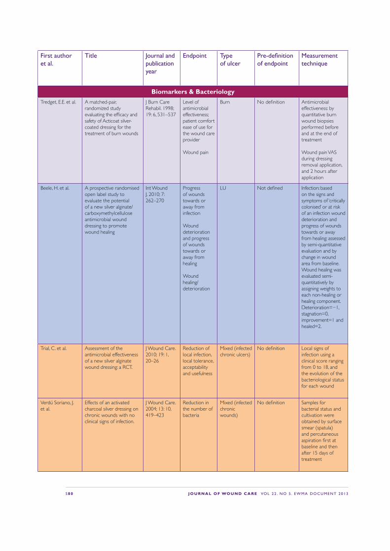
S 8 0 J O U R N A L O F WO U N D C A R E VO L 2 2 . N O 5 . E W M A D O C U M E N T 2 0 1 3
First author et al.
Title Journal and publication year
Endpoint Type of ulcer
Pre-de!nition of endpoint
Measurement technique
Biomarkers & BacteriologyTredget, E.E. et al. A matched-pair,
randomized study evaluating the ef!cacy and safety of Acticoat silver-coated dressing for the treatment of burn wounds
J Burn Care Rehabil. 1998; 19: 6, 531–537
Level of antimicrobial effectiveness; patient comfort ease of use for the wound care provider
Wound pain
Burn No de!nition Antimicrobial effectiveness: by quantitative burn wound biopsies performed before and at the end of treatment
Wound pain VAS during dressing removal application, and 2 hours after application
Beele, H. et al. A prospective randomised open label study to evaluate the potential of a new silver alginate/carboxymethylcellulose antimicrobial wound dressing to promote wound healing
Int Wound J. 2010; 7: 262–270
Progress of wounds towards or away from infection
Wound deterioration and progress of wounds towards or away from healing
Wound healing/deterioration
LU Not de!ned Infection: based on the signs and symptoms of ‘critically colonised’ or at risk of an infection wound deterioration and progress of wounds towards or away from healing: assessed by semi-quantitative evaluation and by change in wound area from baseline. Wound healing was evaluated semi-quantitatively by assigning weights to each non-healing or healing component. Deterioration=−1, stagnation=0, improvement=1 and healed=2.
Trial, C. et al. Assessment of the antimicrobial effectiveness of a new silver alginate wound dressing: a RCT.
J Wound Care. 2010; 19: 1, 20–26
Reduction of local infection, local tolerance, acceptability and usefulness
Mixed (infected chronic ulcers)
No de!nition Local signs of infection using a clinical score ranging from 0 to 18, and the evolution of the bacteriological status for each wound
Verdú Soriano, J. et al.
Effects of an activated charcoal silver dressing on chronic wounds with no clinical signs of infection.
J Wound Care. 2004; 13: 10, 419–423
Reduction in the number of bacteria
Mixed (infected chronic wounds)
No de!nition Samples for bacterial status and cultivation were obtained by surface smear (spatula) and percutaneous aspiration !rst at baseline and then after 15 days of treatment

J O U R N A L O F WO U N D C A R E VO L 2 2 . N O 5 . E W M A D O C U M E N T 2 0 1 3 S 8 1
First author et al.
Title Journal and publication year
Endpoint Type of ulcer
Pre-de!nition of endpoint
Measurement technique
Biomarkers & BacteriologyTredget, E.E. et al. A matched-pair,
randomized study evaluating the ef!cacy and safety of Acticoat silver-coated dressing for the treatment of burn wounds
J Burn Care Rehabil. 1998; 19: 6, 531–537
Level of antimicrobial effectiveness; patient comfort ease of use for the wound care provider
Wound pain
Burn No de!nition Antimicrobial effectiveness: by quantitative burn wound biopsies performed before and at the end of treatment
Wound pain VAS during dressing removal application, and 2 hours after application
Beele, H. et al. A prospective randomised open label study to evaluate the potential of a new silver alginate/carboxymethylcellulose antimicrobial wound dressing to promote wound healing
Int Wound J. 2010; 7: 262–270
Progress of wounds towards or away from infection
Wound deterioration and progress of wounds towards or away from healing
Wound healing/deterioration
LU Not de!ned Infection: based on the signs and symptoms of ‘critically colonised’ or at risk of an infection wound deterioration and progress of wounds towards or away from healing: assessed by semi-quantitative evaluation and by change in wound area from baseline. Wound healing was evaluated semi-quantitatively by assigning weights to each non-healing or healing component. Deterioration=−1, stagnation=0, improvement=1 and healed=2.
Trial, C. et al. Assessment of the antimicrobial effectiveness of a new silver alginate wound dressing: a RCT.
J Wound Care. 2010; 19: 1, 20–26
Reduction of local infection, local tolerance, acceptability and usefulness
Mixed (infected chronic ulcers)
No de!nition Local signs of infection using a clinical score ranging from 0 to 18, and the evolution of the bacteriological status for each wound
Verdú Soriano, J. et al.
Effects of an activated charcoal silver dressing on chronic wounds with no clinical signs of infection.
J Wound Care. 2004; 13: 10, 419–423
Reduction in the number of bacteria
Mixed (infected chronic wounds)
No de!nition Samples for bacterial status and cultivation were obtained by surface smear (spatula) and percutaneous aspiration !rst at baseline and then after 15 days of treatment
First author et al.
Title Journal and publication year
Endpoint Type of ulcer
Pre-de!nition of endpoint
Measurement technique
Biomarkers & BacteriologyDella Paola, L. et al.
Super-oxidised solution (SOS) therapy for infected diabetic foot ulcers
Wounds. 2006; 18: 9, 262–270.
Reduction in bacterial load, healing time, incidence of skin reactions
DFU Probe to bone test, plain radiograph and debridement
Microbiological sample
Change in Wound ConditionCarneiro, P.M. and Nyawawa, E.T.
Topical phenytoin versus EUSOL in the treatment of non-malignant chronic leg ulcers
East Afr Med J. 2003; 80: 3, 124–129
Presence of discharge (purulent, serous, absent), Healthy granulation tissue
LU (various aetiologies)
Presence of discharge (purulent, serous, absent)
Clinical evaluation
Gray, M. and Jones, D.P.
The effect of different formulations of equivalent active ingredients on the performance of two topical wound treatment products
Ostomy Wound Manage. 2004; 50: 3, 34–44
Erythema Mixed: Experimental laser induced partial thickness wounds
Not de!ned Erythema,oedema, scabbing and reepithelialisation
10-point scales for each endpoint
Costs & Resources UsedClay, P.G. et al. Clinical ef!cacy, tolerability,
and cost savings associated with the use of open-label metronidazole plus ceftriaxone once daily compared with ticarcillin/clavulanate every 6 hours as empiric treatment for diabetic lower-extremity infections in older males
Am J Geriat Pharmaco. 2004; 2: 3, 181–189
Institutional cost
DFU (Wagner 1–3, infection)
Cost for antibiotics and treatment days —contract prices
Generalised per patient group not individualised
Jull, A. et al. Randomized clinical trial of honey-impregnated dressings for venous leg ulcers
Br J Surg. 2008; 95: 2, 175–182
Cost LU (VLU) No de!nition (infection, adverse effects QoL, cost/effect)
—
Dressing PerformanceDumville, J.C. et al.
Larval therapy for leg ulcers (VenUS II): randomised controlled trial.
BMJ. 2009; 338: b773 Adverse effects LU (mixed) No de!nition Lab analyses Clinical observation Visual analog scale
Jull, A. et al. Randomized clinical trial of honey-impregnated dressings for venous leg ulcers
Br J Surg. 2008; 95: 2, 175–182
Adverse effects LU (VLU) Infections, adverse effects QoL, cost/effect
Clinical sign of infection
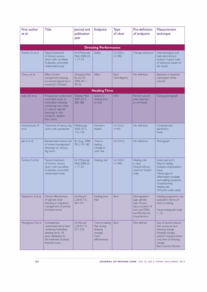
S 8 2 J O U R N A L O F WO U N D C A R E VO L 2 2 . N O 5 . E W M A D O C U M E N T 2 0 1 3
First author et al.
Title Journal and publication year
Endpoint Type of ulcer
Pre-de!nition of endpoint
Measurement technique
Dressing PerformanceTumino, G. et al. Topical treatment
of chronic venous ulcers with sucralfate: A placebo controlled randomized study
Int J Molecular Med. 2008; 22: 1, 17–23
Safety LU (VLU; n=100)
Therapy tolerance Haematological and haematochemical analysis: 4-point scale of tolerance based on lab results
Chen, J. et al. Effect of silver nanoparticle dressing on second degree burn wound [in Chinese]
Zhonghua Wai Ke Za Zhi. 2006; 44: 1, 50–52
Effect Burn (2nd degree)
No de!nition Reduction in bacterial colonisation of the wounds
Healing TimeJude, E.B. et al. Prospective randomized
controlled study of Hydro!ber dressing containing ionic silver or calcium alginate dressings in non-ischaemic diabetic foot ulcers
Diabetic Med. 2007; 24: 3, 280–288
Speed of healing, time to heal
DFU Percent wound area reduction or cm2/week
Tracing photograph
Kucharzewski, M. et al.
Treatment of venous leg ulcers with sulodexide
Phlebologie. 2003; 32: 5, 115–120
Numbers healed
LU (VLU; n=44)
No de!nition Computerised planimetrySwab
Jull, A. et al. Randomized clinical trial of honey-impregnated dressings for venous leg ulcers
Br J Surg. 2008; 95: 2, 175–182
Time to healing Change in ulcer size
LU (VLU) No de!nition Photograph
Tumino, G. et al. Topical treatment of chronic venous ulcers with sucralfate: A placebo-controlled randomized study
Int J Molecular Med. 2008; 22: 1, 17–23
Healing rate LU (VLU; n=100)
Healing rate in daysOverall ef!cacy rated on 4-point scale
Lesion size (cm2)Days to healing Evolution of granulation tissue Clinical signs of in"ammation, exudate and swelling, symptoms of pain/burning Healing rate (3/4-point scales used)
Opasanon, S. et al. Clinical effectiveness of alginate silver dressing in outpatient management of partial-thickness burns.
Int Wound J. 2010; 7: 6, 467–471
Healing timePain
Burn Demographics (age, gender, type of burn injury, location of burn and TBSA burn%) Wound characteristics
Healing progression was assessed in terms of time to healing.
Visual analog pain scale 1–10;
Muangman, P. et al. A prospective, randomized trial of silver containing Hydro!ber dressing versus 1% silver sulfadiazine for the treatment of partial thickness burns.
Int Wound J. 2010; 7: 4, 271–276
Time to healing Pain during dressing changes, Cost-effectiveness.
Burn Not de!ned Day of wound closure Pain scores at each dressing change Hospital charges, patient’s transportation cost, time of dressing change Burn wound infection
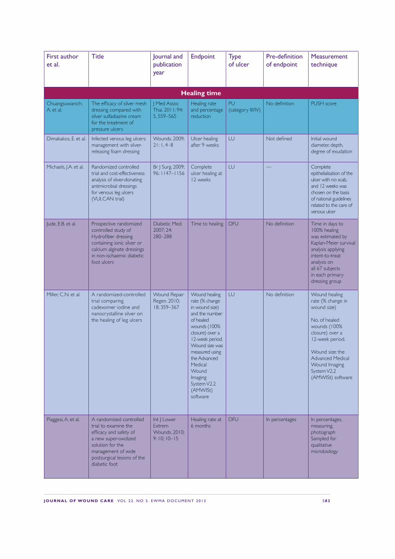
J O U R N A L O F WO U N D C A R E VO L 2 2 . N O 5 . E W M A D O C U M E N T 2 0 1 3 S 8 3
First author et al.
Title Journal and publication year
Endpoint Type of ulcer
Pre-de!nition of endpoint
Measurement technique
Dressing PerformanceTumino, G. et al. Topical treatment
of chronic venous ulcers with sucralfate: A placebo controlled randomized study
Int J Molecular Med. 2008; 22: 1, 17–23
Safety LU (VLU; n=100)
Therapy tolerance Haematological and haematochemical analysis: 4-point scale of tolerance based on lab results
Chen, J. et al. Effect of silver nanoparticle dressing on second degree burn wound [in Chinese]
Zhonghua Wai Ke Za Zhi. 2006; 44: 1, 50–52
Effect Burn (2nd degree)
No de!nition Reduction in bacterial colonisation of the wounds
Healing TimeJude, E.B. et al. Prospective randomized
controlled study of Hydro!ber dressing containing ionic silver or calcium alginate dressings in non-ischaemic diabetic foot ulcers
Diabetic Med. 2007; 24: 3, 280–288
Speed of healing, time to heal
DFU Percent wound area reduction or cm2/week
Tracing photograph
Kucharzewski, M. et al.
Treatment of venous leg ulcers with sulodexide
Phlebologie. 2003; 32: 5, 115–120
Numbers healed
LU (VLU; n=44)
No de!nition Computerised planimetrySwab
Jull, A. et al. Randomized clinical trial of honey-impregnated dressings for venous leg ulcers
Br J Surg. 2008; 95: 2, 175–182
Time to healing Change in ulcer size
LU (VLU) No de!nition Photograph
Tumino, G. et al. Topical treatment of chronic venous ulcers with sucralfate: A placebo-controlled randomized study
Int J Molecular Med. 2008; 22: 1, 17–23
Healing rate LU (VLU; n=100)
Healing rate in daysOverall ef!cacy rated on 4-point scale
Lesion size (cm2)Days to healing Evolution of granulation tissue Clinical signs of in"ammation, exudate and swelling, symptoms of pain/burning Healing rate (3/4-point scales used)
Opasanon, S. et al. Clinical effectiveness of alginate silver dressing in outpatient management of partial-thickness burns.
Int Wound J. 2010; 7: 6, 467–471
Healing timePain
Burn Demographics (age, gender, type of burn injury, location of burn and TBSA burn%) Wound characteristics
Healing progression was assessed in terms of time to healing.
Visual analog pain scale 1–10;
Muangman, P. et al. A prospective, randomized trial of silver containing Hydro!ber dressing versus 1% silver sulfadiazine for the treatment of partial thickness burns.
Int Wound J. 2010; 7: 4, 271–276
Time to healing Pain during dressing changes, Cost-effectiveness.
Burn Not de!ned Day of wound closure Pain scores at each dressing change Hospital charges, patient’s transportation cost, time of dressing change Burn wound infection
First author et al.
Title Journal and publication year
Endpoint Type of ulcer
Pre-de!nition of endpoint
Measurement technique
Healing timeChuangsuwanich, A. et al.
The ef!cacy of silver mesh dressing compared with silver sulfadiazine cream for the treatment of pressure ulcers
J Med Assoc Thai. 2011; 94: 5, 559–565
Healing rate and percentage reduction
PU (category III/IV)
No de!nition PUSH score
Dimakakos, E. et al. Infected venous leg ulcers: management with silver-releasing foam dressing
Wounds. 2009; 21: 1, 4–8
Ulcer healing after 9 weeks
LU Not de!ned Initial wound diameter, depth, degree of exudation
Michaels, J.A. et al. Randomized controlled trial and cost-effectiveness analysis of silver-donating antimicrobial dressings for venous leg ulcers (VULCAN trial)
Br J Surg. 2009; 96: 1147–1156
Complete ulcer healing at 12 weeks
LU — Complete epithelialisation of the ulcer with no scab, and 12 weeks was chosen on the basis of national guidelines related to the care of venous ulcer
Jude, E.B. et al. Prospective randomized controlled study of Hydro!ber dressing containing ionic silver or calcium alginate dressings in non-ischaemic diabetic foot ulcers
Diabetic Med. 2007; 24: 280–288
Time to healing DFU No de!nition Time in days to 100% healing was estimated by Kaplan-Meier survival analysis applying intent-to-treat analysis on all 67 subjects in each primary dressing group
Miller, C.N. et al. A randomized-controlled trial comparing cadexomer iodine and nanocrystalline silver on the healing of leg ulcers
Wound Repair Regen. 2010; 18; 359–367
Wound healing rate (% change in wound size) and the number of healed wounds (100% closure) over a 12-week period. Wound size was measured using the AdvancedMedical Wound Imaging System V2.2 (AMWISt) software
LU No de!nition Wound healing rate (% change in wound size)
No. of healed wounds (100% closure) over a 12-week period.
Wound size: the Advanced Medical Wound Imaging System V2.2 (AMWISt) software
Piaggesi, A. et al. A randomized controlled trial to examine the ef!cacy and safety of a new super-oxidized solution for the management of wide postsurgical lesions of the diabetic foot
Int J Lower Extrem Wounds. 2010; 9: 10; 10–15
Healing rate at 6 months
DFU In percentages In percentages, measuring, photograph Sampled for qualitative microbiology
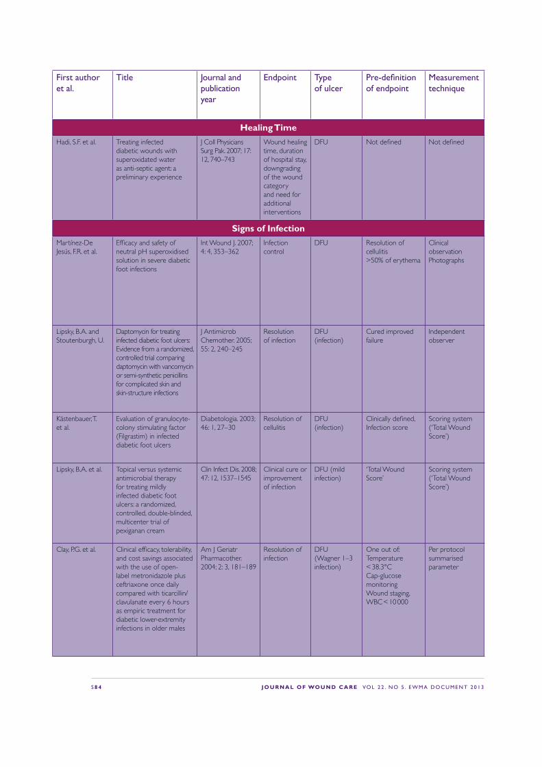
S 8 4 J O U R N A L O F WO U N D C A R E VO L 2 2 . N O 5 . E W M A D O C U M E N T 2 0 1 3
First author et al.
Title Journal and publication year
Endpoint Type of ulcer
Pre-de!nition of endpoint
Measurement technique
Healing TimeHadi, S.F. et al. Treating infected
diabetic wounds with superoxidated water as anti-septic agent: a preliminary experience
J Coll Physicians Surg Pak. 2007; 17: 12, 740–743
Wound healing time, duration of hospital stay, downgrading of the wound category and need for additional interventions
DFU Not de!ned Not de!ned
Signs of InfectionMartínez-De Jesús, F.R. et al.
Ef!cacy and safety of neutral pH superoxidised solution in severe diabetic foot infections
Int Wound J. 2007; 4: 4, 353–362
Infection control
DFU Resolution of cellulitis >50% of erythema
Clinical observation Photographs
Lipsky, B.A. and Stoutenburgh, U.
Daptomycin for treating infected diabetic foot ulcers: Evidence from a randomized, controlled trial comparing daptomycin with vancomycin or semi-synthetic penicillins for complicated skin and skin-structure infections
J Antimicrob Chemother. 2005; 55: 2, 240–245
Resolution of infection
DFU (infection)
Cured improved failure
Independent observer
Kästenbauer, T. et al.
Evaluation of granulocyte-colony stimulating factor (Filgrastim) in infected diabetic foot ulcers
Diabetologia. 2003; 46: 1, 27–30
Resolution of cellulitis
DFU (infection)
Clinically de!ned, Infection score
Scoring system (‘Total Wound Score’)
Lipsky, B.A. et al. Topical versus systemic antimicrobial therapy for treating mildly infected diabetic foot ulcers: a randomized, controlled, double-blinded, multicenter trial of pexiganan cream
Clin Infect Dis. 2008; 47: 12, 1537–1545
Clinical cure or improvement of infection
DFU (mild infection)
‘Total Wound Score’
Scoring system (‘Total Wound Score’)
Clay, P.G. et al. Clinical ef!cacy, tolerability, and cost savings associated with the use of open-label metronidazole plus ceftriaxone once daily compared with ticarcillin/clavulanate every 6 hours as empiric treatment for diabetic lower-extremity infections in older males
Am J Geriatr Pharmacother. 2004; 2: 3, 181–189
Resolution of infection
DFU (Wagner 1–3 infection)
One out of: Temperature < 38.3°C Cap-glucose monitoring Wound staging, WBC < 10 000
Per protocol summarised parameter

J O U R N A L O F WO U N D C A R E VO L 2 2 . N O 5 . E W M A D O C U M E N T 2 0 1 3 S 8 5
First author et al.
Title Journal and publication year
Endpoint Type of ulcer
Pre-de!nition of endpoint
Measurement technique
Healing TimeHadi, S.F. et al. Treating infected
diabetic wounds with superoxidated water as anti-septic agent: a preliminary experience
J Coll Physicians Surg Pak. 2007; 17: 12, 740–743
Wound healing time, duration of hospital stay, downgrading of the wound category and need for additional interventions
DFU Not de!ned Not de!ned
Signs of InfectionMartínez-De Jesús, F.R. et al.
Ef!cacy and safety of neutral pH superoxidised solution in severe diabetic foot infections
Int Wound J. 2007; 4: 4, 353–362
Infection control
DFU Resolution of cellulitis >50% of erythema
Clinical observation Photographs
Lipsky, B.A. and Stoutenburgh, U.
Daptomycin for treating infected diabetic foot ulcers: Evidence from a randomized, controlled trial comparing daptomycin with vancomycin or semi-synthetic penicillins for complicated skin and skin-structure infections
J Antimicrob Chemother. 2005; 55: 2, 240–245
Resolution of infection
DFU (infection)
Cured improved failure
Independent observer
Kästenbauer, T. et al.
Evaluation of granulocyte-colony stimulating factor (Filgrastim) in infected diabetic foot ulcers
Diabetologia. 2003; 46: 1, 27–30
Resolution of cellulitis
DFU (infection)
Clinically de!ned, Infection score
Scoring system (‘Total Wound Score’)
Lipsky, B.A. et al. Topical versus systemic antimicrobial therapy for treating mildly infected diabetic foot ulcers: a randomized, controlled, double-blinded, multicenter trial of pexiganan cream
Clin Infect Dis. 2008; 47: 12, 1537–1545
Clinical cure or improvement of infection
DFU (mild infection)
‘Total Wound Score’
Scoring system (‘Total Wound Score’)
Clay, P.G. et al. Clinical ef!cacy, tolerability, and cost savings associated with the use of open-label metronidazole plus ceftriaxone once daily compared with ticarcillin/clavulanate every 6 hours as empiric treatment for diabetic lower-extremity infections in older males
Am J Geriatr Pharmacother. 2004; 2: 3, 181–189
Resolution of infection
DFU (Wagner 1–3 infection)
One out of: Temperature < 38.3°C Cap-glucose monitoring Wound staging, WBC < 10 000
Per protocol summarised parameter
First author et al.
Title Journal and publication year
Endpoint Type of ulcer
Pre-de!nition of endpoint
Measurement technique
Signs of InfectionLipsky, B.A. et al. Ertapenem versus
piperacillin/tazobactam for diabetic foot infections (SIDESTEP): prospective, randomised, controlled, double-blinded, multicentre trial
Lancet. 2005; 366: 9498, 1695–1703
Resolution of infection
DFU (infection)
Favourable clinical response/cure
At the discretion of the physican
Lipsky, B.A. et al. Treating diabetic foot infections with sequential intravenous to oral moxi"oxacin compared with piperacillin-tazobactam/amoxicillin-clavulanate
J Antimicrob Chemother. 2007; 60: 2, 370–376
Resolution of infection
DFU (infection)
Clinically de!ned At the discretion of the physician
Lipsky, B.A. et al. Treating foot infections in diabetic patients: a randomized, multicenter, open-label trial of linezolid versus ampicillin-sulbactam/amoxicillin- clavulanate
Clin Infect Dis. 2004; 38: 1, 17–24
Resolution of infection
DFU (infection)
Cured, improved or failure
At the discretion of the physician
Jull, A. et al. Randomized clinical trial of honey-impregnated dressings for venous leg ulcers
Br J Surg. 2008; 95: 2, 175–182
Infection LU (VLU) No de!nition Clinical sign of infection
Krishnamoorthy, L. et al.
The clinical and histological effects of Dermagraft in the healing of chronic venous leg ulcers
Phlebology. 2003; 18: 1, 12–22
Wound infection
LU (VLU) No de!nition Clinical sign
Meaume, S. et al. A study to compare a new self-adherent soft silicone dressing with a self-adherent polymer dressing in stage II pressure ulcers.
Ostomy Wound Manage. 2003; 49: 9, 44–51
Signs of in"ammation
PU (category II; n=38)
No de!ntion Size by tracing: other variables as present or absent Exudate: low, moderate or high Granulation tissue as covering 0–25%, 26–50%, 51–75%, 76–100% Surrounding skin damage was described as redness, blisters or other Dressing removal was rated as very easy, easy, minor dif!culties or dif!cult
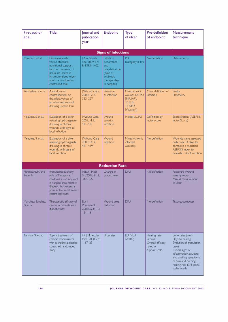
S 8 6 J O U R N A L O F WO U N D C A R E VO L 2 2 . N O 5 . E W M A D O C U M E N T 2 0 1 3
First author et al.
Title Journal and publication year
Endpoint Type of ulcer
Pre-de!nition of endpoint
Measurement technique
Signs of InfectionsCereda, E. et al. Disease-speci!c,
versus standard, nutritional support for the treatment of pressure ulcers in institutionalized older adults: a randomized controlled trial
J Am Geriatr Soc. 2009; 57: 8, 1395–1402
Infection occurrence and hospitalisation (days of antibiotic therapy; days in hospital)
PU (category II–IV)
No de!nition Data records
Kordestani, S. et al. A randomised controlled trial on the effectiveness of an advanced wound dressing used in Iran
J Wound Care. 2008; 17: 7, 323–327
Presence of infection
Mixed: chronic wounds (28 PU [NPUAP], 20 LUs, 12 DFU [Wagner])
Clear de!nition of infection
Swabs Planimetry
Meaume, S. et al. Evaluation of a silver-releasing hydroalginate dressing in chronic wounds with signs of local infection
J Wound Care. 2005; 14: 9, 411–419
Wound severity, infection
Mixed: LU, PU De!nition by index score
Score system (ASEPSIS Index Score)
Meaume, S. et al. Evaluation of a silver-releasing hydroalginate dressing in chronic wounds with signs of local infection
J Wound Care 2005; 14; 9, 411–419
Wound infection
Mixed (chronic infected wounds)
No de!nition Wounds were assessed daily over 14 days to complete a modi!ed ASEPSIS index to evaluate risk of infection
Reduction RatePurandare, H. and Supe, A.
Immunomodulatory role of Tinospora cordifolia as an adjuvant in surgical treatment of diabetic foot ulcers: a prospective randomized controlled study
Indian J Med Sci. 2007; 61: 6, 347–355
Change in wound area
DFU No de!nition Peccoraro Wound severity score Manual measurement of ulcer
Martínez-Sánchez, G. et al.
Therapeutic ef!cacy of ozone in patients with diabetic foot
Eur J Pharmacol. 2005; 523: 1–3, 151–161
Wound area reduction
DFU No de!nition Tracing, computer
Tumino, G. et al. Topical treatment of chronic venous ulcers with sucralfate: a placebo-controlled randomized study
Int J Molecular Med. 2008; 22: 1, 17–23
Ulcer size LU (VLU; n=100)
Healing rate in daysOverall ef!cacy rated on 4-point scale
Lesion size (cm2)Days to healing Evolution of granulation tissue Clinical signs of in"ammation, exudate and swelling: symptoms of pain and burning: healing rate (3/4–point scales used)

J O U R N A L O F WO U N D C A R E VO L 2 2 . N O 5 . E W M A D O C U M E N T 2 0 1 3 S 8 7
First author et al.
Title Journal and publication year
Endpoint Type of ulcer
Pre-de!nition of endpoint
Measurement technique
Signs of InfectionsCereda, E. et al. Disease-speci!c,
versus standard, nutritional support for the treatment of pressure ulcers in institutionalized older adults: a randomized controlled trial
J Am Geriatr Soc. 2009; 57: 8, 1395–1402
Infection occurrence and hospitalisation (days of antibiotic therapy; days in hospital)
PU (category II–IV)
No de!nition Data records
Kordestani, S. et al. A randomised controlled trial on the effectiveness of an advanced wound dressing used in Iran
J Wound Care. 2008; 17: 7, 323–327
Presence of infection
Mixed: chronic wounds (28 PU [NPUAP], 20 LUs, 12 DFU [Wagner])
Clear de!nition of infection
Swabs Planimetry
Meaume, S. et al. Evaluation of a silver-releasing hydroalginate dressing in chronic wounds with signs of local infection
J Wound Care. 2005; 14: 9, 411–419
Wound severity, infection
Mixed: LU, PU De!nition by index score
Score system (ASEPSIS Index Score)
Meaume, S. et al. Evaluation of a silver-releasing hydroalginate dressing in chronic wounds with signs of local infection
J Wound Care 2005; 14; 9, 411–419
Wound infection
Mixed (chronic infected wounds)
No de!nition Wounds were assessed daily over 14 days to complete a modi!ed ASEPSIS index to evaluate risk of infection
Reduction RatePurandare, H. and Supe, A.
Immunomodulatory role of Tinospora cordifolia as an adjuvant in surgical treatment of diabetic foot ulcers: a prospective randomized controlled study
Indian J Med Sci. 2007; 61: 6, 347–355
Change in wound area
DFU No de!nition Peccoraro Wound severity score Manual measurement of ulcer
Martínez-Sánchez, G. et al.
Therapeutic ef!cacy of ozone in patients with diabetic foot
Eur J Pharmacol. 2005; 523: 1–3, 151–161
Wound area reduction
DFU No de!nition Tracing, computer
Tumino, G. et al. Topical treatment of chronic venous ulcers with sucralfate: a placebo-controlled randomized study
Int J Molecular Med. 2008; 22: 1, 17–23
Ulcer size LU (VLU; n=100)
Healing rate in daysOverall ef!cacy rated on 4-point scale
Lesion size (cm2)Days to healing Evolution of granulation tissue Clinical signs of in"ammation, exudate and swelling: symptoms of pain and burning: healing rate (3/4–point scales used)
First author et al.
Title Journal and publication year
Endpoint Type of ulcer
Pre-de!nition of endpoint
Measurement technique
Reduction RateYapucu Günes, U. and Eser, I.
Effectiveness of a honey dressing for healing pressure ulcers
J Wound Ostomy Continence Nurs. 2007; 34: 2, 184–190
Healing PU (category II/III; n=26
Change in PUSH score
Acetate tracing PUSH tool
Meaume, S. et al. Evaluation of a silver-releasing hydroalginate dressing in chronic wounds with signs of local infection
J Wound Care. 2005; 14: 9, 411–419
Closure rate Mixed: LU, PU No de!nition Percentage area reduction Tracing
Harding, K. et al. A prospective, multi-centre, randomised, open label, parallel, comparative study to evaluate effects of AQUACEL Ag and Urgotul Silver dressing on healing of chronic venous leg ulcers
Int Wound J. 2011; doi: 10.1111/j. 1742-481X.2011. 00881.x
Size reduction LU (VLU) No de!nition Photograph Wound status, perilesional skinappearance and condition of the wound were recorded
Lazareth, I. et al. The role of a silver releasing lipido-colloid contact layer in venous leg ulcers presenting in"ammatory signs suggesting heavy bacterial colonization: results of a randomized controlled study
Wounds. 2008; 20: 6, 158–166
Reduction of surface area
LU (VLU) No de!nition No de!nition
Wunderlich, U. and Orfanos, O.E.
Treatment of venous ulcera cruris with dry wound dressings. Phase overlapping use of silver impregnated activated charcoal xerodressing [in German]
Hautarzt. 1991; 42: 7, 446–450
Epithelialisation Reduction of ulcer size
LU No de!nition The parameters of wound healing were documented
Jørgensen, B. et al. The silver-releasing foam dressing, Contreet Foam, promotes faster healing of critically colonised venous leg ulcers: a randomised controlled trial
Int Wound J. 2005; 2: 1, 64–73
Reduction rate LU (VLU) No de!nition Wound size was traced using transparent wound tracing sheets and measured using Image Pro Plus S.O software
Münter, K.C. et al. Effect of a sustained silver-releasing dressing on ulcers with delayed healing: the CONTOP study
J Wound Care. 2006; 15: 5, 199–206
Reduction in wound size
Mixed (chronic wounds)
No de!nition No de!nition

S 8 8 J O U R N A L O F WO U N D C A R E VO L 2 2 . N O 5 . E W M A D O C U M E N T 2 0 1 3
First author et al.
Title Journal and publication year
Endpoint Type of ulcer
Pre-de!nition of endpoint
Measurement technique
Reduction RateRussell, L. The CONTOP
multinational study: preliminary data from the UK arm
Wounds UK. 2005; 1: 44–54
Relative reduction in wound area
Mixed (chronic wounds)
No de!nition No de!nition
Lund-Nielsen, B. et al.
Qualitative bacteriology in malignant wounds—a prospective, randomized, clinical study to compare the effect of honey and silver dressings
Ostomy Wound Manage. 2011; 57: 7, 28–36.
Reduction of wound sizeDressings in"uenced the presence of potential wound pathogens that may increase the risk of wound infection
MFW Swab cultures Digital photographs Swab
Robson, V. et al. Standardized antibacterial honey (Medihoney) with standard therapy in wound care: randomized clinical trail
J Adv Nurs. 2008; 65: 3, 565–575
Healing timeTime to 50% reduction in wound area
Mixed (chronic wounds)
Assessment with report forms
Wound photographs and measurements
Gethin, G. et al. Manuka honey vs. hydrogel—a prospective, open label, multicentre, randomised controlled trail to compare desloughing ef!cacy and healing outcomes in venous ulcers
J Clin Nurs. 2009; 18: 3, 466–474
Wound healing Slough reduction
LU (VLU) No de!nition Measurement using Visitrak digital planimetry
Marshall, C. et al. Honey vs povidone iodine following toenail surgery
Wounds UK. 2005; 5: 10–18
Time for complete re-epitelisation
Other Assessment for toenail surgery
Assessment
Robson, V. et al. Randomised controlled feasibility trail on the use of medical grade honey following microvascular free tissue transfer to reduce the incidence of wound infection
Br J Oral Maxillofac Surg. 2012; 50: 4, 321–527
Reduction of incidence of wound infection
Other Swab Swab
Nagl, M. et al. Tolerability and ef!cacy of N-chlorotaurine in comparison with chloramine T for the treatment of chronic leg ulcers with a purulent coating: a randomized phase II study
Br J Dermatol/ 2003; 149: 3, 590–597
Intensity of pain
LU (not de!ned)
Intensity of pain VAS scale

J O U R N A L O F WO U N D C A R E VO L 2 2 . N O 5 . E W M A D O C U M E N T 2 0 1 3 S 8 9
First author et al.
Title Journal and publication year
Endpoint Type of ulcer
Pre-de!nition of endpoint
Measurement technique
Reduction RateRussell, L. The CONTOP
multinational study: preliminary data from the UK arm
Wounds UK. 2005; 1: 44–54
Relative reduction in wound area
Mixed (chronic wounds)
No de!nition No de!nition
Lund-Nielsen, B. et al.
Qualitative bacteriology in malignant wounds—a prospective, randomized, clinical study to compare the effect of honey and silver dressings
Ostomy Wound Manage. 2011; 57: 7, 28–36.
Reduction of wound sizeDressings in"uenced the presence of potential wound pathogens that may increase the risk of wound infection
MFW Swab cultures Digital photographs Swab
Robson, V. et al. Standardized antibacterial honey (Medihoney) with standard therapy in wound care: randomized clinical trail
J Adv Nurs. 2008; 65: 3, 565–575
Healing timeTime to 50% reduction in wound area
Mixed (chronic wounds)
Assessment with report forms
Wound photographs and measurements
Gethin, G. et al. Manuka honey vs. hydrogel—a prospective, open label, multicentre, randomised controlled trail to compare desloughing ef!cacy and healing outcomes in venous ulcers
J Clin Nurs. 2009; 18: 3, 466–474
Wound healing Slough reduction
LU (VLU) No de!nition Measurement using Visitrak digital planimetry
Marshall, C. et al. Honey vs povidone iodine following toenail surgery
Wounds UK. 2005; 5: 10–18
Time for complete re-epitelisation
Other Assessment for toenail surgery
Assessment
Robson, V. et al. Randomised controlled feasibility trail on the use of medical grade honey following microvascular free tissue transfer to reduce the incidence of wound infection
Br J Oral Maxillofac Surg. 2012; 50: 4, 321–527
Reduction of incidence of wound infection
Other Swab Swab
Nagl, M. et al. Tolerability and ef!cacy of N-chlorotaurine in comparison with chloramine T for the treatment of chronic leg ulcers with a purulent coating: a randomized phase II study
Br J Dermatol/ 2003; 149: 3, 590–597
Intensity of pain
LU (not de!ned)
Intensity of pain VAS scale
First author et al.
Title Journal and publication year
Endpoint Type of ulcer
Pre-de!nition of endpoint
Measurement technique
Symptoms, SignsVaras, R.P. et al. A prospective, randomized
trial of Acticoat versus silver sulfadiazine in the treatment of partial-thickness burns: which method Is less painful?
J Burn Care Rehabil. 2005; 26: 4, 344–347
Pain Burn No de!nition VAS
Romanelli, M. and Price, P.
Health-related quality of life aspects after treatment with a foam dressing and a silver-containing foam dressing in chronic leg ulcers
J Am Acad Dermatol. 2005; 52: 21
Reduction of odour Pain
LU No de!nition No de!nition
Della Paola, L. et al.
Super-oxidized solution (SOS) therapy for infected diabetic foot ulcers
Wounds. 2006; 18: 9, 262–270
Reduction of bacterial load
DFU No de!nition Measuring the number of strains quanti!ed at enrollement and at the time of operative closure
Wound ClosureJude, E.B. et al. Prospective randomized
controlled study of Hydro!ber dressing containing ionic silver or calcium alginate dressings in non-ischaemic diabetic foot ulcers
Diabetic Med. 2007; 24: 3, 280–288
Wound closure DFU Not de!ned Days to closure
Lazareth, I. et al. The role of a silver releasing lipido-colloid contact layer in venous leg ulcers presenting in"ammatory signs suggesting heavy bacterial colonization: results of a randomized controlled study
Wounds, 2008; 20: 6, 158–166
Wound closure LU Yes Clinical evaluation
Jull, A. et al. Randomized clinical trial of honey-impregnated dressings for venous leg ulcers
Br J Surg. 2008; 95: 2, 175–182
Complete healing LU (VLU) Complete epithelialisation, no scab
Complete epithelialisation, no scab
Daróczy, J. Quality control in chronic wound management: the role of local povidone-iodine (Betadine) therapy
Dermatology. 2006; 212: (Suppl. 1), 82–87
Percentage healed Relapse rate of super!cial bacterial skin infections (bacterial culture)
LU (VLU; n=63)
Percentage healed Relapse rate of super!cial bacterial skin infections (bacterial culture)
No de!nition

Journal of Wound Care (JWC) is the de! nitive wound-care journal and the leading source of tissue viability research and clinical information. Launched in 1992, it is designed to meet the needs of the multidisciplinary team. The journal is essential reading for all wound-care specialists — nurses, doctors and researchers — who are keen to keep up-to-date with all developments in wound management and tissue viability, but also appeals to generalists wishing to enhance their practice.
JWC is internationally renowned for its cutting edge and state-of-the-art research and practice articles. The journal also covers management, education and novel therapies. Articles are rigorously peer-reviewed by a panel of international experts.
For information on how to subscribe, call 0800 137 201 or visit our website www.journalofwoundcare.com Here you can also search and access all articles published in the journal since 1999.
j o u rnal of w o u nd ca r e ? ?
&�:�
&�
? ? ? v o l u m e 2 1 . n u m b e r 1 1 . n o v e m b e r 2 0 1 2
Effect of continuous pressure monitoring
on strategic shifting of medical inpatients
Odyssey RCT: Efficacy of two compression systems in the management of VLUs
Yakult: A role in combating multi-drug resistant Pseudomonas aeruginosa?
Psychosocial impact of chronic wounds on patients with epidermolysis bullosa
Red flags and alarm bells: An atypical lesion masquerading as a diabetic foot ulcer
RCT to evaluate a hyaluronic acid-impregnated gauze pad in leg ulcers
Clinical case-series evaluation of a superabsorbent dressing on exuding wounds
-:&B��B��BFRYHU�LQGG����
������������������
&�:�
Effect of continuous pressure monitoring
on strategic shifting of medical inpatients
Odyssey RCT: Efficacy of two compression systems in the management of VLUs
Pseudomonas aeruginosa?
Psychosocial impact of chronic wounds on patients with epidermolysis bullosa
Red flags and alarm bells: An atypical lesion masquerading as a diabetic foot ulcer
RCT to evaluate a hyaluronic acid-impregnated gauze pad in leg ulcers
Clinical case-series evaluation of a superabsorbent dressing on exuding wounds
j o u rnal of w o u nd ca r e
? ? &�:�
&�
? ? ? v o l u m e 2 2 . n u m b e r 1 . j a n u a r y 2 0 1 3
Stem cells in burn care and reconstructionTreatment of hypergranulation at gastrostomy sites with table saltBenefits of active surgical re-closure for disrupted abdominal laparotomy wounds
Economic analysis of soft-heel casting for diabetic foot ulcersA novel surgical approach to calcinosis cutis using a collagen-elastin matrix
More effective cell-based therapy through biofilm suppressionPatients’ experiences of negative pressure wound therapy: A review
-:&B��B�BFRYHU�LQGG����
������������������“JWC publishes independent, accessible, high-quality, evidence-based articles that ful! l the needs of the multidisciplinary team, and provides a forum for the worldwide wound-care community.”

J O U R N A L O F WO U N D C A R E VO L 2 2 . N O 5 . E W M A D O C U M E N T 2 0 1 3 S 9 1
To subscribe, call 0800 137201 or visit www.journalofwoundcare.comQuote promotion code EWMA2013 to receive a 10% discount
Essential reading forall wound care professionals
Benefits of subscription: • Cutting edge research articles
from leading specialists
• Guidance in applying new clinical developments to your day-to-day practice
• Advice on managing tissue viability services
• Coverage of the latest developments in tissue viability education
• Regular supplements, including: venous leg ulcers, pressure ulcers, the diabetic foot, wound infection
“Journal of Wound Care is an extremely valuable resource that contains a
wealth of peer-reviewed papers detailing the latest advances in
wound care research. A must read for clinicians, academics and
researchers who want to advance their own knowledge/ practice
and keep abreast of the wound care literature.”
Caroline McIntosh, Head of Podiatry, National University of Ireland
-:&�68%6�$'����[���BQR�FRXSRQB(:0$�RIIHU�LQGG���� ������������������

A EWMA Document