Embed Size (px)
Citation preview
Anterior and Lateral and Anterior and Lateral and Posterior Compartments of Posterior Compartments of
CalfCalf
By- Dr. Armaan Singh
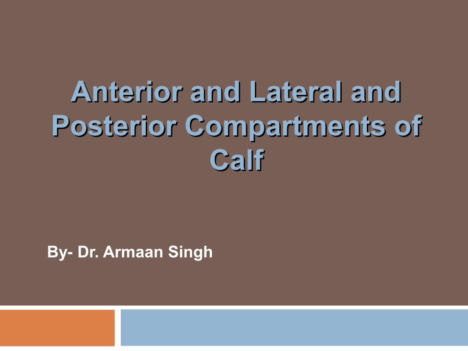
Cutaneous Supply of Calf Saphenous first branch is
infrapatellar branch it Supplies medial side of
the calf and Medial side of foot to ball
of the hallux Lateral cutaneous of calf Superficial peroneal Sural, lateral border of
foot Deep peroneal first cleft
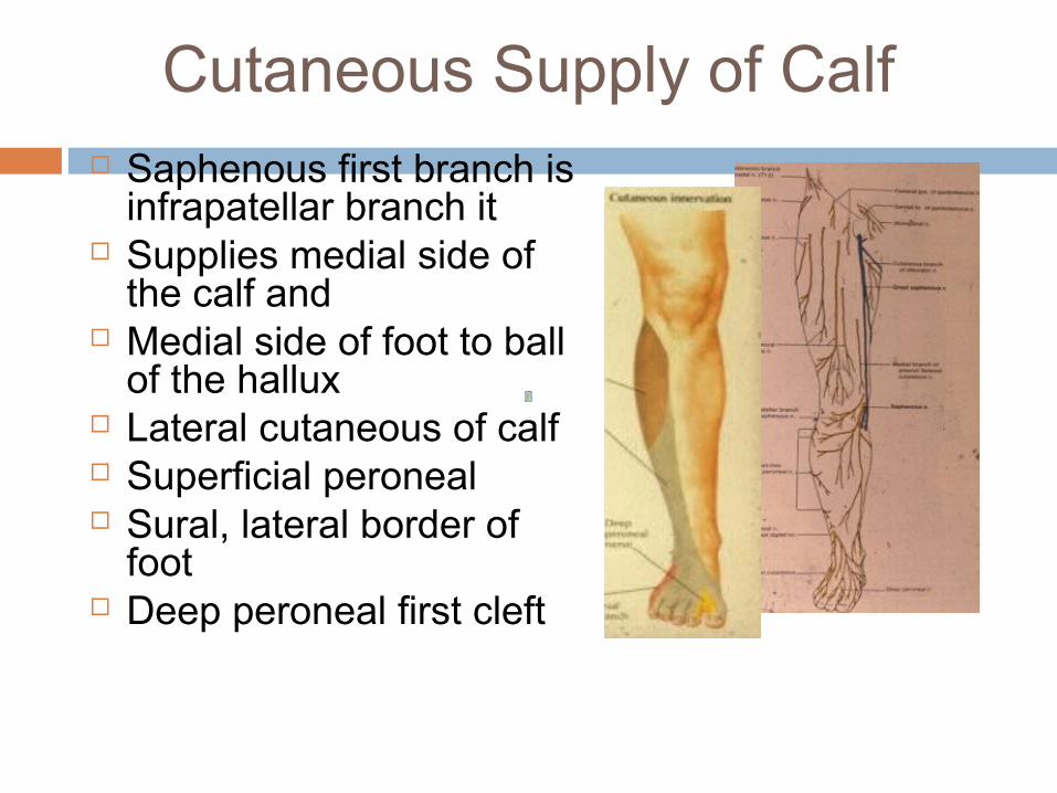
Fascial Compartments in Calf Subcutaneous surface of
tibia Fascia anterior and posterior
intermuscular septa Anterior compartment Lateral compartment Posterior compartmentDivided Superficial Deep Deep posterior
Extensor Retinacula
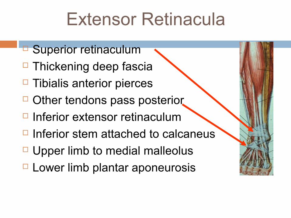
Superior retinaculum Thickening deep fascia Tibialis anterior pierces Other tendons pass posterior Inferior extensor retinaculum Inferior stem attached to calcaneus Upper limb to medial malleolus Lower limb plantar aponeurosis
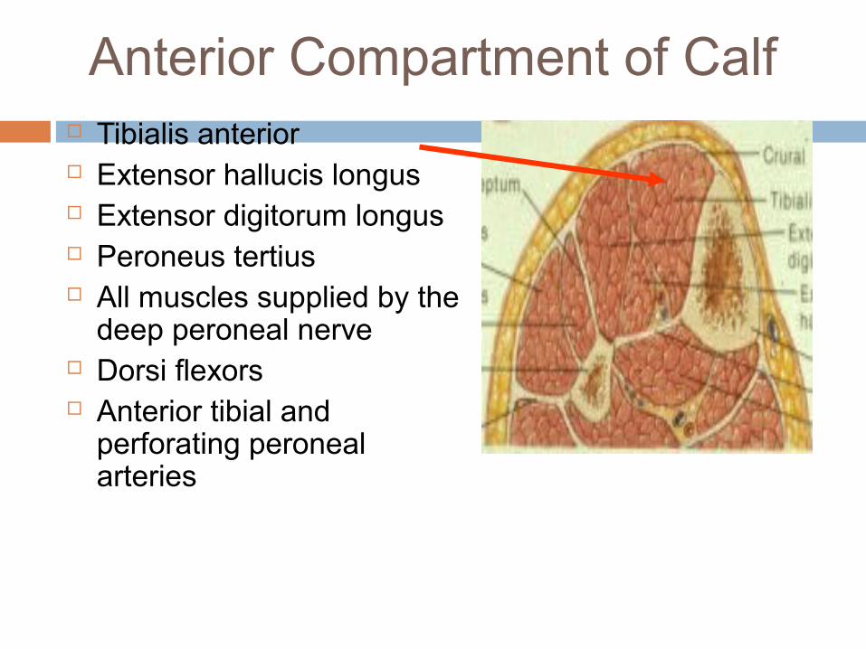
Anterior Compartment of Calf Tibialis anterior Extensor hallucis longus Extensor digitorum longus Peroneus tertius All muscles supplied by the
deep peroneal nerve Dorsi flexors Anterior tibial and
perforating peroneal arteries
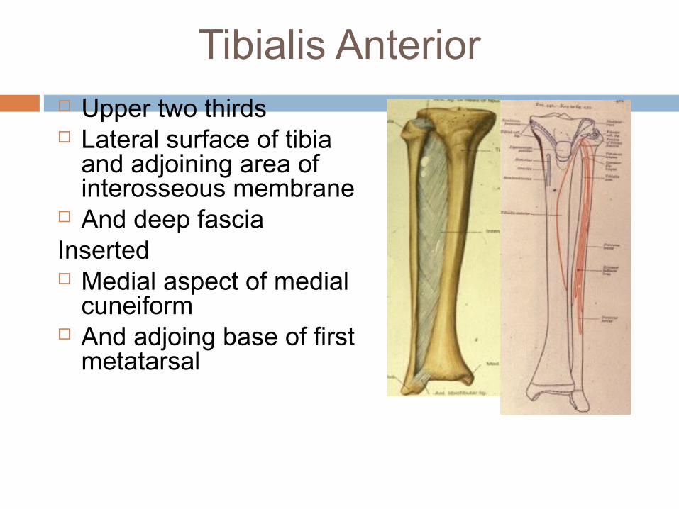
Tibialis Anterior Upper two thirds Lateral surface of tibia
and adjoining area of interosseous membrane
And deep fasciaInserted Medial aspect of medial
cuneiform And adjoing base of first
metatarsal
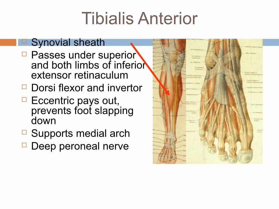
Tibialis Anterior Synovial sheath Passes under superior
and both limbs of inferior extensor retinaculum
Dorsi flexor and invertor Eccentric pays out,
prevents foot slapping down
Supports medial arch Deep peroneal nerve
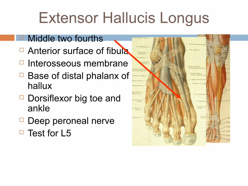
Extensor Hallucis Longus Middle two fourths Anterior surface of fibula Interosseous membrane Base of distal phalanx of
hallux Dorsiflexor big toe and
ankle Deep peroneal nerve Test for L5
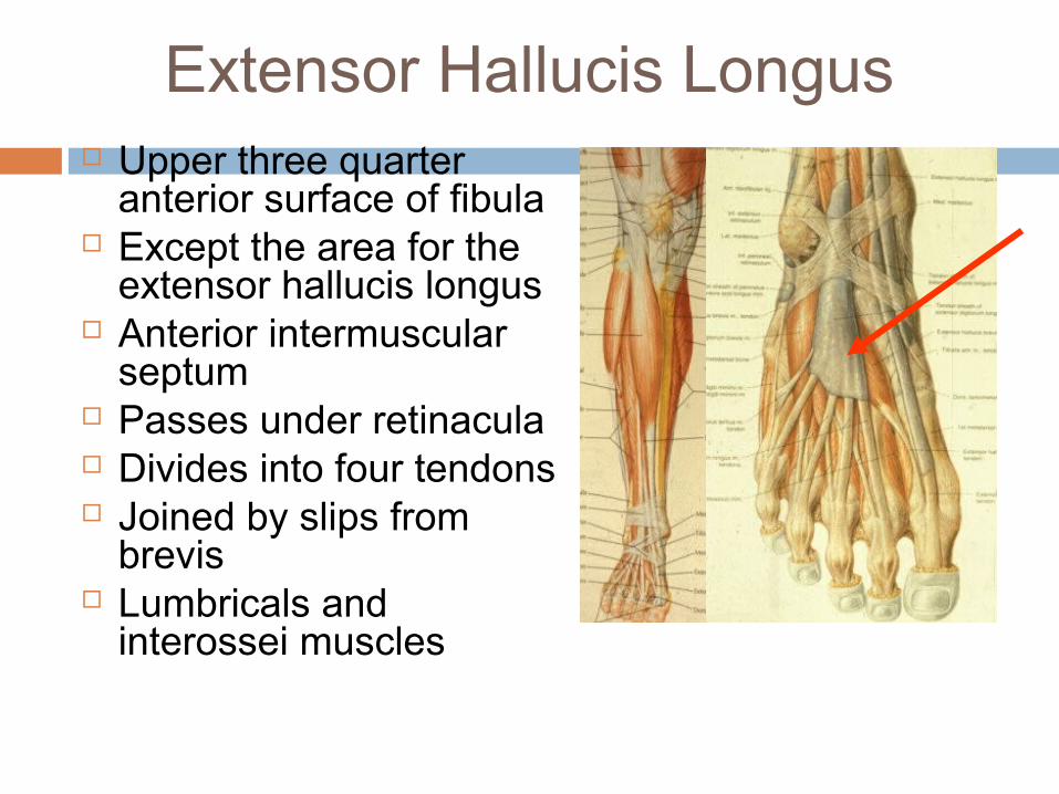
Extensor Hallucis Longus Upper three quarter
anterior surface of fibula Except the area for the
extensor hallucis longus Anterior intermuscular
septum Passes under retinacula Divides into four tendons Joined by slips from
brevis Lumbricals and
interossei muscles
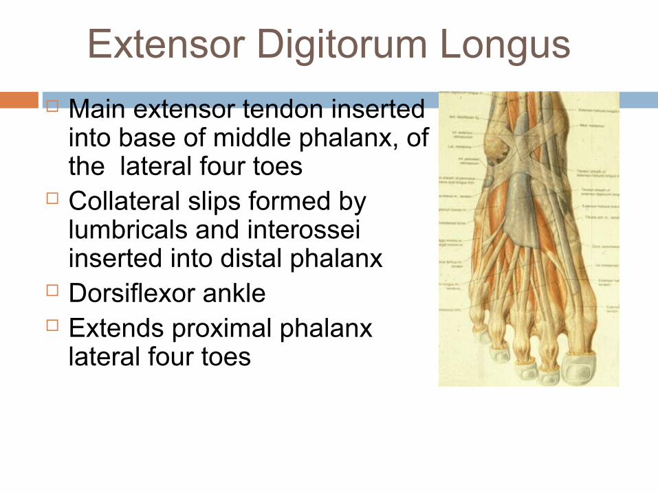
Extensor Digitorum Longus Main extensor tendon inserted
into base of middle phalanx, of the lateral four toes
Collateral slips formed by lumbricals and interossei inserted into distal phalanx
Dorsiflexor ankle Extends proximal phalanx
lateral four toes
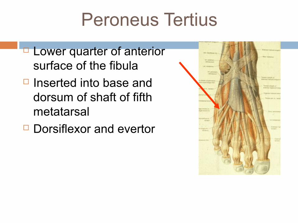
Peroneus Tertius
Lower quarter of anterior surface of the fibula
Inserted into base and dorsum of shaft of fifth metatarsal
Dorsiflexor and evertor
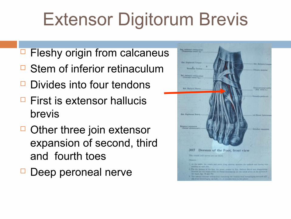
Extensor Digitorum Brevis
Fleshy origin from calcaneus Stem of inferior retinaculum Divides into four tendons First is extensor hallucis
brevis Other three join extensor
expansion of second, third and fourth toes
Deep peroneal nerve
Deep Peroneal Nerve
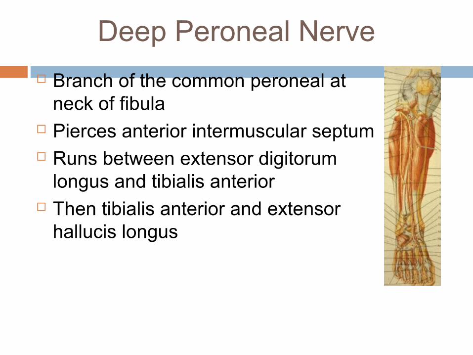
Branch of the common peroneal at neck of fibula
Pierces anterior intermuscular septum Runs between extensor digitorum
longus and tibialis anterior Then tibialis anterior and extensor
hallucis longus
Deep Peroneal Nerve
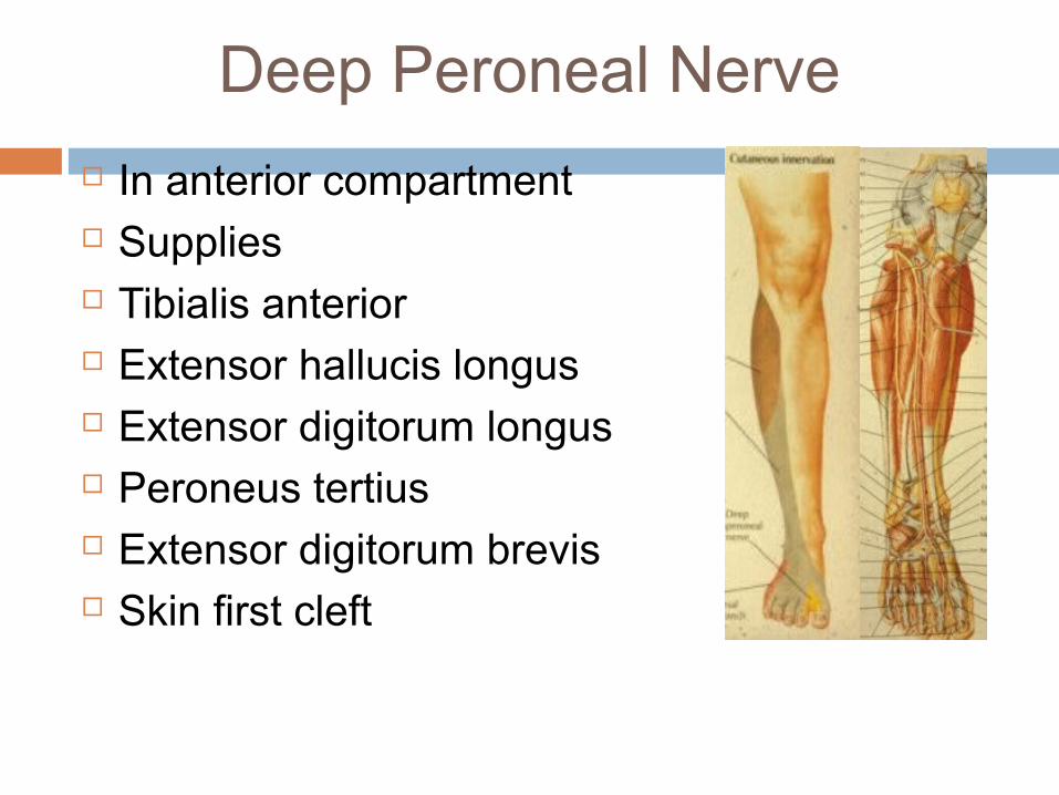
In anterior compartment Supplies Tibialis anterior Extensor hallucis longus Extensor digitorum longus Peroneus tertius Extensor digitorum brevis Skin first cleft
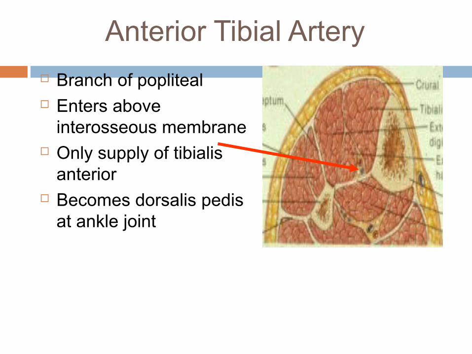
Anterior Tibial Artery
Branch of popliteal Enters above
interosseous membrane Only supply of tibialis
anterior Becomes dorsalis pedis
at ankle joint
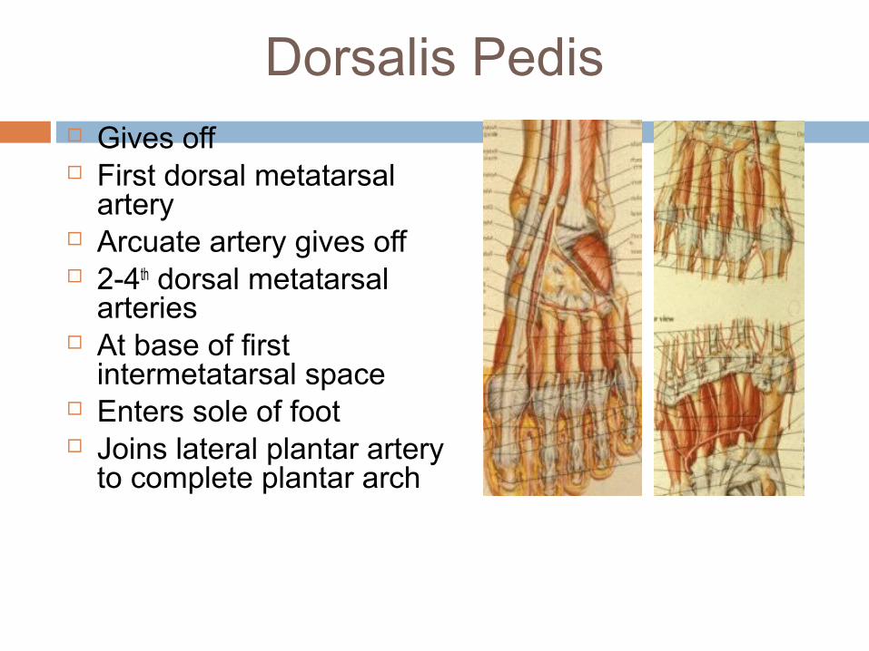
Dorsalis Pedis Gives off First dorsal metatarsal
artery Arcuate artery gives off 2-4th dorsal metatarsal
arteries At base of first
intermetatarsal space Enters sole of foot Joins lateral plantar artery
to complete plantar arch
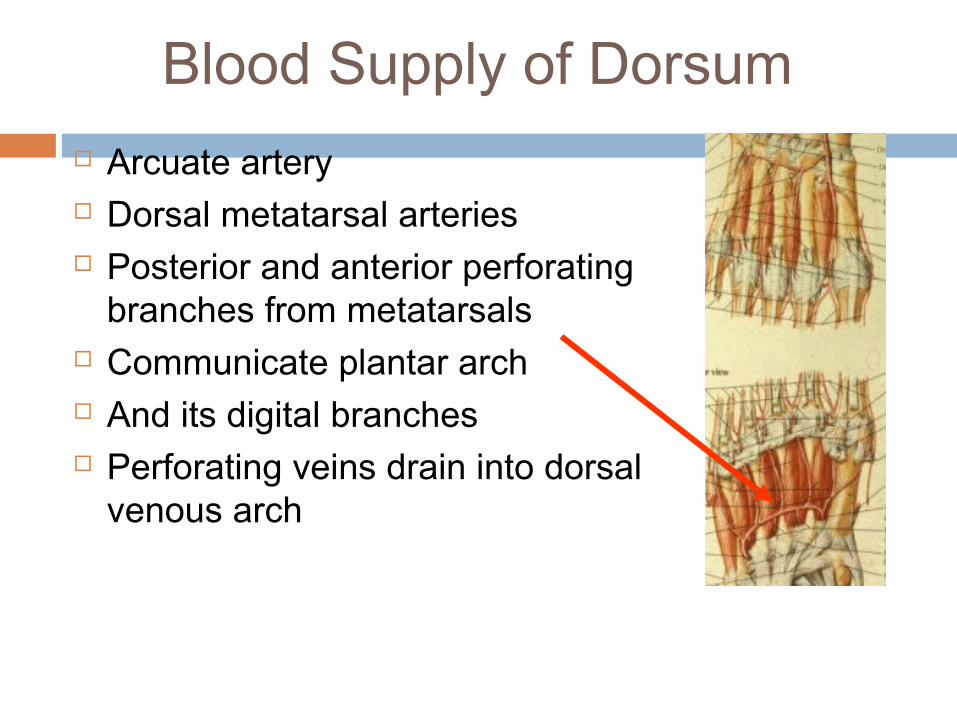
Blood Supply of Dorsum
Arcuate artery Dorsal metatarsal arteries Posterior and anterior perforating
branches from metatarsals Communicate plantar arch And its digital branches Perforating veins drain into dorsal
venous arch
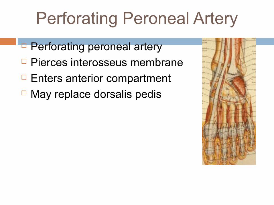
Perforating Peroneal Artery
Perforating peroneal artery Pierces interosseus membrane Enters anterior compartment May replace dorsalis pedis
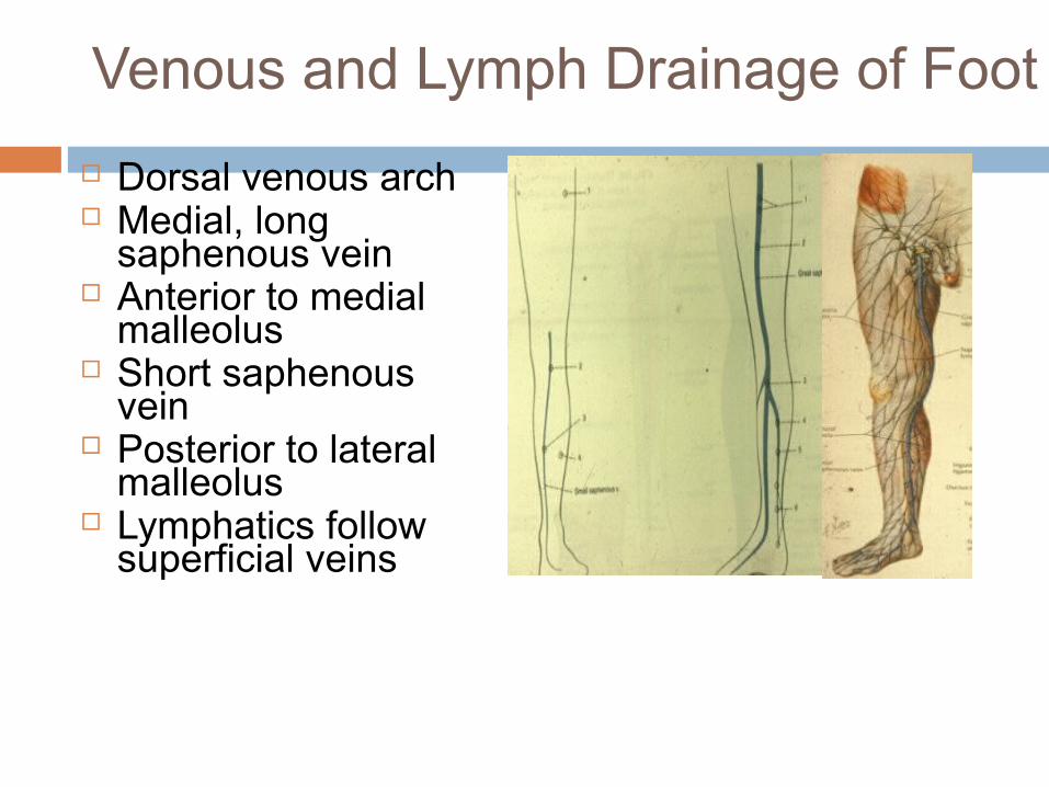
Venous and Lymph Drainage of Foot
Dorsal venous arch Medial, long
saphenous vein Anterior to medial
malleolus Short saphenous
vein Posterior to lateral
malleolus Lymphatics follow
superficial veins
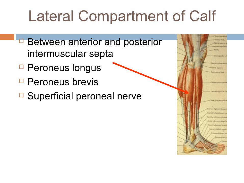
Lateral Compartment of Calf
Between anterior and posterior intermuscular septa
Peroneus longus Peroneus brevis Superficial peroneal nerve
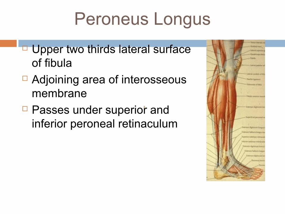
Peroneus Longus
Upper two thirds lateral surface of fibula
Adjoining area of interosseous membrane
Passes under superior and inferior peroneal retinaculum
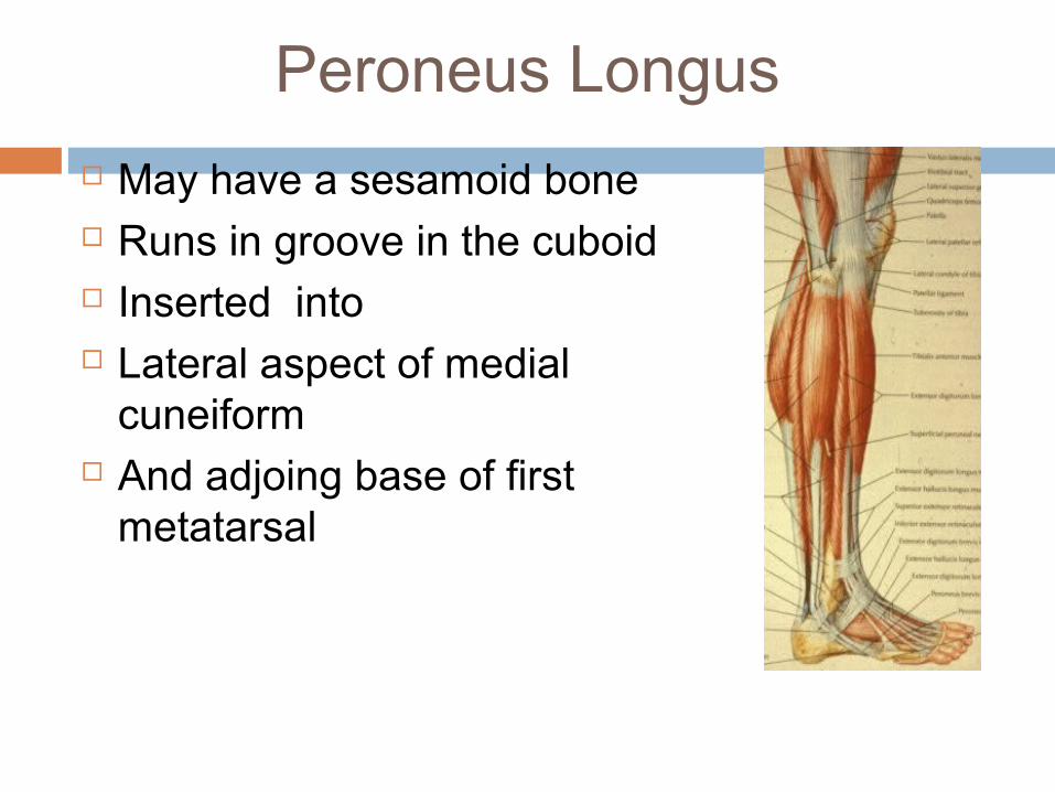
Peroneus Longus
May have a sesamoid bone Runs in groove in the cuboid Inserted into Lateral aspect of medial
cuneiform And adjoing base of first
metatarsal
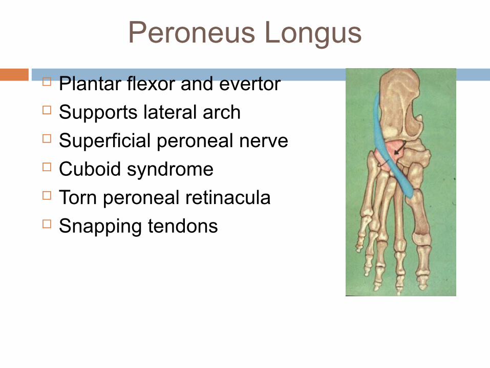
Peroneus Longus
Plantar flexor and evertor Supports lateral arch Superficial peroneal nerve Cuboid syndrome Torn peroneal retinacula Snapping tendons
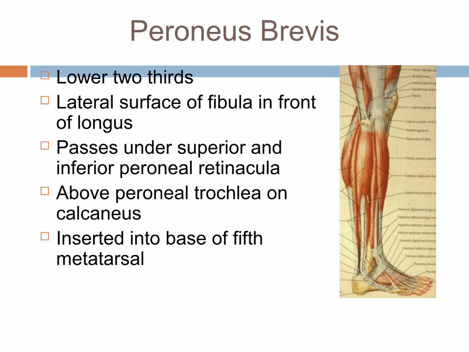
Peroneus Brevis Lower two thirds Lateral surface of fibula in front
of longus Passes under superior and
inferior peroneal retinacula Above peroneal trochlea on
calcaneus Inserted into base of fifth
metatarsal
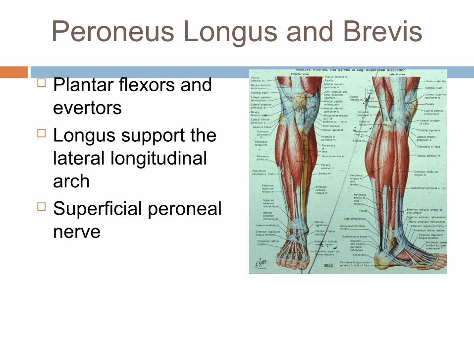
Peroneus Longus and Brevis
Plantar flexors and evertors
Longus support the lateral longitudinal arch
Superficial peroneal nerve
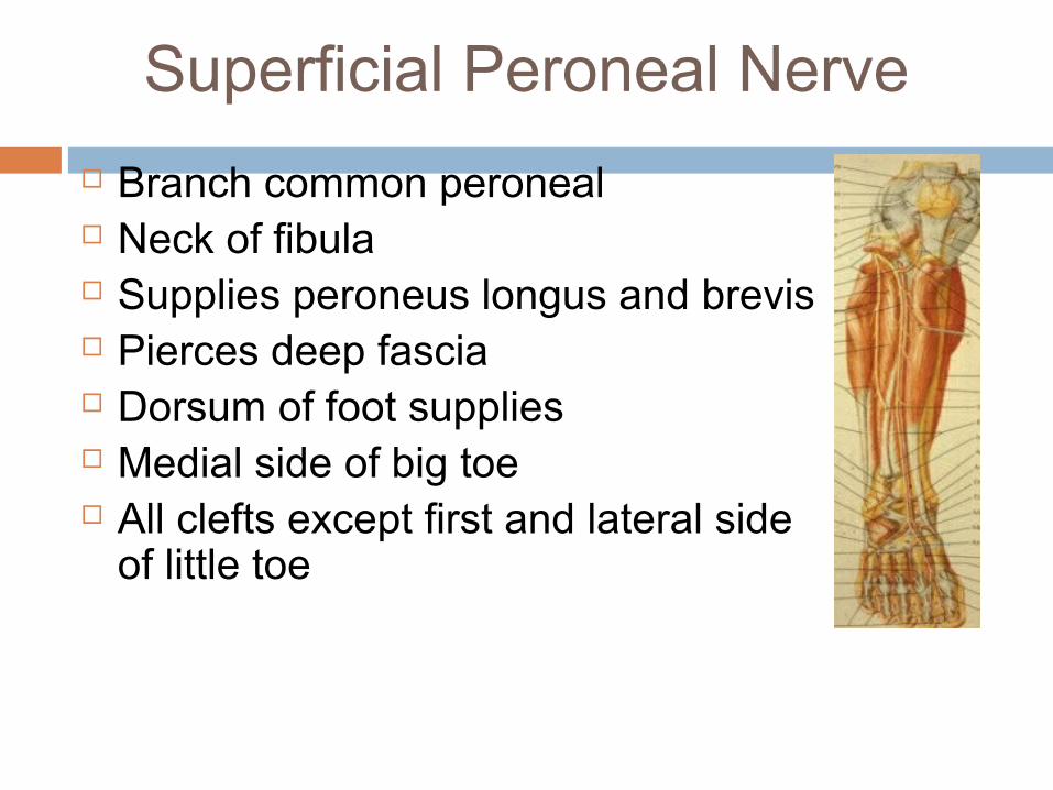
Superficial Peroneal Nerve
Branch common peroneal Neck of fibula Supplies peroneus longus and brevis Pierces deep fascia Dorsum of foot supplies Medial side of big toe All clefts except first and lateral side
of little toe
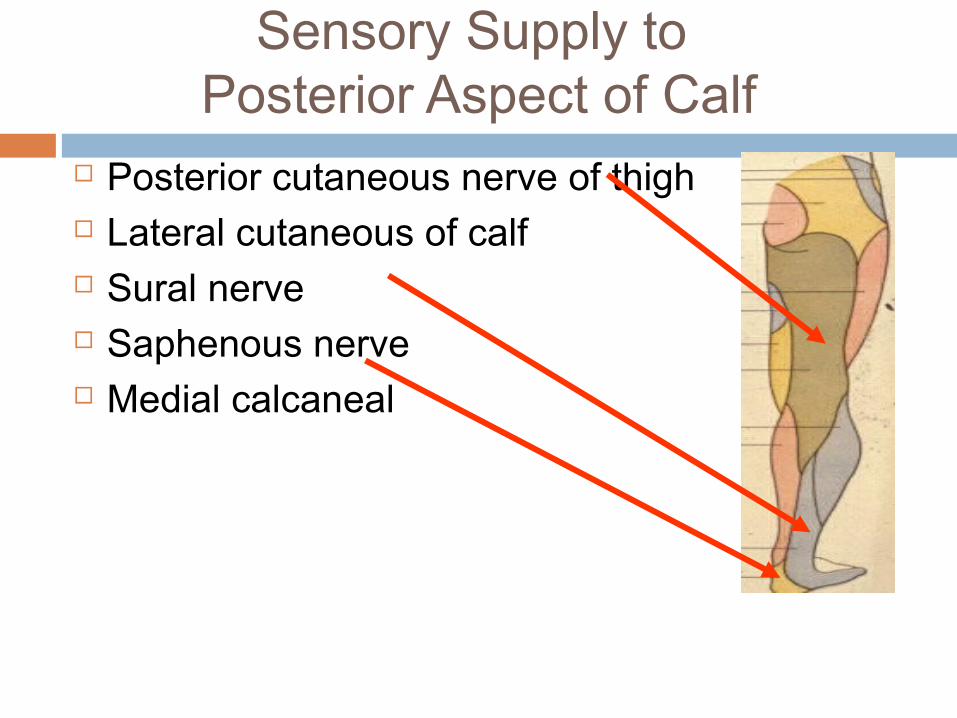
Sensory Supply to Posterior Aspect of Calf
Posterior cutaneous nerve of thigh Lateral cutaneous of calf Sural nerve Saphenous nerve Medial calcaneal
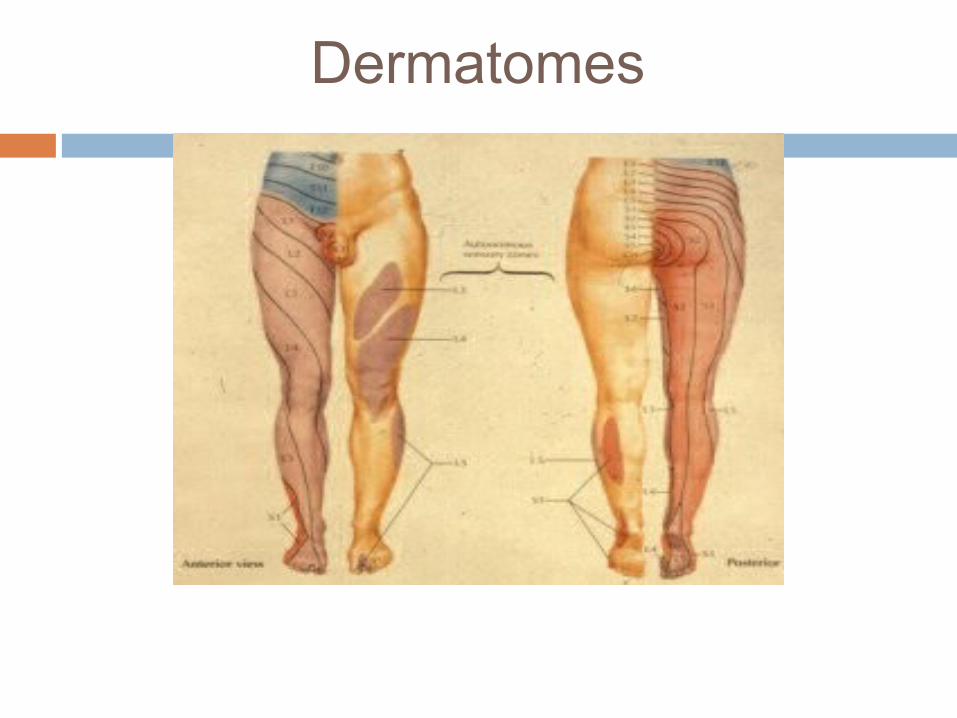
Dermatomes
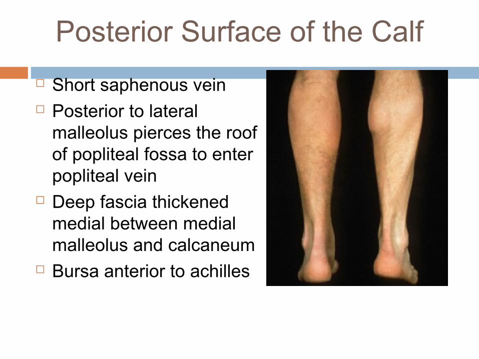
Posterior Surface of the Calf
Short saphenous vein Posterior to lateral
malleolus pierces the roof of popliteal fossa to enter popliteal vein
Deep fascia thickened medial between medial malleolus and calcaneum
Bursa anterior to achilles
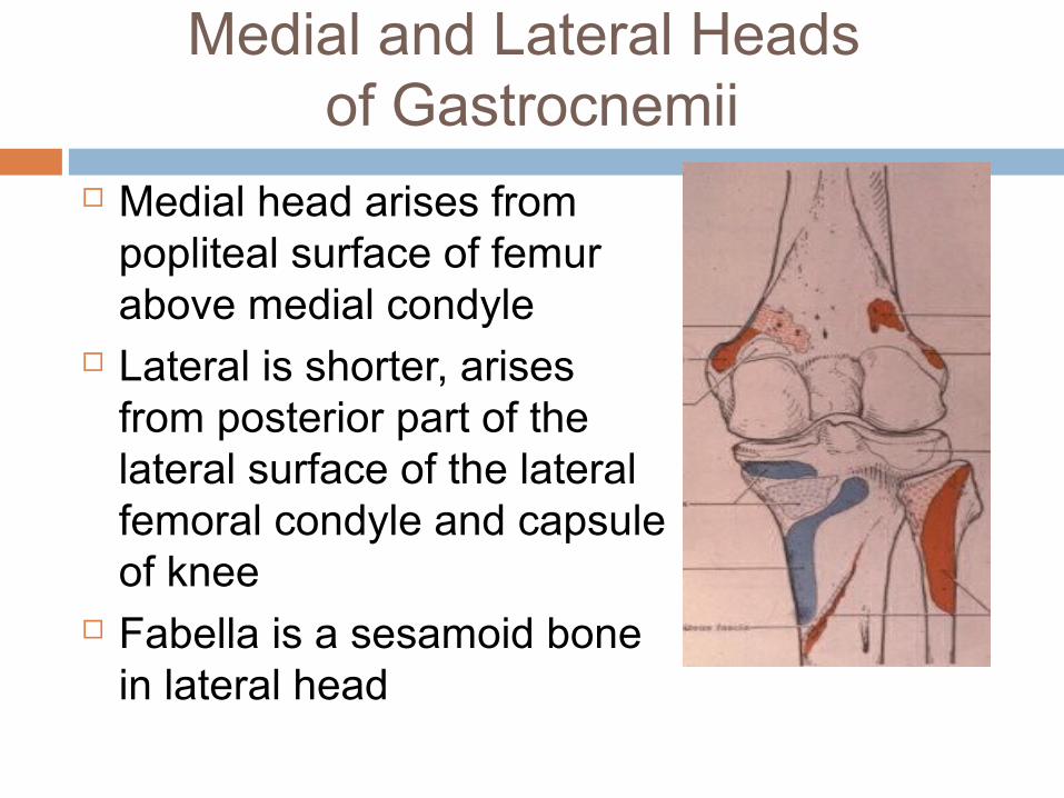
Medial and Lateral Heads of Gastrocnemii
Medial head arises from popliteal surface of femur above medial condyle
Lateral is shorter, arises from posterior part of the lateral surface of the lateral femoral condyle and capsule of knee
Fabella is a sesamoid bone in lateral head
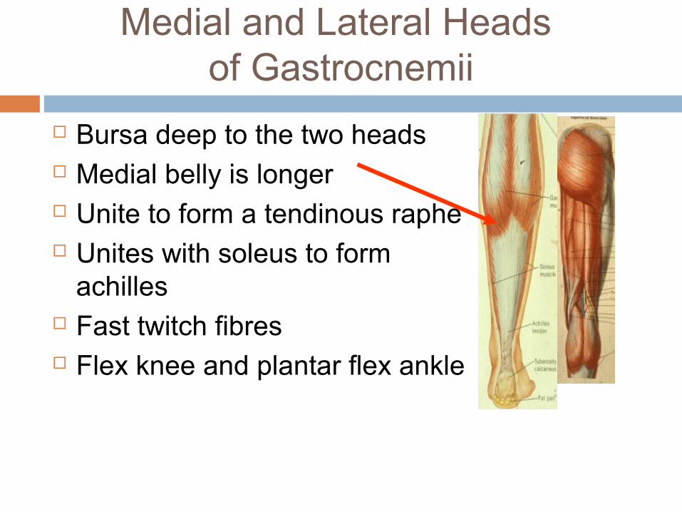
Medial and Lateral Heads of Gastrocnemii
Bursa deep to the two heads Medial belly is longer Unite to form a tendinous raphe Unites with soleus to form
achilles Fast twitch fibres Flex knee and plantar flex ankle
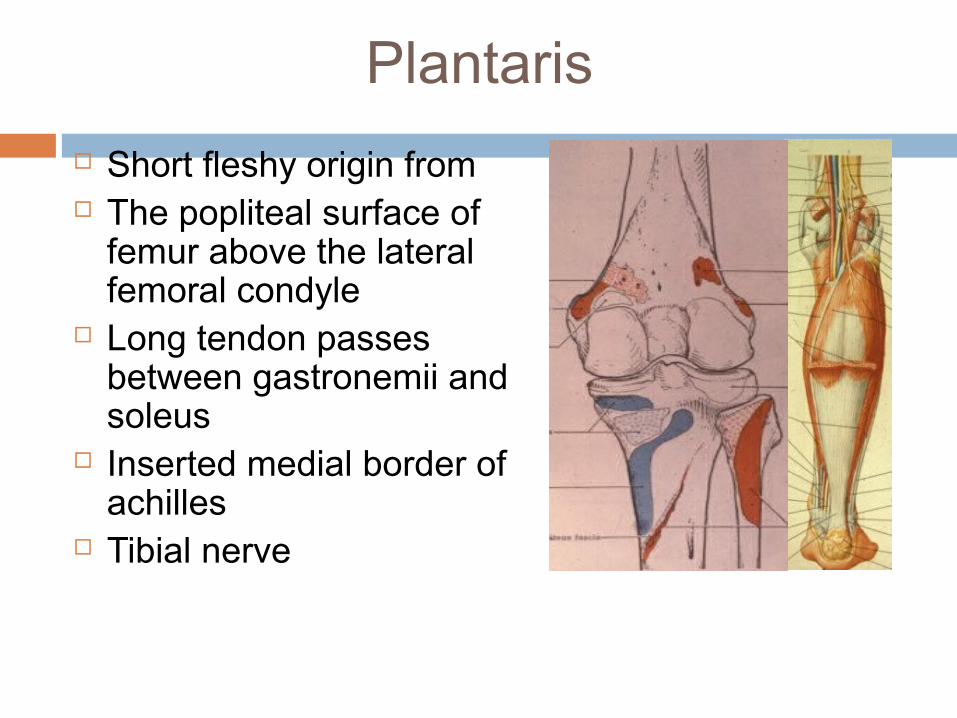
Plantaris
Short fleshy origin from The popliteal surface of
femur above the lateral femoral condyle
Long tendon passes between gastronemii and soleus
Inserted medial border of achilles
Tibial nerve
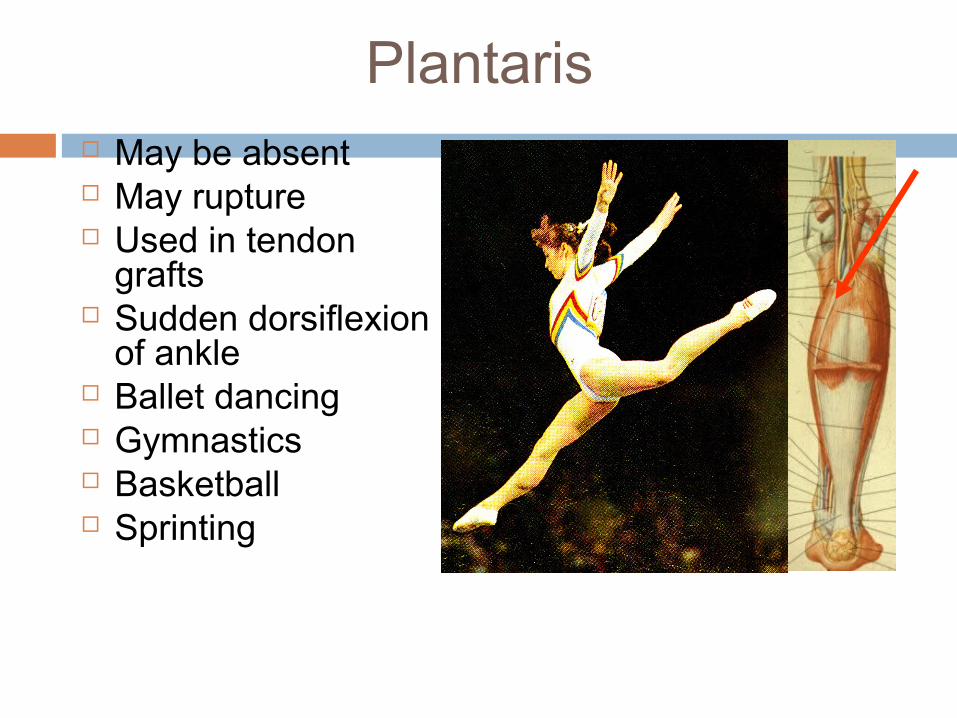
Plantaris May be absent May rupture Used in tendon
grafts Sudden dorsiflexion
of ankle Ballet dancing Gymnastics Basketball Sprinting
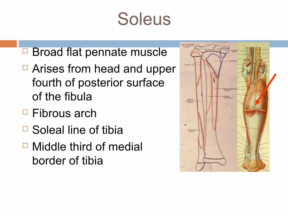
Soleus
Broad flat pennate muscle Arises from head and upper
fourth of posterior surface of the fibula
Fibrous arch Soleal line of tibia Middle third of medial
border of tibia
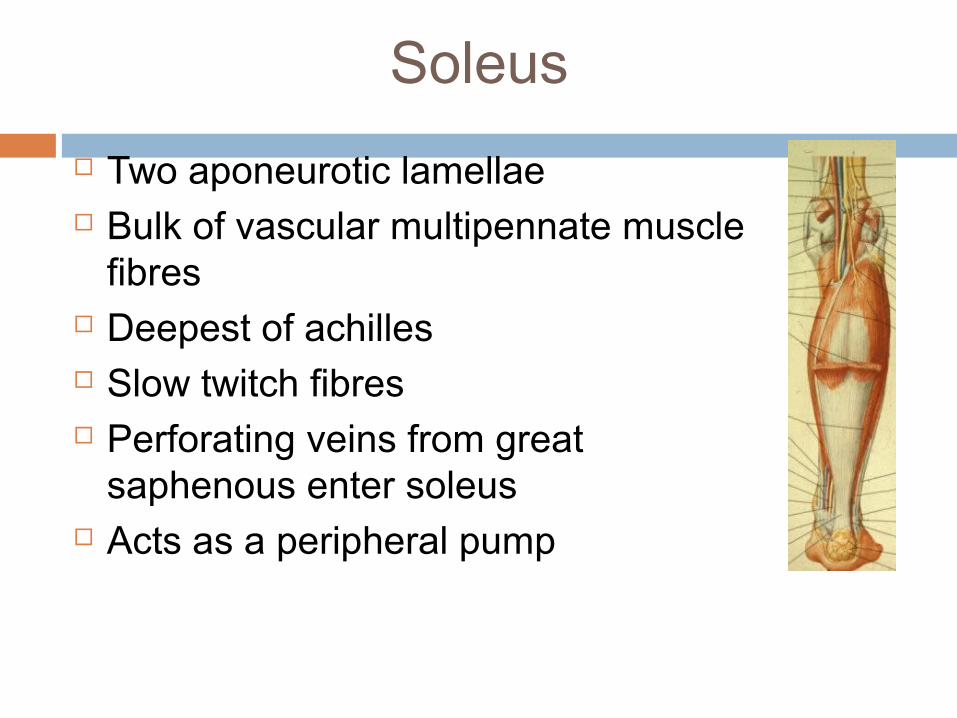
Soleus
Two aponeurotic lamellae Bulk of vascular multipennate muscle
fibres Deepest of achilles Slow twitch fibres Perforating veins from great
saphenous enter soleus Acts as a peripheral pump
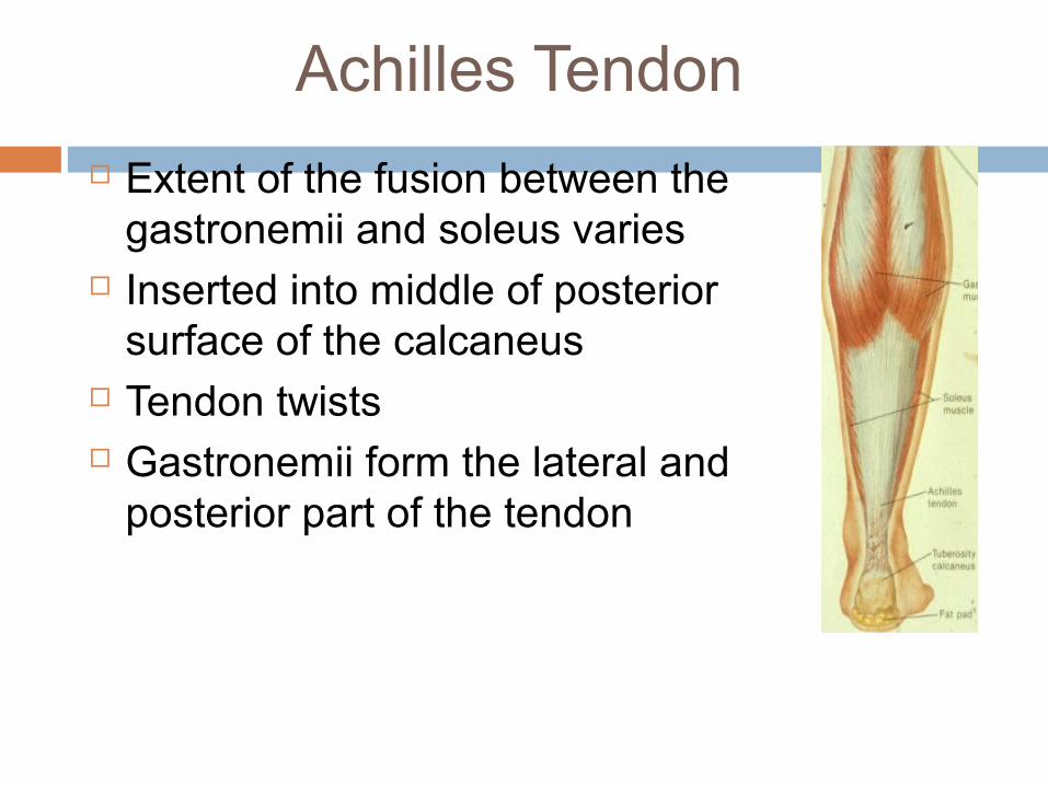
Achilles Tendon
Extent of the fusion between the gastronemii and soleus varies
Inserted into middle of posterior surface of the calcaneus
Tendon twists Gastronemii form the lateral and
posterior part of the tendon
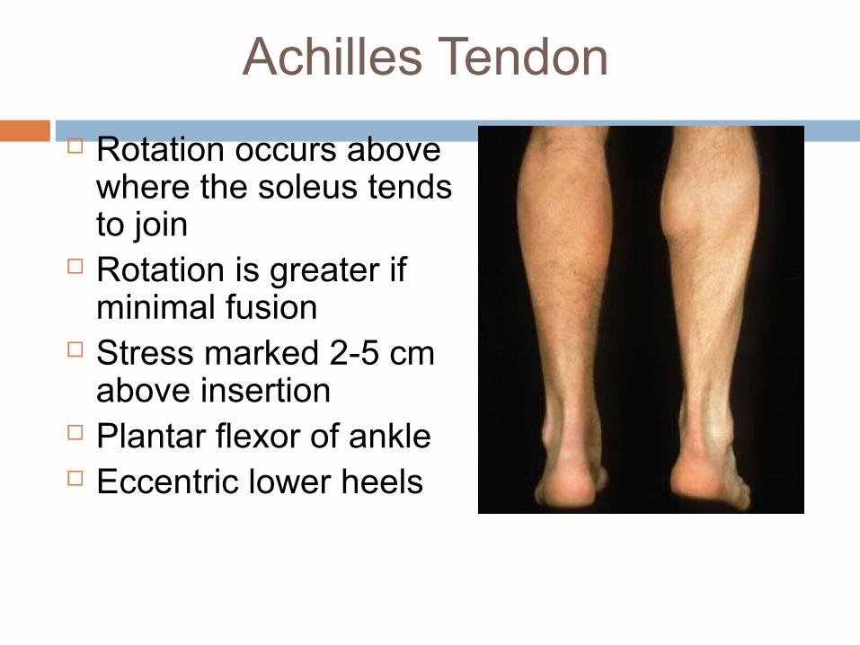
Achilles Tendon
Rotation occurs above where the soleus tends to join
Rotation is greater if minimal fusion
Stress marked 2-5 cm above insertion
Plantar flexor of ankle Eccentric lower heels
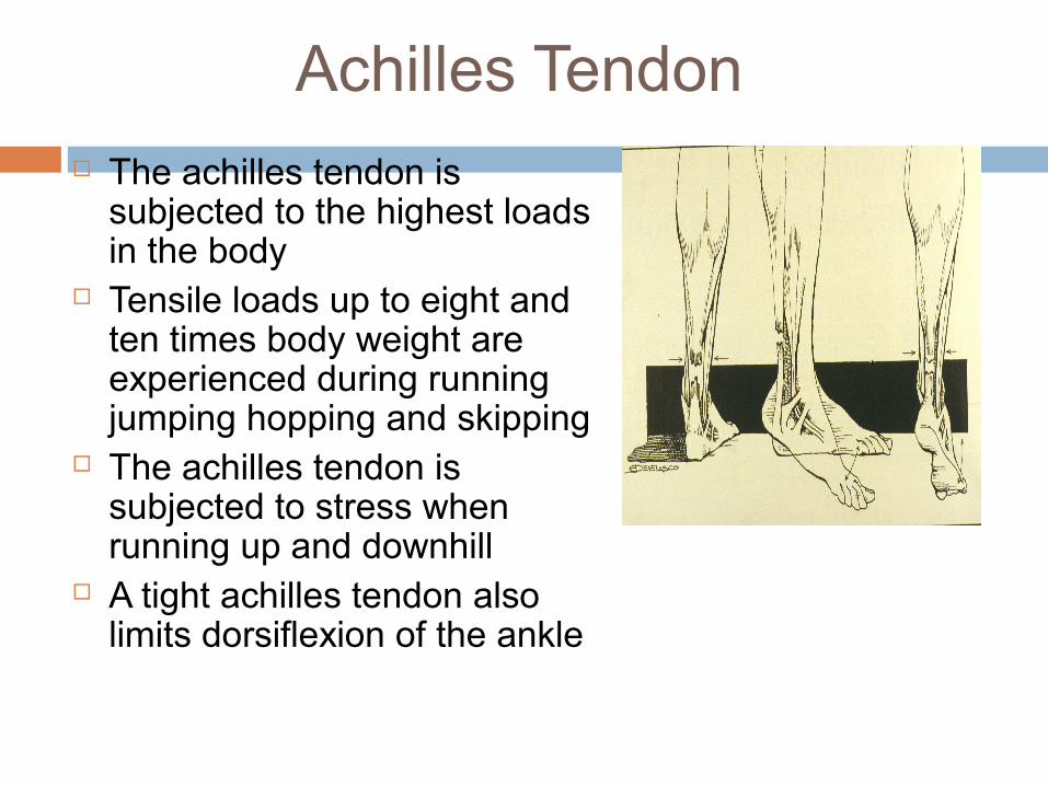
Achilles Tendon The achilles tendon is
subjected to the highest loads in the body
Tensile loads up to eight and ten times body weight are experienced during running jumping hopping and skipping
The achilles tendon is subjected to stress when running up and downhill
A tight achilles tendon also limits dorsiflexion of the ankle
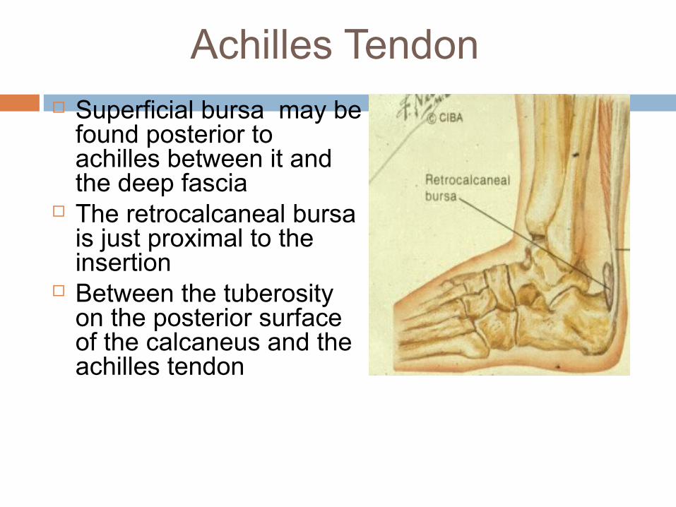
Achilles Tendon Superficial bursa may be
found posterior to achilles between it and the deep fascia
The retrocalcaneal bursa is just proximal to the insertion
Between the tuberosity on the posterior surface of the calcaneus and the achilles tendon
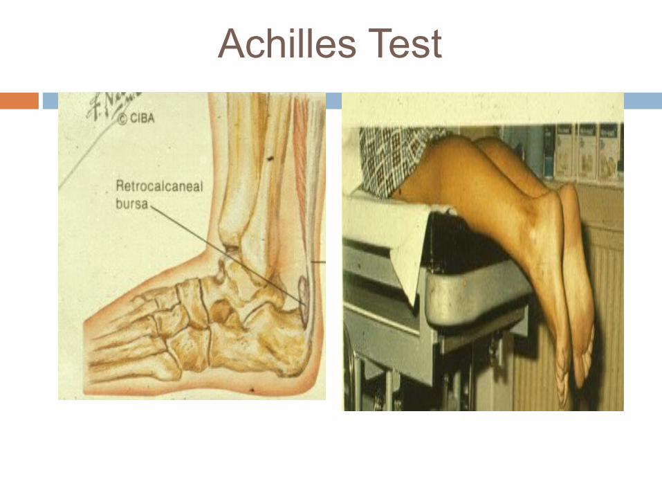
Achilles Test
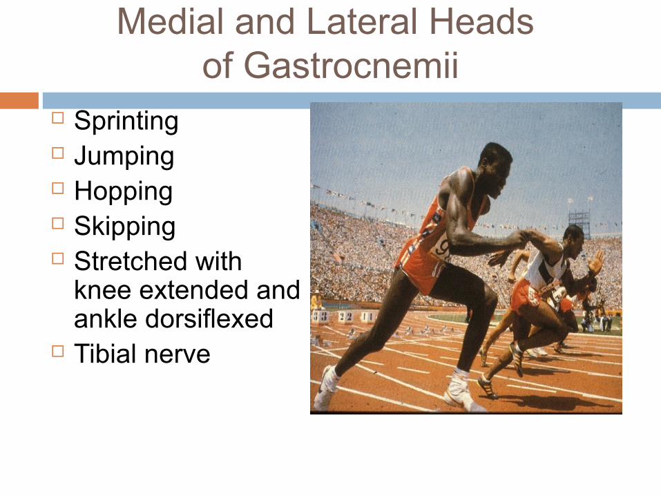
Medial and Lateral Heads of Gastrocnemii
Sprinting Jumping Hopping Skipping Stretched with
knee extended and ankle dorsiflexed
Tibial nerve
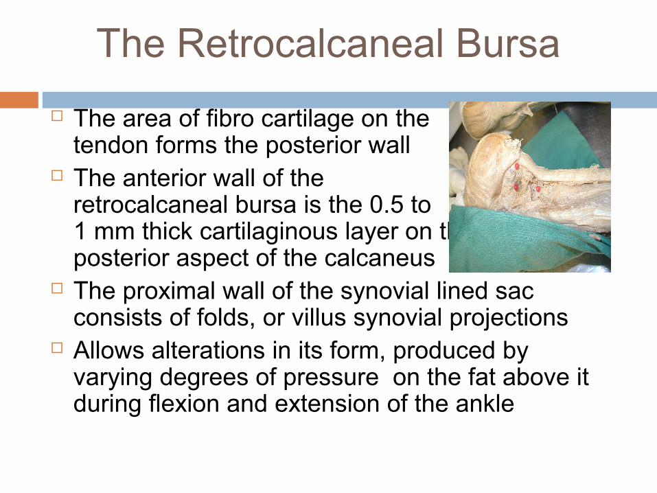
The Retrocalcaneal Bursa
The area of fibro cartilage on the tendon forms the posterior wall
The anterior wall of the retrocalcaneal bursa is the 0.5 to 1 mm thick cartilaginous layer on the posterior aspect of the calcaneus
The proximal wall of the synovial lined sac consists of folds, or villus synovial projections
Allows alterations in its form, produced by varying degrees of pressure on the fat above it during flexion and extension of the ankle
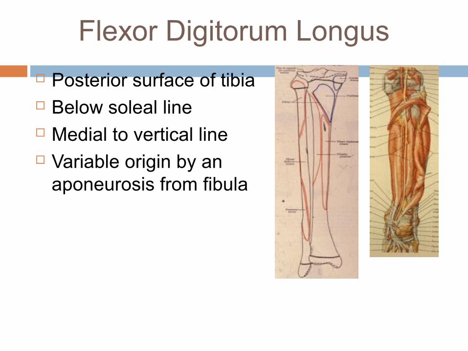
Flexor Digitorum Longus
Posterior surface of tibia Below soleal line Medial to vertical line Variable origin by an
aponeurosis from fibula
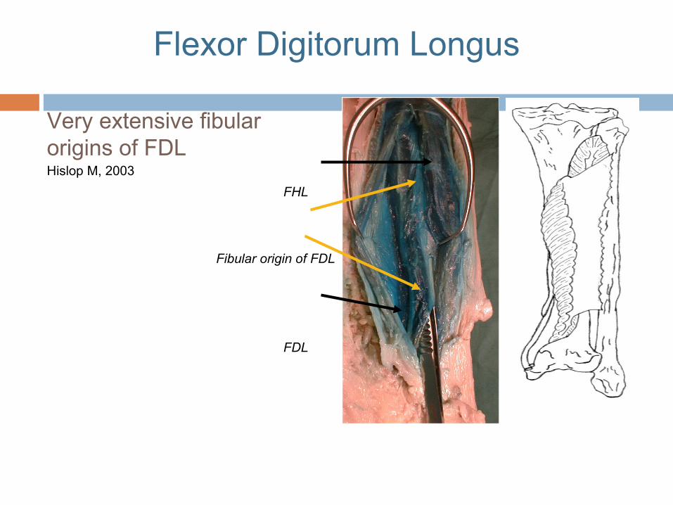
Very extensive fibular origins of FDL
FHL
Fibular origin of FDL
FDL
Hislop M, 2003
Flexor Digitorum Longus
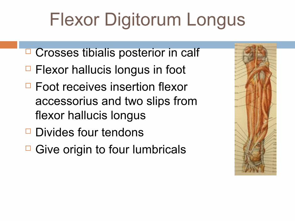
Flexor Digitorum Longus
Crosses tibialis posterior in calf Flexor hallucis longus in foot Foot receives insertion flexor
accessorius and two slips from flexor hallucis longus
Divides four tendons Give origin to four lumbricals
Flexor Digitorum Longus
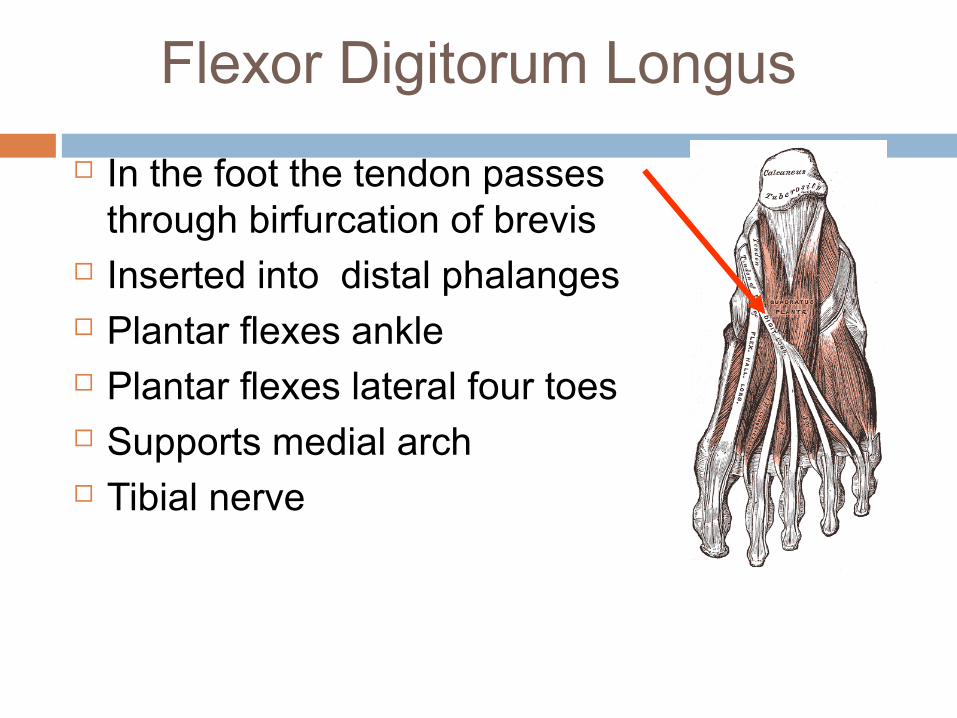
In the foot the tendon passes through birfurcation of brevis
Inserted into distal phalanges Plantar flexes ankle Plantar flexes lateral four toes Supports medial arch Tibial nerve
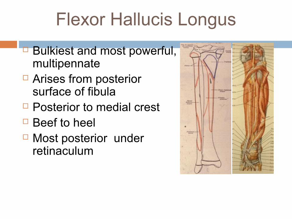
Flexor Hallucis Longus
Bulkiest and most powerful, multipennate
Arises from posterior surface of fibula
Posterior to medial crest Beef to heel Most posterior under
retinaculum
Flexor Hallucis Longus
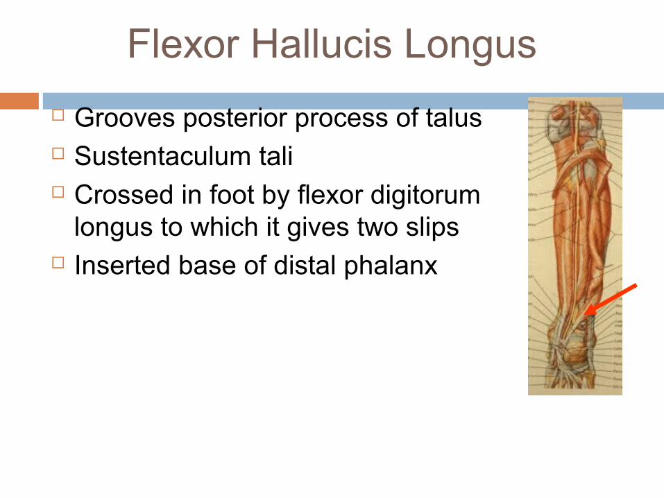
Grooves posterior process of talus Sustentaculum tali Crossed in foot by flexor digitorum
longus to which it gives two slips Inserted base of distal phalanx
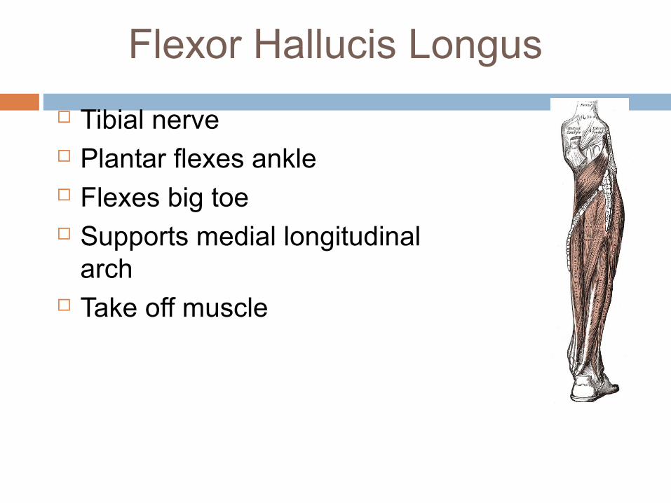
Flexor Hallucis Longus
Tibial nerve Plantar flexes ankle Flexes big toe Supports medial longitudinal
arch Take off muscle
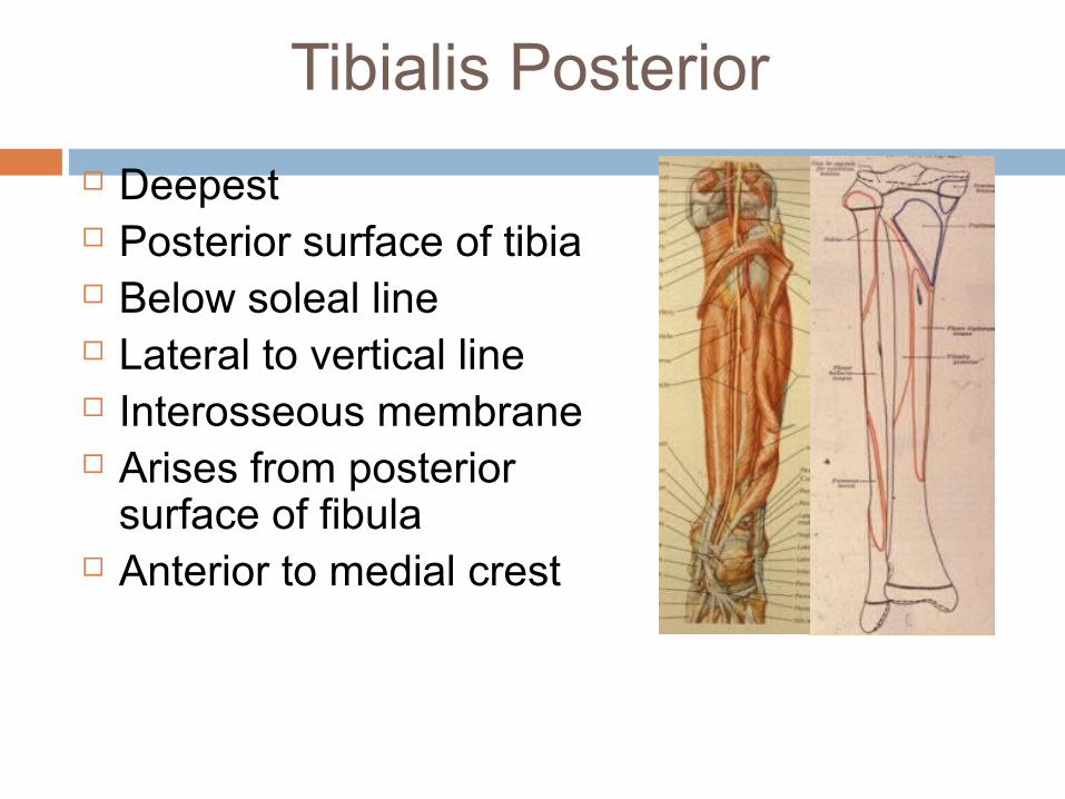
Tibialis Posterior
Deepest Posterior surface of tibia Below soleal line Lateral to vertical line Interosseous membrane Arises from posterior
surface of fibula Anterior to medial crest
Tibialis Posterior
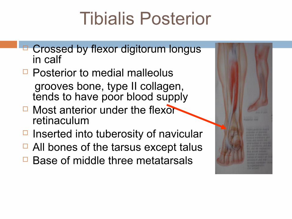
Crossed by flexor digitorum longus in calf
Posterior to medial malleolus grooves bone, type II collagen,
tends to have poor blood supply Most anterior under the flexor
retinaculum Inserted into tuberosity of navicular All bones of the tarsus except talus Base of middle three metatarsals
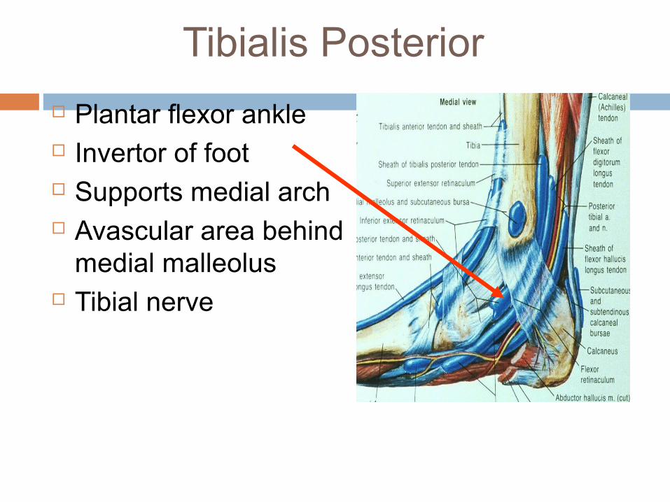
Tibialis Posterior
Plantar flexor ankle Invertor of foot Supports medial arch Avascular area behind
medial malleolus Tibial nerve
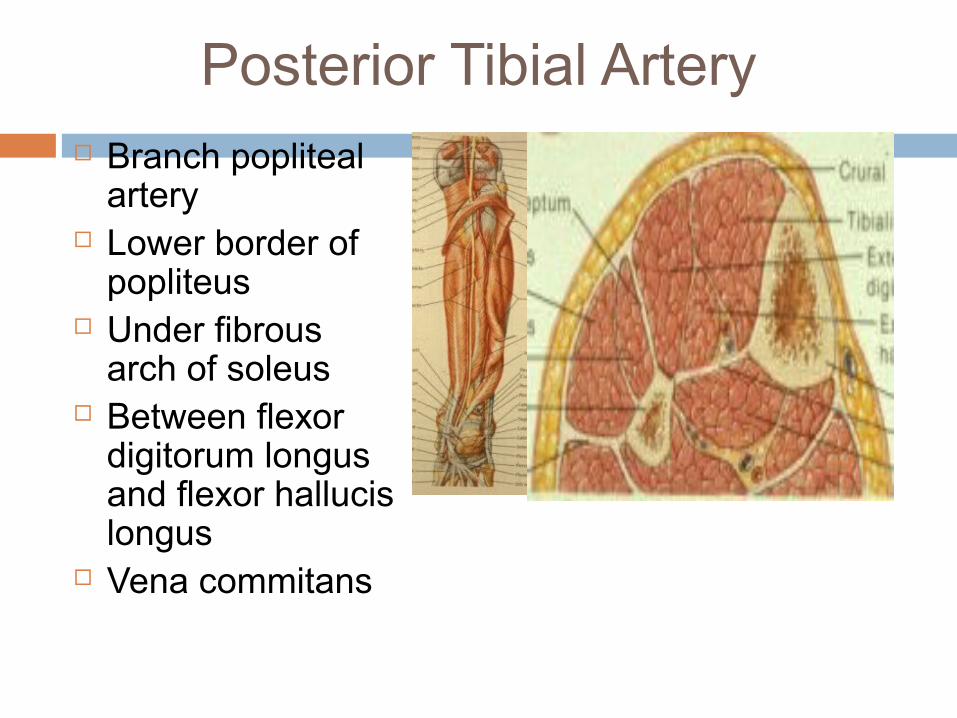
Posterior Tibial Artery Branch popliteal
artery Lower border of
popliteus Under fibrous
arch of soleus Between flexor
digitorum longus and flexor hallucis longus
Vena commitans
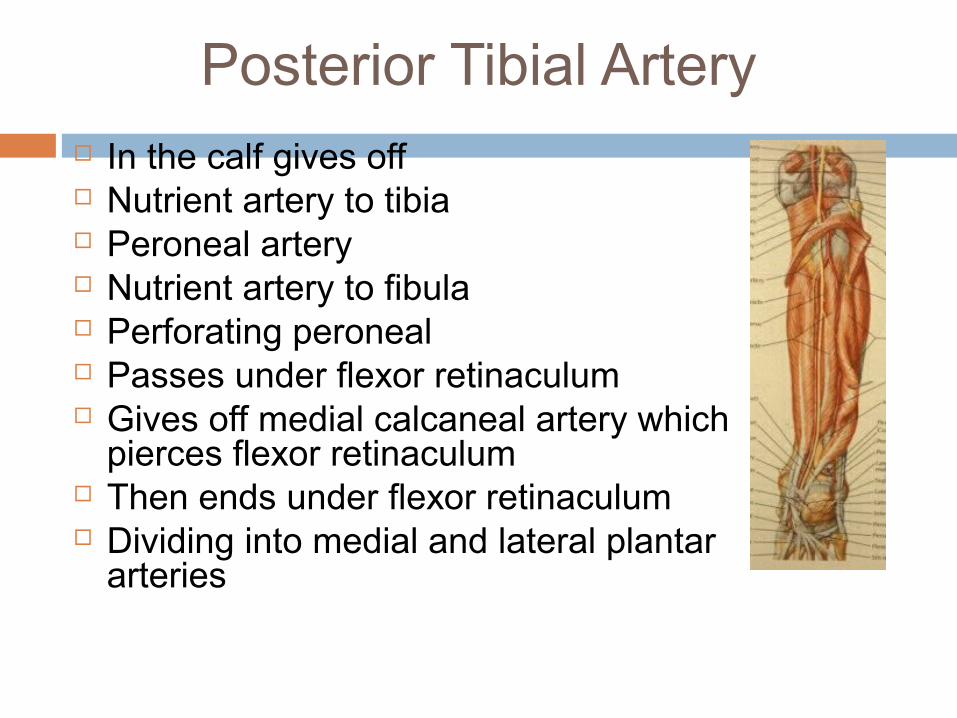
Posterior Tibial Artery In the calf gives off Nutrient artery to tibia Peroneal artery Nutrient artery to fibula Perforating peroneal Passes under flexor retinaculum Gives off medial calcaneal artery which
pierces flexor retinaculum Then ends under flexor retinaculum Dividing into medial and lateral plantar
arteries
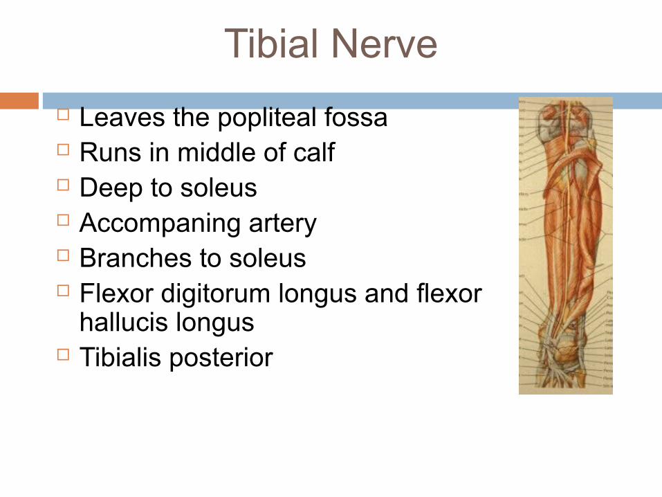
Tibial Nerve
Leaves the popliteal fossa Runs in middle of calf Deep to soleus Accompaning artery Branches to soleus Flexor digitorum longus and flexor
hallucis longus Tibialis posterior
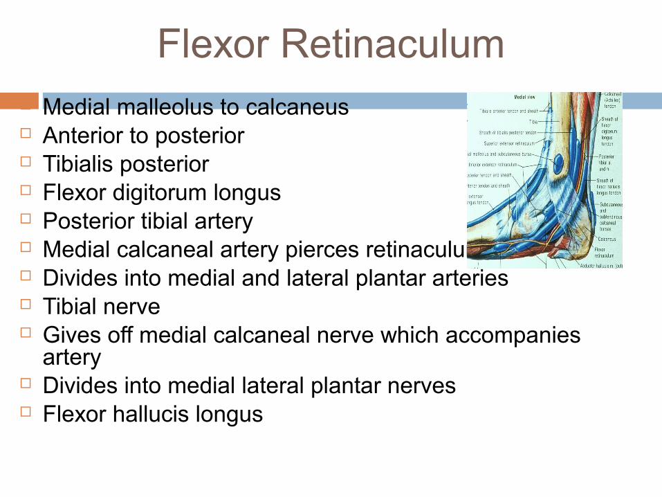
Flexor Retinaculum Medial malleolus to calcaneus Anterior to posterior Tibialis posterior Flexor digitorum longus Posterior tibial artery Medial calcaneal artery pierces retinaculum Divides into medial and lateral plantar arteries Tibial nerve Gives off medial calcaneal nerve which accompanies
artery Divides into medial lateral plantar nerves Flexor hallucis longus
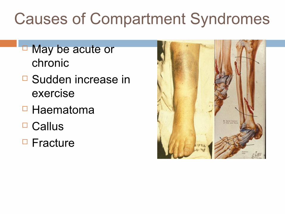
Causes of Compartment Syndromes
May be acute or chronic
Sudden increase in exercise
Haematoma Callus Fracture
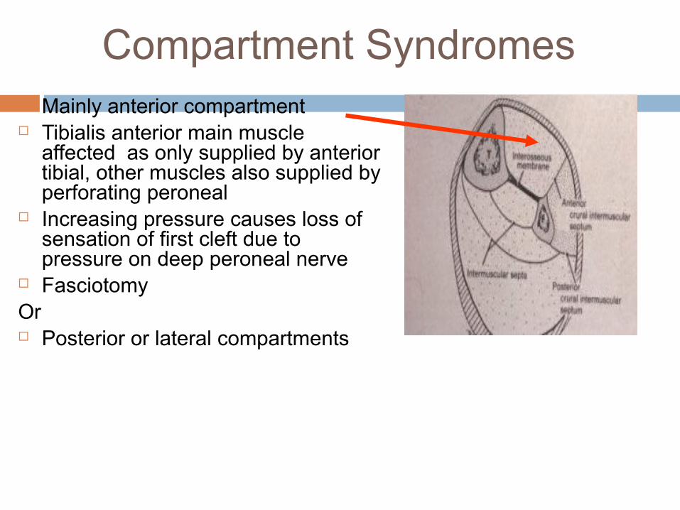
Compartment Syndromes Mainly anterior compartment Tibialis anterior main muscle
affected as only supplied by anterior tibial, other muscles also supplied by perforating peroneal
Increasing pressure causes loss of sensation of first cleft due to pressure on deep peroneal nerve
FasciotomyOr Posterior or lateral compartments
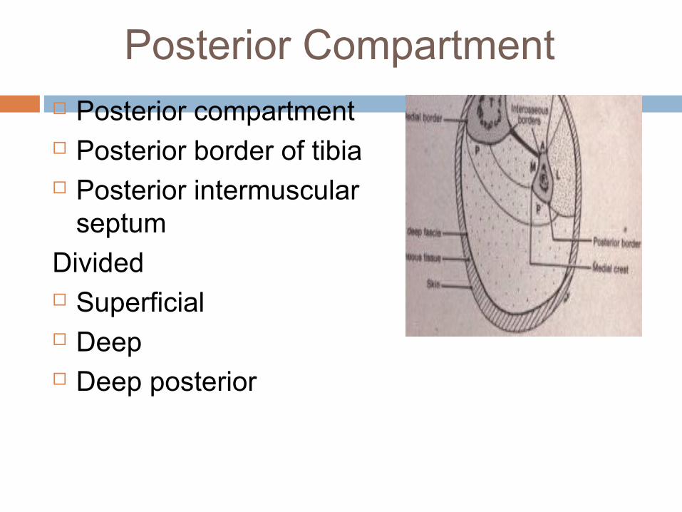
Posterior Compartment Posterior compartment Posterior border of tibia Posterior intermuscular
septum
Divided Superficial Deep Deep posterior
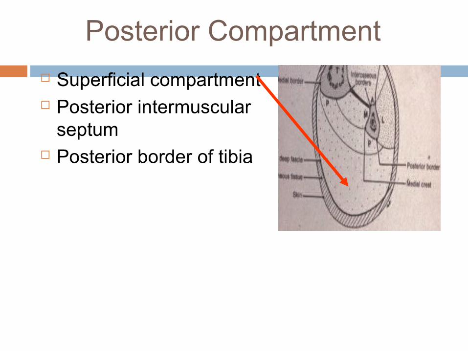
Posterior Compartment
Superficial compartment Posterior intermuscular
septum Posterior border of tibia
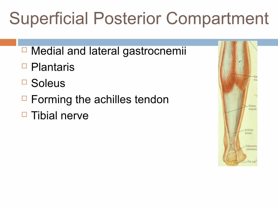
Superficial Posterior Compartment
Medial and lateral gastrocnemii Plantaris Soleus Forming the achilles tendon Tibial nerve
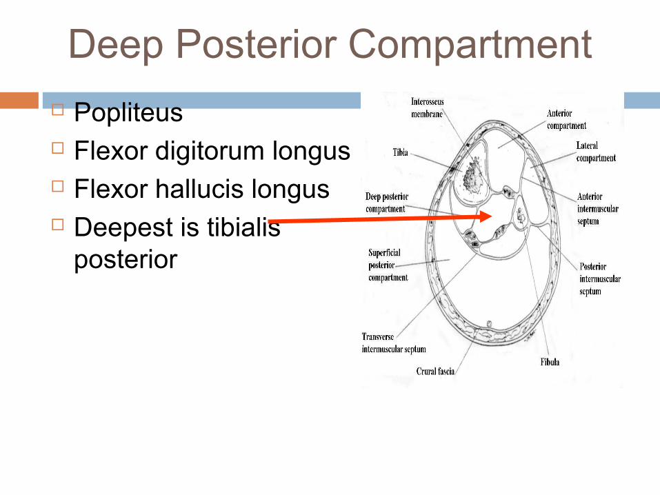
Deep Posterior Compartment
Popliteus Flexor digitorum longus Flexor hallucis longus Deepest is tibialis
posterior
Chronic Exertional Compartment Syndrome (CECS)
• Significant morbidity/limitation activity to athletes
• Activity related, reversible, myofascial intracompartmental pressure increase
• Results in decreased tissue perfusion and neuromuscular function abnormalities
• Predilection for the lower leg
Chronic Exertional Compartment Syndrome (CECS)
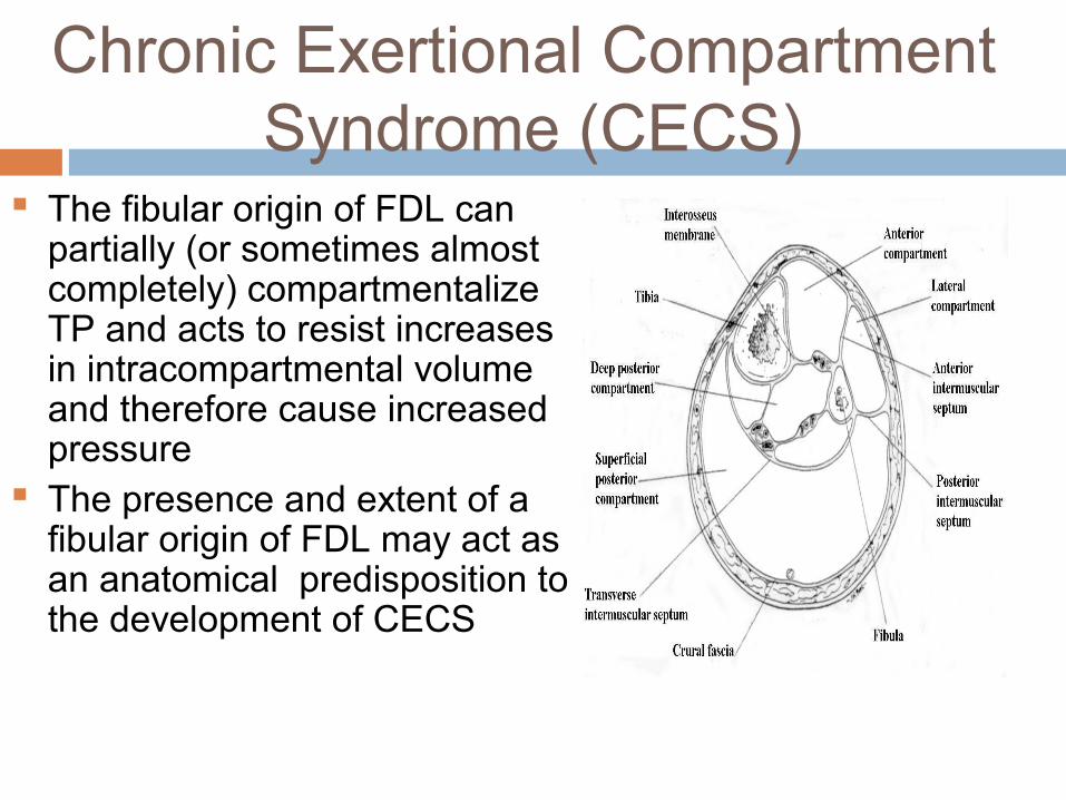
The fibular origin of FDL can partially (or sometimes almost completely) compartmentalize TP and acts to resist increases in intracompartmental volume and therefore cause increased pressure
The presence and extent of a fibular origin of FDL may act as an anatomical predisposition to the development of CECS