Embed Size (px)
Citation preview
PRESENTER
DR TANUJ VERMA

MANJULA 4 years old girl from ANDRA PRADESH addmited in Pediatric casualty with
Cough and cold 4 days
Fever 2 days
Altered behaviour and sensorium 1 day.
1st born to nonconsanguineous parents
Birth history was uneventful
Developmental milestones were normal.

no h/o headache vomiting ,ear discharge,
exanthems , dog bite, recent immunization ,
recent travel or bleeding manifestations

No significant past medical history
No home medications
No prior surgeries

She had status epilepticus and was loaded
with phenytoin, Leviteracetam, and
valproate.
GCS worsened to 5/15, so she was intubated
and shifted to PICU.
She was started on Midazolam infusion,
meningitic doses of Cefotaxime, Acyclovir
and Neuroprotective measures.

PICU ADMISSION GCS 6T/15
SEVERE NEUROLOGICAL COMPROMISE
IN ABSENSE OF NEUROMAUSCULAR BLOCKAGE MO
SPONTENOUS MOVEMENTS WITH MINIMAL WITHDRAWL TO
PAIN
COUGH GAG AND CORNEAL REFLEX ABSENT
B/L PUPIL CONSTRICTED AND REACTING TO LIGHT
NO FACIAL WEAKNESS AND TONE DECRESED IN ALL FOUR
LIMBS
B/L PLANTER EXTENSOR , DTR DEPRESSED
NO NECK RIGIDITY AND KERNIG/ BRUDZENKY ANSENT

ACUTE MENINGOENCEPHALITIS WITH
STATUS EPILEPTICUS

HB 13.2%
WBC 13000
DLC N87 / L4
CRP 6.7
BLOOD CULTURE NO GROWTH
URINE CULTURE NO GROWTH
ABG 7.18 / 73 / 87 / 27.3 / -1.2 / 1.4

CT shows hypodensities involving bilateral thalamic and midbrain, mild prominence of ventricles


VIRAL MENINGOENCAPHLITIS
IEM
ACUTE NECROTIZING MENINGOENCPHLITIS
(Genetic/ infection associated/ metabolic)
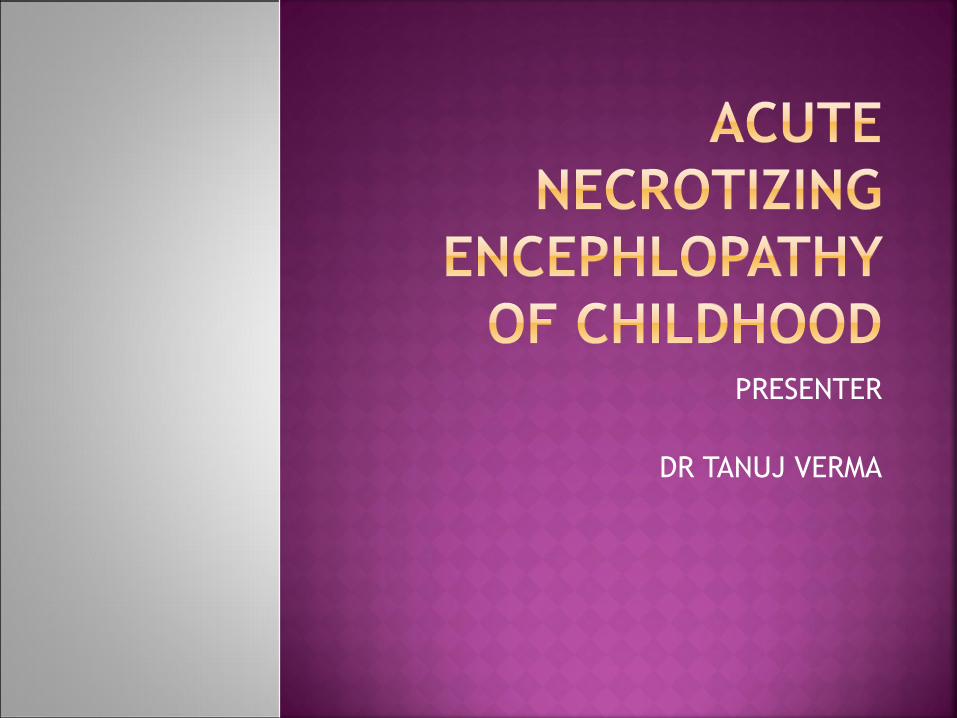
• MRI with contrast
• Etiology work up: blood lactate, ammonia,
acylcarnitine profile, TMS to Clinical
Pharmcological lab, H1N1 screening,
biotinidase assay, serum aminoacids, urine
organic acids
• BBVS screen
• Genetic studies: RNBP gene mutation
• Supportive measures
• HLA DRB1*1401, HLA BRB3*0202, HLA
DQB1*05052

T2 FLAIR hyperintensities involving bilateral symmetrical swelling, haemorrhagic areas and restricted diffusion of thalami. Hyperintensity, swelling and restricted diffusion of posterior putamen, caudate head, hippocampi,pons,dentate nucleus and fornices with haemorrhagic areas in pons and hippocampi.


ESR 30
ANA NEGATIVE
DS DNA 19 IU/ML ( < 100 IU/ML)
S AMMONIA 80mcg%
S LACTATE 1 mmol/l
SE AMINO ACIDS NORMAL
S FREE
ACYLCARNITINE
NORMAL
S TOTAL
CARNITINE
NORMAL
URINE ORGANIC
ACIDS
NOT DETECTED
URINE OROTIC
ACIDS
NORMAL LEVELS

GLUCOSE 52 mg/dl
PROTEIN 152mg/dl
CELLS TLC 5 / CC
CELLS DLC P 60%/ L 40%
LACTATE 1.1 mmol/l
CSF CULTURE NO GROWTH
CSF ACYLCARNITINE NORMAL
CSF TOTAL
ACYLCARNITINE
NORMAL
CSF MULTIPLEX PCR
CMV
NEGATIVE
CSF ENTEROVIRUS NEGATIVE
CSF HHV 6 NEGATIVE
CSF JAP- B NEGATIVE
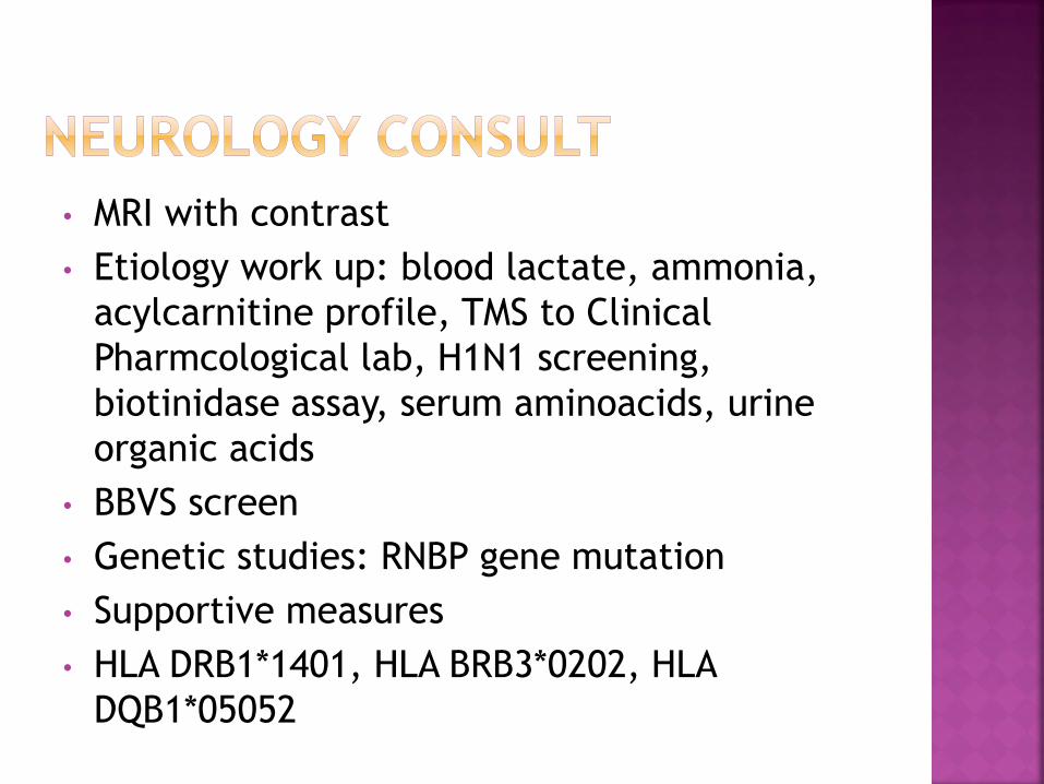
diffuse slowing with no
epileptiform activity

H1N1 POSITIVE

ACUTE NECROTIZING ENCEPHALOPATHY OF
CHILDHOOD (ANEC)

Now not a first reported case of acute
necrotizing encaphlopathy but among
few with H1N1
1st reported in japan by Mizuguzi in 1995
Report on 13 consecutive cases and 28
previous cases

• Acute encephalopathy following viral disease, with seizure and deterioration of consciousness.
• Absence of CSF pleocytosis. CSF protein is commonly increased.
• Neuroimaging findings of symmetric, multifocal brain lesions involving the bilateral thalami, upper brain stem tegmentum, periventricular white matter, internal capsule, putamen and cerebellum.
• Elevation of serum aminotransferase level to a variable degree. No increase in blood ammonia.
• Exclusion of any resembling disease.
Journal of Neurology, Neurosurgery, and Psychiatry 1995;58:555-561

Clinico-radiological diagnosis
Etiology: Mostly associated with Influenza A
and B virus, parainfluenza virus, Mycoplasma,
Herpes simplex virus and Human herpes virus-6.

Journal of Neurology, Neurosurgery, and Psychiatry 1995;

A. Clinical differential diagnosis: toxic shock syndrome, hemolytic uremic syndrome, Reye syndrome, hemorrhagic shock and encephalopathy syndrome, and heat stroke.
B. Radiological (or pathological) differential diagnosis: Leigh encephalopathy, glutaric acidemia, methyl malronic aciduria, infantile bilateral strial necrosis, Wernicke encephalopathy, carbon monoxide poisoning, acute disseminated encephalomyelitis, acute hemorrhagic leukoencephalitis, arterial or venous infarct, severer hypoxic or traumatic injury.
Journal of Neurology, Neurosurgery, and Psychiatry 1995;58:555-561
• Not clear.
• But postulated rapid development of intracranial
cytokine formation which causes blood brain
barrier damage in particular regions of brain
resulting in localized edema, congestion and
hemorrhage, without any signs of direct viral invasion
or post infectious demyelination.
Sugaya N. Influenza associated encephalopathy in Japan: pathogenesis and treatment.
Pediatr Intl 2000; 42: 215-218.

RANBP2 gene mutation*: recurrent episodes
of ANEC and can present as Autosommal
Dominant with incomplete penetrance.
* Neilson DE. Autosomal dominant acute necrotizing encephalopathy.
Neurology 2003; 61: 226–30.
*Gika AD. Recurrent acute necrotizing encephalopathy following Influenza A
in a genetically predisposed family. Dev Med Child Neurol 2010; 52: 99–
102.

Male to female 1:1
Peak incidence age 6-18 months
90% of cases have antecedent infection with
fever, URI symptoms, GI symptoms
Onset of symptoms occur 0.5-3 days
following antecedent infection
Rapidly progressing encephalopathy

Refractory status epilepticus
25% of ANE patients die, and up to 25% of
ANE survivors develop substantial neurologic
sequelae.
The presence of hemorrhage and localized
tissue loss on MRI may suggest a poor
prognosis.

• Supportive: Neuroprotective measures and anticonvulsants
• Antiviral agents/Antibiotics
• *Steroids: Anecdotal reports showed that administration of steroid within 24 hours after the onset was related to better outcome of children with ANEC without brainstem lesions.
• IVIG?
*Okumura A. Outcome of acute necrotizing encephalopathy in relation to treatment with corticosteroids and gammaglobulin. Brain Dev 2008; May 2.


Mizuguchi M. Acute necrotising encephalopathy of childhood: a newsyndrome presenting with multifocal, symmetric brain lesions. Journal ofNeurology, Neurosurgery and Psychiatry 1995;58:555-561
Mizuguchi M. Acute necrotizing encephalopathy of childhood: a novelform of acute encephalopathy prevalent in Japan and Taiwan. Brain andDevelopment 1997; 19:81-92
San Millan B. Acute Necrotizing Encephalopathy of Childhood: Report of aSpanish Case. Pediatric Neurology 2007;37 (6):438.
Kim JH, et al. Acute Necrotizing Encephalopathy in Korean Infants andChildren: Imaging Findings and Diverse Clinical Outcome. Korean Journalof Radiology 2004;5:171-177
Kirton A. Acute Necrotizing Encephalopathy in Caucasian Children: TwoCases and Review of the Literature. J Child Neurol 2005;20:527-532
Centers for Disease Control and Prevention. Neurologic complicationsassociated with novel influenza A (H1N1) virus infection in childrenDallas, Texas, May 2009. MMWR Morb Mortal Wkly Rep 2009; 58: 773–778.
Weitkamp JH, Spring MD, Brogan T, Moses H, Block KC, Wright PF.Influenza A virus–associated acute necrotizing encephalopathy in theUnited States. Pediatr Infect Dis J 2004; 23:259–263.

Thank you


![[1] ANEC position paper on monomers in toys, May 2018 · ANEC Position Paper (Monomers - Proposed requirements for Appendix C of the Toy Safety Directive) 3 1. Background In a German](https://img.pdfslide.us/doc/110x75/5c0cb2ac09d3f2cb6c8c7506/1-anec-position-paper-on-monomers-in-toys-may-2018-anec-position-paper-monomers.jpg)