Embed Size (px)
Citation preview
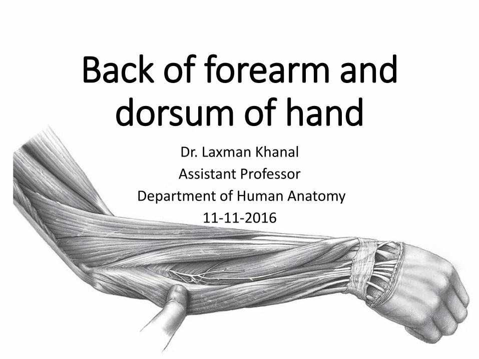
Back of forearm and dorsum of hand
Dr. Laxman Khanal
Assistant Professor
Department of Human Anatomy
11-11-2016
Objectives
• Learn the skeletal components of posterior forearm and dorsum of hand.
• Identify the seven superficial and five deep muscles of posterior forearm.
• Find the posterior interosseous nerve and artery.
• Find extensor retinaculum and identify thestructures on the dorsal aspect of the wrist.
• Know the boundaries and content of anatomicalsnuff box.
• Different components of dorsum of hand.
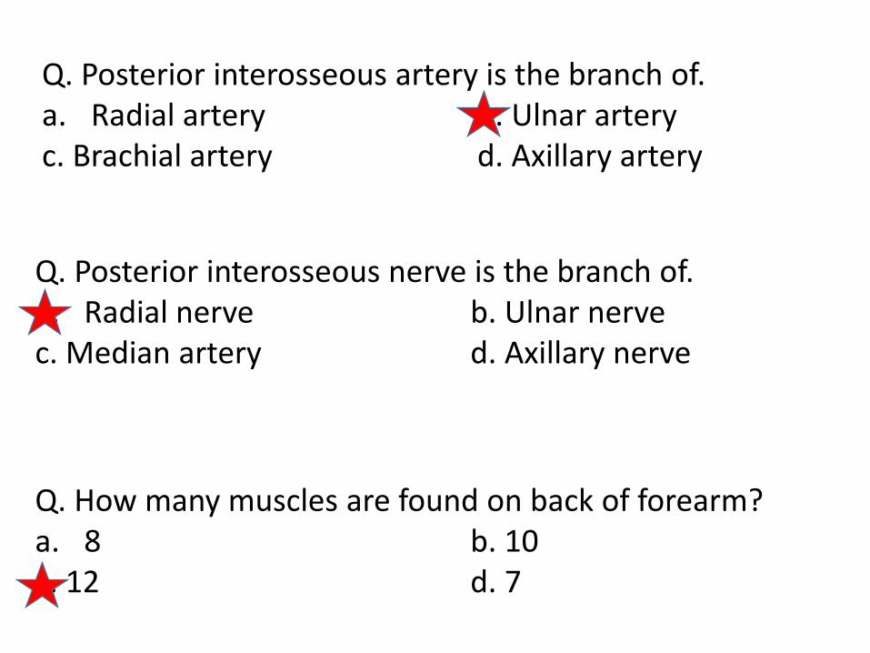
Q. Posterior interosseous artery is the branch of.a. Radial artery b. Ulnar arteryc. Brachial artery d. Axillary artery
Q. How many muscles are found on back of forearm?a. 8 b. 10c. 12 d. 7
Q. Posterior interosseous nerve is the branch of.a. Radial nerve b. Ulnar nervec. Median artery d. Axillary nerve
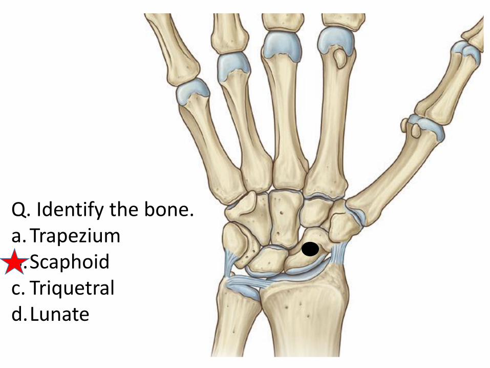
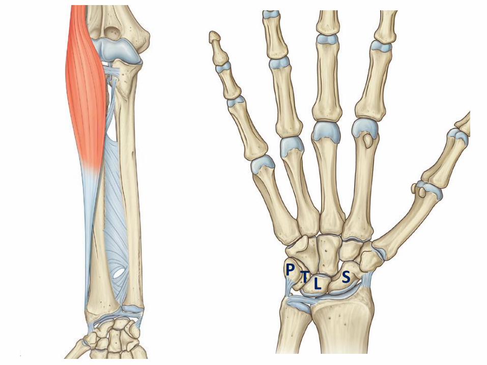
Q. Identify the bone.a.Trapeziumb.Scaphoidc. Triquetral d.Lunate
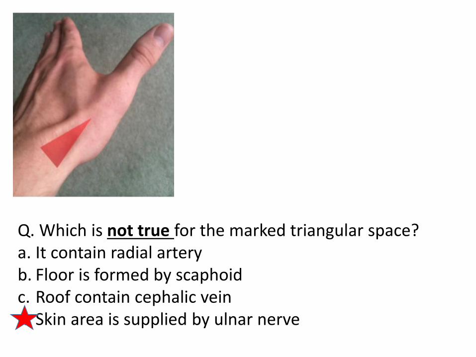
Q. Which is not true for the marked triangular space?a. It contain radial arteryb. Floor is formed by scaphoidc. Roof contain cephalic veind. Skin area is supplied by ulnar nerve
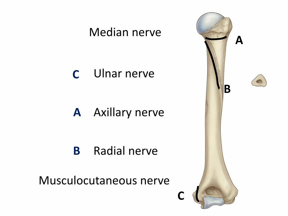
A
B
C
A
B
C
Median nerve
Ulnar nerve
Axillary nerve
Radial nerve
Musculocutaneous nerve
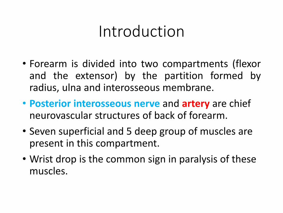
Introduction
• Forearm is divided into two compartments (flexorand the extensor) by the partition formed byradius, ulna and interosseous membrane.
• Posterior interosseous nerve and artery are chief neurovascular structures of back of forearm.
• Seven superficial and 5 deep group of muscles are present in this compartment.
• Wrist drop is the common sign in paralysis of these muscles.
SLTP
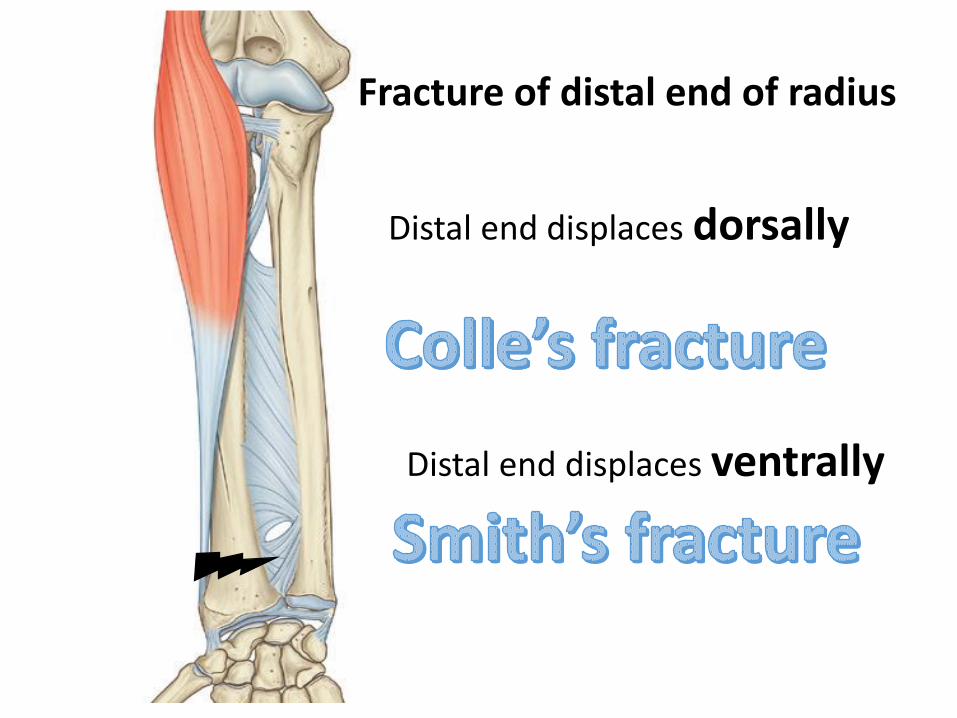
Fracture of distal end of radius
Distal end displaces dorsally
Distal end displaces ventrally
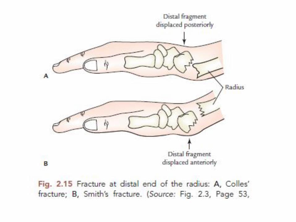

Colle’s or Smith ???
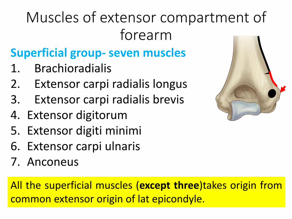
Muscles of extensor compartment of forearm
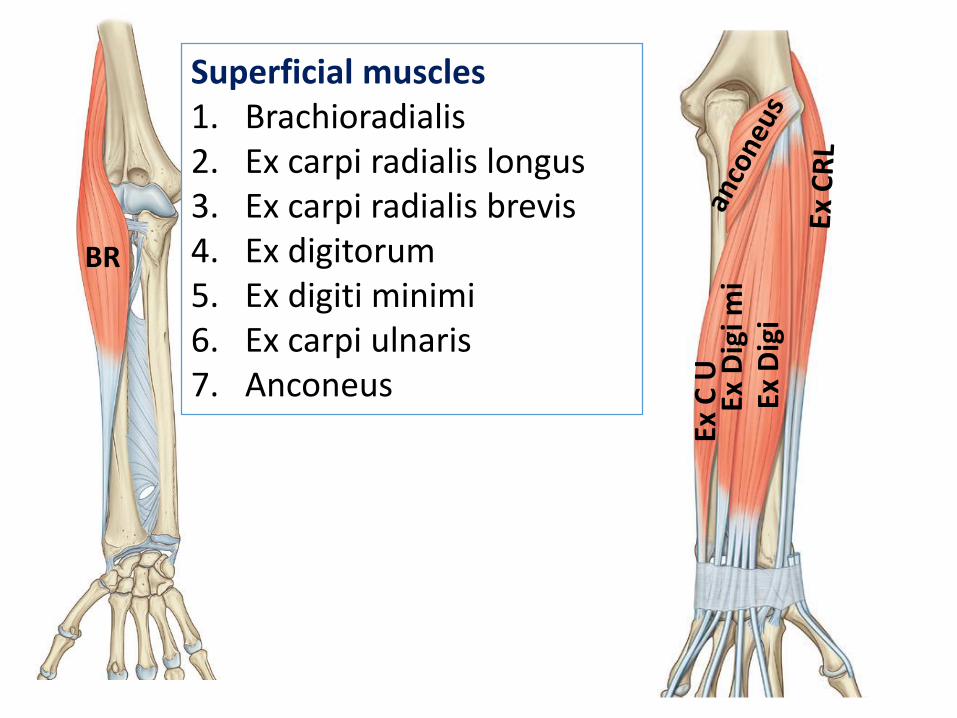
Superficial group- seven muscles1. Brachioradialis2. Extensor carpi radialis longus3. Extensor carpi radialis brevis4. Extensor digitorum5. Extensor digiti minimi6. Extensor carpi ulnaris7. Anconeus
All the superficial muscles (except three)takes origin fromcommon extensor origin of lat epicondyle.
BR
Ex D
igi
Ex D
igi m
iEx
C U
Superficial muscles1. Brachioradialis2. Ex carpi radialis longus3. Ex carpi radialis brevis4. Ex digitorum5. Ex digiti minimi6. Ex carpi ulnaris7. Anconeus
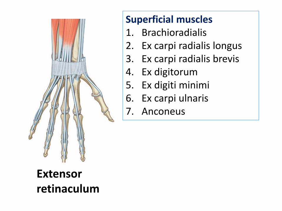
Extensor retinaculum
Superficial muscles1. Brachioradialis2. Ex carpi radialis longus3. Ex carpi radialis brevis4. Ex digitorum5. Ex digiti minimi6. Ex carpi ulnaris7. Anconeus

Superficial muscles1. Brachioradialis2. Ex carpi radialis longus3. Ex carpi radialis brevis4. Ex digitorum5. Ex digiti minimi6. Ex carpi ulnaris7. Anconeus
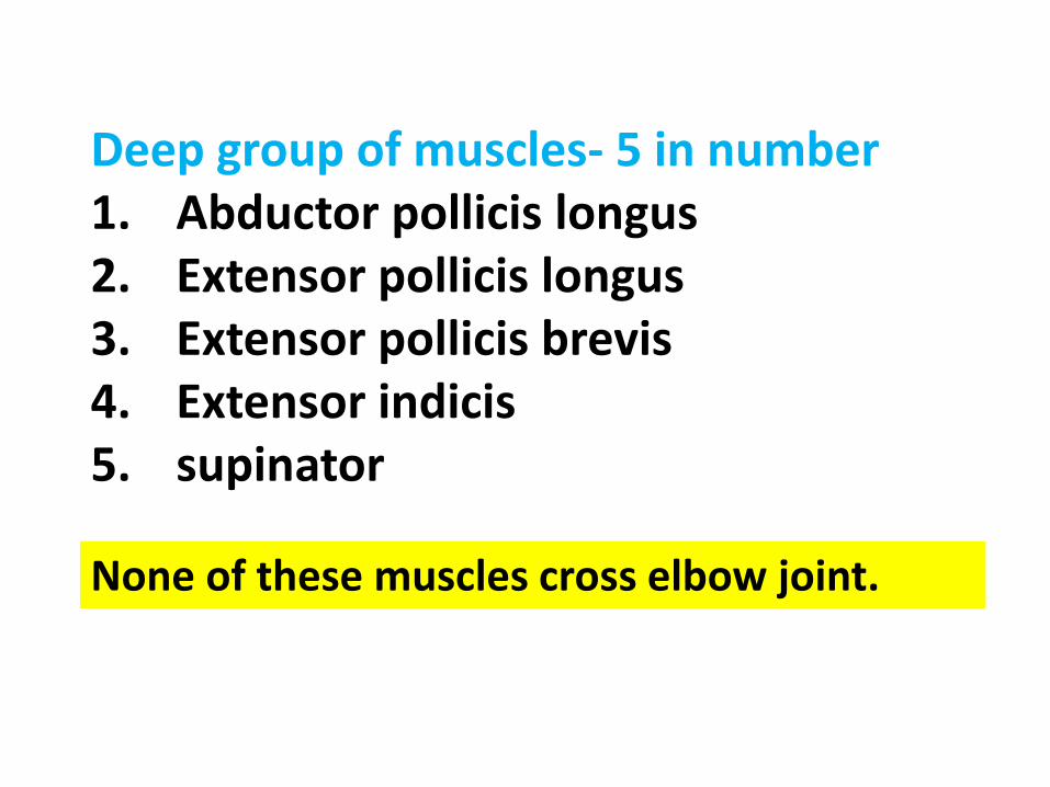
Deep group of muscles- 5 in number1. Abductor pollicis longus2. Extensor pollicis longus3. Extensor pollicis brevis4. Extensor indicis5. supinator
None of these muscles cross elbow joint.
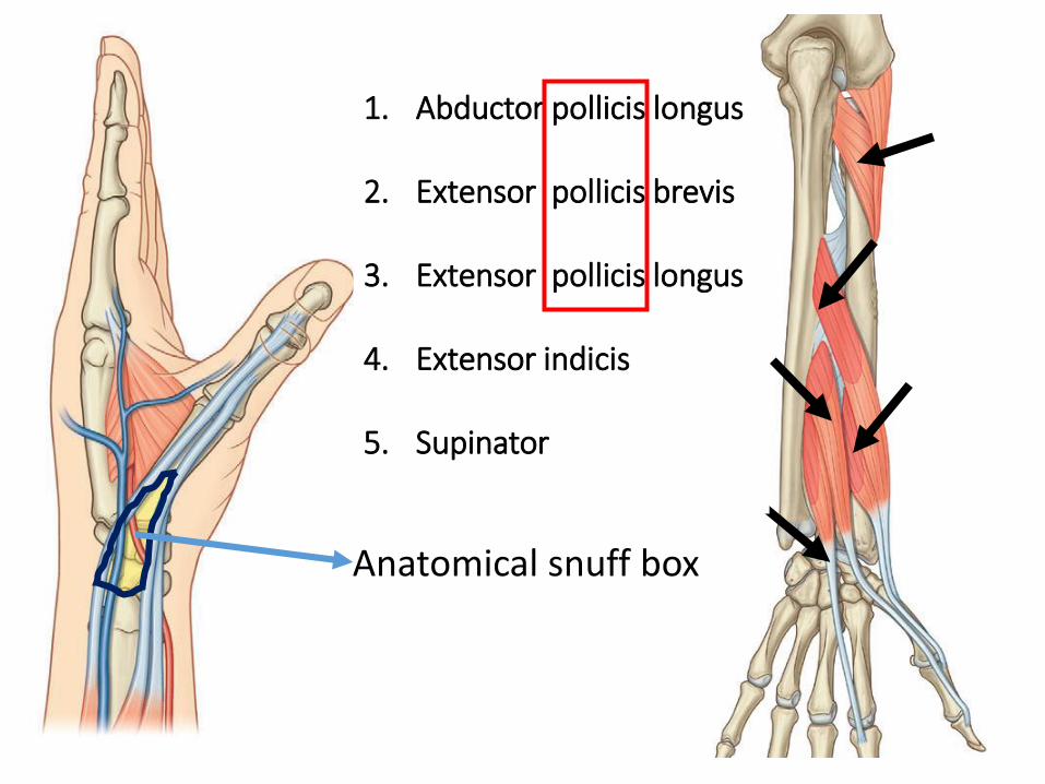
1. Abductor pollicis longus
2. Extensor pollicis brevis
3. Extensor pollicis longus
4. Extensor indicis
5. Supinator
Anatomical snuff box
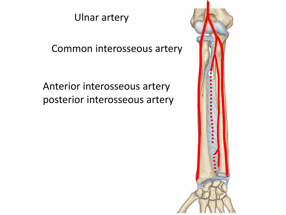
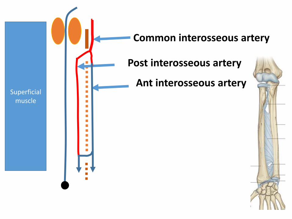
Common interosseous artery
Anterior interosseous arteryposterior interosseous artery
Ulnar artery
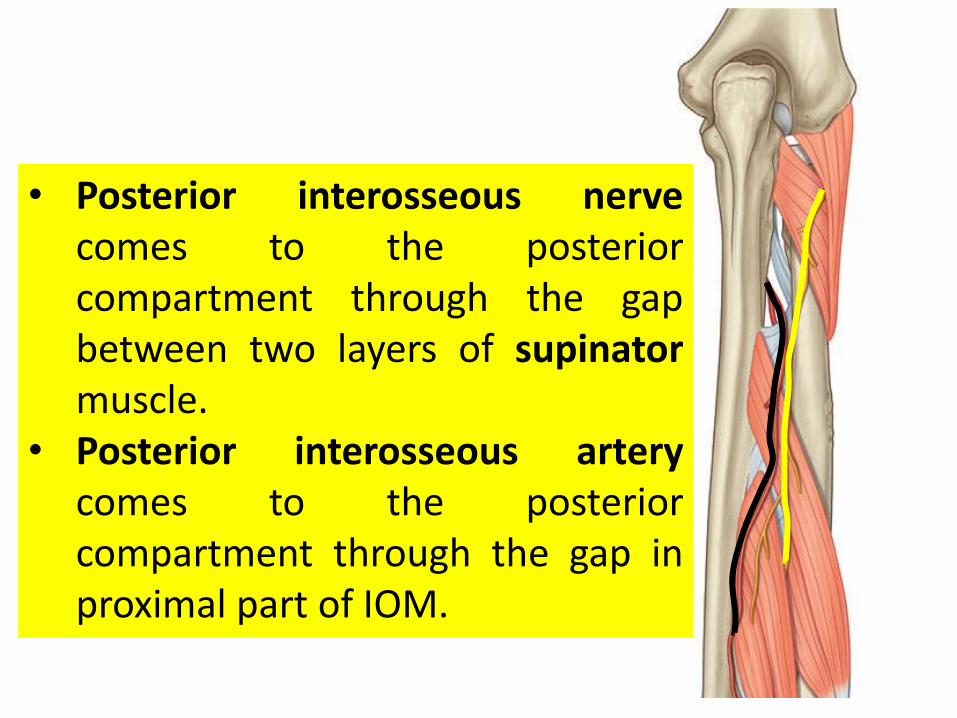
• Posterior interosseous nervecomes to the posteriorcompartment through the gapbetween two layers of supinatormuscle.
• Posterior interosseous arterycomes to the posteriorcompartment through the gap inproximal part of IOM.
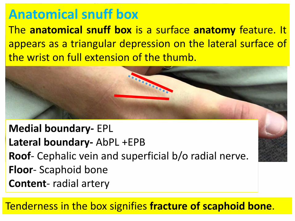
Anatomical snuff boxThe anatomical snuff box is a surface anatomy feature. Itappears as a triangular depression on the lateral surface ofthe wrist on full extension of the thumb.
Medial boundary- EPLLateral boundary- AbPL +EPBRoof- Cephalic vein and superficial b/o radial nerve.Floor- Scaphoid boneContent- radial artery
Tenderness in the box signifies fracture of scaphoid bone.
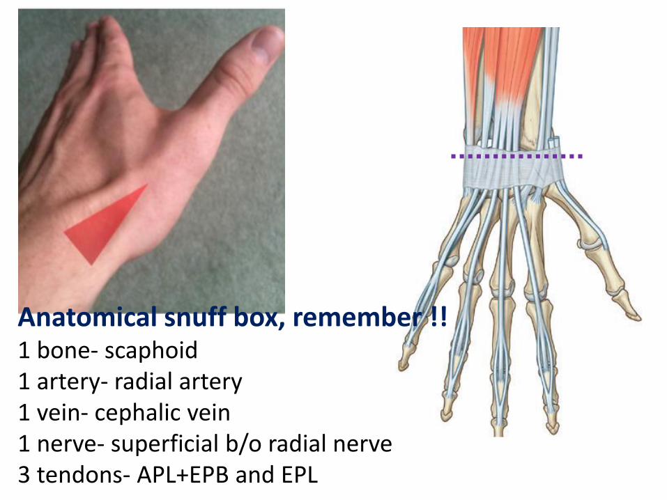
Anatomical snuff box, remember !!1 bone- scaphoid1 artery- radial artery1 vein- cephalic vein1 nerve- superficial b/o radial nerve3 tendons- APL+EPB and EPL
UR
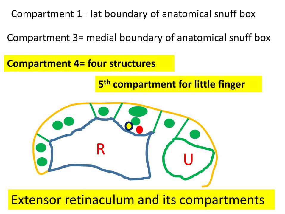
Compartment 1= lat boundary of anatomical snuff box
Compartment 3= medial boundary of anatomical snuff box
Compartment 4= four structures
Extensor retinaculum and its compartments
5th compartment for little finger
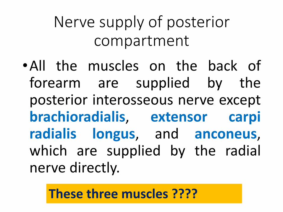
Nerve supply of posterior compartment
•All the muscles on the back offorearm are supplied by theposterior interosseous nerve exceptbrachioradialis, extensor carpiradialis longus, and anconeus,which are supplied by the radialnerve directly.
These three muscles ????
Superficial muscle
Common interosseous artery
Post interosseous artery
Ant interosseous artery
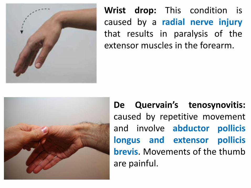
Wrist drop: This condition iscaused by a radial nerve injurythat results in paralysis of theextensor muscles in the forearm.
De Quervain’s tenosynovitis:caused by repetitive movementand involve abductor pollicislongus and extensor pollicisbrevis. Movements of the thumbare painful.
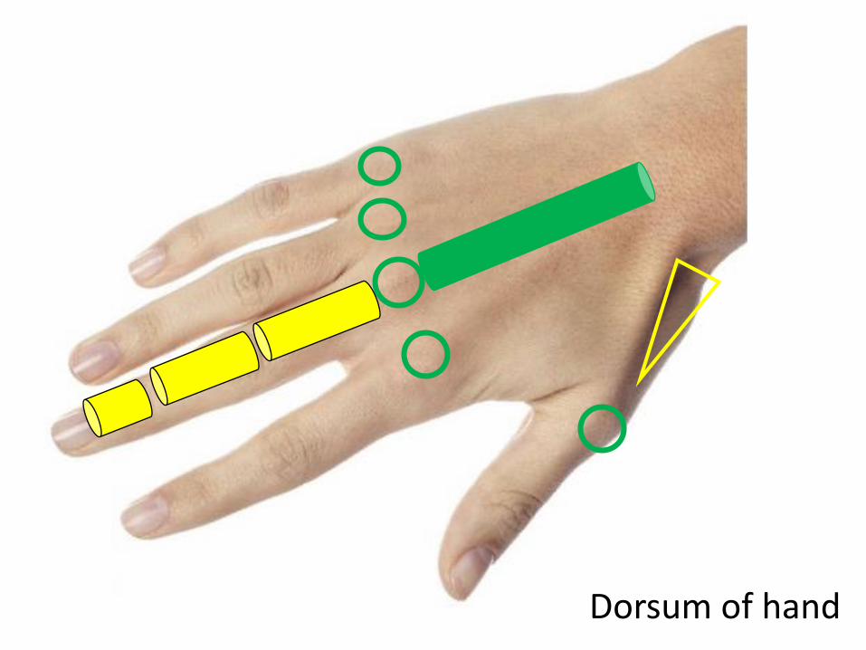
Dorsum of hand
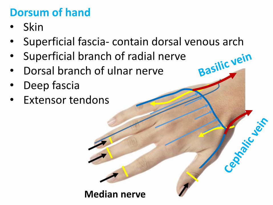
Dorsum of hand• Skin• Superficial fascia- contain dorsal venous arch• Superficial branch of radial nerve• Dorsal branch of ulnar nerve• Deep fascia• Extensor tendons
Median nerve
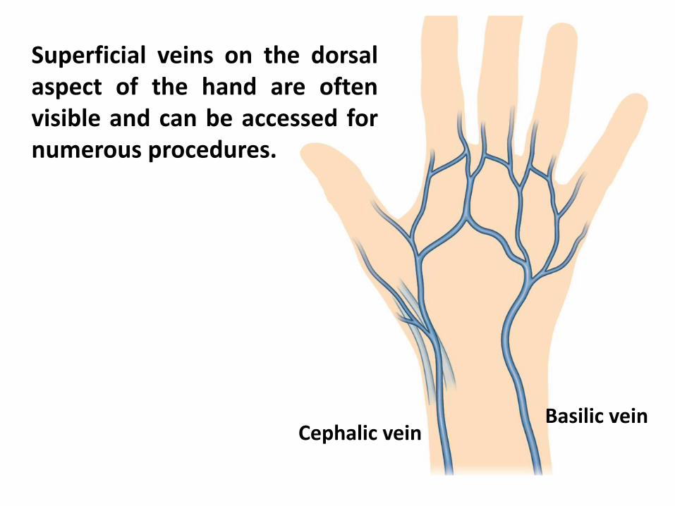
Superficial veins on the dorsalaspect of the hand are oftenvisible and can be accessed fornumerous procedures.
Basilic veinCephalic vein
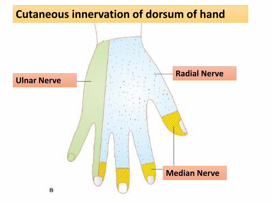
Cutaneous innervation of dorsum of hand
Radial NerveUlnar Nerve
Median Nerve
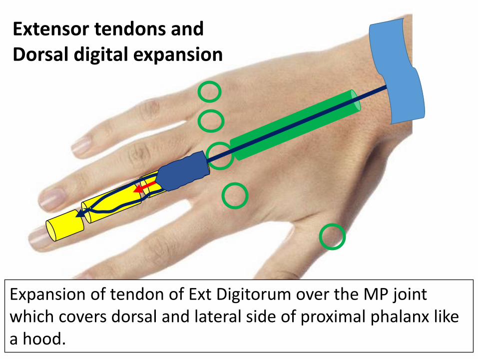
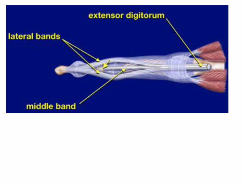
Extensor tendons and Dorsal digital expansion
Expansion of tendon of Ext Digitorum over the MP joint which covers dorsal and lateral side of proximal phalanx like a hood.
Dorsal digital expansion
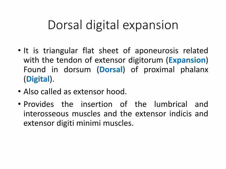
• It is triangular flat sheet of aponeurosis relatedwith the tendon of extensor digitorum (Expansion)Found in dorsum (Dorsal) of proximal phalanx(Digital).
• Also called as extensor hood.
• Provides the insertion of the lumbrical andinterosseous muscles and the extensor indicis andextensor digiti minimi muscles.
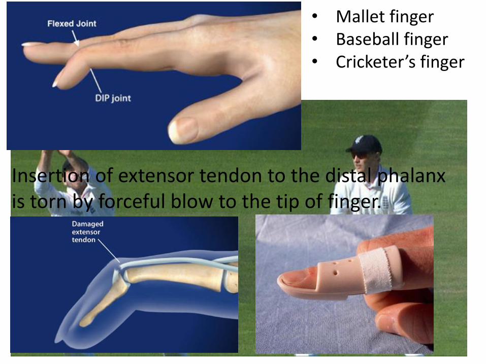
• Mallet finger• Baseball finger• Cricketer’s finger
Insertion of extensor tendon to the distal phalanx is torn by forceful blow to the tip of finger.
Conclusion
• Muscles: 7+5
• Nerve: post interosseous nerve
• Artery: post interosseous artery
• Extensor retinaculum: 6 compartment with 9 muscles+1 artery +1 nerve.
• Anatomical snuff box : boundaries and contents.
• Formation of dorsal digital expansion.