Embed Size (px)
Citation preview

ANALYSIS OF MUTANT PARASITES
THAT HAVE A DEFECT IN ADHESION
Denise MATTEI
Mar08
Cours International « Atelier Paludisme »10 Mars au 18 Avril 1008 – Institut Pasteur de Madagascar

TRAFFICKING OF VIRULENCE FACTORS IN INFECTED
RED BLOOD CELLS
SEARCH FOR TRAFFICKINGMUTANT PARASITES
Mar08

knobs
PfEMP-2
Band 3
Knob-associated proteins
PfHRP-1
Glycophorin C
Adductin
4.9
Tropomodulin
Tropomyosin
and Actin
Spectrin
4.2
4.1
Ankyrin
PfEMP1
PfEMP-3
rifin
other
molecules?PfEMP1
Mar08

OBSERVATIONS
CLAG = cytoadherence linked asexual gene
Identified through the study of parasite lines that no longer bound to C32melanoma cells = subtelomeric deletion in chr 9 (Biggs, BA et al. PNAS,1989)
.
Genetic knock-out (Trenholme, KR et al. PNAS, 2000) or
antisense RNA inhibition Gardiner, DL et al. MBP, 2000) => loss of the CD36 binding phenotype
.CLAG9 may be involved in cytoadhesion by binding directly to CD36or indirectly, through a role in PfEMP1 transport
Mar08

WORKING HYPOTHESISIs CLAG9 another CD36 binding molecule?
EXPERIMENTAL APPROACH
• P. falciparum lines selected to bind to CD36 or to CSA. Adhesion to these receptors occurs in a mutually exclusive manner
⇒ possibility to study the role of CLAG9 in cytoadherent parasites that do not bind to CD36
• Specific antibodies
=> to analyse the location of clag9 during the asexual blood stage development
CSA CD36
CHO
Receptor «panning »
FCR3 CSA FCR3 CD36
PannedFCR3
Scherf et al., 1998, EMBO J. 17 , 5418
monomorphic parasites
Mar08

Transcription of the clag9 gene
--
-2 6 10 14 18 22 26 30 34 38 42 46
9.49
7.46
4.40
3D7
hrs
The clag9 gene is transcribed in theschizont stages of both CD36 andCSA-selected parasites
Mar08
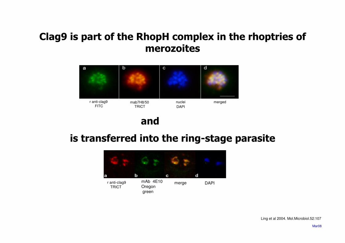
Clag9 is part of the RhopH complex in the rhoptries of merozoites
r anti-clag9FITC
mab7H8/50TRICT
nuclei
DAPI
merged
r anti-clag9 TRICT
mAb 4E10
Oregon green
merge DAPI
Mar08
Ling et al 2004. Mol.Microbiol.52:107
is transferred into the ring-stage parasite
and

Clag9 RhopH2
Localisation of CLAG9 to the rhoptries
Ling et al.2004. Mol.Microbiol.
Mar08

CLAG9
• clag9 gene is transcribed and expressed in late asexual blood stages in CD36 and non-CD36 binders
• Clag9 is present in merozoites as part of the RhopHcomplex in the rhoptries
• Anti-CLAG9 antibodies do not detect it at theerythrocyte membrane of parasitized RBC (trophozoites/schizonts)
Mar08

Analysis of clag9 natural mutant parasites
T9-96 and D10
Mar08

C32 cellsCD36 >> ICAM-1 >>CSA
FCR3CD36 D10
T9-96 and D10 do not cytoadhere to any known adhesion receptor
Mar08
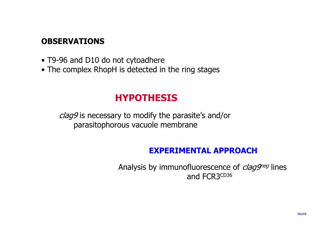
OBSERVATIONS
• T9-96 and D10 do not cytoadhere• The complex RhopH is detected in the ring stages
HYPOTHESIS
clag9 is necessary to modify the parasite’s and/or parasitophorous vacuole membrane
EXPERIMENTAL APPROACH
Analysis by immunofluorescence of clag9neg linesand FCR3CD36
Mar08

RESULTS
The images were similar independently of the genotype
‘PfEMP1’ is located in association with the Maurer’s clefts
PfEMP1Alexa
Pf332TRICT
DAPI merge
FCR3CD36
D10
T9.96
Mar08

CONCLUSIONclag9neg parasites can export polypeptides to the different cellular compartments
FCR3CD36
D10
T9.96
Mab89Alexa
DAPI merge
Mar08
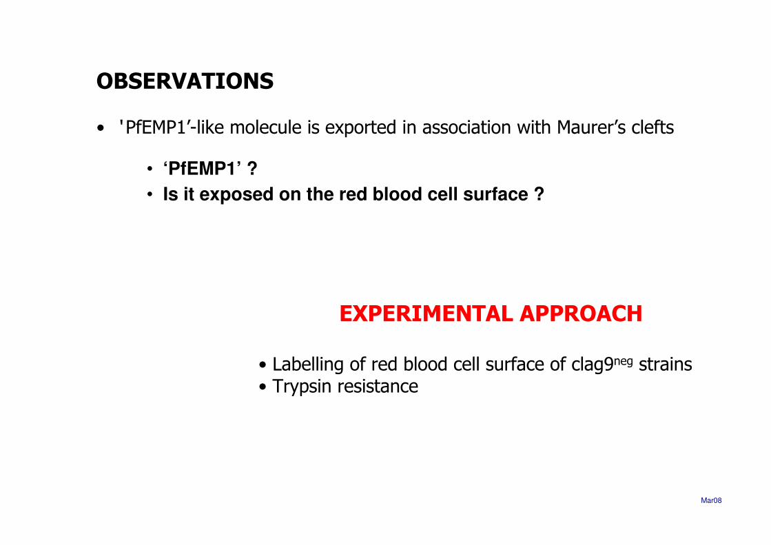
OBSERVATIONS
• ‘ PfEMP1’-like molecule is exported in association with Maurer’s clefts
• ‘PfEMP1’ ?
• Is it exposed on the red blood cell surface ?
EXPERIMENTAL APPROACH
• Labelling of red blood cell surface of clag9neg strains• Trypsin resistance
Mar08
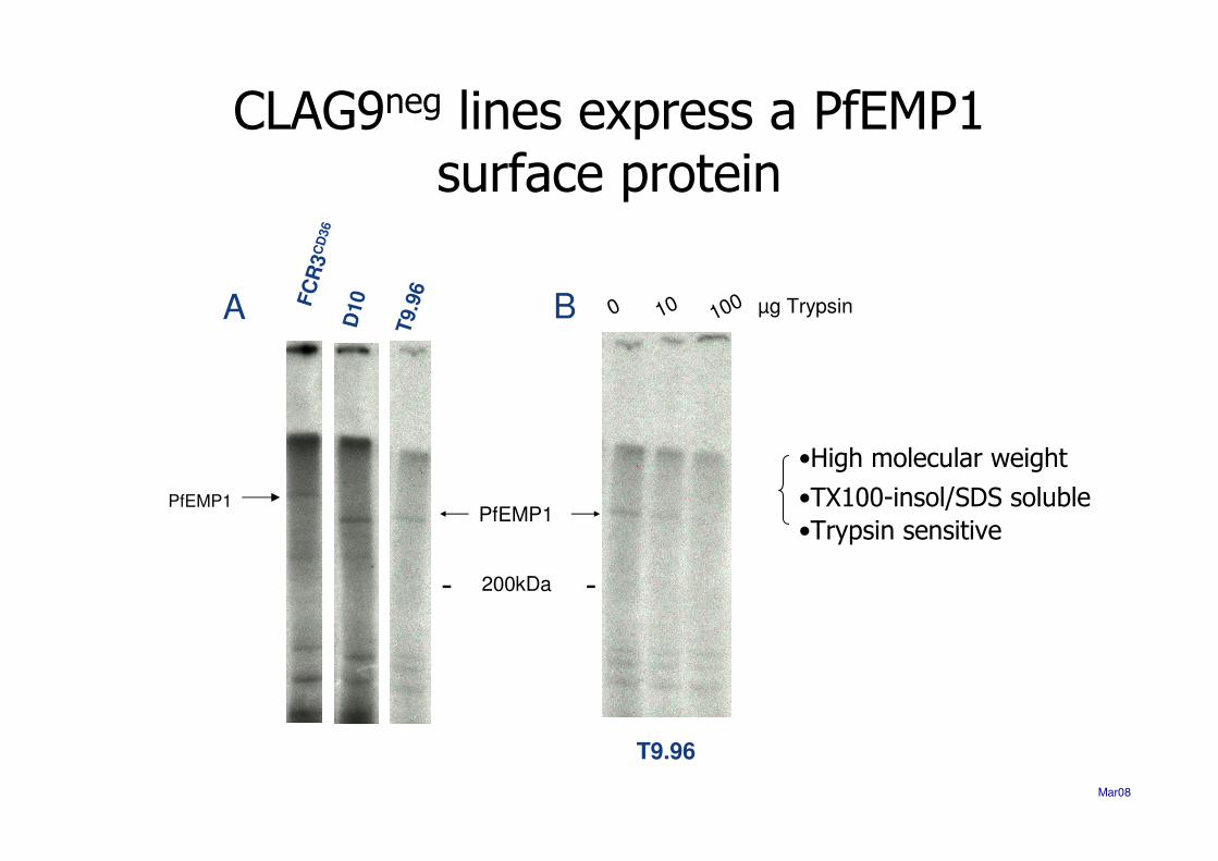
CLAG9neg lines express a PfEMP1surface protein
-200kDa-
PfEMP1
•High molecular weight
•TX100-insol/SDS soluble
•Trypsin sensitive
T9.96
0 10010 µg TrypsinB
T9.9
6
D10FC
R3
CD
36
A
PfEMP1
Mar08

OBSERVATIONS
• The isolates T9.96 and D10 express PfEMP1 on the surface but do notcytoadhere to any known receptor
HYPOTHESIS
• Adhesion negative parasites express a FUNCTIONAL var gene havingunique adhesive properties BUT can not switch expression to classical var genes
• Adhesion negative parasites display a NON FUNCTIONAL conformation* Post-translational modification to establish a functional state (protease, kinase, etc…)
EXPERIMENTAL APPROACH
• Northern blot analysis• Cloning and expression of the CIDR domain BINDING ASSAYS
Mar08

PfEMP1 is transcribed in the clag9neg
parasite lines T9.96 and D10
SR TR S
D10 FCR3CD36
D10cl9
SR TR S
D10 FCR3CD36
ATS
- 7.46 -- 4.40 -
- 9.49 -
- 2.57 -
- 1.35 -
8Kb
Kb
Mar08

Functionality studies of the PfEMP1- CIDR domain in CD36 binding of
T9-96 and D10
Mar08

A classical var gene is transcribed and expressedin mutant parasites
ATS: acidic terminal segment; CIDR (α,β,γ): cisteine-rich interdomain region; DBL (α,β,γ,δ,ε): Duffy-binding-like domain;
NTS: N-terminal segment; TM: transmembrane domain;
Tandem association
DBL1αααα CIDR1ααααNTS
CR1blood gr Aheparinhep sulfate
CD36CD31IgM
Semi-conservedhead structure
TM ATS
CD31
DBL2δδδδ CIDR2ββββ
Mar08

C L K NNKKTCGKKKCNRDCKCYE RW VKRKKEEFKKIKDHFGKQKD MQPYID PDMTLKILLNYVFLQ DMKDANG NPQHIAKIQELLE KKKVELEDNLNKNTIIDYMFEDDLEEINKC
Robinson et alRobinson et al. 2003.. 2003.MolMol..MicrobiolMicrobiol. 47:1265. 47:1265--7878
D10cl9D10cl9
M1 M2 M3C9 C12
The CIDR domain from D10 is predicted to bind to CD36
Mar08
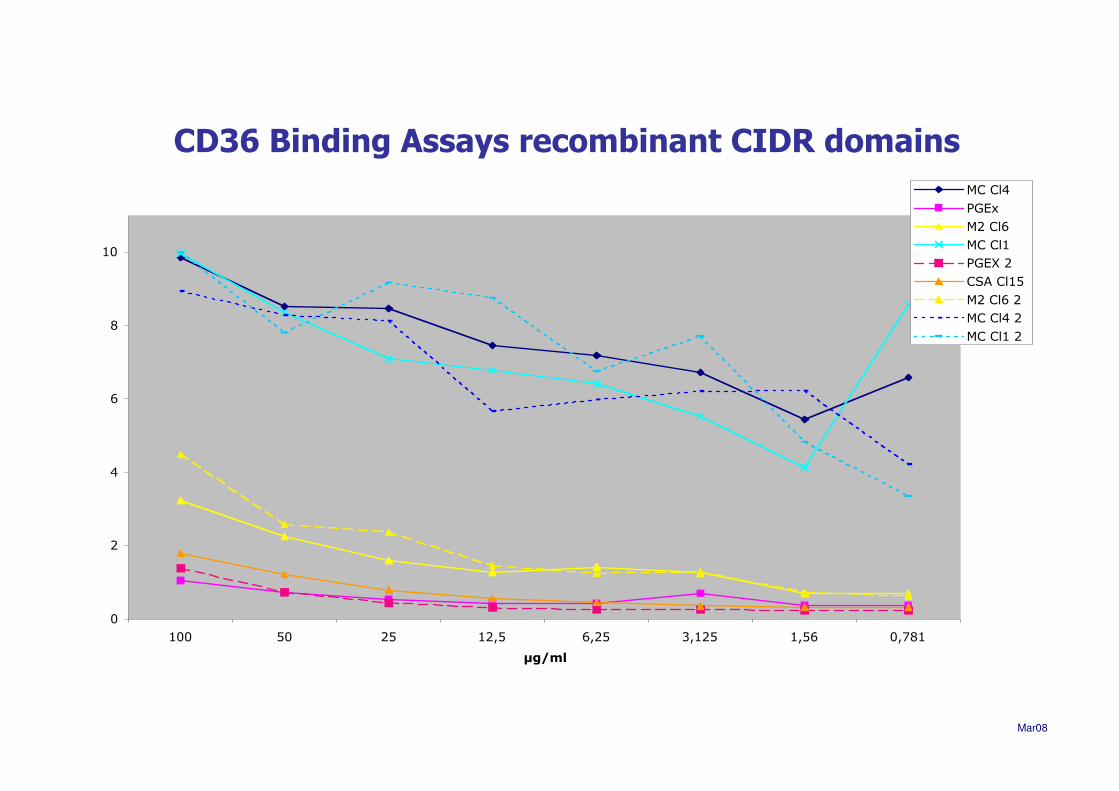
0
2
4
6
8
10
100 50 25 12,5 6,25 3,125 1,56 0,781
µg/ml
MC Cl4
PGEx
M2 Cl6
MC Cl1
PGEX 2
CSA Cl15
M2 Cl6 2
MC Cl4 2
MC Cl1 2
CD36 Binding Assays recombinant CIDR domains
Mar08
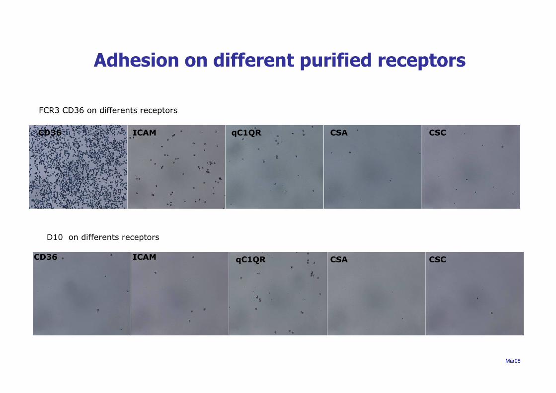
FCR3 CD36 on differents receptors
CD36 ICAM qC1QR CSA CSC
D10 on differents receptors
CD36 ICAM qC1QR CSA CSC
Adhesion on different purified receptors
Mar08

CIDR domain (M2 minimum CD36 binding site)-pDisplay vector transfected in COS-7 cells
M2 domain on the surface
CD36 protein
Anti-CD36 labelled beads
Binding assays with Dynal beads
Mar08

D10
MC+ control
anti-tag
HA merge
Mar08
CD36 Binding Properties of D10 CIDR1
TransfectedCOS7 cells
CSA(-) control

Conclusions
• Cytoadhesion mutants express a PfEMP1 molecule atthe surface of the parasitized red blood cell
• The mutant parasites can not bind to the commonadhesion phenotypes (CD36, ICAM-1, CSA, Rosetting, etc.)
• Panning on endothelial cells does not restore adhesionphenotype
Non-functional PfEMP1 surface expression !
M

Mar08
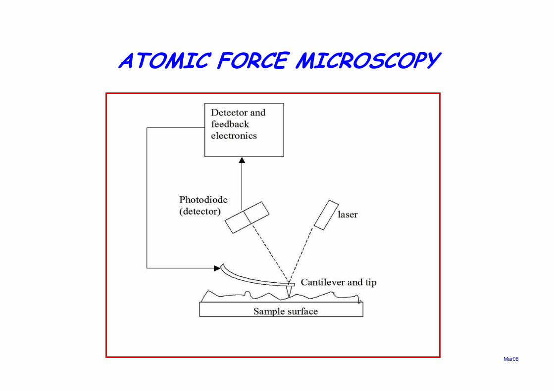
ATOMIC FORCE MICROSCOPY
Mar08

RBC infected by Plasmodium falciparum
Mar08

Control
Mar08

3D7 Parasitised red blood cell (PRBC)
Knobs sizes ø
50-60 nm

FCR3 PRBC
Knobs sizes ø
50-60 nm

D10 PRBC
Height
Amplitude error
Knobs sizes range from ø 20 nm to 100 nm
1.5 µm
Mar08

D10 and T9.96 carry a large chromosome 9 deletion
PF
I1730w
cla
g9
PF
I1725
w
PF
I1715
w
PF
I1720w
gig
PF
I1710w
BP
OR
F
PF
I1760w
rex4
PF
I1770
w
PF
I1765
c
PF
I1775
w
PF
I1780
w
PF
I1785
w
PF
I1790
w
PF
I1800
w
PF
I1795
c
PF
I1805w
rif
in
PF
I1810w
rifi
n
PF
I1825w
rifi
n
PF
I1815c
rifin
Centromere
Telomere
PF
I1820w
PfE
MP
1
PF
I1830c
PfE
MP
1
PF
I1735c
rex
1
PF
I1755c
rex
3
PF
I1740c
rex
2
PF
I1745
c
Chromosome truncation in mutant parasites
PF
I1705
w
PF
I1750
c
Mar08

HYPOTHESIS
• Adhesion negative parasites display a NON FUNCTIONAL conformation* Post-translational modification to establish a functional state(protease, kinase, etc…)
PERSPECTIVES
* Clag 9 KO => clag9 functional role * other KO => chr9 region => genotype and phenotype analysis
* SYSTEMS BIOLOGY => RNAs transcribed/translated ptn-ptn interactions cellular location
Mar08

BIHP (Institut Pasteur, Paris)
Artur ScherfSuwanna ChaorattanakaweJosé Manuel L. NogueiraCaroline DavisChristine Scheidig-BenatarYvon SterkersEmeric Roux
Parasitologie Expérimentale(Université de la Méditerranée, Marseille)
Jürg GysinCatherine Lepolard
National Institute for Medical Research(Mill Hill, London, UK)
Tony HolderIrene T. Ling
Ehime UniversitySchool of Medicine(Ehime, Japan)
Osamu Kaneko
Cellular Microbiology and Infectious Pathogeny(Institute Pasteur Lille)
Frank LafontSebastien Janel

ANALYSIS OF MUTANT PARASITES
THAT HAVE A DEFECT IN ADHESION
Denise MATTEI
Mar08
Cours International « Atelier Paludisme »10 Mars au 18 Avril 1008 – Institut Pasteur de Madagascar


THE PfRhopH COMPLEX
RhopH Complexapprox. 480 kDa
RhopH1/ClagPFI1730w - 155kDa
RhopH2PFI1445w - 140 kDa
RhopH3PFI0265c - 110 kDa
Clag 2
Clag 3.1
Clag 3.2
Clag 8
Clag 9
Kaneko et al. 2005.MBP

Why the PfEMP1expressed by the ‘clagneg’
isolates is non functional ?
HYPOTHESIS
• wrong conformation ?• post-translational modification ?
how can it be tested?
apr07

Perspectives
• A NEW var GENE HAVING UNIQUE ASPECTS?(non CD36, non CSA binding)
AND/OR
• A NEW HOST ENDOTHELIAL ADHESION RECEPTOR ?
OR
•A KNOWN PfEMP1 WHICH DISPLAYS A NON FUNCTIONALCONFORMATION
• clag9 KNOCK-OUT


1000 100 0 1000 100 0
D10FCR3 CD36
HRP
HSP 70
ATSHRP
HSP 70
Trypsin µg
175
83
62
47.5
32.5
kDaN
R
B
C
N
R
B
C
Analysis of surface-exposed PfEMP1 on PRBCby trypsin cleavage

0 100 100 0 100 100
HRP
HSP 70
D10 FCR3 CD36
HSP 70 HRP
ATS
175
83
62
47.5
32.5
kDa N
R
B
C
N
R
B
CTrypsin
Chymotrypsin µg
Analysis of surface-exposed PfEMP1 on PRBCby trypsin and chymotrypsin cleavage
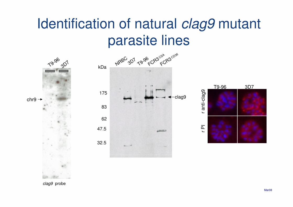
Identification of natural clag9 mutant parasite lines
T9-96
3D7
chr9
NRBCFCR3
CD36
FCR3CSA
T9-96
3D7
175
83
62
47.5
32.5
kDa
clag9
T9-96 3D7
r P
Ir
ant i-c
lag9
clag9 probe
Mar08

Localisation of CLAG9
r anti-clag9 FITC
mab7H8/50TRICT
nuclei
DAPI
merged
pep1 pep2
Clag9-GST 100 aa
Clag9 is encoded by members of the clag multigene family
Ling,I et al. 2004. Mol.Microbiol. 52:107-118
Mar08

r anti-clag9
TRICT
mAb 4E10
Oregon green
merge DAPI
Rabbit anti-CLAG9 reactswith ring-infected erythrocytes
Mar08

Perspectives
• A NEW var GENE HAVING UNIQUE ASPECTS?(non CD36, non CSA binding)
AND/OR
• A NEW HOST ENDOTHELIAL ADHESION RECEPTOR ?
OR
• A KNOWN PfEMP1 WHICH DISPLAYS A NON FUNCTIONAL CONFORMATION
• clag9 KNOCK-OUT
Mar08

Why PfEMP1 expressed by the ‘clag9neg’isolates are non functional ?
HYPOTHESIS
* wrong conformation * post-translational modification
Mar08