Embed Size (px)
Citation preview
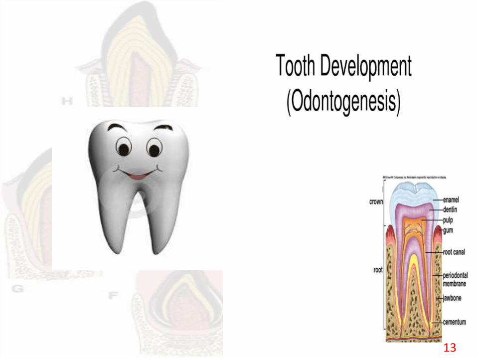
DEVELOPMENT OF TOOTH
CHAITANYA.PI MDSDept of Public Health Dentistry
Previous questions
• Anodontia. (jun 14)
• Write in detail about development of tooth.(may,08).
• Describe the theories of eruption of teeth.
2
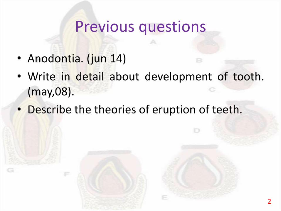
CONTENTS
1. Introduction2. Dental Lamina3. Vestibular Lamina4. Tooth development5. Developmental stages• Bud stage• Cap stage• Bell stage• Advanced bell stage6. Hertwig’s epithelial root sheath and root formation7. Review of literature8. Conclusion9. References
3
INTRODUCTION
4
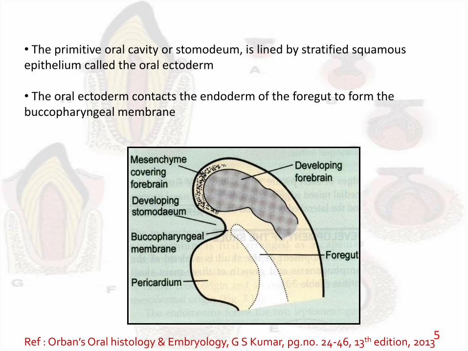
• The primitive oral cavity or stomodeum, is lined by stratified squamous epithelium called the oral ectoderm
• The oral ectoderm contacts the endoderm of the foregut to form the buccopharyngeal membrane
Ref : Orban’sOral histology & Embryology, G S Kumar, pg.no. 24-46, 13th edition, 20135
DENTAL LAMINA
6
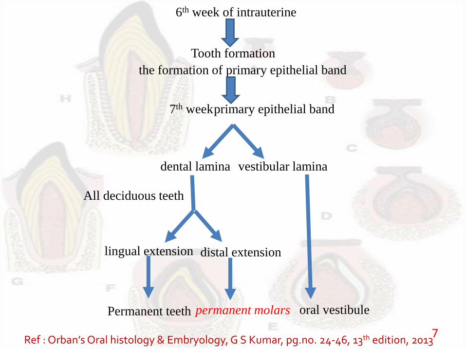
6th week of intrauterine
Tooth formation
the formation of primary epithelial band
7th week primary epithelial band
dental lamina vestibular lamina
lingual extension distal extension
All deciduous teeth
Permanent teeth permanent molars oral vestibule
Ref : Orban’sOral histology & Embryology, G S Kumar, pg.no. 24-46, 13th edition, 20137
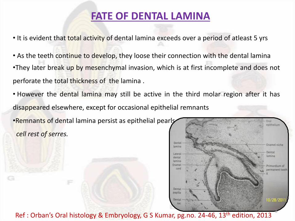
FATE OF DENTAL LAMINA
• It is evident that total activity of dental lamina exceeds over a period of atleast 5 yrs
• As the teeth continue to develop, they loose their connection with the dental lamina
•They later break up by mesenchymal invasion, which is at first incomplete and does not
perforate the total thickness of the lamina .
• However the dental lamina may still be active in the third molar region after it has
disappeared elsewhere, except for occasional epithelial remnants
•Remnants of dental lamina persist as epithelial pearls or
cell rest of serres.
Ref : Orban’s Oral histology & Embryology, G S Kumar, pg.no. 24-46, 13th edition, 20138
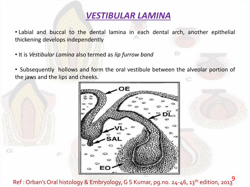
VESTIBULAR LAMINA
• Labial and buccal to the dental lamina in each dental arch, another epithelialthickening develops independently
• It is Vestibular Lamina also termed as lip furrow band
• Subsequently hollows and form the oral vestibule between the alveolar portion ofthe jaws and the lips and cheeks.
Ref : Orban’sOral histology & Embryology, G S Kumar, pg.no. 24-46, 13th edition, 20139
CLINICAL CONSIDERATIONS
10
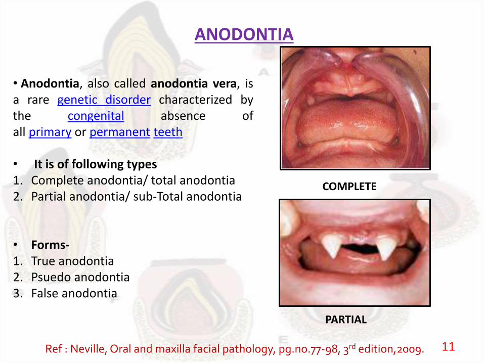
ANODONTIA
• Anodontia, also called anodontia vera, isa rare genetic disorder characterized bythe congenital absence ofall primary or permanent teeth
• It is of following types1. Complete anodontia/ total anodontia2. Partial anodontia/ sub-Total anodontia
• Forms-1. True anodontia2. Psuedo anodontia3. False anodontia
COMPLETE
PARTIAL
Ref : Neville, Oral and maxilla facial pathology, pg.no.77-98, 3rd edition,2009. 11
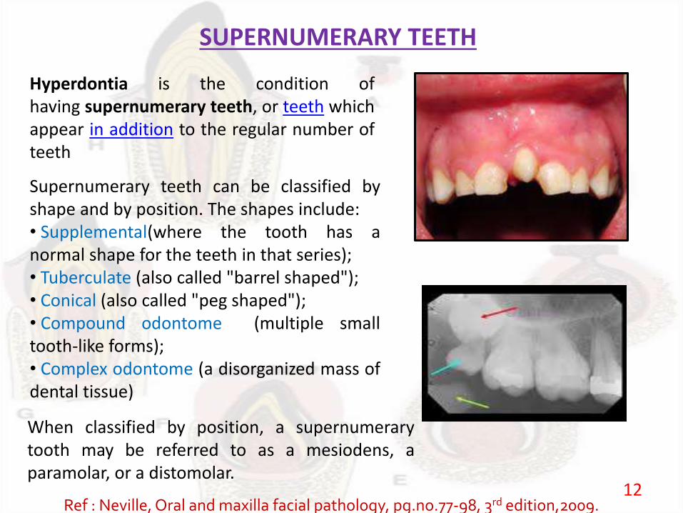
SUPERNUMERARY TEETH
Supernumerary teeth can be classified byshape and by position. The shapes include:• Supplemental(where the tooth has anormal shape for the teeth in that series);• Tuberculate (also called "barrel shaped");• Conical (also called "peg shaped");• Compound odontome (multiple smalltooth-like forms);• Complex odontome (a disorganized mass ofdental tissue)
Hyperdontia is the condition ofhaving supernumerary teeth, or teeth whichappear in addition to the regular number ofteeth
When classified by position, a supernumerarytooth may be referred to as a mesiodens, aparamolar, or a distomolar.
Ref : Neville, Oral and maxilla facial pathology, pg.no.77-98, 3rd edition,2009.12
13
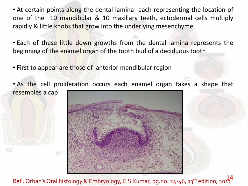
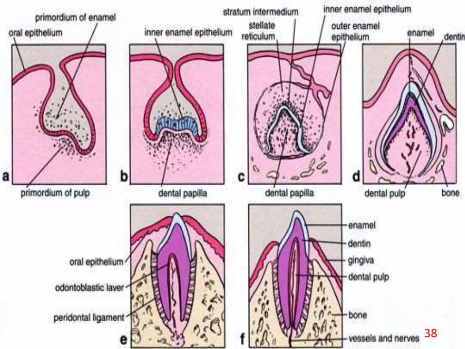
• At certain points along the dental lamina each representing the location ofone of the 10 mandibular & 10 maxillary teeth, ectodermal cells multiplyrapidly & little knobs that grow into the underlying mesenchyme
• Each of these little down growths from the dental lamina represents thebeginning of the enamel organ of the tooth bud of a deciduous tooth
• First to appear are those of anterior mandibular region
• As the cell proliferation occurs each enamel organ takes a shape thatresembles a cap
Ref : Orban’sOral histology & Embryology, G S Kumar, pg.no. 24-46, 13th edition, 201314
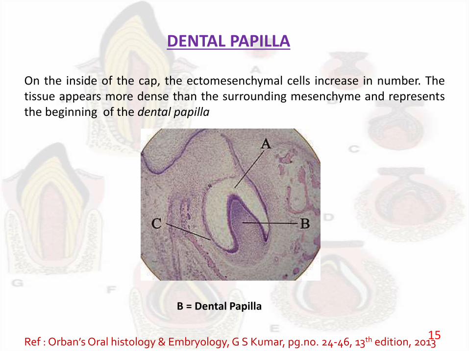
DENTAL PAPILLA
On the inside of the cap, the ectomesenchymal cells increase in number. Thetissue appears more dense than the surrounding mesenchyme and representsthe beginning of the dental papilla
B = Dental Papilla
Ref : Orban’sOral histology & Embryology, G S Kumar, pg.no. 24-46, 13th edition, 201315
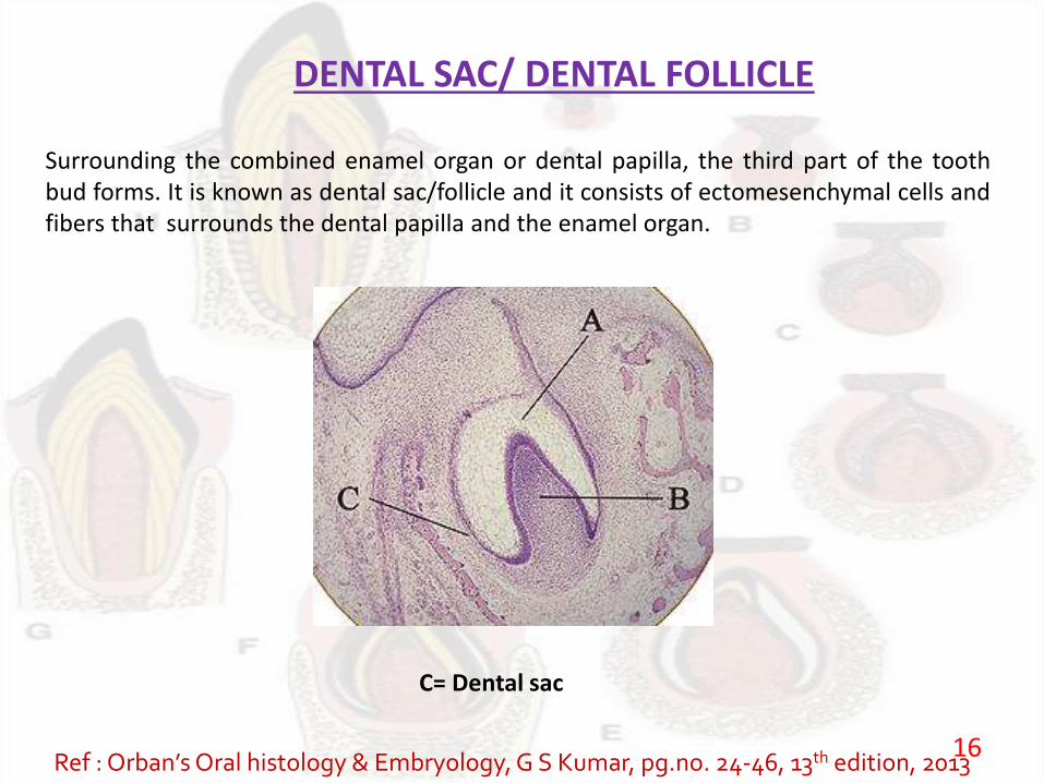
DENTAL SAC/ DENTAL FOLLICLE
Surrounding the combined enamel organ or dental papilla, the third part of the toothbud forms. It is known as dental sac/follicle and it consists of ectomesenchymal cells andfibers that surrounds the dental papilla and the enamel organ.
C= Dental sac
Ref : Orban’sOral histology & Embryology, G S Kumar, pg.no. 24-46, 13th edition, 201316
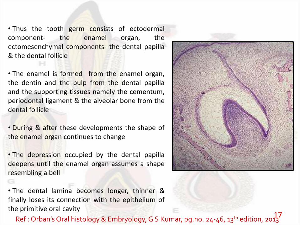
• Thus the tooth germ consists of ectodermalcomponent- the enamel organ, theectomesenchymal components- the dental papilla& the dental follicle
• The enamel is formed from the enamel organ,the dentin and the pulp from the dental papillaand the supporting tissues namely the cementum,periodontal ligament & the alveolar bone from thedental follicle
• During & after these developments the shape ofthe enamel organ continues to change
• The depression occupied by the dental papilladeepens until the enamel organ assumes a shaperesembling a bell
• The dental lamina becomes longer, thinner &finally loses its connection with the epithelium ofthe primitive oral cavity
Ref : Orban’sOral histology & Embryology, G S Kumar, pg.no. 24-46, 13th edition, 201317
DEVELOPMENTAL STAGES
18
MORPHOLOGICAL1. Dental lamina2. Bud stage3. Cap stage4. Early bell stage5. Advanced bell stage6. Formation of enamel and dentin matrix
PHYSIOLOGICALInitiation
ProliferationHistodifferentiationMorphodifferentiationApposition
Ref : Orban’sOral histology & Embryology, G S Kumar, pg.no. 24-46, 13th edition, 201319
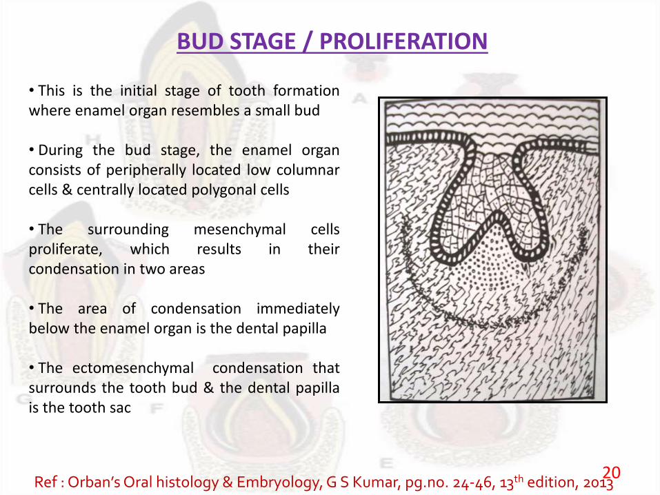
BUD STAGE / PROLIFERATION
• This is the initial stage of tooth formationwhere enamel organ resembles a small bud
• During the bud stage, the enamel organconsists of peripherally located low columnarcells & centrally located polygonal cells
• The surrounding mesenchymal cellsproliferate, which results in theircondensation in two areas
• The area of condensation immediatelybelow the enamel organ is the dental papilla
• The ectomesenchymal condensation thatsurrounds the tooth bud & the dental papillais the tooth sac
Ref : Orban’sOral histology & Embryology, G S Kumar, pg.no. 24-46, 13th edition, 201320
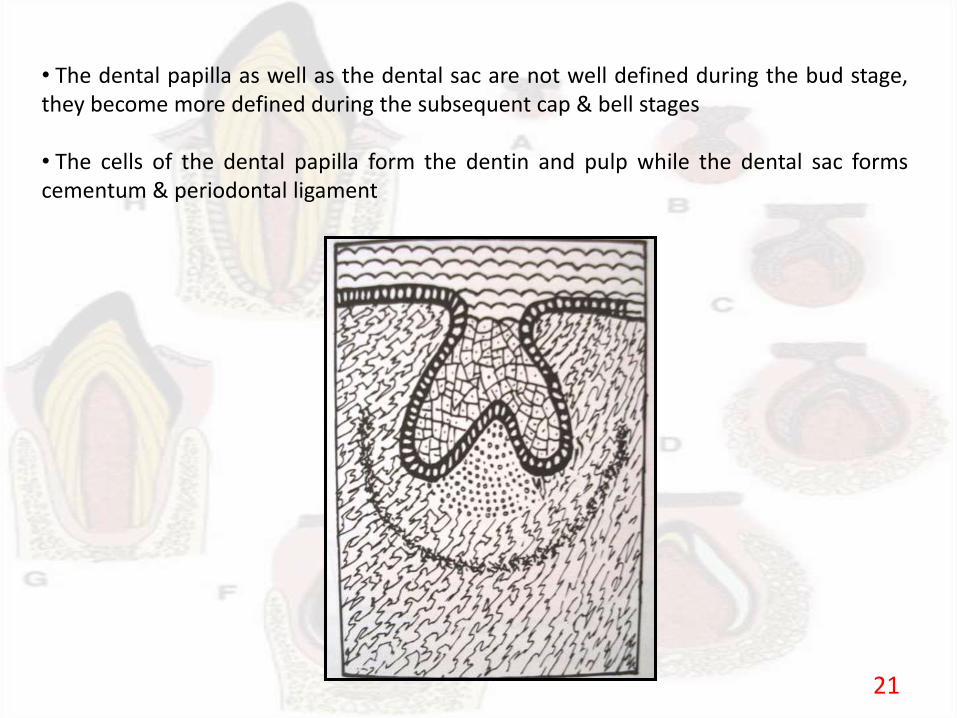
• The dental papilla as well as the dental sac are not well defined during the bud stage,they become more defined during the subsequent cap & bell stages
• The cells of the dental papilla form the dentin and pulp while the dental sac formscementum & periodontal ligament
21
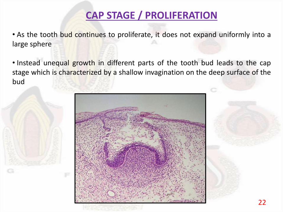
CAP STAGE / PROLIFERATION
• As the tooth bud continues to proliferate, it does not expand uniformly into alarge sphere
• Instead unequal growth in different parts of the tooth bud leads to the capstage which is characterized by a shallow invagination on the deep surface of thebud
22
OUTER & INNER ENAMEL EPITHELIUM
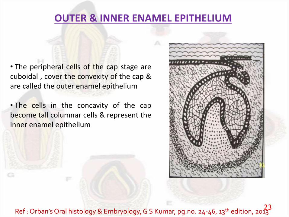
• The peripheral cells of the cap stage arecuboidal , cover the convexity of the cap &are called the outer enamel epithelium
• The cells in the concavity of the capbecome tall columnar cells & represent theinner enamel epithelium
Ref : Orban’sOral histology & Embryology, G S Kumar, pg.no. 24-46, 13th edition, 201323
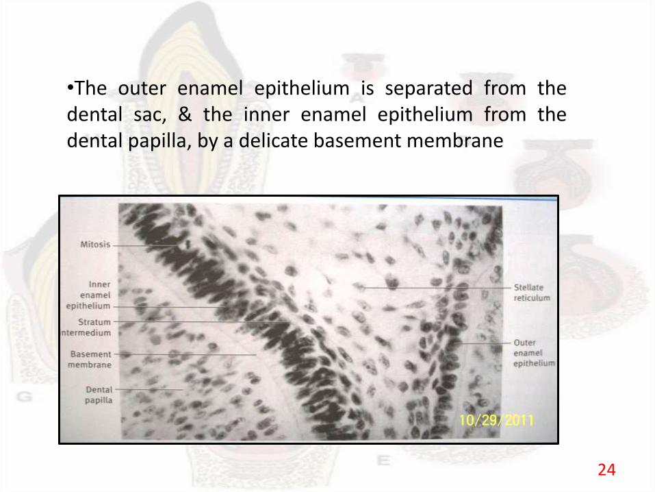
•The outer enamel epithelium is separated from thedental sac, & the inner enamel epithelium from thedental papilla, by a delicate basement membrane
24

STELLATE RETICULUM
• Polygonal cells located between the outer and the inner enamel epithelium, begin toseparate due to water being drawn into the enamel organ from the surrounding dentalpapilla
• As a result the polygonal cells become star shaped but maintain contact with eachother by their cytoplasmic process
• As the star shaped cells form a cellular network, they are called the stellate reticulum
25
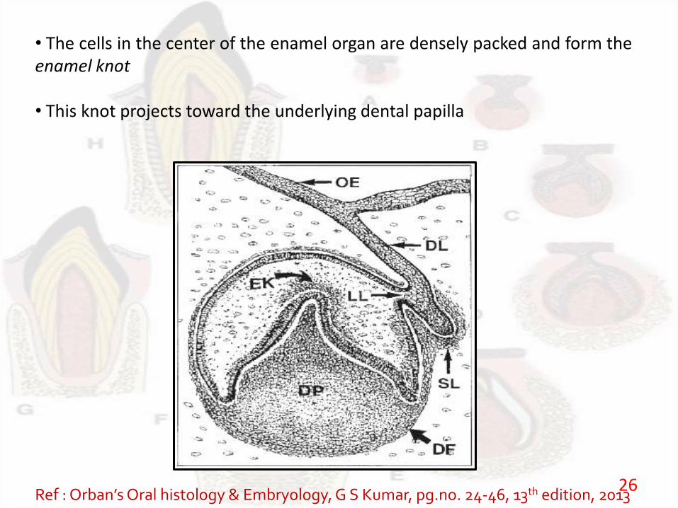
• The cells in the center of the enamel organ are densely packed and form theenamel knot
• This knot projects toward the underlying dental papilla
Ref : Orban’sOral histology & Embryology, G S Kumar, pg.no. 24-46, 13th edition, 201326
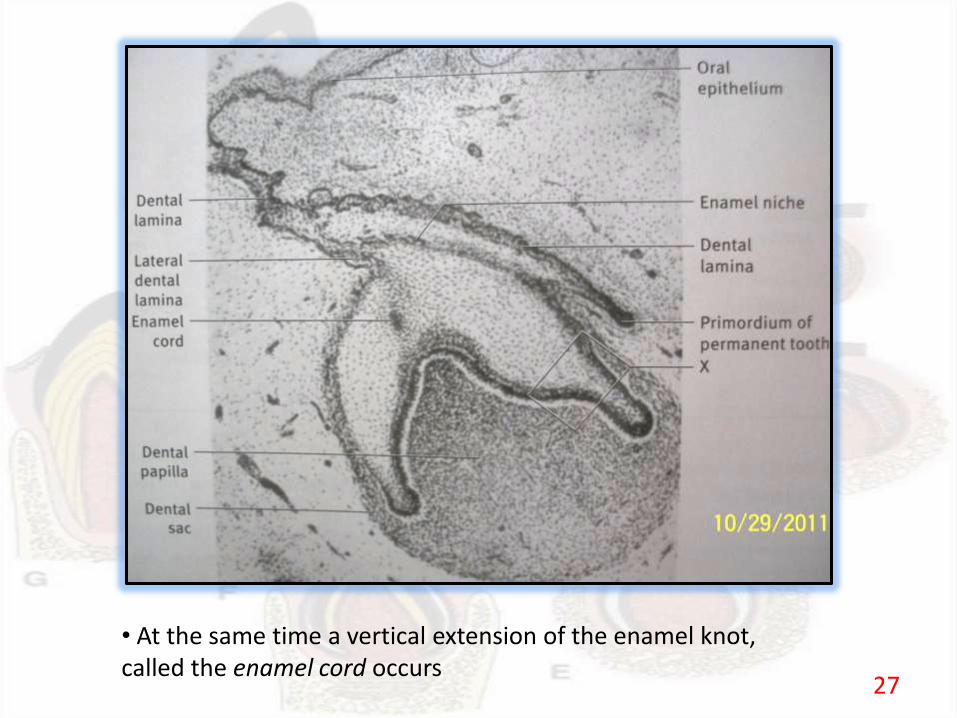
• At the same time a vertical extension of the enamel knot, called the enamel cord occurs
27
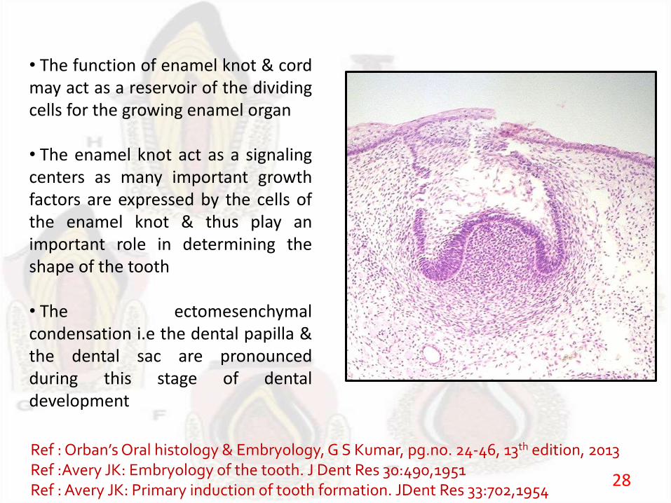
• The function of enamel knot & cordmay act as a reservoir of the dividingcells for the growing enamel organ
• The enamel knot act as a signalingcenters as many important growthfactors are expressed by the cells ofthe enamel knot & thus play animportant role in determining theshape of the tooth
• The ectomesenchymalcondensation i.e the dental papilla &the dental sac are pronouncedduring this stage of dentaldevelopment
Ref :Orban’sOral histology & Embryology, G S Kumar, pg.no. 24-46, 13th edition, 2013Ref :Avery JK: Embryology of the tooth. J Dent Res 30:490,1951Ref : Avery JK: Primary induction of tooth formation. JDent Res 33:702,1954
28
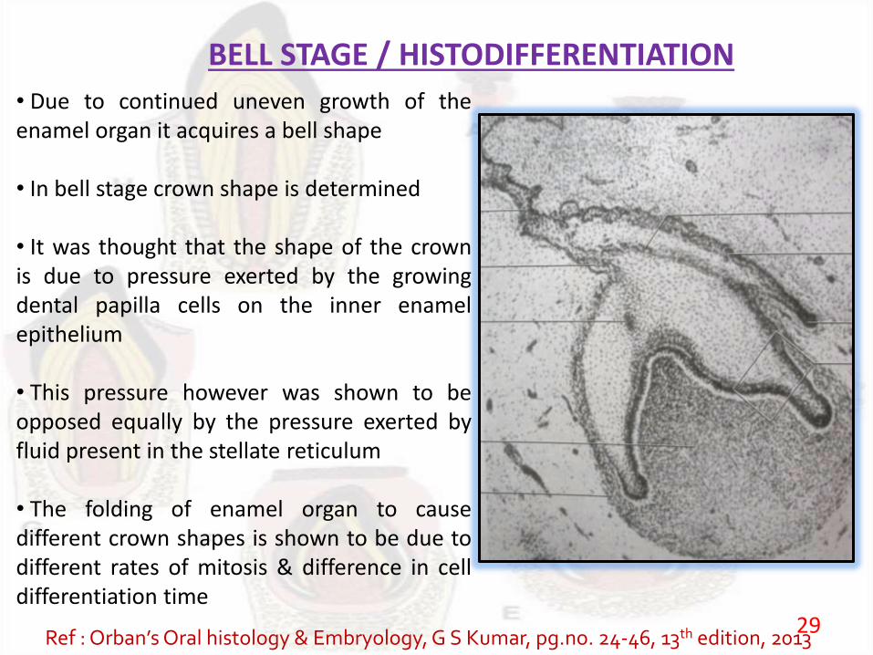
BELL STAGE / HISTODIFFERENTIATION
• Due to continued uneven growth of theenamel organ it acquires a bell shape
• In bell stage crown shape is determined
• It was thought that the shape of the crownis due to pressure exerted by the growingdental papilla cells on the inner enamelepithelium
• This pressure however was shown to beopposed equally by the pressure exerted byfluid present in the stellate reticulum
• The folding of enamel organ to causedifferent crown shapes is shown to be due todifferent rates of mitosis & difference in celldifferentiation time
Ref : Orban’sOral histology & Embryology, G S Kumar, pg.no. 24-46, 13th edition, 201329
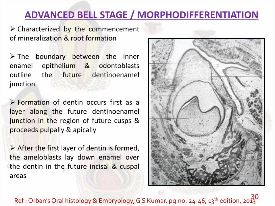
ADVANCED BELL STAGE / MORPHODIFFERENTIATION Characterized by the commencementof mineralization & root formation
The boundary between the innerenamel epithelium & odontoblastsoutline the future dentinoenameljunction
Formation of dentin occurs first as alayer along the future dentinoenameljunction in the region of future cusps &proceeds pulpally & apically
After the first layer of dentin is formed,the ameloblasts lay down enamel overthe dentin in the future incisal & cuspalareas
Ref : Orban’sOral histology & Embryology, G S Kumar, pg.no. 24-46, 13th edition, 201330
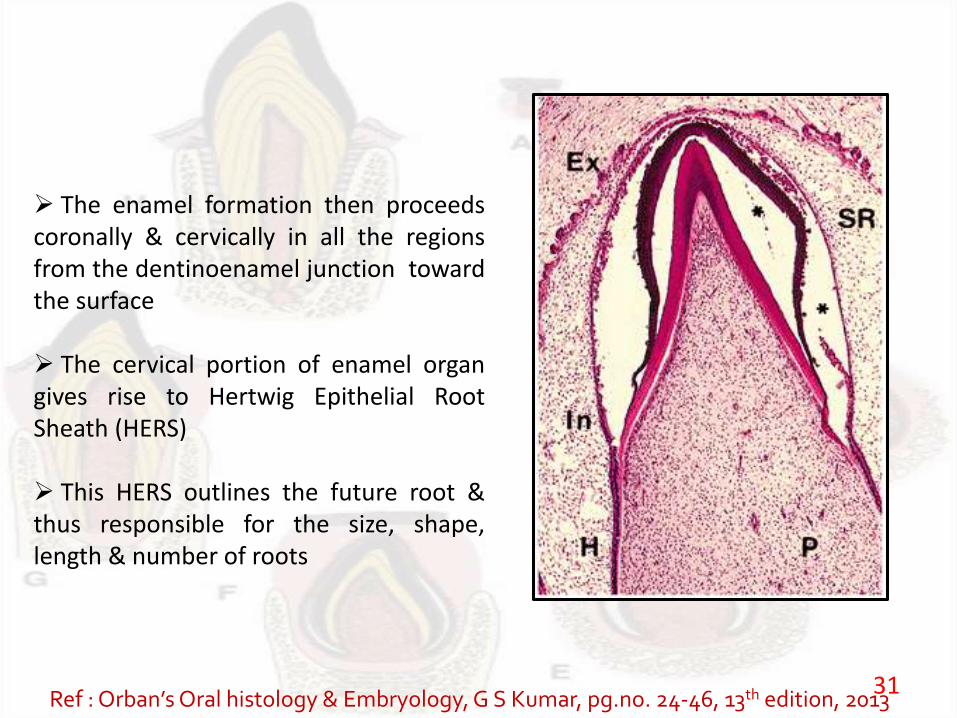
The enamel formation then proceedscoronally & cervically in all the regionsfrom the dentinoenamel junction towardthe surface
The cervical portion of enamel organgives rise to Hertwig Epithelial RootSheath (HERS)
This HERS outlines the future root &thus responsible for the size, shape,length & number of roots
Ref : Orban’sOral histology & Embryology, G S Kumar, pg.no. 24-46, 13th edition, 201331
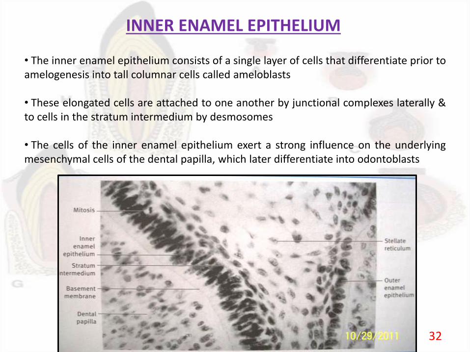
INNER ENAMEL EPITHELIUM
• The inner enamel epithelium consists of a single layer of cells that differentiate prior toamelogenesis into tall columnar cells called ameloblasts
• These elongated cells are attached to one another by junctional complexes laterally &to cells in the stratum intermedium by desmosomes
• The cells of the inner enamel epithelium exert a strong influence on the underlyingmesenchymal cells of the dental papilla, which later differentiate into odontoblasts
32
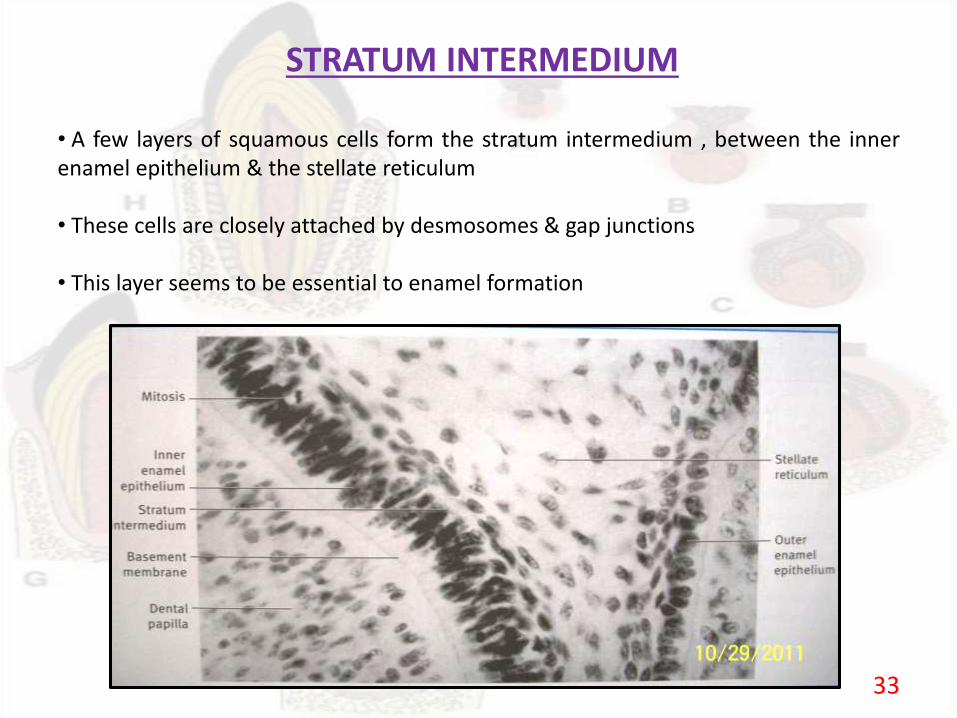
STRATUM INTERMEDIUM
• A few layers of squamous cells form the stratum intermedium , between the innerenamel epithelium & the stellate reticulum
• These cells are closely attached by desmosomes & gap junctions
• This layer seems to be essential to enamel formation
33

STELLATE RETICULUM
• The stellate reticulum expands further due to continued accumulation of intra-cellularfluid
• These star shaped cells, having a large processes anastomose with those of adjacentcells
• As the enamel formation starts., the Stellate reticulum collapses to a narrow zonethereby reducing the distance between the outer & inner enamel epithelium
34
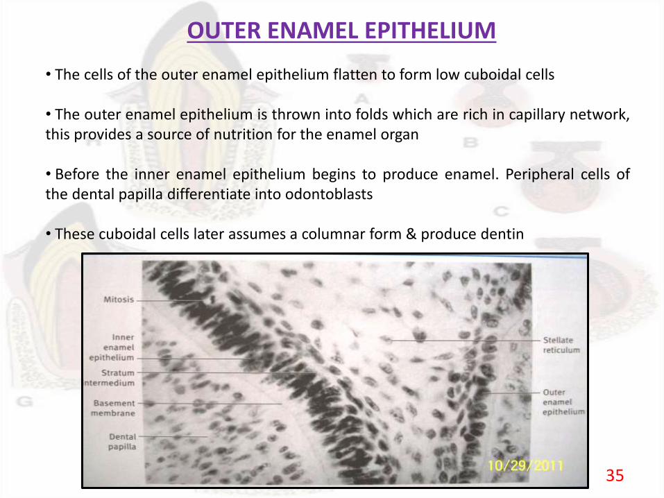
OUTER ENAMEL EPITHELIUM
• The cells of the outer enamel epithelium flatten to form low cuboidal cells
• The outer enamel epithelium is thrown into folds which are rich in capillary network,this provides a source of nutrition for the enamel organ
• Before the inner enamel epithelium begins to produce enamel. Peripheral cells ofthe dental papilla differentiate into odontoblasts
• These cuboidal cells later assumes a columnar form & produce dentin
35
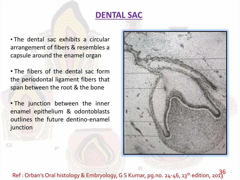
DENTAL SAC
• The dental sac exhibits a circulararrangement of fibers & resembles acapsule around the enamel organ
• The fibers of the dental sac formthe periodontal ligament fibers thatspan between the root & the bone
• The junction between the innerenamel epithelium & odontoblastsoutlines the future dentino-enameljunction
Ref : Orban’sOral histology & Embryology, G S Kumar, pg.no. 24-46, 13th edition, 201336
FORMATION OF ENAMEL & DENTIN MATIX( APPOSITION)
• Apposition is the deposition of the matrix of the hard enamelstructures
• Appositional growth of the enamel & dentin is a layer likedeposition of an extracellular matrix. This type of growth istherefore additive
• Appositional growth is characterised by regular & rhythmicdeposition of the extracellular matrix, which is of itself incapable offurther growth
Ref : Orban’sOral histology & Embryology, G S Kumar, pg.no. 24-46, 13th edition, 201337
38
CLINICAL CONSIDERATIONS
39
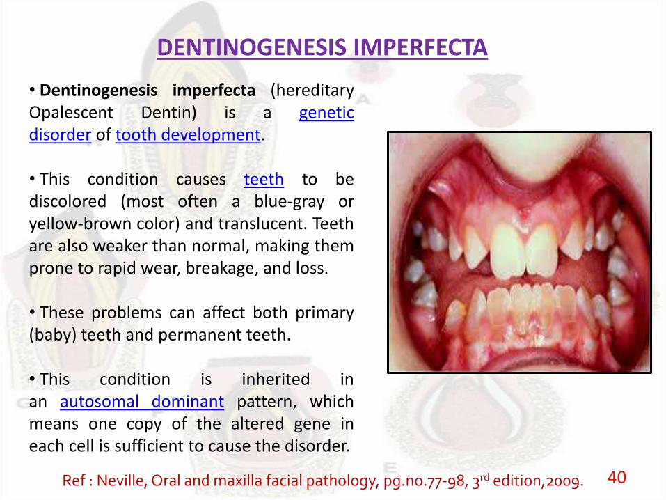
DENTINOGENESIS IMPERFECTA
• Dentinogenesis imperfecta (hereditaryOpalescent Dentin) is a geneticdisorder of tooth development.
• This condition causes teeth to bediscolored (most often a blue-gray oryellow-brown color) and translucent. Teethare also weaker than normal, making themprone to rapid wear, breakage, and loss.
• These problems can affect both primary(baby) teeth and permanent teeth.
• This condition is inherited inan autosomal dominant pattern, whichmeans one copy of the altered gene ineach cell is sufficient to cause the disorder.
Ref : Neville, Oral and maxilla facial pathology, pg.no.77-98, 3rd edition,2009. 40

HUTCHINSON’S INCISOR
MULBERRY MOLARS
Ref : Neville, Oral and maxilla facial pathology, pg.no.77-98, 3rd edition,2009. 41
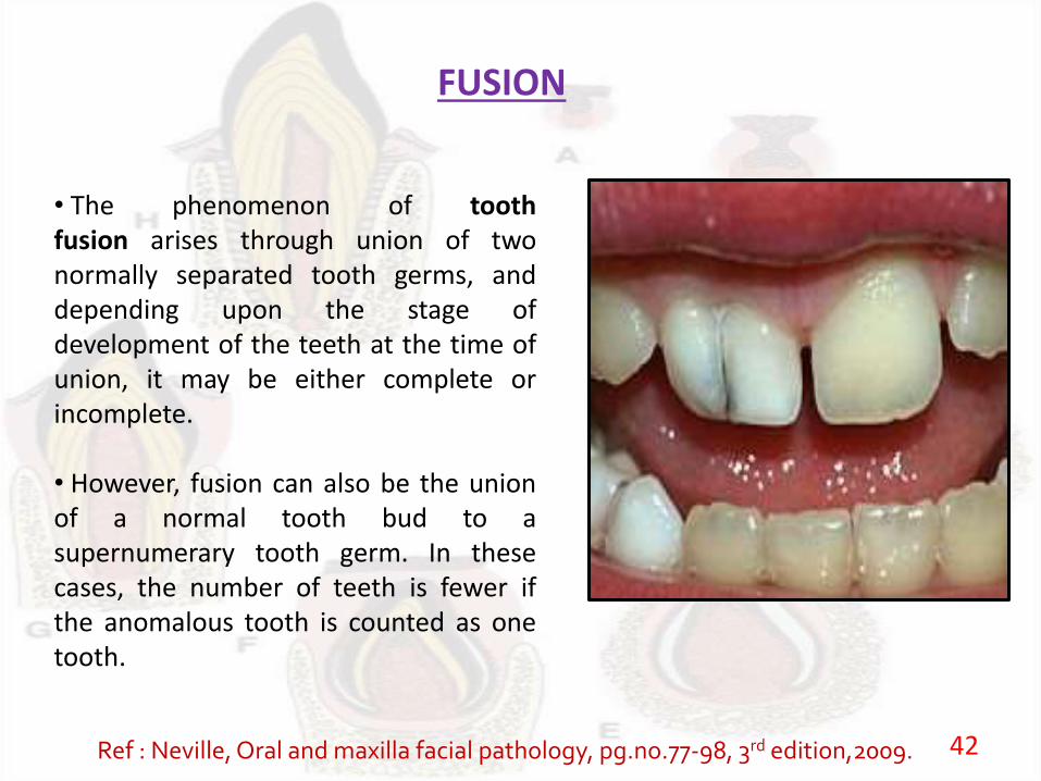
FUSION
• The phenomenon of toothfusion arises through union of twonormally separated tooth germs, anddepending upon the stage ofdevelopment of the teeth at the time ofunion, it may be either complete orincomplete.
• However, fusion can also be the unionof a normal tooth bud to asupernumerary tooth germ. In thesecases, the number of teeth is fewer ifthe anomalous tooth is counted as onetooth.
Ref : Neville, Oral and maxilla facial pathology, pg.no.77-98, 3rd edition,2009. 42
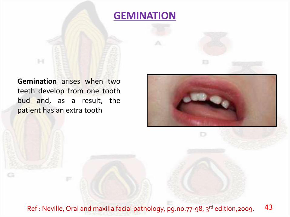
GEMINATION
Gemination arises when twoteeth develop from one toothbud and, as a result, thepatient has an extra tooth
Ref : Neville, Oral and maxilla facial pathology, pg.no.77-98, 3rd edition,2009. 43

ENAMEL HYPOPLASIA
Enamel hypoplasia is thedefect of the teeth in whichthe tooth enamel is hard butthin and deficient in amountThis is caused by defectiveenamel matrix formationwith a deficiency in thecementing substance
Ref : Neville, Oral and maxilla facial pathology, pg.no.77-98, 3rd edition,2009. 44
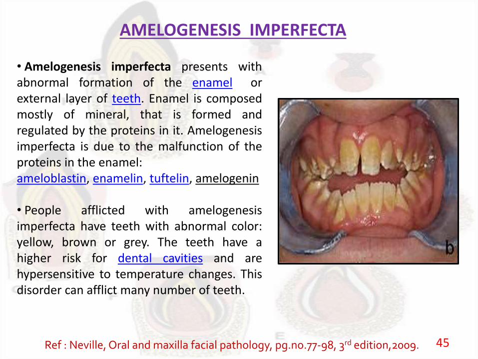
AMELOGENESIS IMPERFECTA
• Amelogenesis imperfecta presents withabnormal formation of the enamel orexternal layer of teeth. Enamel is composedmostly of mineral, that is formed andregulated by the proteins in it. Amelogenesisimperfecta is due to the malfunction of theproteins in the enamel:ameloblastin, enamelin, tuftelin, amelogenin
• People afflicted with amelogenesisimperfecta have teeth with abnormal color:yellow, brown or grey. The teeth have ahigher risk for dental cavities and arehypersensitive to temperature changes. Thisdisorder can afflict many number of teeth.
Ref : Neville, Oral and maxilla facial pathology, pg.no.77-98, 3rd edition,2009. 45
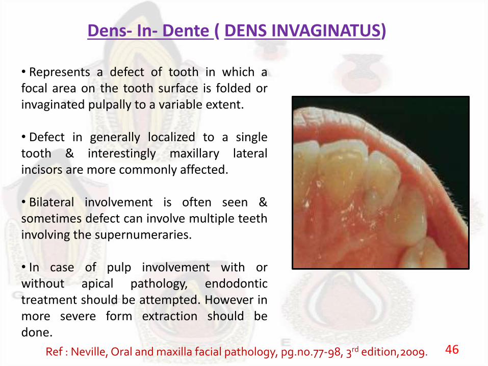
Dens- In- Dente ( DENS INVAGINATUS)
• Represents a defect of tooth in which afocal area on the tooth surface is folded orinvaginated pulpally to a variable extent.
• Defect in generally localized to a singletooth & interestingly maxillary lateralincisors are more commonly affected.
• Bilateral involvement is often seen &sometimes defect can involve multiple teethinvolving the supernumeraries.
• In case of pulp involvement with orwithout apical pathology, endodontictreatment should be attempted. However inmore severe form extraction should bedone.
Ref : Neville, Oral and maxilla facial pathology, pg.no.77-98, 3rd edition,2009. 46

DENS EVAGINATUS• Dens evaginatus is a condition foundin teeth where the outer surface appearsto form an extra bump or cusp.
• Premolars are more likely to beaffected than any other tooth. This maybe seen more frequently in Asians
• The pulp of the tooth may extend intothe dens evaginatus.
• There is a risk of the dens evaginatuschipping off in normal function
• Hence this condition requiresmonitoring as the tooth can lose its bloodand nerve supply as a result and mayneed root canal treatment.
Ref : Neville, Oral and maxilla facial pathology, pg.no.77-98, 3rd edition,2009. 47
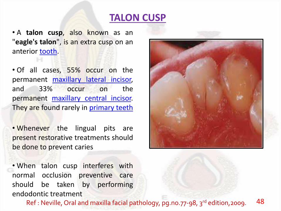
TALON CUSP
• A talon cusp, also known as an"eagle's talon", is an extra cusp on ananterior tooth.
• Of all cases, 55% occur on thepermanent maxillary lateral incisor,and 33% occur on thepermanent maxillary central incisor.They are found rarely in primary teeth
• Whenever the lingual pits arepresent restorative treatments shouldbe done to prevent caries
• When talon cusp interferes withnormal occlusion preventive careshould be taken by performingendodontic treatment
Ref : Neville, Oral and maxilla facial pathology, pg.no.77-98, 3rd edition,2009. 48
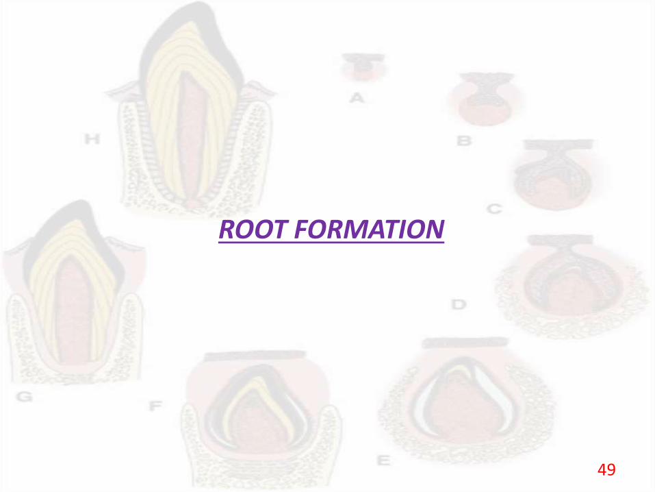
ROOT FORMATION
49
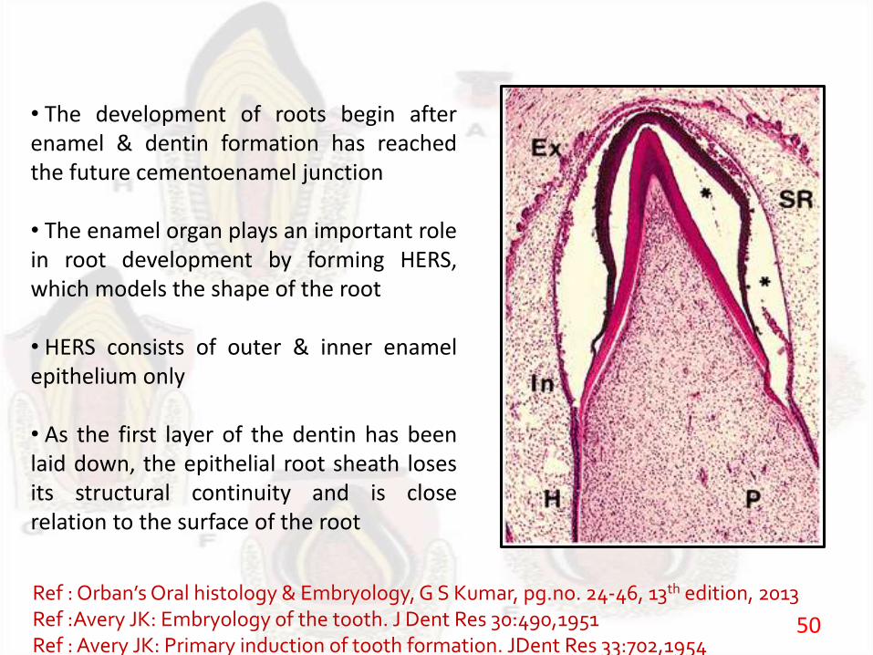
• The development of roots begin afterenamel & dentin formation has reachedthe future cementoenamel junction
• The enamel organ plays an important rolein root development by forming HERS,which models the shape of the root
• HERS consists of outer & inner enamelepithelium only
• As the first layer of the dentin has beenlaid down, the epithelial root sheath losesits structural continuity and is closerelation to the surface of the root
Ref :Orban’sOral histology & Embryology, G S Kumar, pg.no. 24-46, 13th edition, 2013Ref :Avery JK: Embryology of the tooth. J Dent Res 30:490,1951Ref : Avery JK: Primary induction of tooth formation. JDent Res 33:702,1954
50
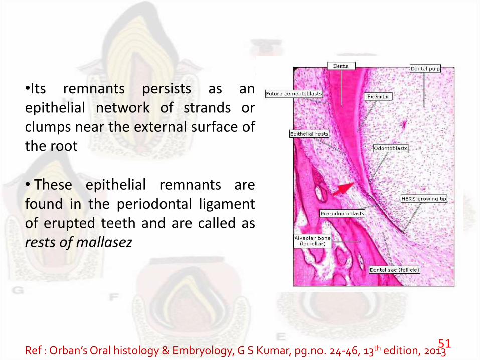
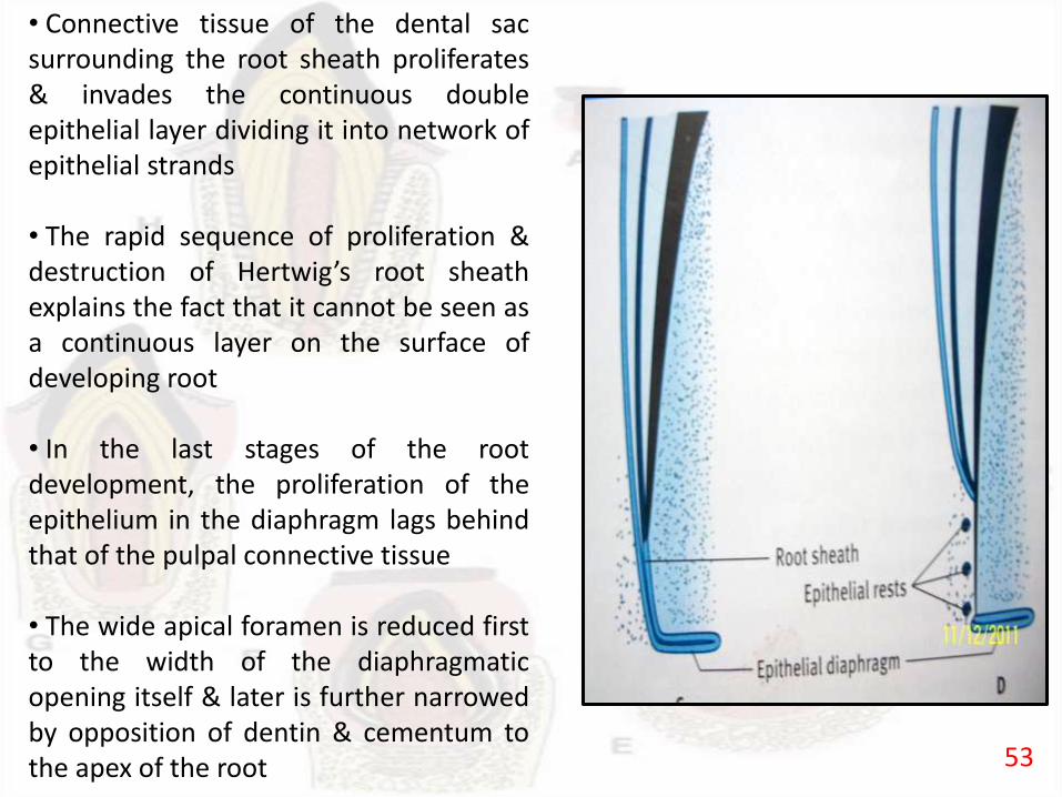
•Its remnants persists as anepithelial network of strands orclumps near the external surface ofthe root
• These epithelial remnants arefound in the periodontal ligamentof erupted teeth and are called asrests of mallasez
Ref : Orban’sOral histology & Embryology, G S Kumar, pg.no. 24-46, 13th edition, 201351
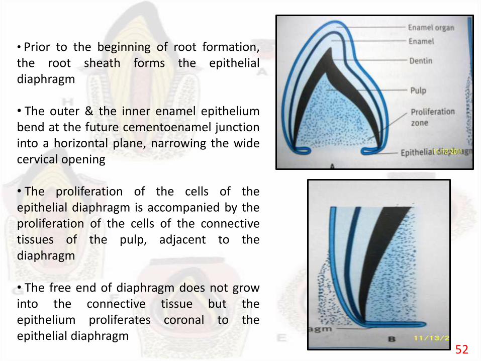
• Prior to the beginning of root formation,the root sheath forms the epithelialdiaphragm
• The outer & the inner enamel epitheliumbend at the future cementoenamel junctioninto a horizontal plane, narrowing the widecervical opening
• The proliferation of the cells of theepithelial diaphragm is accompanied by theproliferation of the cells of the connectivetissues of the pulp, adjacent to thediaphragm
• The free end of diaphragm does not growinto the connective tissue but theepithelium proliferates coronal to theepithelial diaphragm
52
• Connective tissue of the dental sacsurrounding the root sheath proliferates& invades the continuous doubleepithelial layer dividing it into network ofepithelial strands
• The rapid sequence of proliferation &destruction of Hertwig’s root sheathexplains the fact that it cannot be seen asa continuous layer on the surface ofdeveloping root
• In the last stages of the rootdevelopment, the proliferation of theepithelium in the diaphragm lags behindthat of the pulpal connective tissue
• The wide apical foramen is reduced firstto the width of the diaphragmaticopening itself & later is further narrowedby opposition of dentin & cementum tothe apex of the root 53
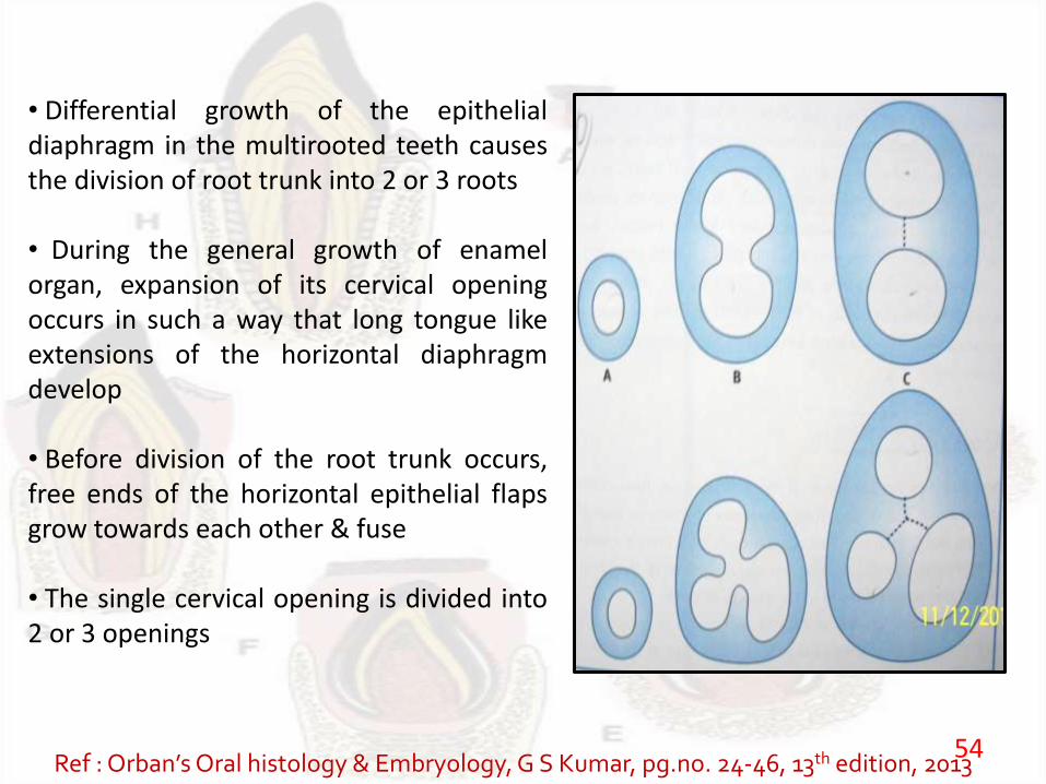
• Differential growth of the epithelialdiaphragm in the multirooted teeth causesthe division of root trunk into 2 or 3 roots
• During the general growth of enamelorgan, expansion of its cervical openingoccurs in such a way that long tongue likeextensions of the horizontal diaphragmdevelop
• Before division of the root trunk occurs,free ends of the horizontal epithelial flapsgrow towards each other & fuse
• The single cervical opening is divided into2 or 3 openings
Ref : Orban’sOral histology & Embryology, G S Kumar, pg.no. 24-46, 13th edition, 201354
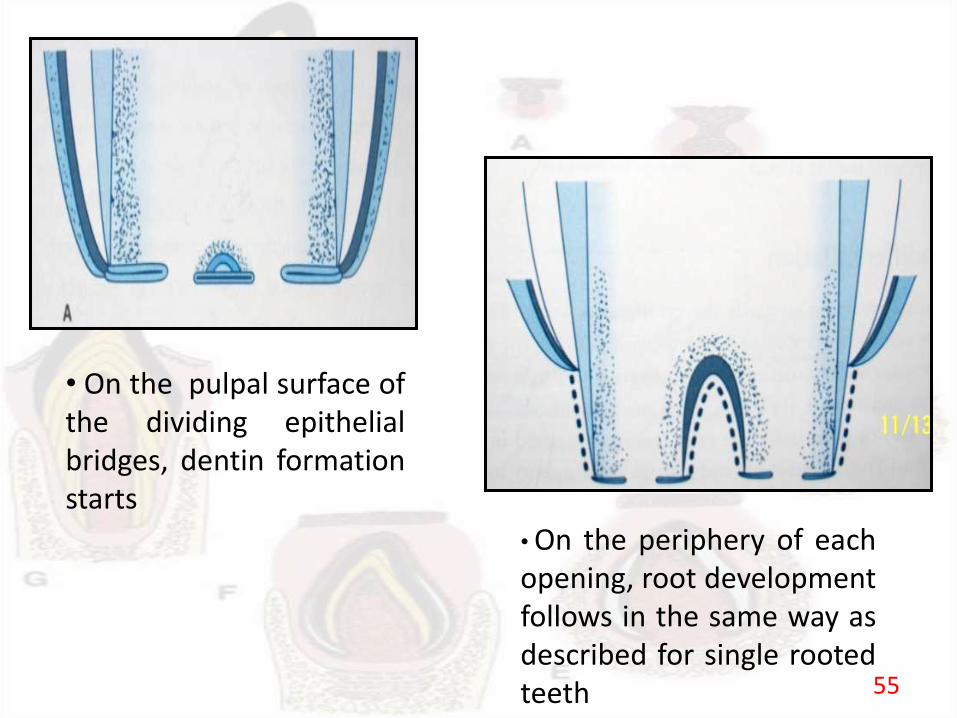
• On the pulpal surface ofthe dividing epithelialbridges, dentin formationstarts
• On the periphery of eachopening, root developmentfollows in the same way asdescribed for single rootedteeth 55
CLINICAL CONSIDERATIONS
56
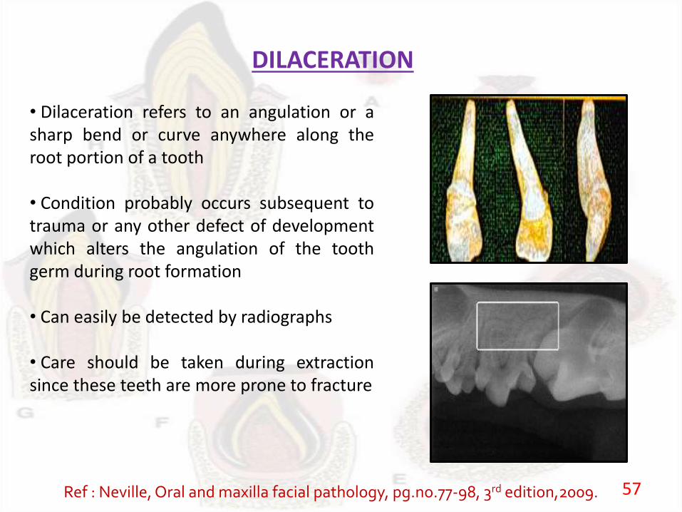
DILACERATION
• Dilaceration refers to an angulation or asharp bend or curve anywhere along theroot portion of a tooth
• Condition probably occurs subsequent totrauma or any other defect of developmentwhich alters the angulation of the toothgerm during root formation
• Can easily be detected by radiographs
• Care should be taken during extractionsince these teeth are more prone to fracture
Ref : Neville, Oral and maxilla facial pathology, pg.no.77-98, 3rd edition,2009. 57
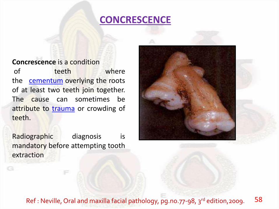
CONCRESCENCE
Concrescence is a conditionof teeth where
the cementum overlying the rootsof at least two teeth join together.The cause can sometimes beattribute to trauma or crowding ofteeth.
Radiographic diagnosis ismandatory before attempting toothextraction
Ref : Neville, Oral and maxilla facial pathology, pg.no.77-98, 3rd edition,2009. 58
Tooth Eruption
59

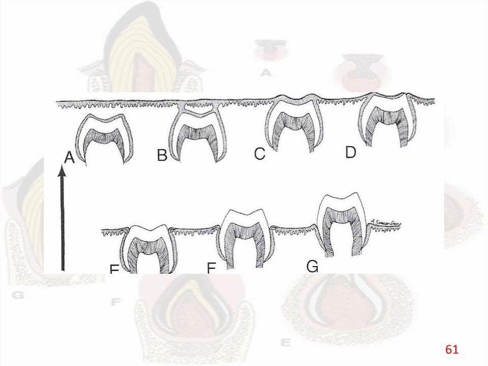
1 Phases of tooth eruption
Eruption is the movement of the developing teeth through the boneand the overlying mucosa of the jaws to appear in the oral cavityand reach the occlusal plane
Movements of teeth leading to eruption take place in threephases
1. Preeruptive phase
2. Eruptive phase
3. Functional phase
Ref :Orban’sOral histology & Embryology, G S Kumar, pg.no. 24-46, 13th edition, 2013Ref :Avery JK: Embryology of the tooth. J Dent Res 30:490,1951Ref : Avery JK: Primary induction of tooth formation. JDent Res 33:702,1954 60
61

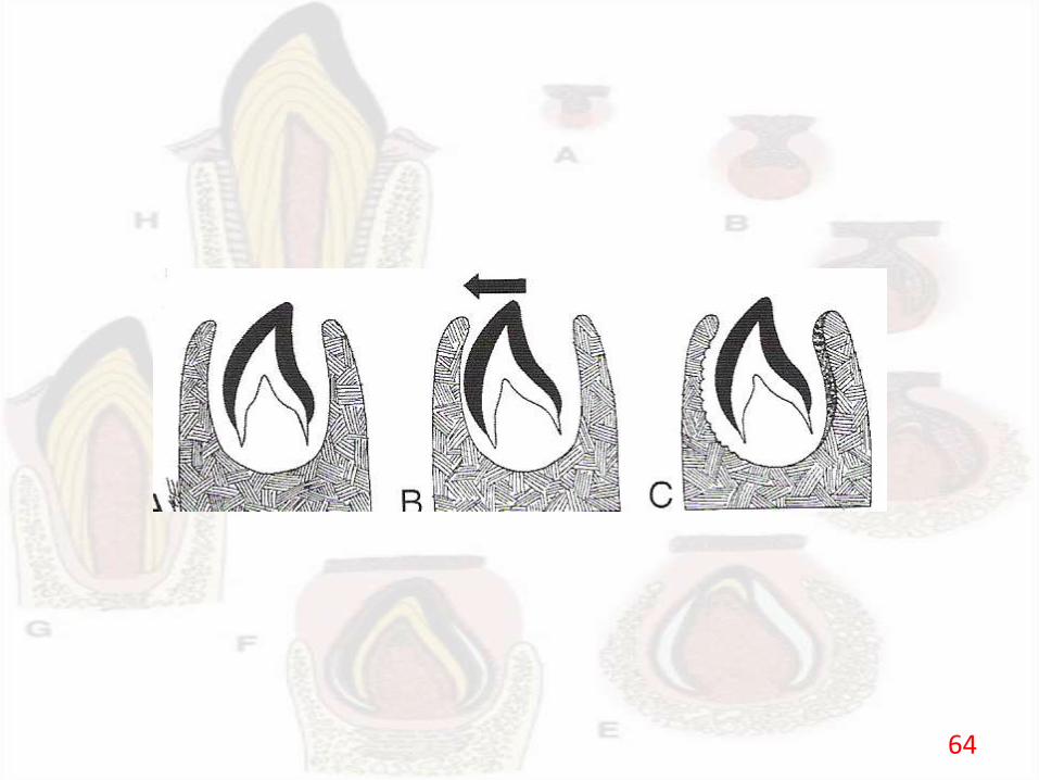
1. Preeruptive phase
The movements in preeruptive phase are the movements of thedeveloping tooth before the root begins to form.
It consists of movements of the developing tooth within thealveolar process.
The tooth germs move outward and upward/downward withthe increasing length, width and height of the jaws.
62
Preeruptive movements involve bodily movement and eccentricmovement of the developing tooth germ.
Bodily movement is the shift of the entire tooth germ.
Eccentric movement is the relative growth in one part of thetooth, leading to a change in the centre of the tooth germ.
Ref :Orban’sOral histology & Embryology, G S Kumar, pg.no. 24-46, 13th edition, 2013Ref :Avery JK: Embryology of the tooth. J Dent Res 30:490,1951Ref : Avery JK: Primary induction of tooth formation. JDent Res 33:702,1954
63
64
2. Eruptive phase (Prefunctional phase)
Eruptive phase begins when the root starts to form and endswhen the tooth reaches occlusal plane.
After initiation of root formation PDL also starts to develop.
PDL is remodelled continuously to accommodate the eruptivetooth movement.
The end of secretory phase of amelogenesis also coincides withthe start of the eruptive phase.
65
During the eruptive phase the tooth germ undergoesintraosseous movements and supraosseous movements.
The rate of supraosseous movements is a lot faster than theintraosseous movements.
The tissue in front of an erupting primary tooth is different fromthat of a permanent tooth.
A strand of fibrous tissue containing the remnants of the dentallamina, know as gobernacular cord forms a pathway in advanceof developing permanent tooth.
66
During the eruptive stage the crown breaks the double layerepithelium overlying it and enters the oral cavity.
The eruption causes the tissue around it form the junctionalepithelium and the gingiva.
This phase is also called as prefunctional phase.
Ref :Orban’sOral histology & Embryology, G S Kumar, pg.no. 24-46, 13th edition, 2013Ref :Avery JK: Embryology of the tooth. J Dent Res 30:490,1951Ref : Avery JK: Primary induction of tooth formation. JDent Res 33:702,1954
67
3. Functional phase (Post eruptive phase)
The functional phase begins when the tooth reaches theocclusal plane and continues as long as the tooth remains in theoral cavity.
Movements in the early stages of this phase accommodates thegrowth of root and the jaws.
This phase is also called the post eruptive phase.
Ref :Orban’sOral histology & Embryology, G S Kumar, pg.no. 24-46, 13th edition, 2013Ref :Avery JK: Embryology of the tooth. J Dent Res 30:490,1951Ref : Avery JK: Primary induction of tooth formation. JDent Res 33:702,1954
68
2 Mechanism of tooth eruption
There are many theories that explain the mechanism of tootheruption
Vascularity
Vascularity plays an important role in tooth eruption
Sufficient blood supply to the tooth germ has proven to causeeruptive tooth movement
Localized hyperamia has shown to causes increased vascularityof the periodontal tissue and also increased eruption of adjacenttooth
69
Pressure
Decreased pressure overlying a tooth and increasedpressure around the tooth are major factors in tooth eruption.
When the root formation begins an eruption pathwaydevelops overlying the tooth.
Remodelling of tissue around the developing tooth bringsabout an increase in pressure tooth which causes the toothmovement.
70
Root formation
Root formation causes an overall increase in the length ofthe tooth.
It produces enough force that leads to the resorption ofbone.
However, this force in itself does not cause tooth movement.
Rootless teeth also erupt.
71
Bone remodelling
The selective resorption and formation of bone surroundingthe tooth cause its movement.
This theory also explains the tooth movement duringpreeuptive phase.
Dental Follicle
There is signalling between the reduced enamel epitheliumand the dental follicle.
This signalling regulates the timing of eruption of teeth –known as ‘biologic clock’.
72
Periodontal ligament
The remodelling of PDL has also been considered as afactor for tooth eruption.
The fibroblasts possess traction power that causes toothmovement.
The PDL helps lift the tooth to its occlusal plane duringthe supraosseous phase of eruption.
Ref :Orban’sOral histology & Embryology, G S Kumar, pg.no. 24-46, 13th edition, 2013Ref :Avery JK: Embryology of the tooth. J Dent Res 30:490,1951Ref : Avery JK: Primary induction of tooth formation. JDent Res 33:702,1954
73
Review of literature
Ref. K. Heikinheimo Stage-specific Expression of Decapentaplegic-Vg-related Genes 2, 4, and 6 (Bone Morphogenetic Proteins 2, 4,and 6) During Human Tooth Morphogenesis, J Dent Res 73(3): 590-597, March, 1994
74
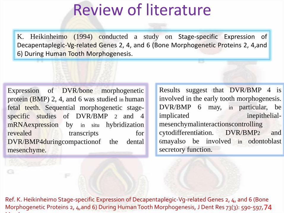
K. Heikinheimo (1994) conducted a study on Stage-specific Expression ofDecapentaplegic-Vg-related Genes 2, 4, and 6 (Bone Morphogenetic Proteins 2, 4,and6) During Human Tooth Morphogenesis.
Expression of DVR/bone morphogenetic
protein (BMP) 2, 4, and 6 was studied in human
fetal teeth. Sequential morphogenetic stage-
specific studies of DVR/BMP 2 and 4
mRNAexpression by in situ hybridization
revealed transcripts for
DVR/BMP4duringcompactionof the dental
mesenchyme.
Results suggest that DVR/BMP 4 is
involved in the early tooth morphogenesis.
DVR/BMP 6 may, in particular, be
implicated inepithelial-
mesenchymalinteractionscontrolling
cytodifferentiation. DVR/BMP2 and
6mayalso be involved in odontoblast
secretory function.
75
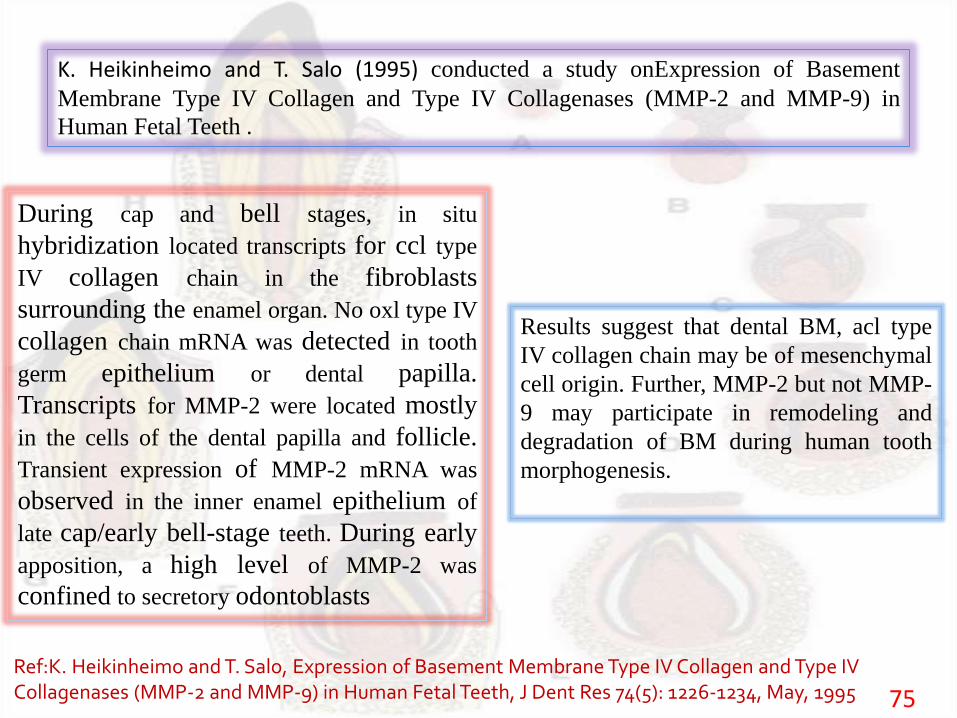
K. Heikinheimo and T. Salo (1995) conducted a study onExpression of Basement
Membrane Type IV Collagen and Type IV Collagenases (MMP-2 and MMP-9) in
Human Fetal Teeth .
During cap and bell stages, in situ
hybridization located transcripts for ccl type
IV collagen chain in the fibroblasts
surrounding the enamel organ. No oxl type IV
collagen chain mRNA was detected in tooth
germ epithelium or dental papilla.
Transcripts for MMP-2 were located mostly
in the cells of the dental papilla and follicle.
Transient expression of MMP-2 mRNA was
observed in the inner enamel epithelium of
late cap/early bell-stage teeth. During early
apposition, a high level of MMP-2 was
confined to secretory odontoblasts
Results suggest that dental BM, acl type
IV collagen chain may be of mesenchymal
cell origin. Further, MMP-2 but not MMP-
9 may participate in remodeling and
degradation of BM during human tooth
morphogenesis.
Ref:K. Heikinheimo and T. Salo, Expression of Basement Membrane Type IV Collagen and Type IV Collagenases (MMP-2 and MMP-9) in Human Fetal Teeth, J Dent Res 74(5): 1226-1234, May, 1995
76
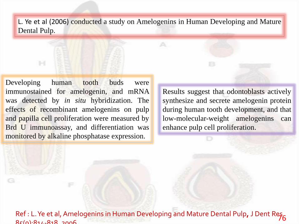
L. Ye et al (2006) conducted a study on Amelogenins in Human Developing and Mature
Dental Pulp.
Developing human tooth buds were
immunostained for amelogenin, and mRNA
was detected by in situ hybridization. The
effects of recombinant amelogenins on pulp
and papilla cell proliferation were measured by
Brd U immunoassay, and differentiation was
monitored by alkaline phosphatase expression.
Results suggest that odontoblasts actively
synthesize and secrete amelogenin protein
during human tooth development, and that
low-molecular-weight amelogenins can
enhance pulp cell proliferation.
Ref : L. Ye et al, Amelogenins in Human Developing and Mature Dental Pulp, J Dent Res 85(9):814-818, 2006
CONCLUSION
Since development of tooth forms the base ofdentistry, a thorough understanding and a soundknowledge is required by a dentist regarding thedevelopment stages of tooth & the anomalies relatedto it, so as to identify & treat them in a proper fashion.
77
REFERENCES1. Orban’s Oral histology & Embryology, G S Kumar, pg.no. 24-46, 13th edition,
2013.
2. Neville, Oral and maxilla facial pathology, pg.no.77-98, 3rd edition,2009
3. Avery JK: Embryology of the tooth. J Dent Res 30:490,1951
4. Avery JK: Primary induction of tooth formation. JDent Res 33:702,1954
5. L. Ye et al, Amelogenins in Human Developing and Mature Dental Pulp, J
Dent Res 85(9):814-818, 2006
6. K. Heikinheimo and T. Salo, Expression of Basement Membrane Type IV
Collagen and Type IV Collagenases (MMP-2 and MMP-9) in Human Fetal
Teeth, J Dent Res 74(5): 1226-1234, May, 1995
7. K. Heikinheimo Stage-specific Expression of Decapentaplegic-Vg-related
Genes 2, 4, and 6 (Bone Morphogenetic Proteins 2, 4,and 6) During Human
Tooth Morphogenesis, J Dent Res 73(3): 590-597, March, 1994