Embed Size (px)
Citation preview
Review
1460 www.thelancet.com Vol 385 April 11, 2015
IgG4-related diseaseTerumi Kamisawa, Yoh Zen, Shiv Pillai, John H Stone
IgG4-related disease is a protean condition that mimics many malignant, infectious, and infl ammatory disorders. This multi-organ immune-mediated condition links many disorders previously regarded as isolated, single-organ diseases without any known underlying systemic condition. It was recognised as a unifi ed entity only 10 years ago. Histopathology is the key to diagnosis. The three central pathology features of IgG4-related disease are lymphoplasmacytic infi ltration, storiform fi brosis, and obliterative phlebitis. The extent of fi brosis is an important determinant of responsiveness to immunosuppressive therapies. IgG4-related disease generally responds to glucocorticoids in its infl ammatory stage, but recurrent or refractory cases are common. Important mechanistic insights have been derived from studies of patients treated by B-cell depletion. Greater awareness of this disease is needed to ensure earlier diagnoses, which can prevent severe organ damage, disabling tissue fi brosis, and even death. Identifi cation of specifi c antigens and T-cell clones that drive the disease will be the fi rst steps to elucidate the pathogenesis of IgG4-related disease.
IntroductionIgG4-related disease is a multi-organ immune-mediated condition that mimics many malignant, infectious, and infl ammatory disorders.1–3 The diagnosis links many conditions once regarded as isolated, single-organ diseases without any known underlying systemic condition (panel 1). IgG4-related disease, unrecognised as a unifi ed disease for well over a century, has been likened to a “black crow fl ying through the dark night”.4 The disease has many similarities to sarcoidosis and some forms of systemic vasculitis, other protean diseases in which the histopathological fi ndings are consistent across a wide range of organ systems.
Two introductory points deserve emphasis. First, awareness of IgG4-related disease is essential because the disorder is treatable. The therapeutic approaches contrast starkly with those of some of the disorders in the diff erential diagnosis (panel 2), especially malignant disorders but also autoimmune diseases, such as Sjögren’s syndrome, granulomatosis with polyangiitis, and membranous nephropathy. Second, knowledge of the immune dysregulation associated with IgG4-related disease explains much about the human immune system. Progress in elucidation of the basis of IgG4-related disease has been swift.
EpidemiologyUnderstanding of the epidemiology of IgG4-related disease is hampered by insuffi cient awareness of the diagnosis, because the disease did not appear in medical
publications until 2003.5,6 Defi nitive diagnosis generally necessitates a biopsy, insightful interpretation of the pathology, and rigorous clinicopathological correlation. Although the overall prevalence of type 1 (IgG4-related) autoimmune pancreatitis in Japan has been estimated as 2·2 cases per 100 000 population,7 the pancreas is only one of more than a dozen organs aff ected by IgG4-related disease. Therefore, this is surely a substantial under-estimate of the true prevalence, especially because the study from which this estimate was derived was done early in the development of knowledge about IgG4-related disease. The prevalence of various organ manifestations also remains unclear, but autoimmune pancreatitis, sialadenitis (particularly of the submandibular gland), dacryoadenitis, and IgG4-related retroperitoneal fi brosis are the most common disease features.
The typical patient with IgG4-related disease is a middle-aged to elderly man.7,8 For autoimmune pancreatitis, the mean age at diagnosis is 67 years and the male to female ratio is three to one.7 The male predilection contrasts strikingly with classic autoimmune diseases, for which female patients can outnumber male cases by nine to one. For organs of the head and neck, however—the orbits, salivary glands, and sinuses—the proportions of male and female patients are roughly equal.9 The reasons for diff erential organ expression in the two sexes are unclear.
We know of no reports of familial cases of IgG4-related disease. More extensive studies of patients from several ethnic backgrounds are needed before any conclusions can be drawn about genetic susceptibility.10–13
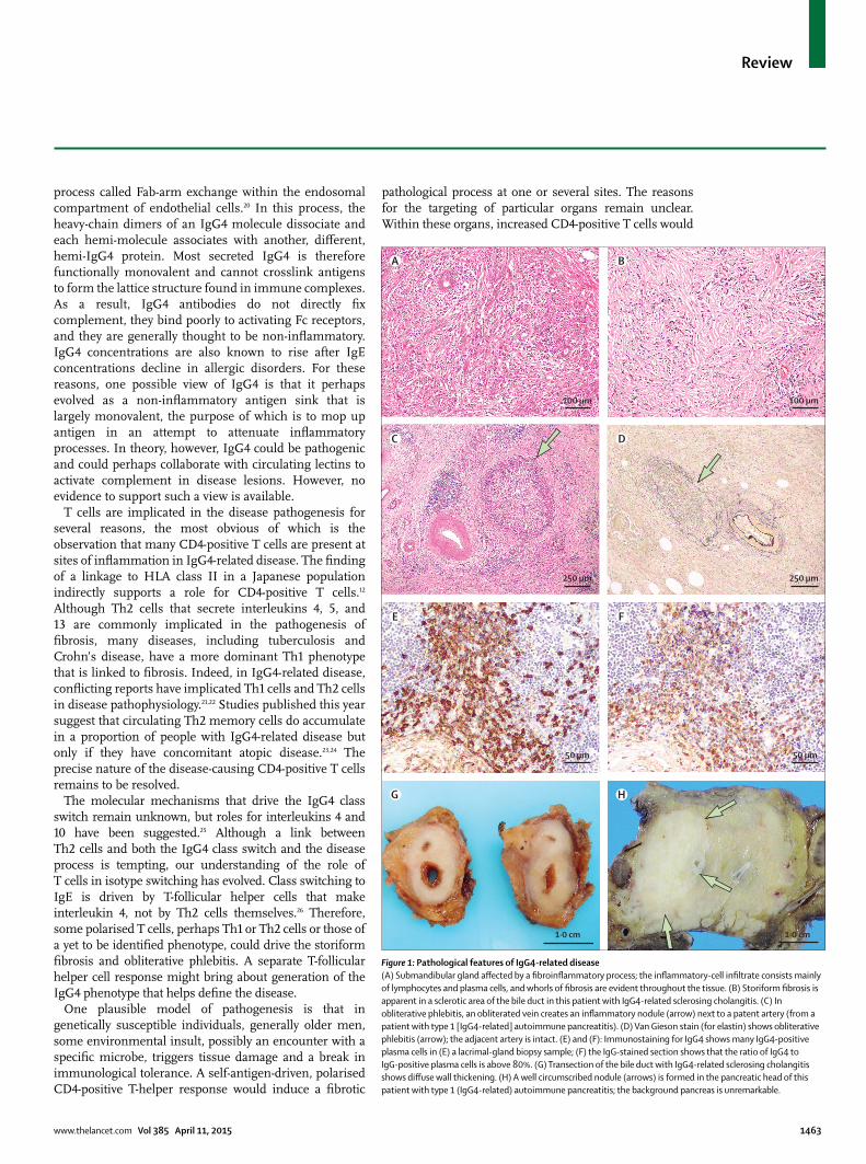
PathologyHistology featuresHistopathology is the key to diagnosis of IgG4-related disease. Three central pathology features are lympho-plasmacytic infi ltration, obliterative phlebitis, and storiform fi brosis (fi gure 1).14 The lymphocytes and plasma cells are polyclonal. Eosinophils are also commonly present and extreme examples can resemble eosinophilic organopathy, but neutrophilic infi ltration is
Lancet 2015; 385: 1460–71
Published OnlineDecember 4, 2014
http://dx.doi.org/10.1016/S0140-6736(14)60720-0
Department of Internal Medicine, Tokyo Metropolitan
Komagome Hospital, Tokyo, Japan (Dr T Kamisawa MD); Department of Pathology,
Kobe University, Kobe, Japan (Prof Y Zen MD); and Center for
Cancer Research (Prof S Pillai MBBS) and Division of Rheumatology, Allergy, and
Immunology (Prof J H Stone MD),
Massachusetts General Hospital, Harvard Medical
School, Boston, MA, USA
Correspondence to:Dr John H Stone, Rheumatology
Unit/Yawkey 2, Massachusetts General Hospital, 55 Fruit St,
Boston, MA 02114, [email protected]
Search strategy and selection criteria
Data for this Review were identifi ed by searches of Medline, PubMed, and references from relevant articles with the search terms “IgG4”, “IgG4-related”, and “autoimmune pancreatitis”. We focused on publications since the year 2000, since the multiorgan nature of IgG4-related disease was not recognised until 2003. We also cited other important publications from earlier years pertaining to conditions now recognised as part of the IgG4-related disease spectrum.
Review
www.thelancet.com Vol 385 April 11, 2015 1461
rare in IgG4-related disease. Necrosis, discrete granulomata, and xanthogranulomatous changes are atypical and, when present, suggest other diagnoses.9,14
Fibrosis is a histological prerequisite for the diagnosis. Some fi brosis is present in all cases, even in patients who present shortly after symptom onset. Storiform fi brosis, characterised by radially arranged collagen fi bres that seem to weave through the tissue, typifi es the unique pattern associated with IgG4-related disease (fi gure 1).9,14 Because of its typically patchy distribution, however, storiform fi brosis sometimes escapes detection through sampling error, especially if the tissue is obtained by needle biopsy. Acellular, keloidal fi brosis is not characteristic of IgG4-related disease.
The characteristic venous lesion, obliterative phlebitis, is defi ned as the partial or complete obliteration of medium-sized veins.9,14 This fi nding should be distinguished from fi brous venous occlusion with no infl ammation, which is known to occur in other conditions (eg, primary sclerosing cholangitis). Obliterated veins commonly appear as an infl ammatory nodule next to a patent artery (fi gure 1) and sometimes can be identifi ed as veins only through elastin staining (fi gure 1).
The histological appearance is similar for all organs. Some more organ-specifi c changes, however, are noteworthy. Both obliterative arteritis and focal neutrophilic infi ltration, rare in other organs, can occur in the lungs. Obliterative arteritis lacks the vascular-wall necrosis typical of many systemic vasculitides. The neutrophilic infi ltration in IgG4-related pulmonary disease is typically seen in alveolar spaces.15 Other minor pathological diff erences between organs include the absence of storiform fi brosis within lacrimal glands and lymph nodes, and the lower frequency of obliterative phlebitis in salivary glands, lacrimal glands, lymph nodes, and kidneys.9,14 The rarity of fi brosis in lymph nodes means that the diagnosis of IgG4-related disease is diffi cult on the basis of lymph-node pathology alone.
ImmunostainingHigh numbers of IgG4-positive plasma cells at tissue sites are a disease hallmark, even when serum IgG4 concentrations are normal. The fi nding of IgG4-positive plasma cells is helpful in diff erentiating IgG4-related disease from other plasma-cell-rich disorders, such as primary sclerosing cholangitis and multicentric Castleman’s disease.16,17
In interpretation of tissue IgG4 stains, several caveats must be borne in mind.14 First, IgG4-positive plasma cells are generally present diff usely throughout lesions of IgG4-related disease. Focal aggregations of IgG4-positive cells are atypical. Second, the absolute number of IgG4-positive plasma cells must be interpreted according to the specifi c tissue. An international pathology consensus statement proposed, for example, that for sialadenitis the cutoff value should be at least 100 cells per high-power fi eld, but that in the pancreas more than
50 cells per high-power fi eld is compatible with a diagnosis of autoimmune pancreatitis.14 Third, the ratio of IgG4 to IgG-positive plasma cells must be at least 40% (it is typically 70% or higher) (fi gure 1). Finally, and most importantly, IgG4-related disease cannot be diagnosed on the basis of infi ltration by IgG4-positive cells alone, because these plasma cells can be present in other infl ammatory and neoplastic disorders.18
Fibrosis commonly predominates over a long disease course, and the histological features can become less specifi c in patients with longstanding disease. Thus, some undiagnosed or untreated cases of IgG4-related disease are consigned to categories such as so-called idiopathic end-stage diseases—for example, chronic pancreatitis, cryptogenic cirrhosis, or honeycomb lung. Review of biopsy samples taken earlier in the course, however, could document the progression of IgG4-related disease from a lymphoplasmacytic infi ltrate to one characterised mainly by fi brosis.
Morphological change of aff ected organsTransformations in the gross pathology of aff ected organs occur. The pancreas and kidneys become diff usely enlarged (appendix). By contrast, ductal organs (eg, bile duct, bronchus) assume the appearance of a pipe stem, with diff use wall-thickening (fi gure 1).19 In IgG4-related disease, discrete small nodules within an otherwise unremarkable organ are seen occasionally, indicating site-selective immune reactions. The background tissue is histologically not infl amed, even though its tissue constituents are the same as those of aff ected regions (fi gure 1). This feature contrasts with those of classic autoimmune disorders such as autoimmune hepatitis and Graves’ disease, in which the organs are diff usely infl amed and the cells targeted are injured non-selectively.
See Online for appendix
Panel 1: Conditions once regarded as individual disorders now recognised to be part of IgG4-related disease
• Autoimmune pancreatitis (lymphoplasmacytic sclerosing pancreatitis)• Eosinophilic angiocentric fi brosis (aff ecting the orbits and upper respiratory tract)• Fibrosing mediastinitis• Hypertrophic pachymeningitis• Idiopathic hypocomplementaemic tubulointerstitial nephritis with extensive
tubulointerstitial deposits• Infl ammatory pseudotumour (aff ecting the orbits, lungs, kidneys, and other organs)• Küttner’s tumour (aff ecting the submandibular glands)• Mikulicz’s disease (aff ecting the salivary and lacrimal glands)• Multifocal fi brosclerosis (commonly aff ecting the orbits, thyroid gland,
retroperitoneum, mediastinum, and other tissues and organs)• Periaortitis and periarteritis• Infl ammatory aortic aneurysm• Retroperitoneal fi brosis (Ormond’s disease)• Riedel’s thyroiditis• Sclerosing mesenteritis
Review
1462 www.thelancet.com Vol 385 April 11, 2015
PathophysiologyTwo parallel processes could underlie the observed pathological features in IgG4-related disease. The fi rst is the induction of a polarised CD4-positive T-cell population, yet to be conclusively characterised, which activates innate immune cells, including macrophages, myofi broblasts, and fi broblasts to drive fi brosis. This process could involve the collaboration of activated
B-lineage cells, possibly expanded plasmablasts that enter the damaged tissue along with activated CD4-positive T cells. The second is a feedback negative regulatory process, which might involve the generation of IgG4-secreting plasmablasts, plasma cells, and IgG4 antibodies.
Several reasons lead us to believe that IgG4 itself is not a driver of pathogenesis. IgG4 antibodies undergo a
Panel 2: Diff erential diagnosis of IgG4-related disease, by organ system
Orbits and periorbital tissues• Lymphoma• Graves’ orbitopathy• Granulomatosis with polyangiitis• Sarcoidosis
Ears, nose, and sinuses• Allergic disease• Churg-Strauss syndrome• Granulomatosis with polyangiitis• Sarcoma• Chronic infection
Salivary glands• Lymphoma• Sjögren’s syndrome• Sarcoidosis• Sialodocholithiasis
Meninges• Idiopathic hypertrophic pachymeningitis• Infl ammatory myofi broblastic tumour• Lymphoma• Granulomatosis with polyangiitis• Giant-cell arteritis• Langerhans-cell histiocytosis• Sarcoidosis
Pituitary• Neoplasms• Histiocytosis• Primary hypophysitis• Secondary hypophysitis (sarcoidosis, ipilimumab-induced)
Lymph nodes• Multicentric Castleman’s disease• Lymphoma• Sarcoidosis• Systemic lupus erythematosus
Thyroid gland• Thyroid lymphoma• Diff erentiated thyroid carcinoma (papillary variant)• Other malignant disease
Lungs• Malignancy (adenocarcinoma or bronchioloalveolar
carcinoma)• Infl ammatory myofi broblastic tumour
• Sarcoidosis• Granulomatosis with polyangiitis• Castleman’s disease• Lymphomatoid granulomatosis• Idiopathic interstitial pneumonitis• Erdheim-Chester disease
Aorta• Primary large-vessel vasculitis (giant-cell or Takayasu’s arteritis)• Sarcoidosis • Erdheim-Chester disease• Histiocytosis• Lymphoma• Infectious aortitis
Retroperitoneum• Lymphoma• Sarcoma• Methysergide-induced retroperitoneal fi brosis• Idiopathic retroperitoneal fi brosis
Kidney• Lymphoma• Renal-cell carcinoma• Drug-induced tubulointerstitial nephritis• Idiopathic membranous glomerulonephritis• Pauci-immune, necrotising glomerulonephritis• Sarcoidosis• Sjögren’s syndrome• Systemic lupus erythematosus (membranous nephropathy)
Pancreas• Pancreatic cancer
Biliary tree• Pancreatic cancer• Cholangiocarcinoma• Primary sclerosing cholangitis
Liver• Cholangiocarcinoma• Hepatocellular carcinoma• Primary sclerosing cholangitis
Prostate• Benign prostatic hypertrophy
Skin• Cutaneous lymphoma
Review
www.thelancet.com Vol 385 April 11, 2015 1463
process called Fab-arm exchange within the endosomal compartment of endothelial cells.20 In this process, the heavy-chain dimers of an IgG4 molecule dissociate and each hemi-molecule associates with another, diff erent, hemi-IgG4 protein. Most secreted IgG4 is therefore functionally monovalent and cannot crosslink antigens to form the lattice structure found in immune complexes. As a result, IgG4 antibodies do not directly fi x complement, they bind poorly to activating Fc receptors, and they are generally thought to be non-infl ammatory. IgG4 concentrations are also known to rise after IgE concentrations decline in allergic disorders. For these reasons, one possible view of IgG4 is that it perhaps evolved as a non-infl ammatory antigen sink that is largely monovalent, the purpose of which is to mop up antigen in an attempt to attenuate infl ammatory processes. In theory, however, IgG4 could be pathogenic and could perhaps collaborate with circulating lectins to activate complement in disease lesions. However, no evidence to support such a view is available.
T cells are implicated in the disease pathogenesis for several reasons, the most obvious of which is the observation that many CD4-positive T cells are present at sites of infl ammation in IgG4-related disease. The fi nding of a linkage to HLA class II in a Japanese population indirectly supports a role for CD4-positive T cells.12 Although Th2 cells that secrete interleukins 4, 5, and 13 are commonly implicated in the pathogenesis of fi brosis, many diseases, including tuberculosis and Crohn’s disease, have a more dominant Th1 phenotype that is linked to fi brosis. Indeed, in IgG4-related disease, confl icting reports have implicated Th1 cells and Th2 cells in disease pathophysiology.21,22 Studies published this year suggest that circulating Th2 memory cells do accumulate in a proportion of people with IgG4-related disease but only if they have concomitant atopic disease.23,24 The precise nature of the disease-causing CD4-positive T cells remains to be resolved.
The molecular mechanisms that drive the IgG4 class switch remain unknown, but roles for interleukins 4 and 10 have been suggested.25 Although a link between Th2 cells and both the IgG4 class switch and the disease process is tempting, our understanding of the role of T cells in isotype switching has evolved. Class switching to IgE is driven by T-follicular helper cells that make interleukin 4, not by Th2 cells themselves.26 Therefore, some polarised T cells, perhaps Th1 or Th2 cells or those of a yet to be identifi ed phenotype, could drive the storiform fi brosis and obliterative phlebitis. A separate T-follicular helper cell response might bring about generation of the IgG4 phenotype that helps defi ne the disease.
One plausible model of pathogenesis is that in genetically susceptible individuals, generally older men, some environmental insult, possibly an encounter with a specifi c microbe, triggers tissue damage and a break in immunological tolerance. A self-antigen-driven, polarised CD4-positive T-helper response would induce a fi brotic
pathological process at one or several sites. The reasons for the targeting of particular organs remain unclear. Within these organs, increased CD4-positive T cells would
Figure 1: Pathological features of IgG4-related disease(A) Submandibular gland aff ected by a fi broinfl ammatory process; the infl ammatory-cell infi ltrate consists mainly of lymphocytes and plasma cells, and whorls of fi brosis are evident throughout the tissue. (B) Storiform fi bros is is apparent in a sclerotic area of the bile duct in this patient with IgG4-related sclerosing cholangitis. (C) In obliterative phlebitis, an obliterated vein creates an infl ammatory nodule (arrow) next to a patent artery (from a patient with type 1 [IgG4-related] autoimmune pancreatitis). (D) Van Gieson stain (for elastin) shows obliterative phlebitis (arrow); the adjacent artery is intact. (E) and (F): Immunostaining for IgG4 shows many IgG4-positive plasma cells in (E) a lacrimal-gland biopsy sample; (F) the IgG-stained section shows that the ratio of IgG4 to IgG-positive plasma cells is above 80%. (G) Transection of the bile duct with IgG4-related sclerosing cholangitis shows diff use wall thickening. (H) A well circumscribed nodule (arrows) is formed in the pancreatic head of this patient with type 1 (IgG4-related) autoimmune pancreatitis; the background pancreas is unremarkable.
A B
C D
E F
G H
100 µm100 µm
250 µm250 µm
50 µm50 µm
1·0 cm 1·0 cm
Review
1464 www.thelancet.com Vol 385 April 11, 2015
activate innate immune cells that secrete other cytokines and drive the pathology. The memory CD4-positive T cells that orchestrate the disease are presumably sustained by antigen-presenting B cells, which would explain the clinical improvement after B-cell depletion.27,28 Either the same antigen or some event triggered by fi brosis could trigger a parallel T-follicular helper response that would induce the development of germinal centres within lymph nodes and the generation of IgG4-secreting plasmablasts and long-lived plasma cells. The existence of these cells can be inferred because rituximab does not completely attenuate IgG4 concen trations in treated patients.
DiagnosisTissue biopsy is the gold standard for diagnosis in most settings. Review of archived pathology samples can confi rm the diagnosis of IgG4-related disease on histological fi ndings alone, if large specimens such as submandibular gland resections are available. Even with supporting histopathological evidence, however, clinicopathological correlation is needed to confi rm the diagnosis.
Imaging is an important part of the diagnostic approach in many organs. Under some circumstances, the imaging fi ndings in autoimmune pancreatitis (appendix) can be regarded as diagnostic, provided that the clinical presentation is also straightforward. Because imaging fi ndings elsewhere in the body are less specifi c, tissue diagnosis is important for patients with no pancreatic involvement. Several samples or repeat biopsy procedures might be needed. PET can help to defi ne the extent of organ involvement and can also be helpful in monitoring disease activity after treatment.29
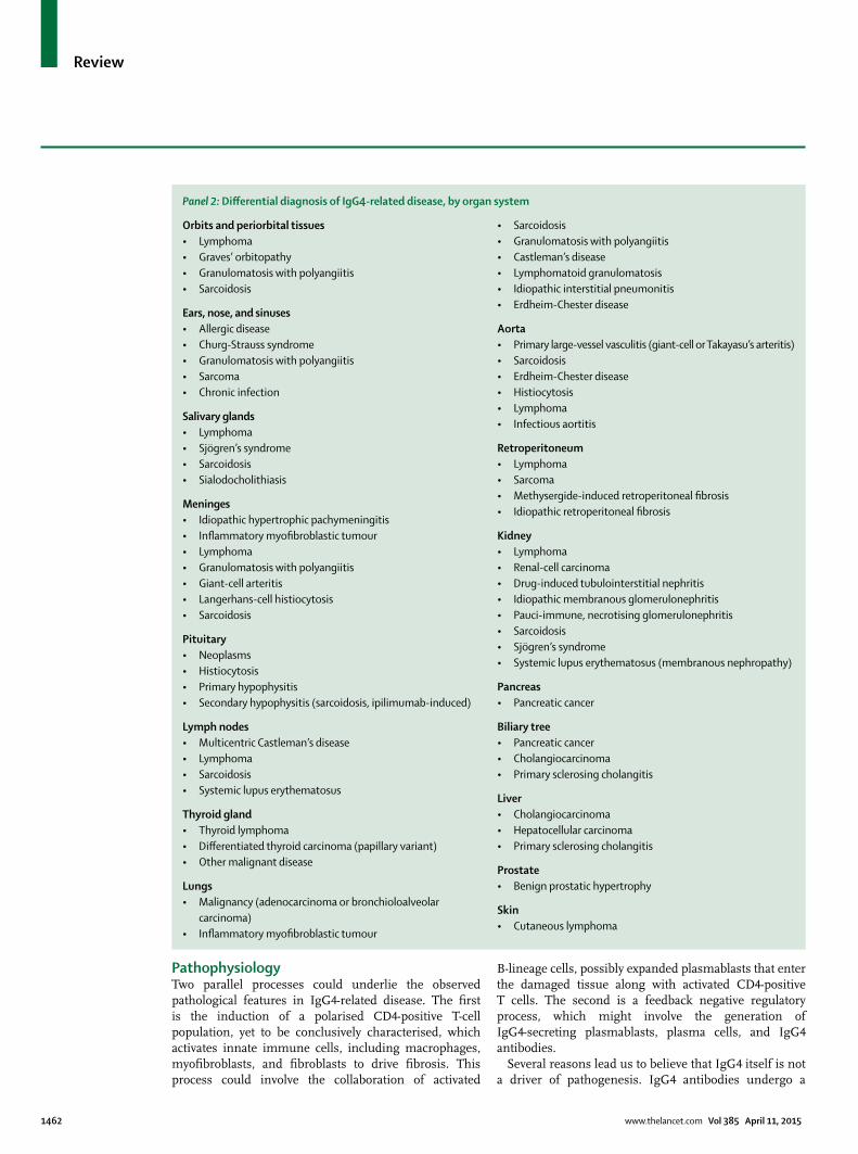
Diff erentiation of IgG4-related disease from malignant tumours is crucial. Common mimics of multi-organ IgG4-related disease are Sjögren’s syndrome, granulo-matosis with polyangiitis, eosinophilic granulo matosis with polyangiitis (formerly Churg-Strauss syndrome), sarcoidosis, and multicentric Castleman’s disease. Single-organ diseases such as primary sclerosing cholangitis must also be excluded (panel 2).
Four sets of diagnostic criteria for specifi c organs have been devised.30–33 Comprehensive diagnostic criteria for IgG4-related disease have been proposed for practical use by non-specialists.34
SerologyHigh serum IgG4 concentrations are neither suffi ciently sensitive nor specifi c for diagnosis. Serum IgG4 concentrations are useful for screening but are unreliable as a single diagnostic marker. About 20% of patients with type 1 autoimmune pancreatitis have normal serum IgG4 concentrations at presentation.35,36 The proportion with normal concentrations can be somewhat lower among patients with multi-organ disease,37 but many diagnoses can be associated with high serum IgG4 concentrations. In one study, 22% of
patients who did not have IgG4-related disease had serum IgG4 concentrations higher than twice normal.37 Other studies have shown that 4–10% of both healthy and disease controls, including patients with pancreatic cancer, have high serum IgG4 concentrations.36,38,39 Increased ratios of IgG4 to total IgG (>10%) or IgG1 (>24%) increase diagnostic specifi city, especially when IgG4 concentrations are only slightly raised.40 The identifi cation of high numbers of plasmablasts within blood by fl ow cytometry is more sensitive than serum IgG4 concentrations,41,42 but such assays are not yet widely available.
Monitoring of serum IgG4 concentrations seems useful in assessment of disease activity in some patients, but this measurement should never be used as the sole determinant in treatment decisions. The serum IgG4 concentration declines substantially after glucocorticoid treatment in most patients, but in one study did not return to the normal range in 115 (63%) of 182 patients.43 Clinical relapses occurred in 10% of patients who had persistently normal IgG4 concentrations.43
Nephelometry assays for IgG4 are prone to error in the presence of large antigen excess, potentially leading to gross underestimates of the serum IgG4 concentration because fl occulation does not occur. This eff ect, known as the prozone phenomenon, can lead to false reports of normal serum IgG4 concentrations and has been observed frequently in patients with IgG4-related disease with serum IgG4 concentrations many times higher than the upper limit of normal.44 Appropriate dilution of the serum sample during the assay process prevents the prozone eff ect.
Organ involvementConstitutional and musculoskeletal symptomsThe presentation of IgG4-related disease is typically subacute, with symptoms and organ dysfunction evident for months or even years before diagnosis. Disease can progress haltingly, with occasional spontaneous impro vements (generally temporary) or long plateaus of disease quiescence in a specifi c organ. In such cases, disease recurrence in an organ known to be aff ected or the emergence of new organ involvement can lead to diagnosis.
Weight loss of 5–10 kg can occur over months, but fevers and hectic presentations are unusual. Fatigue commonly accompanies IgG4-related disease, especially when the disease aff ects several organ systems. We have observed a diff use array of musculo skeletal symptoms, including arthralgias and enthesopathy (infl ammation in the site at which a tendon inserts into a bone). To date, however, no histopathological abnormalities of synovium or tenosynovium have been confi rmed.
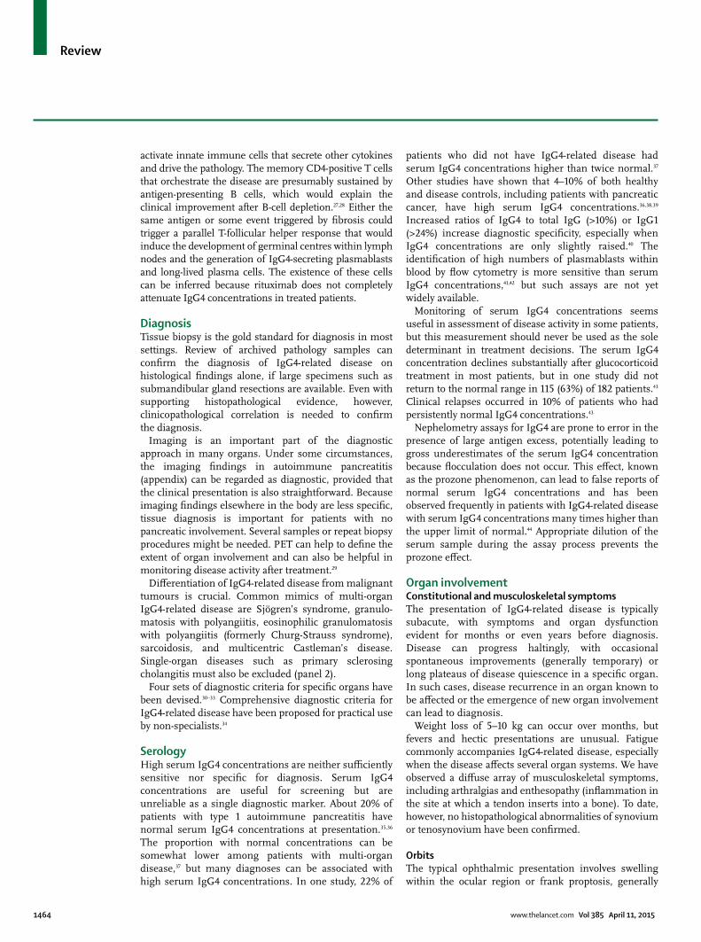
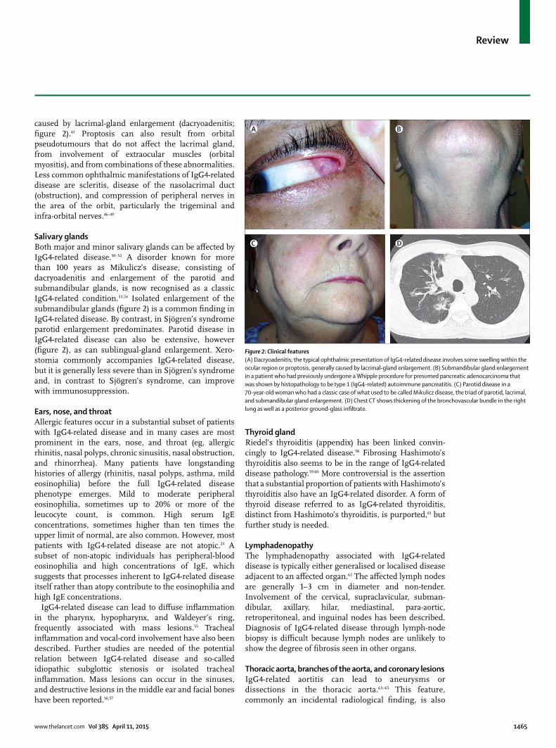
OrbitsThe typical ophthalmic presentation involves swelling within the ocular region or frank proptosis, generally
Review
www.thelancet.com Vol 385 April 11, 2015 1465
caused by lacrimal-gland enlargement (dacryoadenitis; fi gure 2).45 Proptosis can also result from orbital pseudotumours that do not aff ect the lacrimal gland, from involvement of extraocular muscles (orbital myositis), and from combinations of these abnormalities. Less common ophthalmic manifest ations of IgG4-related disease are scleritis, disease of the nasolacrimal duct (obstruction), and compression of peripheral nerves in the area of the orbit, particularly the trigeminal and infra-orbital nerves.46–49
Salivary glandsBoth major and minor salivary glands can be aff ected by IgG4-related disease.50–52 A disorder known for more than 100 years as Mikulicz’s disease, consisting of dacryo adenitis and enlargement of the parotid and submandibular glands, is now recognised as a classic IgG4-related condition.53,54 Isolated enlargement of the submandibular glands (fi gure 2) is a common fi nding in IgG4-related disease. By contrast, in Sjögren’s syndrome parotid enlargement predominates. Parotid disease in IgG4-related disease can also be extensive, however (fi gure 2), as can sublingual-gland enlargement. Xero-stomia commonly accompanies IgG4-related disease, but it is generally less severe than in Sjögren’s syndrome and, in contrast to Sjögren’s syndrome, can improve with immunosuppression.
Ears, nose, and throatAllergic features occur in a substantial subset of patients with IgG4-related disease and in many cases are most prominent in the ears, nose, and throat (eg, allergic rhinitis, nasal polyps, chronic sinusitis, nasal obstruction, and rhinorrhea). Many patients have longstanding histories of allergy (rhinitis, nasal polyps, asthma, mild eosinophilia) before the full IgG4-related disease phenotype emerges. Mild to moderate peripheral eosinophilia, sometimes up to 20% or more of the leucocyte count, is common. High serum IgE concentrations, sometimes higher than ten times the upper limit of normal, are also common. However, most patients with IgG4-related disease are not atopic.24 A subset of non-atopic individuals has peripheral-blood eosinophilia and high concentrations of IgE, which suggests that processes inherent to IgG4-related disease itself rather than atopy contribute to the eosinophilia and high IgE concentrations.
IgG4-related disease can lead to diff use infl ammation in the pharynx, hypopharynx, and Waldeyer’s ring, frequently associated with mass lesions.55 Tracheal infl ammation and vocal-cord involvement have also been described. Further studies are needed of the potential relation between IgG4-related disease and so-called idiopathic subglottic stenosis or isolated tracheal infl ammation. Mass lesions can occur in the sinuses, and destructive lesions in the middle ear and facial bones have been reported.56,57
Thyroid glandRiedel’s thyroiditis (appendix) has been linked convin-cingly to IgG4-related disease.58 Fibrosing Hashimoto’s thyroiditis also seems to be in the range of IgG4-related disease pathology.59,60 More controversial is the assertion that a substantial proportion of patients with Hashimoto’s thyroiditis also have an IgG4-related disorder. A form of thyroid disease referred to as IgG4-related thyroiditis, distinct from Hashimoto’s thyroiditis, is purported,61 but further study is needed.
LymphadenopathyThe lymphadenopathy associated with IgG4-related disease is typically either generalised or localised disease adjacent to an aff ected organ.62 The aff ected lymph nodes are generally 1–3 cm in diameter and non-tender. Involvement of the cervical, supraclavicular, subman-dibular, axillary, hilar, mediastinal, para-aortic, retroperitoneal, and inguinal nodes has been described. Diagnosis of IgG4-related disease through lymph-node biopsy is diffi cult because lymph nodes are unlikely to show the degree of fi brosis seen in other organs.
Thoracic aorta, branches of the aorta, and coronary lesionsIgG4-related aortitis can lead to aneurysms or dissections in the thoracic aorta.63–65 This feature, commonly an incidental radiological fi nding, is also
Figure 2: Clinical features(A) Dacryoadenitis; the typical ophthalmic presentation of IgG4-related disease involves some swelling within the ocular region or proptosis, generally caused by lacrimal-gland enlargement. (B) Submandibular gland enlargement in a patient who had previously undergone a Whipple procedure for presumed pancreatic adenocarcinoma that was shown by histopathology to be type 1 (IgG4-related) autoimmune pancreatitis. (C) Parotid disease in a 70-year-old woman who had a classic case of what used to be called Mikulicz disease, the triad of parotid, lacrimal, and submandibular gland enlargement. (D) Chest CT shows thickening of the bronchovascular bundle in the right lung as well as a posterior ground-glass infi ltrate.
A B
C D
Review
1466 www.thelancet.com Vol 385 April 11, 2015
sometimes an unexpected fi nding at surgery. In contrast to giant-cell and Takayasu’s arteritis, which mainly aff ect the primary aortic branches, especially the subclavian arteries, IgG4-related disease tends to spare these vessels, at least in terms of clinical manifestations. No defi nitive histopathological investigations of primary aortic branch vessels have been undertaken, but small case series substantiate the concept that medium-sized blood vessels can also be aff ected by IgG4-related disease.66,67 Coronary artery lesions in IgG4-related disease are rare but documented.68
Chronic periaortitis and retroperitoneal fi brosisSo-called idiopathic retroperitoneal fi brosis, known for decades as Ormond’s disease,69 is now classifi ed within a larger disease grouping known as chronic periaortitis (appendix). The three major components of chronic periaortitis are IgG4-related retroperitoneal fi brosis, IgG4-related abdominal aortitis, and IgG4-related perianeurysmal fi brosis.65,70
The presentations of IgG4-related chronic periaortitis can be subtle and non-specifi c, leading to diagnostic delay. Common presentations are: a poorly localised pain in the back, fl anks, lower abdomen, or thighs; leg oedema; and hydronephrosis from ureteral involvement. The disease targets three sites: periaortic/arterial regions, involving connective tissue around the abdominal aorta or its fi rst branches (appendix); periureteral areas, tending to cause ureteral obstruction and hydronephrosis; and a plaque-like mass that broadly involves the retroperitoneum.
IgG4-related disease is the cause of up to two-thirds of cases of idiopathic retroperitoneal fi brosis.69,70 In advanced disease, the ratio of IgG4-positive plasma cells to the total number of plasma cells in tissue can be more helpful diagnostically than the overall number of IgG4-positive plasma cells per high-power fi eld. Even if the classic lymphoplasmacytic infi ltrate is not evident in longstanding cases, both storiform fi brosis and obliterative phlebitis are commonly identifi ed (appendix).
LungsThe greatest diversity of clinical and radiological presentations is seen in the lungs.71 Thickening of the bronchovascular bundle, best shown by CT, is a characteristic lesion (fi gure 2); it shows the tendency of IgG4-related disease to track along bronchi and blood vessels, which course together.15 Other radiological features of IgG4-related disease include pulmonary nodules, ground-glass opacities, pleural thickening, and interstitial lung disease. The last of these, which mimics non-specifi c interstitial pneumonitis and other forms of interstitial fi brosis, emphasises the fi brotic tendencies of IgG4-related disease.
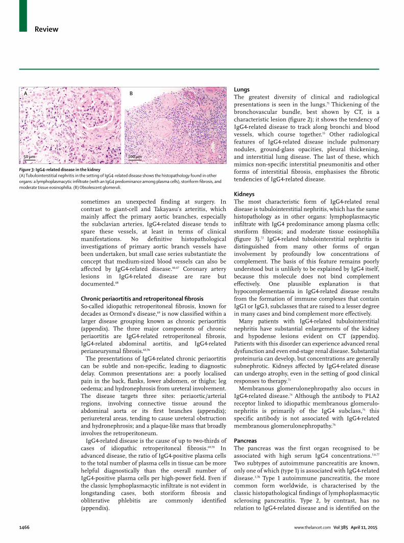
KidneysThe most characteristic form of IgG4-related renal disease is tubulointerstitial nephritis, which has the same histopathology as in other organs: lymphoplasmacytic infi ltrate with IgG4 predominance among plasma cells; storiform fi brosis; and moderate tissue eosinophilia (fi gure 3).72 IgG4-related tubulointerstitial nephritis is distinguished from many other forms of organ involvement by profoundly low concentrations of complement. The basis of this feature remains poorly understood but is unlikely to be explained by IgG4 itself, because this molecule does not bind complement eff ectively. One plausible explanation is that hypocomplementaemia in IgG4-related disease results from the formation of immune complexes that contain IgG1 or IgG3, subclasses that are raised to a lesser degree in many cases and bind complement more eff ectively.
Many patients with IgG4-related tubulointerstitial nephritis have substantial enlargements of the kidney and hypodense lesions evident on CT (appendix). Patients with this disorder can experience advanced renal dysfunction and even end-stage renal disease. Substantial proteinuria can develop, but concentrations are generally subnephrotic. Kidneys aff ected by IgG4-related disease can undergo atrophy, even in the setting of good clinical responses to therapy.73
Membranous glomerulonephropathy also occurs in IgG4-related disease.74 Although the antibody to PLA2 receptor linked to idiopathic membranous glomerulo-nephritis is primarily of the IgG4 subclass,75 this specifi c antibody is not associated with IgG4-related membranous glomerulonephropathy.76
PancreasThe pancreas was the fi rst organ recognised to be associated with high serum IgG4 concentrations.5,6,77 Two subtypes of autoimmune pancreatitis are known, only one of which (type 1) is associated with IgG4-related disease.3,78 Type 1 autoimmune pancreatitis, the more common form worldwide, is characterised by the classic histopathological fi ndings of lymphoplasmacytic sclerosing pancreatitis. Type 2, by contrast, has no relation to IgG4-related disease and is identifi ed on the
Figure 3: IgG4-related disease in the kidney(A) Tubulointerstitial nephritis in the setting of IgG4-related disease shows the histopathology found in other organs: a lymphoplasmacytic infi ltrate (with an IgG4 predominance among plasma cells), storiform fi brosis, and moderate tissue eosinophilia. (B) Obsolescent glomeruli.
A B
50 µm 100 µm
Review
www.thelancet.com Vol 385 April 11, 2015 1467
basis of histological features of neutrophilic infi ltration into the epithelium of the pancreatic duct.79–81
The most common clinical presentation of auto-immune pancreatitis is obstructive jaundice, induced by con comitant IgG4-related sclerosing cholangitis. Secondary diabetes mellitus occurs in about half of cases, which makes treatment with glucocorticoids diffi cult in many patients. Diff erentiation of autoimmune pancreatitis from pancreatic cancer is crucial to avoid unnecessary surgery. The nearly diagnostic CT features of autoimmune pancreatitis include diff use pancreatic enlargement with delayed enhancement and a capsule-like low-density rim (appendix).80,81 Diff use, irregular narrowing of the main pancreatic duct on endoscopic retrograde and magnetic resonance cholangiopancreatography is also highly specifi c for autoimmune pancreatitis. In cases of segmental autoimmune pancreatitis, skipped narrowed lesions, side-branch derivation from the narrowed portion, and relatively less upstream dilatation on pancreatography suggest autoimmune pancreatitis rather than pancreatic cancer (appendix).81,82 In PET studies, uptake of fl uoro-deoxyglucose in organs other than the pancreas known to be aff ected by IgG4-related disease suggests autoimmune pancreatitis.29,83
International consensus diagnostic criteria for autoimmune pancreatitis were proposed in 2011.30 Under these criteria, the diagnosis can be made by a combination of parenchymal and ductal imaging, serum IgG4 concentrations, pancreatic histology, extra-pancreatic disease, and glucocorticoid responsive ness. Endoscopic ultrasonography-guided fi ne-needle aspir-ation is a useful diagnostic approach to exclude pancreatic cancer and should be attempted before any empirical trial of glucocorticoid treatment is undertaken. Several cases of pancreatic cancer have been reported in patients with type 1 autoimmune pancreatitis.84,85 Pancreatic stones occur with increased frequency among these patients.79,86
IgG4-related sclerosing cholangitis and cholecystitisType 1 autoimmune pancreatitis is commonly accompanied by IgG4-related sclerosing cholangitis.19 Whether the limited intrapancreatic bile-duct stricture associated with autoimmune pancreatitis should be regarded as a biliary manifestation of IgG4-related disease is controversial, because such stenoses can be induced by compression from the swollen pancreas.87 The histology of IgG4-related sclerosing cholangitis includes obliterative phlebitis and transmural fi brosis with dense infi ltration of IgG4-positive plasma cells and T cells.
IgG4-related sclerosing cholangitis must be diff erentiated from both primary sclerosing cholangitis and hilar cholangiocarcinoma. Neither serum IgG4 concentrations nor cholangiographic or cholangioscopic fi ndings diff erentiate these disorders clearly.88–91 Thus, endoscopic transpapillary biopsy is generally needed.
Although cholangiocarcinoma can be excluded by endoscopic biopsy, the superfi cial nature of samples obtained by this procedure limits their usefulness for diagnosis of IgG4-related sclerosing cholangitis.92
IgG4-related cholecystitis can occur with sclerosing cholangitis. Thickening of the gallbladder wall is detected on imaging, but it is asymptomatic in most cases.81
Other organsIgG4-related disease seldom, if ever, aff ects the brain parenchyma but it is one of the most common causes of hypertrophic pachymeningitis.93 IgG4-related disease is also an unheralded cause of hypophysitis. IgG4-related hypophysitis can lead to hormone defi ciencies from both the anterior and posterior pituitary.94 MRI shows sellar enlargement and thickening of the pituitary stalk.
Sclerosing lesions of both the mediastinum and mesentery have been described.95,96 In fi brosing mediastinitis, compression of vital mediastinal structures can result from proliferation of invasive fi brous tissue within the mediastinum. A review of 15 patients with fi brosing mediastinitis showed that a substantial proportion of cases are within the IgG4-related disease spectrum.95 The relation between these cases and antecedent infections with histoplasma, if any, remains unclear.
The infl ammatory process in sclerosing mesenteritis seems to originate at the mesenteric root.96 The ensuing process merges imperceptibly with retroperitoneal fi brosis and can evolve in a devastating manner, encasing vital organs and obviating any attempt at surgical resection.
Several clinical presentations of IgG4-related skin disease have been reported. The most common is the presence of erythematous papules. These lesions typically aff ect the head and neck but have also been described on the trunk and limbs.97 Among individuals with darkly pigmented skin, hyperpigmented lesions have been observed. Peripheral-nerve lesions typically consist of perineural masses, up to 3 cm in diameter. These are commonly seen on MRI in the absence of overt clinical manifestations.48
The diagnosis of IgG4-related prostate disease is commonly made presumptively when the initiation of treatment for IgG4-related disease in other organs mediates abrupt symptomatic relief of apparently benign prostatic hypertrophy. Both radiological demonstration of prostatic enlargement and biopsy-proven IgG4-related prostatic disease have been reported.98
TreatmentGlucocorticoidsMost clinical manifestations of IgG4-related disease respond to glucocorticoids. These agents are the fi rst-line, standard-of-care approach for most patients.43,99 However, no randomised treatment trials have been done, and few large retrospective examinations have been reported. One treatment approach uses a starting prednisolone dose of
Review
1468 www.thelancet.com Vol 385 April 11, 2015
0·6–1·0 mg/kg daily.30,43 After 2–4 weeks, the dose is tapered by 5 mg every 1–2 weeks according to clinical responses (eg, clinical mani festations, blood tests, and follow-up imaging studies). Practice varies as to whether the prednisolone is discontinued entirely after 2 or 3 months or maintained at a low dose. A single-group trial of prednisolone in Japan showed complete remissions in only 61% of patients at 1 year despite continuation of maintenance doses of prednisone in all patients.100
Clinical improvement after the start of glucocorticoid therapy is rapid, and a follow-up serological assessment should be done about 2 weeks after treatment initiation. Follow-up radiological assessment is also appropriate for some types of organ involvement, such as the pancreas, biliary tree, lungs, and kidneys. PET with fl uoro-deoxyglucose is useful to assess treatment response.29 A swift response to glucocorticoids is reassuring and provides further diagnostic confi rmation if a tissue diagnosis was not possible before the start of therapy. A poor response to glucocorticoids, however, should raise the possibility of other diagnoses, particularly cancer.
The response to glucocorticoids varies according to the aff ected organs and the degree of fi brosis.8 Both endocrine and exocrine pancreatic function can improve in autoimmune pancreatitis, and salivary secretion in IgG4-related sialadenitis is more likely to improve after glucocorticoid therapy than is the glandular function of Sjögren’s syndrome.100–103 By contrast, retroperitoneal fi brosis, sclerosing mesenteritis, and fi brosing mediasti-nitis are less amenable to therapy with glucocorticoids, underscoring the importance of early diagnosis and treatment.104
Conventional steroid-sparing agentsDrugs such as azathioprine, mycophenolate mofetil, and methotrexate, all used widely in gastroenterology, rheumatology, and transplant medicine as means of achieving additional immunosuppression and sparing patients the eff ects of long-term glucocorticoids, are commonly chosen for this purpose in IgG4-related disease.79,105 However, none has been tested in prospective, controlled studies, and evidence for their effi cacy beyond that off ered by concomitant glucocorticoid therapy is scarce. Rigorous assessment of these treatments in IgG4-related disease is needed.
B-cell depletionRituximab was used initially in patients who did not respond to glucocorticoids, conventional steroid-sparing agents, or both, under the assumption that B-cell depletion might ameliorate the condition putatively mediated by high serum concentrations of IgG4.27,48,106 The fundamental assumption underlying this approach now seems incorrect or at least not entirely true, but careful mechanistic studies of patients with IgG4-related disease treated with rituximab have led to several important observations and novel insights about the pathophysiology of this disorder. First,
B-cell depletion targets the subset of plasma cells that produce IgG4 in IgG4-related disease.27,28 They seem to achieve this action by depleting all circulating CD20-positive cells (ie, B cells), which interferes in turn with the repletion of short-lived plasma cells making IgG4. In other words, the plasma cells generating IgG4 in IgG4-related disease are mainly of the short-lived type that naturally undergo apoptosis within weeks. Once these cells disappear as programmed, they cannot be repleted after rituximab administration because their precursors—CD20-positive B cells—are not available.
Second, IgG4-positive plasmablasts (positive for IgG4, CD38, CD37, and CD19lo cells) seem to be a good biomarker for IgG4-related disease and are probably superior to serum IgG4 concentrations for diagnosis and monitoring of disease activity.41,42 We have seen patients with substantially raised numbers of IgG4-positive plasmablasts whose serum IgG4 concentrations were normal in the setting of active disease. These plasmablasts decline quickly after B-cell depletion and can be useful in identifying when to readminister rituximab in some patients, but this question needs further study.
Future perspectivesIn only 10 years since the recognition of extrapancreatic features in patients with autoimmune pancreatitis signalled a systemic, multi-organ disease, substantial progress has been achieved in IgG4-related disease. The disease has been identifi ed in nearly every organ system and most of its clinical features have been mapped. Nomenclature has been standardised, and a consensus has been achieved about the major and minor pathological manifestations.3,14 Eff ective treatments have been identifi ed and important advances have been made in understanding of disease pathophysiology through mechanistic studies of B-cell depletion. Greater awareness in the medical community of this protean disease is needed to ensure earlier diagnoses, which can prevent severe organ damage, disabling tissue fi brosis, and death. The epidemiology of IgG4-related disease remains poorly understood, mainly because of challenges in recognition and diff erentiation from the many disorders it mimics. Blood-based diagnostic tests through serology or fl ow cytometry would be a step forward in case identifi cation. Greater understanding of the immunopathology of IgG4-related disease promises new insights into human immunology and interactions between various T-cell pathways, as well as the possibility of new mechanisms of disease centred around novel T-cell phenotypes. Identifi cation of specifi c antigens and T-cell clones that drive the disease will be crucial steps in elucidating the pathogenesis of IgG4-related disease.ContributorsAll the authors contributed equally to the literature search, planning, writing, and editing of the Review and all have approved the submission of this version.
Review
www.thelancet.com Vol 385 April 11, 2015 1469
Declaration of interestsJHS is the principal investigator in a Genentech-funded trial of rituximab in IgG4-related disease and has consulted for Genentech on this disease. The other authors declare no competing interests.
References1 Mahajan VS, Mattoo H, Deshpande V, Pillai SS, Stone JH.
IgG4-related disease. Annu Rev Pathol 2014; 9: 315–47.2 Umehara H, Okazaki K, Masaki Y, et al, and the Research Program
for Intractable Disease by Ministry of Health, Labor and Welfare (MHLW) Japan G4 team. A novel clinical entity, IgG4-related disease (IgG4RD): general concept and details. Mod Rheumatol 2012; 22: 1–14.
3 Stone JH, Khosroshahi A, Deshpande V, et al. Recommendations for the nomenclature of IgG4-related disease and its individual organ system manifestations. Arthritis Rheum 2012; 64: 3061–67.
4 Kawa S, Kawano M. IgG4-related disease: an overview. In: Umehara H, Okazaki K, Stone JH, Kawa S, Kawano M, eds. IgG4-related disease. Berlin: Springer, 2013.
5 Kamisawa T, Egawa N, Nakajima H. Autoimmune pancreatitis is a systemic autoimmune disease. Am J Gastroenterol 2003; 98: 2811–12.
6 Kamisawa T, Funata N, Hayashi Y, et al. A new clinicopathological entity of IgG4-related autoimmune disease. J Gastroenterol 2003; 38: 982–84.
7 Kanno A, Nishimori I, Masamune A, et al, and the Research Committee on Intractable Diseases of Pancreas. Nationwide epidemiological survey of autoimmune pancreatitis in Japan. Pancreas 2012; 41: 835–39.
8 Stone JH, Zen Y, Deshpande V. IgG4-related disease. N Engl J Med 2012; 366: 539–51.
9 Zen Y, Nakanuma Y. IgG4-related disease: a cross-sectional study of 114 cases. Am J Surg Pathol 2010; 34: 1812–19.
10 Ota M, Ito T, Umemura T, et al. Polymorphism in the KCNA3 gene is associated with susceptibility to autoimmune pancreatitis in the Japanese population. Dis Markers 2011; 31: 223–29.
11 Umemura T, Ota M, Hamano H, et al. Association of autoimmune pancreatitis with cytotoxic T-lymphocyte antigen 4 gene polymorphisms in Japanese patients. Am J Gastroenterol 2008; 103: 588–94.
12 Ota M, Katsuyama Y, Hamano H, et al. Two critical genes (HLA-DRB1 and ABCF1)in the HLA region are associated with the susceptibility to autoimmune pancreatitis. Immunogenetics 2007; 59: 45–52.
13 Umemura T, Ota M, Hamano H, Katsuyama Y, Kiyosawa K, Kawa S. Genetic association of Fc receptor-like 3 polymorphisms with autoimmune pancreatitis in Japanese patients. Gut 2006; 55: 1367–68.
14 Deshpande V, Zen Y, Chan JK, et al. Consensus statement on the pathology of IgG4-related disease. Mod Pathol 2012; 25: 1181–92.
15 Zen Y, Inoue D, Kitao A, et al. IgG4-related lung and pleural disease: a clinicopathologic study of 21 cases. Am J Surg Pathol 2009; 33: 1886–93.
16 Hamano H, Kawa S, Ochi Y, et al. Hydronephrosis associated with retroperitoneal fi brosis and sclerosing pancreatitis. Lancet 2002; 359: 1403–04.
17 Kamisawa T, Funata N, Hayashi Y, et al. Close relationship between autoimmune pancreatitis and multifocal fi brosclerosis. Gut 2003; 52: 683–87.
18 Strehl JD, Hartmann A, Agaimy A. Numerous IgG4-positive plasma cells are ubiquitous in diverse localised non-specifi c chronic infl ammatory conditions and need to be distinguished from IgG4-related systemic disorders. J Clin Pathol 2011; 64: 237–43.
19 Zen Y, Harada K, Sasaki M, et al. IgG4-related sclerosing cholangitis with and without hepatic infl ammatory pseudotumor, and sclerosing pancreatitis-associated sclerosing cholangitis: do they belong to a spectrum of sclerosing pancreatitis? Am J Surg Pathol 2004; 28: 1193–203.
20 Aalberse RC, Stapel SO, Schuurman J, Rispens T. Immunoglobulin G4: an odd antibody. Clin Exp Allergy 2009; 39: 469–77.
21 Okazaki K, Uchida K, Ohana M, et al. Autoimmune-related pancreatitis is associated with autoantibodies and a Th1/Th2-type cellular immune response. Gastroenterology 2000; 118: 573–81.
22 Zen Y, Fujii T, Harada K, et al. Th2 and regulatory immune reactions are increased in immunoglobin G4-related sclerosing pancreatitis and cholangitis. Hepatology 2007; 45: 1538–46.
23 Mattoo H, Della-Torre E, Mahajan VS, Stone JH, Pillai S. Circulating Th2 memory cells in IgG4-related disease are restricted to a defi ned subset of subjects with atopy. Allergy 2014; 69: 399–402.
24 Della Torre E, Mattoo H, Mahajan VS, Carruthers M, Pillai S, Stone JH. Prevalence of atopy, eosinophilia, and IgE elevation in IgG4-related disease. Allergy 2014; 69: 269–72.
25 Tsuboi H, Matsuo N, Iizuka M, et al. Analysis of IgG4 class switch-related molecules in IgG4-related disease. Arthritis Res Ther 2012; 14: R171.
26 King C, Tangye SG, Mackay CR. T follicular helper (TFH) cells in normal and dysregulated immune responses. Annu Rev Immunol 2008; 26: 741–66.
27 Khosroshahi A, Bloch DB, Deshpande V, Stone JH. Rituximab therapy leads to rapid decline of serum IgG4 levels and prompt clinical improvement in IgG4-related systemic disease. Arthritis Rheum 2010; 62: 1755–62.
28 Khosroshahi A, Carruthers MN, Deshpande V, Unizony S, Bloch DB, Stone JH. Rituximab for the treatment of IgG4-related disease: lessons from 10 consecutive patients. Medicine (Baltimore) 2012; 91: 57–66.
29 Ebbo M, Grados A, Guedj E, et al. Usefulness of 2-[18F]-fl uoro-2-deoxy-D-glucose-positron emission tomography/computed tomography for staging and evaluation of treatment response in IgG4-related disease: a retrospective multicenter study. Arthritis Care Res (Hoboken) 2014; 66: 86–96.
30 Shimosegawa T, Chari ST, Frulloni L, et al, and the International Association of Pancreatology. International consensus diagnostic criteria for autoimmune pancreatitis: guidelines of the International Association of Pancreatology. Pancreas 2011; 40: 352–58.
31 Ohara H, Okazaki K, Tsubouchi H, et al, and the Research Committee of IgG4-related Diseases, and the Research Committee of Intractable Diseases of Liver and Biliary Tract, and the Ministry of Health, Labor and Welfare, Japan, and the Japan Biliary Association. Clinical diagnostic criteria of IgG4-related sclerosing cholangitis 2012. J Hepatobiliary Pancreat Sci 2012; 19: 536–42.
32 Kawano M, Saeki T, Nakashima H, et al. Proposal for diagnostic criteria for IgG4-related kidney disease. Clin Exp Nephrol 2011; 15: 615–26.
33 Masaki Y, Sugai S, Umehara H. IgG4-related diseases including Mikulicz’s disease and sclerosing pancreatitis: diagnostic insights. J Rheumatol 2010; 37: 1380–85.
34 Umehara H, Okazaki K, Masaki Y, et al. Comprehensive diagnostic criteria for IgG4-related disease (IgG4-RD), 2011. Mod Rheumatol 2012; 22: 21–30.
35 Sah RP, Chari ST. Serologic issues in IgG4-related systemic disease and autoimmune pancreatitis. Curr Opin Rheumatol 2011; 23: 108–13.
36 Tabata T, Kamisawa T, Takuma K, et al. Serum IgG4 concentrations and IgG4-related sclerosing disease. Clin Chim Acta 2009; 408: 25–28.
37 Carruthers MN, Khosroshahi A, Augustin T, Deshpande V, Stone JH. The diagnostic utility of serum IgG4 concentrations in patients with potential IgG4-related disease. Ann Rheum Dis 2014; published online March 20. DOI:10.1136/annrheumdis-2013-204907.
38 Ghazale A, Chari ST, Smyrk TC, et al. Value of serum IgG4 in the diagnosis of autoimmune pancreatitis and in distinguishing it from pancreatic cancer. Am J Gastroenterol 2007; 102: 1646–53.
39 Sadler R, Chapman RW, Simpson D, et al. The diagnostic signifi cance of serum IgG4 levels in patients with autoimmune pancreatitis: a UK study. Eur J Gastroenterol Hepatol 2011; 23: 139–45.
40 Boonstra K, Culver EL, de Buy Wenniger LM, et al. Serum IgG4 and IgG1 for distinguishing IgG4-associated cholangitis from primary sclerosing cholangitis. Hepatology 2014; 59: 1954–63.
41 Wallace ZS, Mattoo H, Carruthers MN, et al. Plasmablasts as a biomarker for IgG4-related disease, independent of serum IgG4 concentrations. Ann Rheum Dis 2014; published online May 9. DOI:10.1136/annrheumdis-2014-205233.rint.
42 Mattoo H, Mahajan VS, Della Torre E, et al. De novo oligoclonal expansions of circulating plasmablasts in active and relapsing IgG4-related disease. J Allergy Clin Immunol 2014; published online May 6. DOI:10.1016/j.jaci.2014.03.034.
43 Kamisawa T, Shimosegawa T, Okazaki K, et al. Standard steroid treatment for autoimmune pancreatitis. Gut 2009; 58: 1504–07.
Review
1470 www.thelancet.com Vol 385 April 11, 2015
44 Khosroshahi A, Cheryk LA, Carruthers MN, Edwards JA, Bloch DB, Stone JH. Brief Report: spuriously low serum IgG4 concentrations caused by the prozone phenomenon in patients with IgG4-related disease. Arthritis Rheum (Munch) 2014; 66: 213–17.
45 Wallace ZS, Deshpande V, Stone JH. Ophthalmic manifestations of IgG4-related disease: single-center experience and literature review. Semin Arthritis Rheum 2013; 43: 806–17.
46 Ohno K, Sato Y, Ohshima K, et al. IgG4-related disease involving the sclera. Mod Rheumatol 2014; 24: 195–98.
47 Wallace ZS, Khosroshahi A, Jakobiec FA, et al. IgG4-related systemic disease as a cause of “idiopathic” orbital infl ammation, including orbital myositis, and trigeminal nerve involvement. Surv Ophthalmol 2012; 57: 26–33.
48 Inoue D, Zen Y, Sato Y, et al. IgG4-related perineural disease. Int J Rheumatol 2012; 29: 212–18.
49 Takahira M, Ozawa Y, Kawano M, et al. Clinical aspects of IgG4-related orbital infl ammation in a case series of ocular adnexal lymphoproliferative disorders. Int J Rheumatol 2012; published online April 2. DOI:10.1155/2012/635473.
50 Baer AN, Gourin CG, Westra WH, Cox DP, Greenspan JS, Daniels TE, and the Sjögren’s International Collaborative Alliance. Rare diagnosis of IgG4-related systemic disease by lip biopsy in an international Sjögren syndrome registry. Oral Surg Oral Med Oral Pathol Oral Radiol 2013; 115: e34–39.
51 Hermet M, André M, Kémény JL, et al. Is IgG4-related disease a cause of xerostomia? A cohort study of 60 patients. Int J Rheumatol 2012; published online Oct 16. DOI:10.1155/2012/303506.
52 Geyer JT, Ferry JA, Harris NL, et al. Chronic sclerosing sialadenitis (Küttner tumor) is an IgG4-associated disease. Am J Surg Pathol 2010; 34: 202–10.
53 Yao Q, Wu G, Hoschar A. IgG4-related Mikulicz’s disease is a multiorgan lymphoproliferative disease distinct from Sjögren’s syndrome: a Caucasian patient and literature review. Clin Exp Rheumatol 2013; 31: 289–94.
54 Himi T, Takano K, Yamamoto M, Naishiro Y, Takahashi H. A novel concept of Mikulicz’s disease as IgG4-related disease. Auris Nasus Larynx 2012; 39: 9–17.
55 Fatemi G, Fang MA. IgG4-related pharyngitis-an addition to the nomenclature of IgG4-related disease: comment on the article by Stone et al. Arthritis Rheum 2013; 65: 2217.
56 Hu EK, Parrish C, Wrobel B, Deshpande V, Stone JH. Immunoglobulin G4-related disease presenting as an ethmoid and maxillary mass. Ann Allergy Asthma Immunol 2013; 111: 75–77.
57 Schiff enbauer AI, Gahl WA, Pittaluga S, et al. IgG4-related disease presenting as recurrent mastoiditis. Laryngoscope 2012; 122: 681–84.
58 Dahlgren M, Khosroshahi A, Nielsen GP, Deshpande V, Stone JH. Riedel’s thyroiditis and multifocal fi brosclerosis are part of the IgG4-related systemic disease spectrum. Arthritis Care Res (Hoboken) 2010; 62: 1312–18.
59 Deshpande V, Huck A, Ooi E, Stone JH, Faquin WC, Nielsen GP. Fibrosing variant of Hashimoto thyroiditis is an IgG4 related disease. J Clin Pathol 2012; 65: 725–28.
60 Pusztaszeri M, Triponez F, Pache JC, Bongiovanni M. Riedel’s thyroiditis with increased IgG4 plasma cells: evidence for an underlying IgG4-related sclerosing disease? Thyroid 2012; 22: 964–68.
61 Watanabe T, Maruyama M, Ito T, et al. Clinical features of a new disease concept, IgG4-related thyroiditis. Scand J Rheumatol 2013; 42: 325–30.
62 Cheuk W, Chan JK. Lymphadenopathy of IgG4-related disease: an underdiagnosed and overdiagnosed entity. Semin Diagn Pathol 2012; 29: 226–34.
63 Kasashima S, Zen Y, Kawashima A, et al. A clinicopathologic study of immunoglobulin G4-related sclerosing disease of the thoracic aorta. J Vasc Surg 2010; 52: 1587–95.
64 Stone JH, Patel VI, Oliveira GR, Stone JR. Case records of the Massachusetts General Hospital: case 38-2012, a 60-year-old man with abdominal pain and aortic aneurysms. N Engl J Med 2012; 367: 2335–46.
65 Zen Y, Kasashima S, Inoue D. Retroperitoneal and aortic manifestations of immunoglobulin G4-related disease. Semin Diagn Pathol 2012; 29: 212–18.
66 Stone JR. Aortitis, periaortitis, and retroperitoneal fi brosis, as manifestations of IgG4-related systemic disease. Curr Opin Rheumatol 2011; 23: 88–94.
67 Inoue D, Zen Y, Abo H, et al. Immunoglobulin G4-related periaortitis and periarteritis: CT fi ndings in 17 patients. Radiology 2011; 261: 625–33.
68 Inokuchi G, Hayakawa M, Kishimoto T, Makino Y, Iwase H. A suspected case of coronary periarteritis due to IgG4-related disease as a cause of ischemic heart disease. Forensic Sci Med Pathol 2014; 10: 103–08.
69 Khosroshahi A, Carruthers MN, Stone JH, et al. Rethinking Ormond’s disease: “idiopathic” retroperitoneal fi brosis in the era of IgG4-related disease. Medicine (Baltimore) 2013; 92: 82–91.
70 Zen Y, Onodera M, Inoue D, et al. Retroperitoneal fi brosis: a clinicopathologic study with respect to immunoglobulin G4. Am J Surg Pathol 2009; 33: 1833–39.
71 Inoue D, Zen Y, Abo H, et al. Immunoglobulin G4-related lung disease: CT fi ndings with pathologic correlations. Radiology 2009; 251: 260–70.
72 Saeki T, Nishi S, Imai N, et al. Clinicopathological characteristics of patients with IgG4-related tubulointerstitial nephritis. Kidney Int 2010; 78: 1016–23.
73 Saeki T, Kawano M, Mizushima I, et al. The clinical course of patients with IgG4-related kidney disease. Kidney Int 2013; 84: 826–33.
74 Alexander MP, Larsen CP, Gibson IW, et al. Membranous glomerulonephritis is a manifestation of IgG4-related disease. Kidney Int 2013; 83: 455–62.
75 Beck LH Jr, Bonegio RG, Lambeau G, et al. M-type phospholipase A2 receptor as target antigen in idiopathic membranous nephropathy. N Engl J Med 2009; 361: 11–21.
76 Khosroshahi A, Ayalon R, Beck LH Jr, Salant DJ, Bloch DB, Stone JH. IgG4-related disease is not associated with antibody to the phospholipase A2 receptor. Int J Rheumatol 2012; published online May 10. DOI:10.1155/2012/139409.
77 Hamano H, Kawa S, Horiuchi A, et al. High serum IgG4 concentrations in patients with sclerosing pancreatitis. N Engl J Med 2001; 344: 732–38.
78 Sah RP, Chari ST, Pannala R, et al. Diff erences in clinical profi le and relapse rate of type 1 versus Type 2 autoimmune pancreatitis. Gastroenterology 2010; 139: 140–48, quiz e12–13.
79 Hart PA, Kamisawa T, Brugge WR, et al. Long-term outcomes of autoimmune pancreatitis: a multicentre, international analysis. Gut 2013; 62: 1771–76.
80 Kamisawa T, Chari ST, Lerch MM, Kim MH, Gress TM, Shimosegawa T. Recent advances in autoimmune pancreatitis: type 1 and Type 2. Gut 2013; 62: 1373–80.
81 Kamisawa T, Takuma K, Egawa N, Tsuruta K, Sasaki T. Autoimmune pancreatitis and IgG4-related sclerosing disease. Nat Rev Gastroenterol Hepatol 2010; 7: 401–09.
82 Sugumar A, Levy MJ, Kamisawa T, et al. Endoscopic retrograde pancreatography criteria to diagnose autoimmune pancreatitis: an international multicentre study. Gut 2011; 60: 666–70.
83 Ozaki Y, Oguchi K, Hamano H, et al. Diff erentiation of autoimmune pancreatitis from suspected pancreatic cancer by fl uorine-18 fl uorodeoxyglucose positron emission tomography. J Gastroenterol 2008; 43: 144–51.
84 Pezzilli R, Vecchiarelli S, Di Marco MC, et al. Pancreatic ductal adenocarcinoma associated with autoimmune pancreatitis. Case Rep Gastroenterol 2011; 5: 378–85.
85 Gupta R, Khosroshahi A, Shinagare S, et al. Does autoimmune pancreatitis increase the risk of pancreatic carcinoma?: a retrospective analysis of pancreatic resections. Pancreas 2013; 42: 506–10.
86 Maruyama M, Arakura N, Ozaki Y, et al. Type 1 autoimmune pancreatitis can transform into chronic pancreatitis: a long-term follow-up study of 73 Japanese patients. Int J Rheumatol 2013; published online May 16. DOI:10.1155/2013/272595.
87 Hirano K, Tada M, Isayama H, et al. Endoscopic evaluation of factors contributing to intrapancreatic biliary stricture in autoimmune pancreatitis. Gastrointest Endosc 2010; 71: 85–90.
88 Mendes FD, Jorgensen R, Keach J, et al. Elevated serum IgG4 concentration in patients with primary sclerosing cholangitis. Am J Gastroenterol 2006; 101: 2070–75.
Review
www.thelancet.com Vol 385 April 11, 2015 1471
89 Oseini AM, Chaiteerakij R, Shire AM, et al. Utility of serum immunoglobulin G4 in distinguishing immunoglobulin G4-associated cholangitis from cholangiocarcinoma. Hepatology 2011; 54: 940–48.
90 Kalaitzakis E, Levy M, Kamisawa T, et al. Endoscopic retrograde cholangiography does not reliably distinguish IgG4-associated cholangitis from primary sclerosing cholangitis or cholangiocarcinoma. Clin Gastroenterol Hepatol 2011; 9: 800–03, e2.
91 Itoi T, Kamisawa T, Igarashi Y, et al. The role of peroral video cholangioscopy in patients with IgG4-related sclerosing cholangitis. J Gastroenterol 2013; 48: 504–14.
92 Nakazawa T, Ando T, Hayashi K, Naitoh I, Ohara H, Joh T. Diagnostic procedures for IgG4-related sclerosing cholangitis. J Hepatobiliary Pancreat Sci 2011; 18: 127–36.
93 Wallace ZS, Carruthers MN, Khosroshahi A, et al. IgG4-related disease and hypertrophic pachymeningitis. Medicine (Baltimore) 2013; 92: 206–16.
94 Leporati P, Landek-Salgado MA, Lupi I, Chiovato L, Caturegli P. IgG4-related hypophysitis: a new addition to the hypophysitis spectrum. J Clin Endocrinol Metab 2011; 96: 1971–80.
95 Peikert T, Shrestha B, Aubry MC, et al. Histopathologic overlap between fi brosing mediastinitis and IgG4-related disease. Int J Rheumatol 2012; published online May 10. DOI:10.1155/2012/207056.
96 Salvarani C, Valli R, Boiardi L, Pipitone N, Nicoli F, Muratore F. IgG4-associated sclerosing mesenteritis. Clin Exp Rheumatol 2011; 29 (suppl 64): S79–80.
97 Ikeda T, Oka M, Shimizu H, et al. IgG4-related skin manifestations in patients with IgG4-related disease. Eur J Dermatol 2013; 23: 241–45.
98 Hart PA, Smyrk TC, Chari ST. IgG4-related prostatitis: a rare cause of steroid-responsive obstructive urinary symptoms. Int J Urol 2013; 20: 132–34.
99 Kamisawa T, Okazaki K, Kawa S, Shimosegawa T, Tanaka M, and the Research Committee for Intractable Pancreatic Disease and Japan Pancreas Society. Japanese consensus guidelines for management of autoimmune pancreatitis: III. Treatment and prognosis of AIP. J Gastroenterol 2010; 45: 471–77.
100 Masaki Y, for the All-Japan Team for the Prospective Treatment Study of IgG4-RD. A trial of corticosteroids for IgG4-related disease. Second International Symposium on IgG4-related Disease & Associated Conditions, Honolulu, HA, USA, February 16–19, 2014.
101 Kamisawa T, Egawa N, Inokuma S, et al. Pancreatic endocrine and exocrine function and salivary gland function in autoimmune pancreatitis before and after steroid therapy. Pancreas 2003; 27: 235–38.
102 Ito T, Kawabe K, Arita Y, et al. Evaluation of pancreatic endocrine and exocrine function in patients with autoimmune pancreatitis. Pancreas 2007; 34: 254–59.
103 Ko SB, Mizuno N, Yatabe Y, et al. Corticosteroids correct aberrant CFTR localization in the duct and regenerate acinar cells in autoimmune pancreatitis. Gastroenterology 2010; 138: 1988–96.
104 Shimizu Y, Yamamoto M, Naishiro Y, et al. Necessity of early intervention for IgG4-related disease–delayed treatment induces fi brosis progression. Rheumatology (Oxford) 2013; 52: 679–83.
105 Hart PA, Topazian MD, Witzig TE, et al. Treatment of relapsing autoimmune pancreatitis with immunomodulators and rituximab: the Mayo Clinic experience. Gut 2013; 62: 1607–15.
106 Carruthers MN, Khosroshahi A, Topazian M, et al. Rituximab for IgG4-related disease: a prospective treatment trial. Arthritis Rheum 2013; 10: 2649.