Embed Size (px)
Citation preview
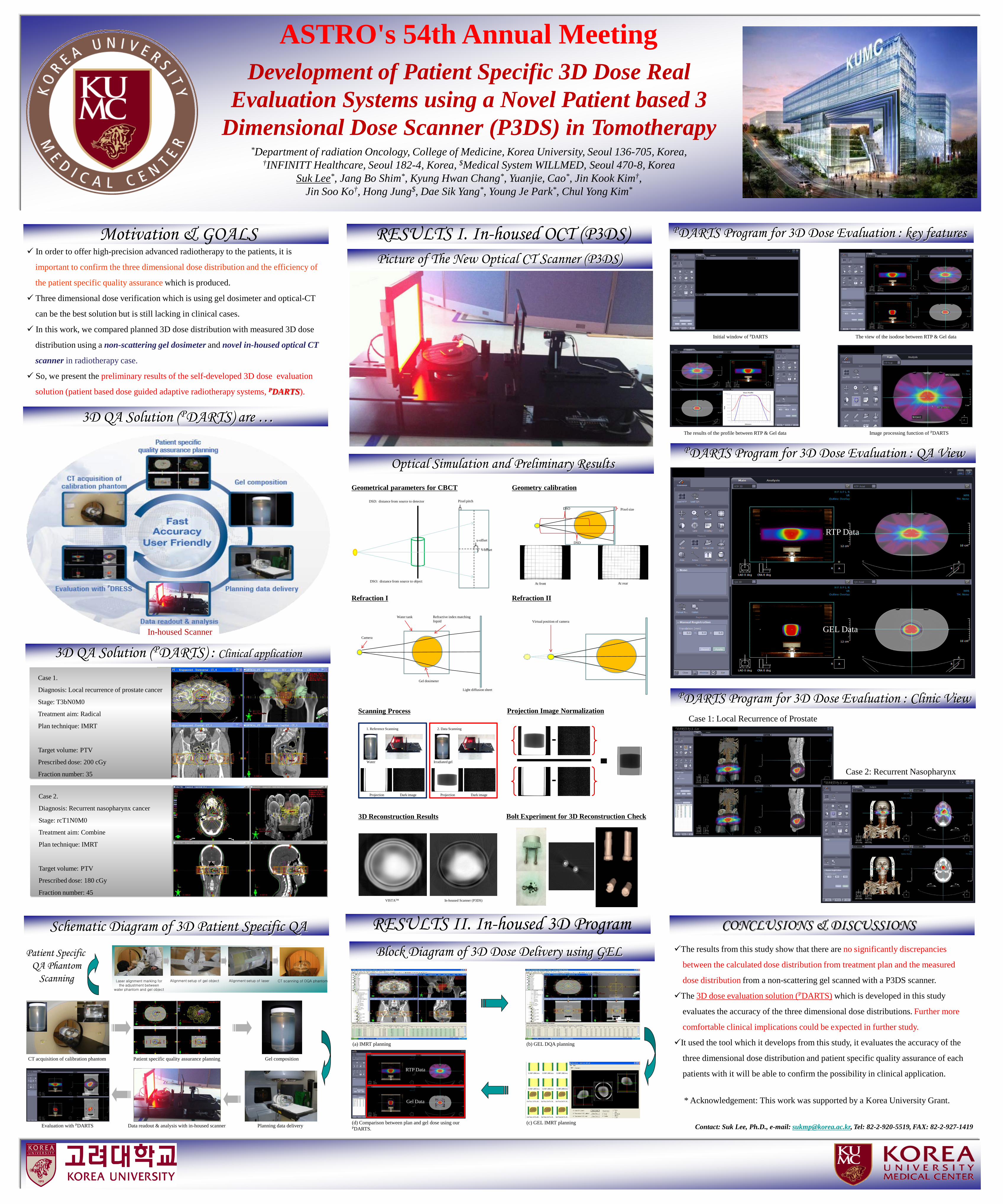
Development of Patient Specific 3D Dose Real Evaluation Systems using a Novel Patient based 3
Dimensional Dose Scanner (P3DS) in Tomotherapy*Department of radiation Oncology, College of Medicine, Korea University, Seoul 136-705, Korea,
†INFINITT Healthcare, Seoul 182-4, Korea, $Medical System WILLMED, Seoul 470-8, KoreaSuk Lee*, Jang Bo Shim*, Kyung Hwan Chang*, Yuanjie, Cao*, Jin Kook Kim†,
Jin Soo Ko†, Hong Jung$, Dae Sik Yang*, Young Je Park*, Chul Yong Kim*
3D QA Solution (PDARTS) are …
CONCLUSIONS & DISCUSSIONS
Contact: Suk Lee, Ph.D., e-mail: [email protected], Tel: 82-2-920-5519, FAX: 82-2-927-1419
RESULTS I. In-housed OCT (P3DS)
Schematic Diagram of 3D Patient Specific QA
PDARTS Program for 3D Dose Evaluation : key features In order to offer high-precision advanced radiotherapy to the patients, it is
important to confirm the three dimensional dose distribution and the efficiency of
the patient specific quality assurance which is produced.
Three dimensional dose verification which is using gel dosimeter and optical-CT
can be the best solution but is still lacking in clinical cases.
In this work, we compared planned 3D dose distribution with measured 3D dose
distribution using a non-scattering gel dosimeter and novel in-housed optical CT
scanner in radiotherapy case.
So, we present the preliminary results of the self-developed 3D dose evaluation
solution (patient based dose guided adaptive radiotherapy systems, PDARTS).
Motivation & GOALS
(a) IMRT planning (b) GEL DQA planning
(d) Comparison between plan and gel dose using our PDARTS.
(c) GEL IMRT planning
RTP Data
Gel Data
Block Diagram of 3D Dose Delivery using GEL The results from this study show that there are no significantly discrepancies
between the calculated dose distribution from treatment plan and the measured
dose distribution from a non-scattering gel scanned with a P3DS scanner.
The 3D dose evaluation solution (PDARTS) which is developed in this study
evaluates the accuracy of the three dimensional dose distributions. Further more
comfortable clinical implications could be expected in further study.
It used the tool which it develops from this study, it evaluates the accuracy of the
three dimensional dose distribution and patient specific quality assurance of each
patients with it will be able to confirm the possibility in clinical application.
3D QA Solution (PDARTS) : Clinical application
Case 1.
Diagnosis: Local recurrence of prostate cancer
Stage: T3bN0M0
Treatment aim: Radical
Plan technique: IMRT
Target volume: PTV
Prescribed dose: 200 cGy
Fraction number: 35
Case 2.
Diagnosis: Recurrent nasopharynx cancer
Stage: rcT1N0M0
Treatment aim: Combine
Plan technique: IMRT
Target volume: PTV
Prescribed dose: 180 cGy
Fraction number: 45
CT acquisition of calibration phantom Patient specific quality assurance planning Gel composition
Planning data deliveryData readout & analysis with in-housed scannerEvaluation with PDARTS
Laser alignment marking forthe adjustment between
water phantom and gel object
Alignment setup of gel object Alignment setup of laser CT scanning of DQA phantom
Patient Specific QA Phantom
Scanning
Picture of The New Optical CT Scanner (P3DS)
PDARTS Program for 3D Dose Evaluation : QA View
PDARTS Program for 3D Dose Evaluation : Clinic View
PDARTS by S. Lee
Initial window of PDARTS The view of the isodose between RTP & Gel data
The results of the profile between RTP & Gel data Image processing function of PDARTS
Case 1: Local Recurrence of Prostate
Case 2: Recurrent Nasopharynx
RTP Data
GEL Data
RTP Data
GEL DataIn-housed Scanner
RESULTS II. In-housed 3D Program
Optical Simulation and Preliminary Results
1. Reference Scanning 2. Data Scanning
Projection Dark image Projection Dark image
Water Irradiated gel
VISTATM In-housed Scanner (P3DS)
Scanning Process Projection Image Normalization
3D Reconstruction Results Bolt Experiment for 3D Reconstruction Check
u-offset
v-offset
DSO: distance from source to object
DSD: distance from source to detector Pixel pitch
Water tank Refractive index matching liquid
Gel dosimeter
Camera
Light diffusion sheet
Virtual position of camera
DSD
DSO Pixel size
At front At rear
Geometrical parameters for CBCT Geometry calibration
Refraction I Refraction II
PDARTS by S. Lee
* Acknowledgement: This work was supported by a Korea University Grant.
ASTRO's 54th Annual Meeting