Embed Size (px)
Citation preview

Chapter X
Culturing and maintaining mammalian cell culture
X.1 Introduction
Mammalian cell culture was first developed in the early twentieth century where
initially the tissue cultures were made by the isolation of individual cells from
fragments of tissue or organs for the use in laboratory. In the early years of tissue
culture, yielded tissue had very limited growth capabilities and limited by a finite
. The discovery of 1 number of divisions, a phenomenon known as Hayflick’s Limit
HeLa, a unique cervical carcinoma cell lines which has limitless lifespan, harvested
from patient called Henrietta Lawson, at John Hopkins Hospital, Baltimore in the
1950s, embarked modern advancement in clinical research for drug development and
. 2 therapy, where cells can be easily grown in large number and stored indefinitely
Cell culture technology has established wide applications in the field of cell biology
such as the study of intra and intercellular activities and flux, genetic and phenotyping
analysis, proteomics, toxicology studies, drug development, recombinant protein
development of Significant advancement and . 3treatment production, diagnosis and
mammalian cell culture had enabled researchers to further understand the underlying
mechanism of diseases in the surge for cure and therapy.
The routine for cell culture maintenance for feeding and subculturing should be
conducted in a sterile and in a laminar flow hood condition. Understanding of cellular
mitosis and morphological changes is important in the observation and critical
examination of cellular behavior in the culture. This begin with newly plated cells
attachment to the flask/plate surface to the cellular division until they reach
confluency and form a monolayer. At this stage, the cells will stop dividing to enter
resting period due to lack of space, nutrient and oxygen, a phenomenon known as
density-dependent growth inhibition. However, cancerous cells will continue to divide
on the top of the monolayer to form multilayers, even with the lack of nutrients and
oxygen supplies.
Subculturing is performed when cells attachment and proliferation in a culture reach
more than 70% of the surface area. Subculturing involves the removal of the media,
detachment of cells from the surface by the use of dissociating enzymes such as
trypsin and collagenase and chelator, Ethylene diamine tetra acetic acid (EDTA) for
ions. Long term exposure to trypsin is harmful to the cells as the 2+ the removal of Ca
enzymes may affect the essential extracellular matrix proteins such as laminin and
fibronectin required for cellular attachment and proliferation.

X.1.1 Laboratory safety and Professional Conduct
Safety policies are important in the laboratory to ensure safety and sterility conditions.
Failure to comply with these procedures may lead to accidents, contaminations and
health complications. The following policies must be adhered while in the laboratory.
1. No drink or food is allowed in the laboratory.
2. Wear a laboratory coat, long pants, clean and closed-toe shoes during the
laboratory session.
3. Use goggles when handling with hazardous chemicals and fumes.
4. Disinfect all benches and laboratory working area with 70% ethanol prior
work.
5. Wash hands before and after working in the laboratory, Gloves should be wear
all the time in the laboratory.
6. Running and playing in the laboratory is not allowed.
7. All accidents involving spillage, injuries, contaminations and break of
glasswares and apparatus should be brought to the attention of the instructors
and lab technicians.
8. Dispose all chemicals, biological, biohazard, domestics and accordingly to its
container box and to the instruction of sterilization and disposal.
9. Cell phone is not allowed in the laboratory.
10. Place the aspirate liquid waste from the cell culture flask in a beaker and then
bleached for 10 minutes before discard into a sink.
11. All lab users must familiarize with the locations of emergency safety
equipment in the laboratory such as first aid kit, eye-wash station and fire
extinguisher
X.1.2 Lab contamination
Cell-culture contamination is a very common problem in all cell-culture facilities.
Contamination can be very frustrating and will lead to the loss of the culture, time,
effort and cost. The risk of contamination can be reduced and managed by careful and
aseptic handling of the cultures and reagent preparation, good laboratory practice and
systematic procedures. Cell culture contaminations can be divided into two major
categories; chemical and microbial. Chemical contamination occurs when unwanted
chemical substance is present in the media or any of the reagent by leakage, spillage
or in direct contact. Chemical contamination may be harmful to the cell culture and
may alter cellular behavior, morphologies and biological effect and is usually
undetectable due to the low concentration of contaminants. Water, media components,
unsterile surface and chemical reagents are the examples source of contaminants.
Therefore, it is very important to everyone to take precaution and care when handling,
measuring and preparing the media and other solutions for cell culture. Alcohol
contamination is a common chemical contamination problem in the laboratory due to
routine aseptic procedure that involve spraying alcohol on bench and media/reagent
bottles before working with cells in a fume hood.
Microbial contamination happens when bacteria, fungus, viruses, mycoplasma and
other microorganism are present in cell culture medium. Cell culture medium rich
with nutrients, growth factors and energy source and warm temperature of the
incubators provide an ideal environment for the growth of microbial contaminants.
Some microbial contaminations such as bacteria and fungus are visible can be
detected easily by the changes in media coloration. Phenol red is the component in
culture media that is a pH marker. A yellow coloration indicates of a low pH and
possible bacteria contamination as bacterial growth often acidify the culture media. In
the later stage of bacteria contamination, the culture media will turn cloudy and
visible to the naked eye. Confirmation of bacterial contamination can be done under
the microscope. As bacteria cells are much smaller than mammalian cells, their
presence will appear as tiny dots on the cell surface and cover the empty spaces of the
plate/flask in between the cells. The use of antibiotic such as penicillin and
streptomycin in the media can prevent the growth of some bacteria. However, the
practice is not recommended due to the risk of more aggressive, antibiotic-resistant
bacteria.
Mold contamination is easily detectable from the naked eye by the fuzzy-looking
hyphae strands and spores in the culture media. Another form of fungus which is
circular and smaller in size than mammalian cells, yeast cells is also another
contaminant to the cell culture media and easily detectable under the microscope.
Virus and mycoplasma contaminations are invisible to the naked eye and difficult to
determine without thorough screening of molecular test and investigations.
Mycoplasma are very small microorganisms that can grow rapidly inside the cells
without any visible appearance and do not cause any alteration to cellular
morphology. Virus, however is a very small particles that infect cells and causing cell
death. Cultures that are found with these contaminants should be destroyed
immediately and routine cleaning of incubator has to be done to ensure sterility and
safety of future cell culture works.
Cross-contamination is another form of cell culture contamination due to the presence
of two or more cell lines in the laboratory. Contamination of cell culture media with
different type of cell lines, laboratory accidents, human error that involve of
negligence and mismanagement of cell lines, could be the factors for cross-
contamination. This impose a serious problem to the research work and will
invalidate/compromise with the experimental results. Proper labeling of the
vials/flask/culture dish is critical and can significantly reduce the risk.
X.1.2.1 Prevention of contamination and aseptic techniques
Aseptic techniques are a series of techniques and practices used to lower the risk of
contamination and to protect lab users from health hazard by the contaminants and
other potentially hazardous materials. Common contaminant sources in the laboratory
are non-sterile solutions and supplies, air-borne dust, laboratory personnel and
unclean equipment such as water bath, incubator and laminar flow hood.
Below is a list of laboratory practices and aseptic techniques that can reduce the risk
of contamination:
Always maintain and clean the equipment routinely with 70% ethanol before
and after every use. Laminar flow hood, incubator and water bath should be
cleaned extensively once in every week, depending on the heavy usage. The
water in the incubator and water bath need to be replaced every week and low

concentration of surfactant (160 μl/1000 ml) should be added into the water to
prevent microbial growth and to ensure sterility. In case of spillage and
contamination, the equipment must be cleaned thoroughly and aseptically
accordingly to the procedure.
All equipment and supplies must be wiped with 70% ethanol before
commencing work on cell culture.
Never put your note books, papers and stationery inside the laminar flow hood
to avoid outside contamination.
Do not cough or sneeze in the direction of the hood.
All chemical solutions and reagents for cell culture works must be sterile and
aseptic prior work.
Hands should be washed before and after handling cell culture. Gloves and a
clean lab coat should be worn all the time. The lab coat use for cell culture
works should be kept hanged in the laboratory to avoid possible contamination
from the outside.
Users should have back up cultures in cryovials and cell stocks to avoid long
period of cultivation and lag in research.
Always aliquot (distribute) solutions and reagents into smaller volumes
accordingly to usage concentrations in several sterile containers to lower the
risk of contamination.
Open the wrapped and sterile serological pipettes inside the hood. Be careful
not to touch the tip of the pipette. If doing so, the direct contact of the pipette
tip with the culture media will possess high risk of contamination to the
culture work.
Micropipettes can be cleaned and wiped with 70% ethanol prior use for aseptic
procedure in the fume hood. For cell culture work, you should ensure that only
the tip of micropipette, which is autoclaved and sterile should touch the
interior of the containers.
Users should always read the instruction and read the protocols prior working
in the laboratory,
Disorganization and mismanagement increase the risk and changes of
contamination.
The chemicals and apparatus for cell culture works should be organized
properly to not blocking the flow of air in the hood.
Always observe your cells under the microscope prior working to ensure
sterility and destroy contaminated samples immediately accordingly to
procedure.
Never leave the top of cell culture medium vessel or reagent bottle open when
they are not in use (even in the fume hood).
Wipe and clean the spillage inside the fume hood with 70% ethanol
immediately after an accident.
Never share or use other people’s media and solutions. This will only increase
risk of cross contaminations.
Do not use suspicious chemicals, reagents, equipment or cell culture media for
any cell culture works. You can put aside the non-sterile solutions and pipettes
to be used for other procedures in the laboratory.

X.1.2.2 In case of microbial contamination
Contaminated cell culture should be destroyed and disposed immediately to prevent
further contamination. Below is the procedure of disinfection and decontamination of
a cell culture:
1. Sterilize the contaminated culture vessel with10% bleach and leave for
10 minutes.
2. Discard the bleach culture in the sink with a running tap water.
Dispose of the flask/plate in a domestic waste disposal container.
3. Clean your work area and equipment that has been in touch with the
contaminated culture.
4. Record and inform other lab/incubator users to raise awareness of
possible contamination and risk to the research work.
5. If the contamination problem is widespread among your work and cell
cultures, discard all media and reagents that you have been using and
start new.
X.2 Cell culture laboratory equipment
Equipment commonly used in a cell culture laboratory
X.2.1 Laminar-flow hood
Many of cell culture procedures are conducted inside laminar flow hoods or biological
safety cabinets (Figure X.1). The hood provides a clean working environment to
prevent contamination of cell culture by filtration of circulating air and particles
inside the hood. The flow of air inside the hood is in smooth parallel lines, creating
movement that separates the inside from the outside. Some laminar hoods are
equipped with a UV-germicidal lamp to sterilize the working bench and the content
inside while not in use. The UV lamp must be turned off prior working in the hood to
prevent exposure to hazardous UV light.
The following guidelines must be followed while working inside the hood:
1. Ensure that the UV-germicidal lamp is turn off before any work.
2. Open the glass shield to allowable level and switch on the blower. Wait for 10
minutes before starting your work to allow for air filtration and clean air
circulation.
3. Wipe the working surface with 70% ethanol.
4. Keep your media, reagents and equipment organized. Do not block the air
filter and blowers. Ensure that all apparatus are placed in an undisturbed area
in the fume hood to not interfere your work. This is to avoid risk of spillage
and cross contamination.
5. Wipe any spillage immediately with 70% ethanol.
6. Clean the working bench with 70% ethanol after work, turn off the blower,
close the glass shield and turn on the UV-germicidal lamp before you leave.

Figure X.1 Laminar flow hood for biological works on
cell culture.
X.2.2 Inverted microscope
Inverted microscopes are used to observe the cells in culture (Figure X.2). It is the
type of microscope with objective lenses below the stage and the light source and the
condenser above the stage. The microscope is suitable for the observations of cellular
attachment and morphologies at the bottom of the plates and/or flasks. The image can
be focused by turning the focus knob located at the right side of the microscope.
There are 3 types of objective lens for magnifications, 4X, 10X and 20X which are
located at the turret below the stage. The phase rings located above the stage allow the
the change of phase of light when going through different structures of cells in order
to make the transparent structures more visible to the eyes. It also can be placed in the
light path for a clearer image. Whereas the condenser above the stage, concentrates
and focuses on the light from the light source.
Figure X.2 Inverted microscope
X.2.3 Clinical centrifuge
Clinical centrifuge are used to concentrate the cells and to separate the cells from
culture media and other reagents. An optimum slow rotational speed should be used to
prevent damage to the cells. A gravitational force between 80-100g is sufficient for
the separation of cells and culture media.
Following are the general rules for using a clinical centrifuge:
1. Transfer the liquid suspension to the appropriate size of centrifugal tubes.
2. Weight your tube contents on a pan-balance and make sure that the tubes are
of equal weight.
3. In case of uneven number of samples, you can prepare a balancing tube of the
same size by filling it with tap water.
4. Place the balanced tubes into two opposing slots of the centrifuge.
5. Close the safety lid and set the centrifuge to the appropriate speed and time.
Then press on.
6. Stay close to the centrifuge at the first minutes to ensure the centrifuge is
running smoothly. If the centrifuge is not well-balanced, it will vibrate and
some centrifuge will turn off immediately. In case of older version of
centrifuge, you may have to turn off the machine manually.
7. Do not open the safety lid while the motor is running.
8. Wipe any spillage that might have occurred after the centrifugation process.
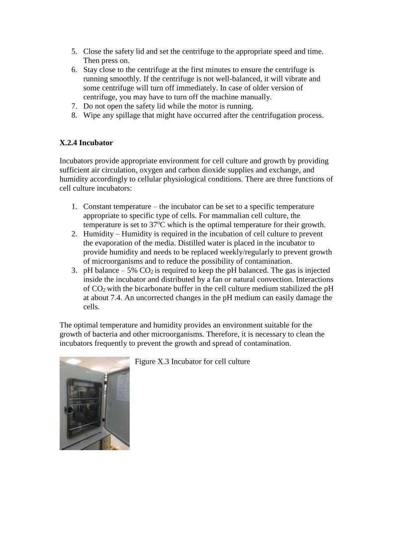
X.2.4 Incubator
Incubators provide appropriate environment for cell culture and growth by providing
sufficient air circulation, oxygen and carbon dioxide supplies and exchange, and
humidity accordingly to cellular physiological conditions. There are three functions of
cell culture incubators:
1. Constant temperature – the incubator can be set to a specific temperature
appropriate to specific type of cells. For mammalian cell culture, the
temperature is set to 37oC which is the optimal temperature for their growth.
2. Humidity – Humidity is required in the incubation of cell culture to prevent
the evaporation of the media. Distilled water is placed in the incubator to
provide humidity and needs to be replaced weekly/regularly to prevent growth
of microorganisms and to reduce the possibility of contamination.
3. pH balance – 5% CO2 is required to keep the pH balanced. The gas is injected
inside the incubator and distributed by a fan or natural convection. Interactions
of CO2 with the bicarbonate buffer in the cell culture medium stabilized the pH
at about 7.4. An uncorrected changes in the pH medium can easily damage the
cells.
The optimal temperature and humidity provides an environment suitable for the
growth of bacteria and other microorganisms. Therefore, it is necessary to clean the
incubators frequently to prevent the growth and spread of contamination.
Figure X.3 Incubator for cell culture

X.2.5 37oC water bath
Culture media and supplementations are usually kept in the 4oC refrigerator, whereas
the cells are kept in the 37oC incubator. In order to prevent abrupt temperature
elevation, the media and reagents are warmed up in the water bath prior use.
The warm water in the water bath is an ideal environment for the growth of
microorganism and other contaminants. Therefore the water bath need to be cleaned
and the water need to be replaced routinely. Bottles and containers that have been
warmed up in the water bath must be wiped down carefully with 70% ethanol before
being transferred into the hood.
X.2.6 Refrigerator and freezer
Most reagents and solutions used for cell culture are kept in the refrigerator for short
term storage. Some reagents are kept in the -20oC freezer for longer term storage. Cell
culture facilities often have -80oC freezer to store reagents and cryopreserved cells.
For long term storage, cells are kept in liquid nitrogen tanks of the temperature -
180oC. Cells can be kept frozen in liquid nitrogen for many years. Extreme cold
temperature of liquid nitrogen is hazardous and may cause burn to skin when exposed.
Therefore, thick gloves must be worn when handling the tank with great care.
X.2.7 Biohazard waste container
Hazardous and potentially hazardous materials must be disposed properly and
accordingly in biohazard waste container. The materials need to be sterilized before
the disposal. The biohazard waster must be handled accordingly to the procedures and
municipal council, federal and institutional law. It is very important to adhere with the
rules to prevent the spread of potentially hazardous materials to the environment.
X.3 Mammalian cell culture
X.3.1 Media Preparation
Cell culture is a method of multiplying cells under sterile and controlled laboratory
condition. It is used by the scientists to study cellular biological activities, functions,
morphologies and behavior of cells. The most important factor influencing the
mammalian cell culture is the choice of the culture medium as the cells required an
abundant source of easy to use nutrients for their viability and growth4. The
formulations for the medium should mimic natural conditions of systemic interactions
that normally occur in the body of an organism to support their biological activities
and viability. There are many different types of culture media are available in the
market, in which vary accordingly to their sugar, protein, minerals supplemented with
animal serums, growth factors and hormones as different cell types may favor
different formulations for optimum growth and differentiation5.
Most cells in are grown in a basal media containing nutrient, vitamins and mineral
supplemented with animal serums of different type such as horse, calf and fetal
bovine, This type of media is called ‘undefined media’ due to its unknown exact
components of the supplemental serum. Examples of readily available cell culture
medium are Dulbecco’s Modified Eagle’s Medium (DMEM)6, α-Eagle’s Minimal
Essential Medium (α –MEM)4, and RPMI 16407.
All basal media should contain the following components:
Source of energy/carbon – glucose/glutamine
The building blocks of proteins – amino acids
Supplementation to support cell survival and growth – vitamin
Isotonic mixture of ions to act as cofactors for enzymatic reactions and cellular
activities – balanced salt solution
pH indicator that changes from orange/red at pH 7-7.4 to yellow at acidic
lower pH and purple at basic higher pH environment,
Buffer – HEPES or bicarbonate to maintain a balanced pH in the media.
In the preparation of ‘complete media’, supplementations of serum is added into the
basal media. Antibiotics such as Penicillin and Streptomycin and fungicides (i.e.
Fungizone) may be added to prevent bacterial and fungi growth. This supplementation
is however not recommended due to increase susceptibility of antibiotic resistant
bacteria/fungi. Alternatively, glutamine, epithelial growth factor and cholera toxin can
be added into the basal media and is said to be ‘defined media’. This defined media
can be customized and selective for specific cell type and experimental conditions.

X.3 Thawing and recovering mammalian cells from cryopreservation
Cells thawing from cryopreservation is the most critical procedure in the cell culture
routine that has to be done quickly with care. Cryopreserved cells are kept in
complete media with 5-10% of dimethyl sulfoxide (DMSO) to prevent crystallization
and breakage of the frozen cellular components. Although DMSO is harmful to cells
in culture medium it helps to maintain cellular membrane during the freezing-thawing
process. From this procedure, it is estimated that only 50% of the cryopreserved cells
will survive. Therefore, higher number of cells (>1,000,000 cells/ml) in the
cryopreservation will have higher chance for cell survival.
Materials and Equipment
1. Cryopreserved cultures of cells
2. Complete media (Dulbecco’s Modified Eagle’s Medium, DMEM supplemented
with 10% fetal bovine serum, FBS), 37°C
3. 70% ethanol
4. 25cm2 T-flasks or 60-mm petri plates, sterile
5. Sterile centrifugal tube
6. Sterile serological pipettes
7. Incubator with 5% CO2 and 37°C
8. Cooling centrifuge, 4oC
9. Water bath, 37oC
Method
1. Remove the cryopreserved vials from the -80oC freezer or the liquid nitrogen
tank. Examine the labels – cell type, passage number and date of storage to
ensure that you have the correct cell line. Place the frozen vials in a small
container in the 37oC water bath and make sure that the caps do not get wet to
avoid possible contamination. The procedure should be done very quickly
(about 1-2 minutes) to prevent the breakage of cellular membrane due to the
formation of DMSO crystallization.
2. Remove the vials from the water bath and wipe down well with 70% ethanol.
The vials then should be quickly transferred into a fume hood and mixed well
with 9 ml of a complete cell culture medium in a 15 ml tube.
3. The tubes should be closed properly and labelled. Then using a balanced tube,
centrifuge the cells at about 120g (1000-1500 rpm) in a cooling centrifuge (4°C)
for about 3 minutes.
4. The heavy cells will form a pallet and are separated from the media
(supernatant)
5. Using a sterile pipette, remove the supernatant carefully without affecting the
pellet which contains the cells at the bottom side of the tube.
6. Then, using another sterile glass pipette, resuspend the cell pellet with 5 ml of
a complete medium and transfer the mixture into a culture plate or 25cm2 T-
flask containing 5 ml complete medium.
7. Swirl the flask gently and be careful not to wet the flask’s cap.
8. Observe the cells using an inverted microscope. You should observe plenty of
floating cells in the media.
9. Place the flask in an incubator with 5% CO2 at 37°C.

10. Cells may take up about 5-24 hours or more to attach to the bottom of the flask
and to start dividing. Many cells may not survive the freezing-thawing process
and will not attach onto the flask. Therefore, after 24 hours, the cell culture
media should be changed and replenish with a new culture media.
Notes and Tips
Protective clothing, gloves and goggles, should be worn while removing the vials
from -80oC freezer and liquid nitrogen tank.
X.4 Detachment and subculturing of mammalian cells from flask/plates.
When a cell line is cultured, a lag period after seeding usually followed by
exponential growth, called the log phase. When the cell density reaches a level such
that all the available cell growth area is occupied (100% confluence), or when the cell
concentration exceeds the capacity of the medium, the growth ceases or is greatly
decreases, Then either the medium should be changed or the culture must be divided
(subculture). X.4.1 and X.4.2, subculturing of two types of culture, monolayer and
suspension culture, respectively are discussed.
X.4.1 Subculture of cells in monolayer cultures
The first step in subculturing cells from monolayer cell culture is to detach cells from
the surface of the primary culture vessel by the use of accutase, trypsine or
mechanical means. This is followed by transferring fraction of the detached cells to
new culture medium to grow inside new culture vessel.
Materials and Equipment
1. 100% confluence cultures of cells
2. Complete media (DMEM supplemented with 10% FBS), 37°C
3. Sterile serological pipettes
4. Incubator with 5% CO2, 37°C
5. Cooling centrifuge, 4oC
6. 25cm2 T-flasks or 60-mm petri plates, sterile
7. PBS (Phosphate Buffer Saline), 37°C
8. Accutase or trypsin
Method
1. Remove the medium from the 100% confluence cultures of cells with a sterile
Pasteur pipette. Wash the monolayer cells with 5 ml (for culture in 25cm2 flask)
of 37°C PBS to remove the residual FBS.
2. Add 1 ml of 37°C Accutase/trypsin solution to the culture which cover adhering
cell layer.
3. Place plate in incubator with 5% CO2 at 37°C for 2 to 3 minutes. Check culture
with microscope to make sure that cells are rounded up and detached from the
surface.
4. Add 8 ml of 37°C complete medium. Draw cell suspension into a Pasteur pipet
and rinse the cell layer three or four times to dissociate remaining adherent cells.
Transfer cell suspension to centrifuge tube (sterile) and centrifuge it for 5-6

minutes at 120g cooling centrifuge (4°C). Discard the supernatant and
resuspend the cell pellet with complete media.
5. Add an equal volume of cell suspension (1 ml) to fresh culture vessels that are
well labeled.
6. Add 4 ml fresh complete media to each culture and incubate them in a 37°C,
5% CO2 incubator.
7. In some cases, feed cultures after 3 or 4 days by removing old media and adding
fresh media.
8. Passage the culture when it becomes confluent by repeating steps 1 to 6.
Notes and Tips
- Cells can be counted using a hemacytometer then diluted to the desired density
so a specific number of cells can be added to each culture vessel. - For primary cultures and early subcultures, 60-mm petri plates or 25-cm2
flasks are generally used; larger vessels (e.g., 150-mm plates or 75-cm2 flasks)
may be used for the following subcultures. - Culture vessels should be labeled with the passage number and date of
subculture. - For culturing cells using 75 cm2 culture flasks, 9 ml medium should be added
per flask. - CO2 incubators with 5% CO2 and 4% O2 should be used because the low
oxygen concentration simulate the in vivo environment of cells and enhance
cell growth.
X.4.2 Subculture of cells in suspension cultures
A suspension culture requires the same incubation conditions as in the monolayer
culture. Fortunately, subculturing of suspension cultures is less complicated than
subculturing of monolayer cultures because the cells are not adhering to the vessel
surface. There is no necessity for the detachment or dispersion prior to subculturing.
Materials
1. 100% confluence cultures of cells
2. Complete media (DMEM supplemented with 10% FBS), 37°C
3. Sterile serological pipettes
4. Incubator with 5% CO2 and 37°C
5. 25 cm2 T-flasks, sterile
6. 70% (v/v) ethanol
7. Isopropanol
8. Mr. Frosty container
Method
1. Cells should be fed every 2 to 3 days as follows until the cultures reaches
confluency:
a. Take out the flask of suspension cells from the CO2 incubator, making
sure not to disturb cells that have settled at the 25cm2 T-flasks bottom.
b. Remove and discard about one-third of the medium (under sterile
conditions) from flask and replace with an equal volume of (37°C)
medium. Gently stir the flask to homogenize cells with the media.
c. Return flask to the incubator. If there is <20 ml of medium in the flask,
the flask should be incubated in horizontal position to assist
cell/medium contact. On higher volume of media, vertical incubation
of flask can be applied.
2. Culture should be checked regularly. Gentle swirling and stirring should be
applied to resuspend cells.
3. When suspension cultures reaches confluency (∼2 × 10⁶cells/ml), subculturing
should be performed as follows:
a. Take out the flask from incubator and gently swirl flask in order to
distribute the cells evenly in the media.
b. Remove half of the volume of cell suspension and place into new
sterile flask.
c. Add 7 to 10 ml medium to each flask and return flasks to incubator.
X.5 Cryopreservation of mammalian cells in cell suspension
Cryopreservation of cells involve cell suspension in 10% DMSO in FBS or complete
medium to prevent crystallization and damage to the cells. To maintain cellular
viability, the cells has to be placed in a freezing container called Mr. Frosty (Nalgene
Ltd, USA) before being placed in the -80oC freezer. The container is filled with
isopropanol to prevent rapid cooling and slowly decreasing at the rate of -1oC per
minute. As many of the cells die in the process of freezing and thawing, higher
concentrations of 1x10⁶ cells/ml is preferred to ensure cell survival.
X.5.1 Cell cryopreservation from monolayer cultures
Materials
1. 100% confluence culture of cells
2. Complete media (DMEM supplemented with 10% FBS), 37°C
3. Freezing medium (FBS with 10% (v/v) DMSO)
4. Incubator with 5% CO2, 37°C
5. Cooling centrifuge, 4°C
6. 25 cm2 T-flasks or 60-mm petri plates, sterile
7. 70% (v/v) ethanol
8. PBS (Phosphate Buffer Saline), 37°C
9. Accutase or trypsin
10. Sterile serological pipettes.
11. Isopropanol
12. Mr. Frosty container

Method
1. Remove the cell culture medium from the flask with a sterile pipette. Wash the
monolayer cells with PBS twice (5 ml for 25 cm flask) to remove the residual
FBS.
2. Add 3 ml of accutase/trypsin solution to the culture to cover the attached cell
surface.
3. Place the flask in an incubator with 5% CO2 (37°C) for 2 to 3 minutes. Check
the cellular detachment from the surface with an microscope. Tap the side of
the flask and observe under the microscope again to make sure that all of the
cells arerounded up and detached from the surface.
4. Quench trypsin by adding 8 ml of a complete cell culture medium and mix well
by pipetting up and down to dissociate remaining adherent cells. Transfer cell
suspension to 15 ml centrifuge tube (sterile) and centrifuge it for 5-6 minutes at
120g cooling centrifuge (4°C). Discard the supernatant that contains accutase
or trypsin. Resuspend the cell pellet with a complete cell culture media.
5. Count cells using a hemacytometer, dilute with media to get a final cell
concentration of 106 or 107 cells/ml and transfer into a 15 ml tube.
6. Centrifuge the cell suspension at 120g for 3 minutes (4oC) to separate the cell
pellet from the supernatant. Then remove the supernantant and add 4 ml of cell
cryopreserved medium containing 10% DMSO in FBS. Mix the medium and
the pellet well by pipetting up and down slowly. Transfer 1 ml of the mixture
into a labelled 2 ml cryovials. (freezing cells from 25 cm2 flask requires 2 ml
freezing solution).
7. Place the vials in Mr. Frosty for overnight in a −80°C freezer before storage in
liquid nitrogen tank or in a -140oC freezer.

X.5.2 Cell cryopreservation of suspension cultures
The method of freezing cells from suspension culture is almost similar to freezing
cells from monolayer cultures. The major difference is that suspension cultures do not
requires cellular detachment from the surface using accutase or trypsin.
Materials
1. Suspension cultures of cells
2. Complete media (DMEM supplemented with 10% FBS), 37°C
3. Freezing medium (FBS with 10% (v/v) DMSO
4. Cooling Benchtop clinical centrifuge (4°C)
5. 25 cm2 T-flasks or 60-mm petri plates, sterile
6. 70% (v/v) ethanol
7. Sterile serological pipettes.
8. Isopropanol
9. Mr. Frosty container
Method
1. Transfer the cell suspension to 15 ml centrifuge tube (sterile) and centrifuge
it for 5-6 minutes at 120g cooling centrifuge (4°C). Discard the supernatant
and resuspend the cell pellet with a known volume of complete cell culture
media for cell counting.
2. Count cells using a hemacytometer, dilute with media to get a final cell
concentration of 106 or 107 cells/ml and transfer into a 15 ml tube.
3. Centrifuge the cell suspension at 120g for 3 minutes (4oC) to separate the
cell pellet from the supernatant. Then remove the supernantant and add 4 ml
of cell cryopreserved medium containing 10% DMSO in FBS. Mix the
medium and the pellet well by pipetting up and down slowly. Transfer 1 ml
of the mixture into a labelled 2 ml cryovials. (freezing cells from 25 cm2
flask requires 2 ml freezing solution).
4. Place the vials in Mr. Frosty for overnight in a −80°C freezer before storage
in liquid nitrogen tank or in a -140oC freezer.
Notes and tips
Styrofoam boxes can be used as substitute to Mr.Frosty (with isopropanol) for
freezing cells in a -80oC freezer for overnight. The cryovials should be
covered at all sides in wet wipes/tissues and placed in the container. Label
with your name, date, cell type and passage.
For routine freezing procedure, estimation of cell concentration can be
predicted. Normally, for 100% confluent T25 flask, >4x10⁶ can be harvested
and placed in 4 cryovials with 4 ml of cryopreserved medium. (Note that the
cell concentration varies with cell types)
Freezing media of DMSO and FBS can be stored in a refrigerator.
DMSO is light sensitive. Cover the freezing media with aluminium foil to
keep it in the dark.

X.6 Mammalian cell counting
Cell counting is the most important step in standardization of cell culture.
Hemacytometer is a device invented by Louis-Charles Malasses 8 to measure cell
concentration. It is made of a glass slide with a grid on each half (Figure X.7). Each
grid is made of nine squares and each square is subdivided into smaller squares. The
grid is covered by a cover slip and creates a chamber which can hold up to 0.1 μl of
liquid. It is very important to use the hemacytometer cover which is thick and evenly
surfaced cover slip because ordinary cover slips have uneven surfaces that may cause
errors in cell counting. In order to count, a small sample (0.1 μl) of known volume of
trypsinized cells will be placed in the hemacytometer chamber. Since the volume in
each square is known, therefore, the concentration of cells can be calculated.
Materials
1. Suspension cultures of cells
2. 0.4% (w/v) trypan blue
3. Hemacytometer with coverslip.
4. Hand counter
5. 70% (v/v) ethanol
Method
1. Prepare hemacytometer by cleaning surface of hemacytometer slide and
coverslip with 70% ethanol.
2. Prepare cell suspension (preparation of cell suspension depends on the type of
culture (monolayer or suspension) by following the protocols above).
3. Stain cells with trypan blue (to determine cell viability) by adding an equal
volume of trypan blue to cell suspension on an ordinary slide. Mix thoroughly
using pipette and let stand 5 min before loading hemacytometer.
4. Loading the hemacytometer using a sterile Pasteur pipette to transfer cell
suspension to the edge of hemacytometer counting chamber. Hold tip of pipette
under the coverslip and dispense one drop of cell suspension.
5. Allow the cells to settle for a 2-3 minutes before beginning to count and remove
excess liquid.
6. Using the hand counter, count cells in the four corners and central squares of
one counting chamber and also other counting chamber of the hemacytometer
(Figure 1).
7. Calculate cell number by determine cells per milliliter using the following
calculations:
cells/ml = average count per square *dilution factor *104.

Notes and Tips
The hemacytometer is a non sterile equipment. The pipette that was used to
transport cells to the hemacytometer should not be reused in the original
suspension.
Hemacytometers are fragile and expensive, please be careful not to drop or
break them.
Settlement of cells at the bottom of the flask and tubes tend to form clumps. It
is very important to mix the cells well by pipetting up and downwards to
dissociate the clumps and to ensure homogenous cell suspension.
Cells at outside incubator and trypsinized are under stress and its viability may
deteriorate over time. It is important to speed up the procedure rather than be
accurate in cell counting.
Observe your cells in the culture prior trypsinization to ensure higher number
of cells for calculation.
Trypan blue is a blue stain that can transverse inside the dead cells through
their porous membranes. Live cells do not allow the stain to get in, therefore
the dead cells will appear dark blue as opposed to the clear live cells.

Glossary:
Aseptic techniques: Series of techniques and practices used to reduce the chances of
contamination
Basal media: Basic media required for cellular growth and metabolism that contains
basic nutrients, vitamins and mineral necessary for cell culture. The media needs to be
supplemented with additional growth hormones, growth factors and proteins for
efficient cell growth.
Bicarbonate buffer: The buffer used in cell culture media to keep the pH balance at
7.4 by interaction with CO2
Chelator: Molecules like Ethylene diamine tetra acetic acid (EDTA) that bind to
certain metal ions and inactivate the ions to prevent their interactions with other
elements
Complete-media: Basal media that contains additional proteins, growth factors and
components necessary for efficient cell growth for specific cell type.
Confluency: The percentage of the cell culture surface that is covered by the cells.
Cytotoxic studies: The measurement of cellular viability and indicator of cellular
metabolism when interacting with a toxic factor.
Defined media: Basal media supplemented with additional growth factors, proteins
and other components without the use of serum.
Density-dependent growth inhibition: Cell growth arrest due to insufficient space,
nutrients or oxygen.
Differentiation: A process when cellular enters maturity and specialized into specific
cells.
Extracellular matrix: Chemical components and structural support for the cellular
attachment, proliferation and differentiation in a tissue.
Hayflick’s limit: The limit for normal cells in culture to divide.
Undefined media: A basal media with serum supplementation without
acknowledging the exact components of the serum.

References
1. Hayflick, L. & Moorhead, P. S. The serial cultivation of human diploid cell
strains. Exp. Cell Res. 25, 585–621 (1961).
2. Masters, J. R. HeLa cells 50 years on: the good, the bad and the ugly. Nat. Rev.
Cancer 2, 315–319 (2002).
3. Ozturk, S. & Hu, W.-S. Cell culture technology for pharmaceutical and cell-
based therapies. (CRC Press, 2005).
4. Eagle, H. Amino acid metabolism in mammalian cell cultures. Science (80-. ).
130, 432–437 (1959).
5. Freshney, R. I. Culture of specific cell types. (Wiley Online Library, 2005).
6. Dulbecco, R. & Freeman, G. Plaque production by the polyoma virus. Virology
8, 396–397 (1959).
7. Moore, G. E., Gerner, R. E. & Franklin, H. A. Culture of normal human
leukocytes. Jama 199, 519–524 (1967).
8. Verso, M. L. Some nineteenth-century pioneers of haematology. Med. Hist. 15,
55–67 (1971).


![Application of rDNA in animal cell culture [Animal Biotech]](https://img.pdfslide.us/doc/110x75/546a7726b4af9f83288b499c/application-of-rdna-in-animal-cell-culture-animal-biotech.jpg)