Embed Size (px)
Citation preview
ANIMAL TRANSPORT SYSTEM
CHAPTER 6
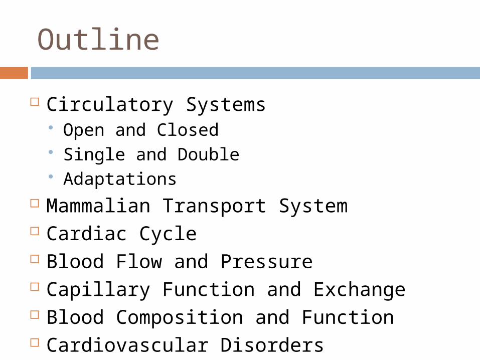
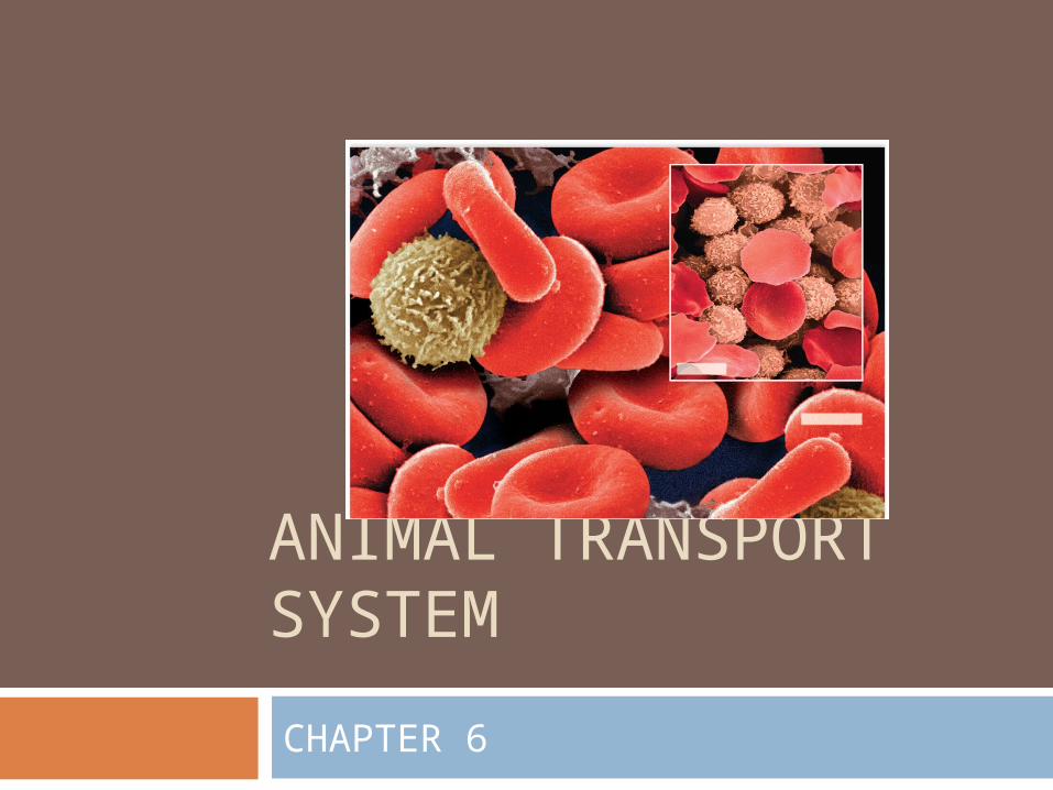
Outline
Circulatory Systems Open and Closed Single and Double Adaptations
Mammalian Transport System Cardiac Cycle Blood Flow and Pressure Capillary Function and Exchange Blood Composition and Function Cardiovascular Disorders
Overview: Transport and Exchange
Every organism must exchange materials with its environment
Exchanges ultimately occur at the cellular level
In unicellular organisms, these exchanges occur directly with the environment
For most cells making up multicellular organisms, direct exchange with the environment is not possible
Gills are an example of a specialized exchange system in animals
Internal transport and gas exchange are functionally related in most animals
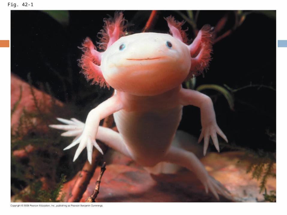
Fig. 42-1
Circulatory systems
In small and/or thin animals, cells can exchange materials directly with the surrounding medium
In most animals, transport systems connect the organs of exchange with the body cells
Most complex animals have internal transport systems that circulate fluid
Gastrovascular Cavities
Simple animals, such as cnidarians and some aquatic animals, have a body wall that is only two cells thick and that encloses a gastrovascular cavity
This cavity functions in both digestion and distribution of substances throughout the body
Some cnidarians, such as jellies, have elaborate gastrovascular cavities
Flatworms have a gastrovascular cavity and a large surface area to volume ratio
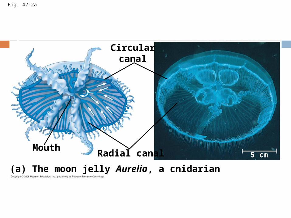
Fig. 42-2a
Circularcanal
Radial canalMouth
(a) The moon jelly Aurelia, a cnidarian
5 cm
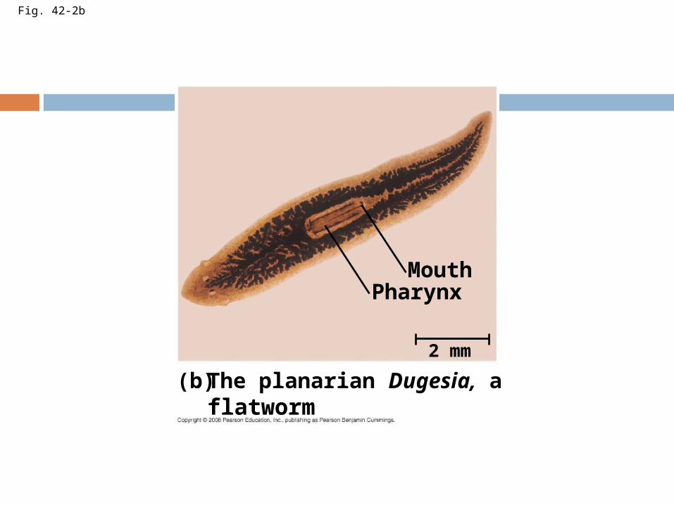
Fig. 42-2b
The planarian Dugesia, aflatworm
(b)
MouthPharynx
2 mm
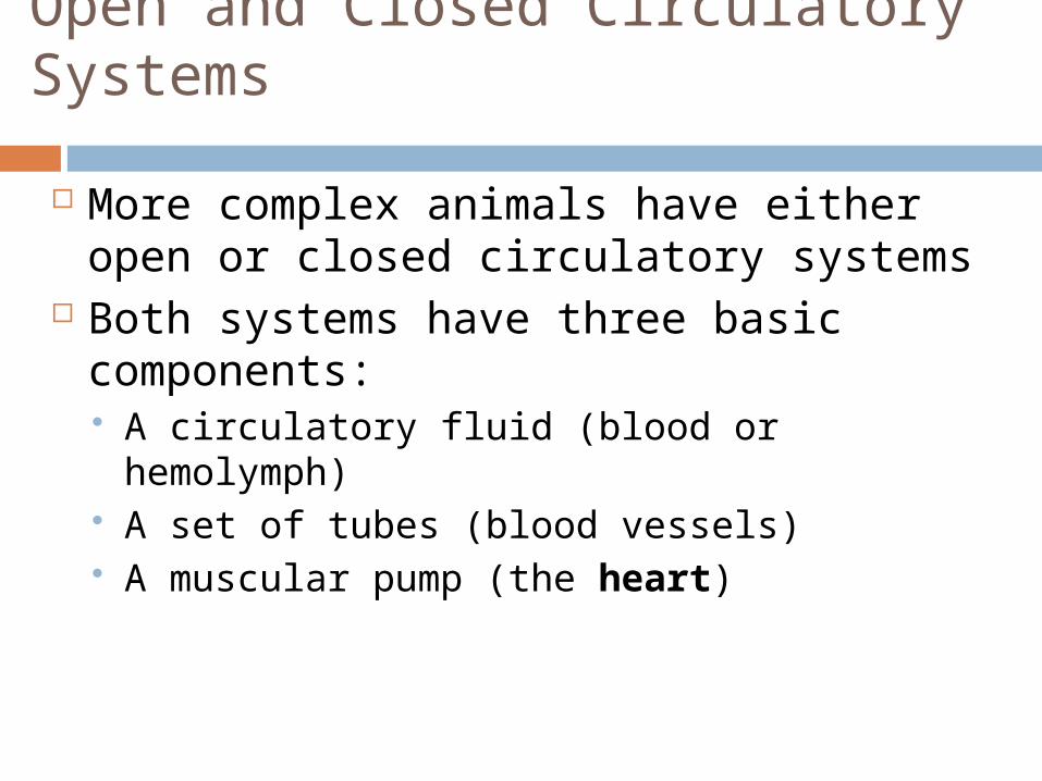
Open and Closed Circulatory Systems
More complex animals have either open or closed circulatory systems
Both systems have three basic components: A circulatory fluid (blood or hemolymph) A set of tubes (blood vessels) A muscular pump (the heart)
In insects, other arthropods, and most molluscs, blood bathes the organs directly in an open circulatory system
In an open circulatory system, there is no distinction between blood and interstitial fluid, and this general body fluid is more correctly called hemolymph
In a closed circulatory system, blood is confined to vessels and is distinct from the interstitial fluid
Closed systems are more efficient at transporting circulatory fluids to tissues and cells
Open and Closed Circulatory Systems
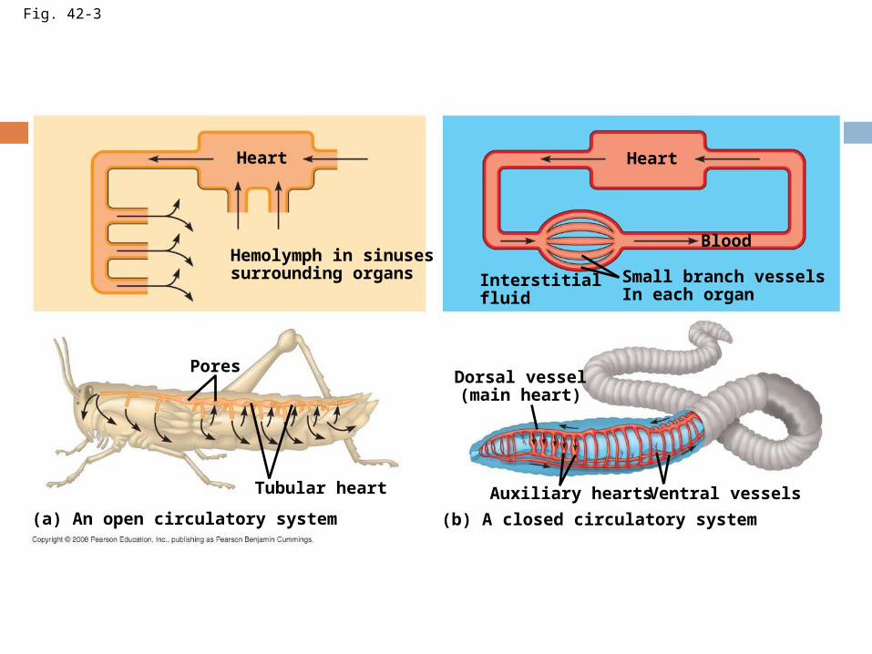
Fig. 42-3
Heart
Hemolymph in sinusessurrounding organs
Heart
Interstitialfluid
Small branch vesselsIn each organ
Blood
Dorsal vessel(main heart)
Auxiliary hearts Ventral vessels
(b) A closed circulatory system(a) An open circulatory system
Tubular heart
Pores
Organization of Vertebrate Circulatory Systems
Humans and other vertebrates have a closed circulatory system, often called the cardiovascular system
The three main types of blood vessels are arteries, veins, and capillaries
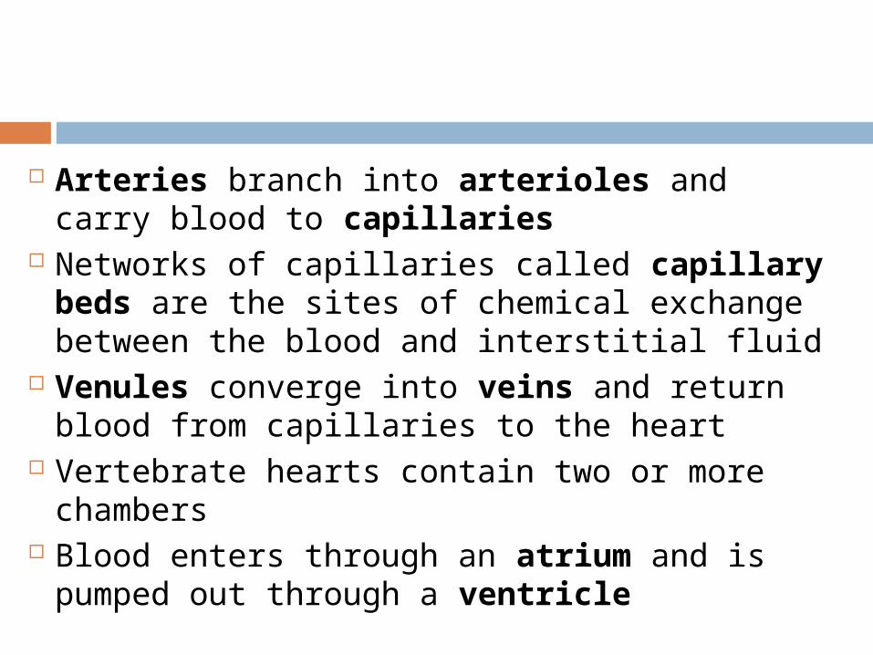
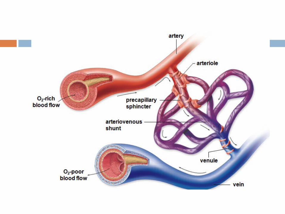
Arteries branch into arterioles and carry blood to capillaries
Networks of capillaries called capillary beds are the sites of chemical exchange between the blood and interstitial fluid
Venules converge into veins and return blood from capillaries to the heart
Vertebrate hearts contain two or more chambers
Blood enters through an atrium and is pumped out through a ventricle
Single Circulation
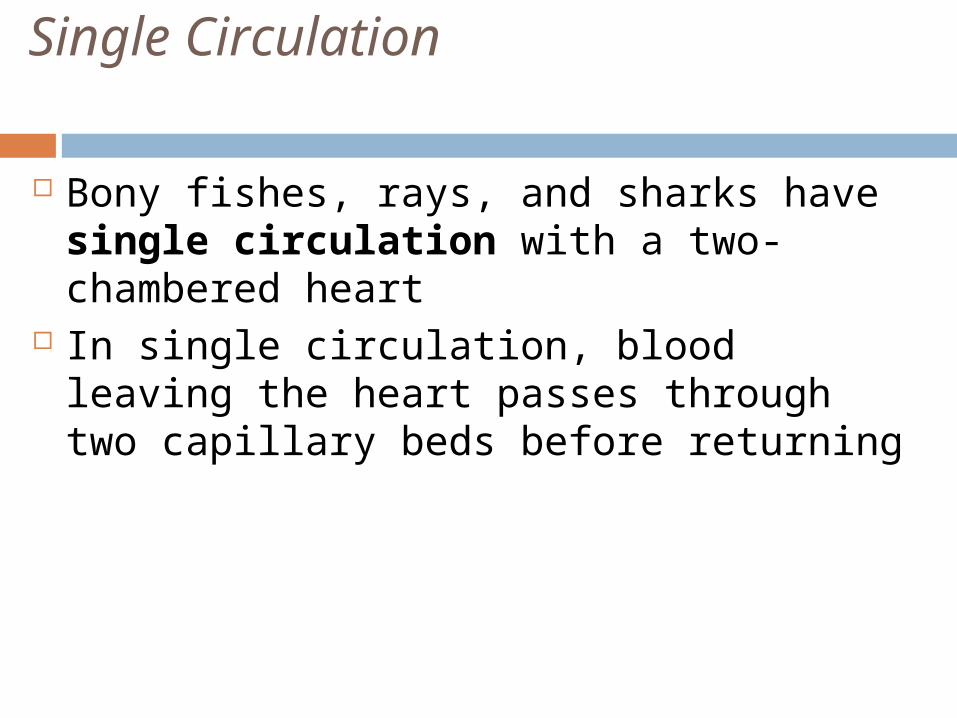
Bony fishes, rays, and sharks have single circulation with a two-chambered heart
In single circulation, blood leaving the heart passes through two capillary beds before returning
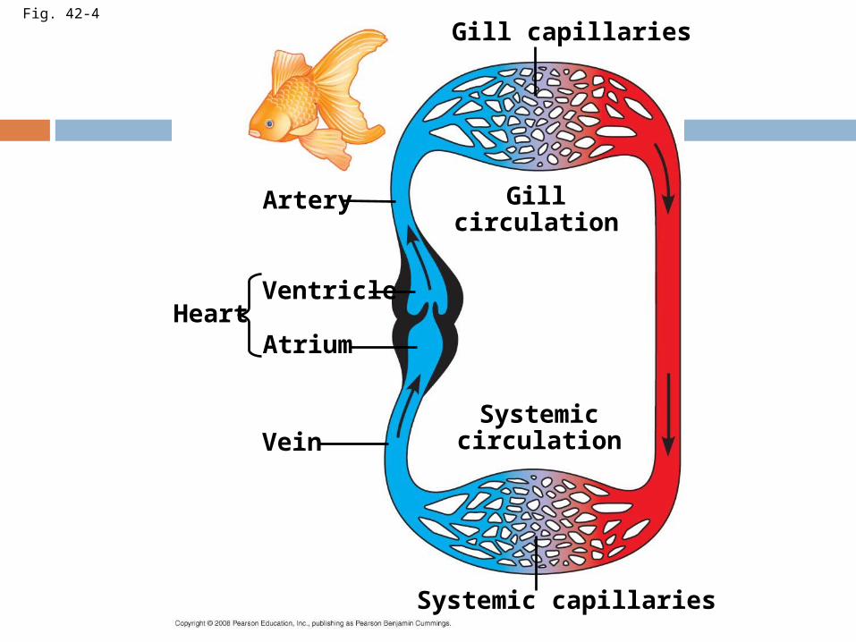
Fig. 42-4
Artery
Ventricle
AtriumHeart
Vein
Systemic capillaries
Systemiccirculation
Gillcirculation
Gill capillaries
Double Circulation
Amphibian, reptiles, and mammals have double circulation
Oxygen-poor and oxygen-rich blood are pumped separately from the right and left sides of the heart
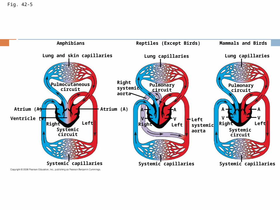
Fig. 42-5
Amphibians
Lung and skin capillaries
Pulmocutaneouscircuit
Atrium (A)
Ventricle (V)
Atrium (A)
Systemiccircuit
Right Left
Systemic capillaries
Reptiles (Except Birds)
Lung capillaries
Pulmonarycircuit
Rightsystemicaorta
Right LeftLeftsystemicaorta
Systemic capillaries
A A
VV
Systemic capillaries
Pulmonarycircuit
Systemiccircuit
Right Left
A A
VV
Lung capillaries
Mammals and Birds
In reptiles and mammals, oxygen-poor blood flows through the pulmonary circuit to pick up oxygen through the lungs
In amphibians, oxygen-poor blood flows through a pulmocutaneous circuit to pick up oxygen through the lungs and skin
Oxygen-rich blood delivers oxygen through the systemic circuit
Double circulation maintains higher blood pressure in the organs than does single circulation
Adaptations of Double Circulatory Systems
Hearts vary in different vertebrate groups
Amphibians Frogs and other amphibians have a three-
chambered heart: two atria and one ventricle
The ventricle pumps blood into a forked artery that splits the ventricle’s output into the pulmocutaneous circuit and the systemic circuit
Underwater, blood flow to the lungs is nearly shut off
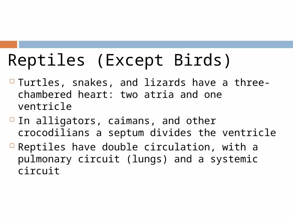
Reptiles (Except Birds) Turtles, snakes, and lizards have a three-
chambered heart: two atria and one ventricle
In alligators, caimans, and other crocodilians a septum divides the ventricle
Reptiles have double circulation, with a pulmonary circuit (lungs) and a systemic circuit
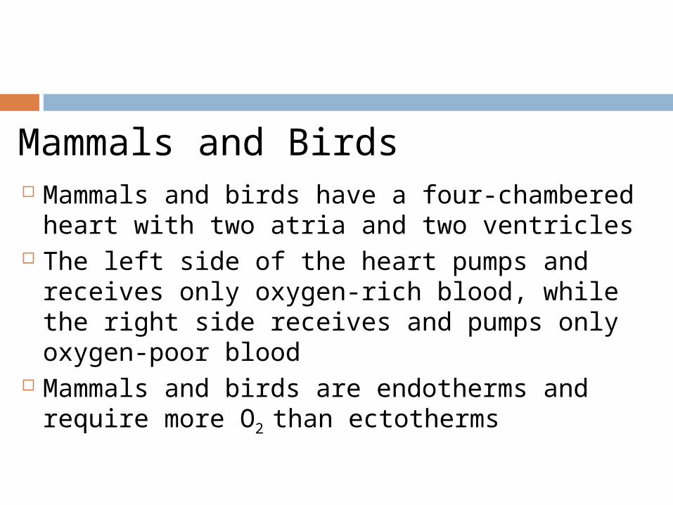
Mammals and Birds Mammals and birds have a four-chambered
heart with two atria and two ventricles The left side of the heart pumps and
receives only oxygen-rich blood, while the right side receives and pumps only oxygen-poor blood
Mammals and birds are endotherms and require more O2 than ectotherms
Mammalian Transport
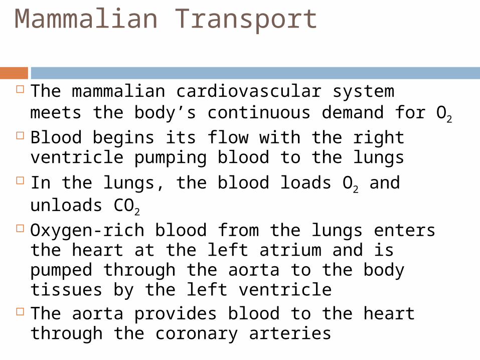
The mammalian cardiovascular system meets the body’s continuous demand for O2
Blood begins its flow with the right ventricle pumping blood to the lungs
In the lungs, the blood loads O2 and unloads CO2
Oxygen-rich blood from the lungs enters the heart at the left atrium and is pumped through the aorta to the body tissues by the left ventricle
The aorta provides blood to the heart through the coronary arteries
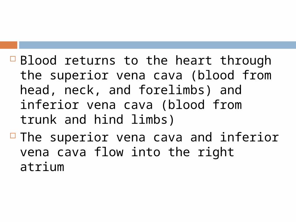
Blood returns to the heart through the superior vena cava (blood from head, neck, and forelimbs) and inferior vena cava (blood from trunk and hind limbs)
The superior vena cava and inferior vena cava flow into the right atrium
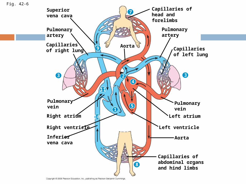
Fig. 42-6
Superiorvena cava
Pulmonaryartery
Capillariesof right lung
3
7
3
8
9
24
11
51
10
Aorta
Pulmonaryvein
Right atrium
Right ventricle
Inferiorvena cava
Capillaries ofabdominal organsand hind limbs
Pulmonaryvein
Left atrium
Left ventricle
Aorta
Capillariesof left lung
Pulmonaryartery
Capillaries ofhead andforelimbs
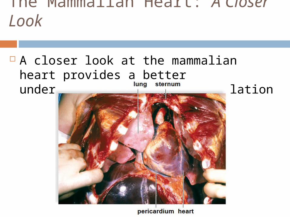
The Mammalian Heart: A Closer Look
A closer look at the mammalian heart provides a better understanding of double circulation
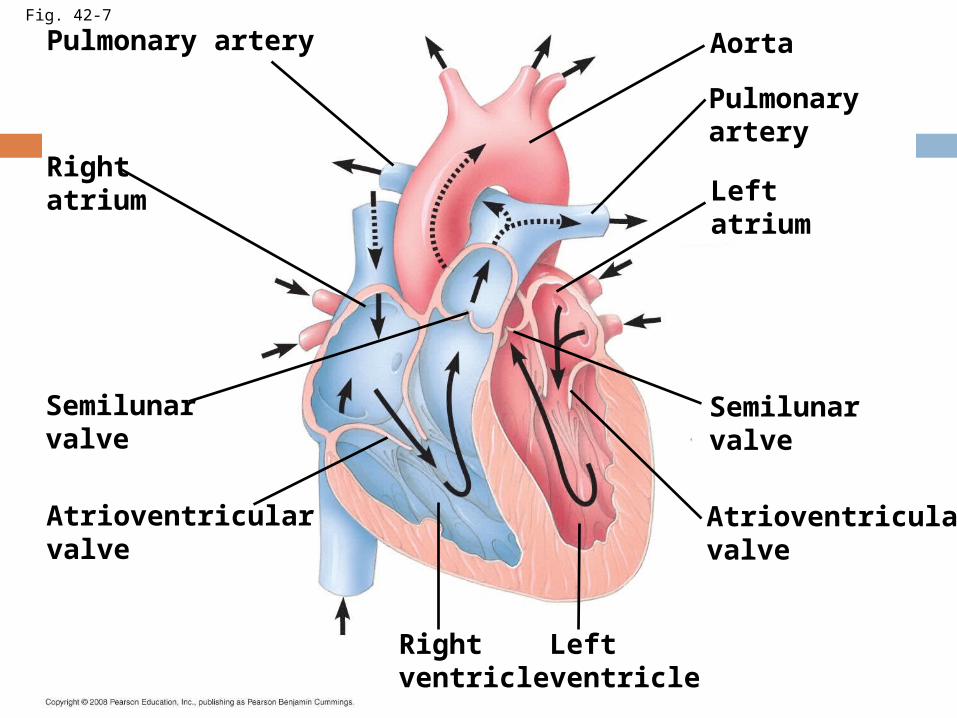
Fig. 42-7
Pulmonary artery
Rightatrium
Semilunarvalve
Atrioventricularvalve
Rightventricle
Leftventricle
Atrioventricularvalve
Leftatrium
Semilunarvalve
Pulmonaryartery
Aorta
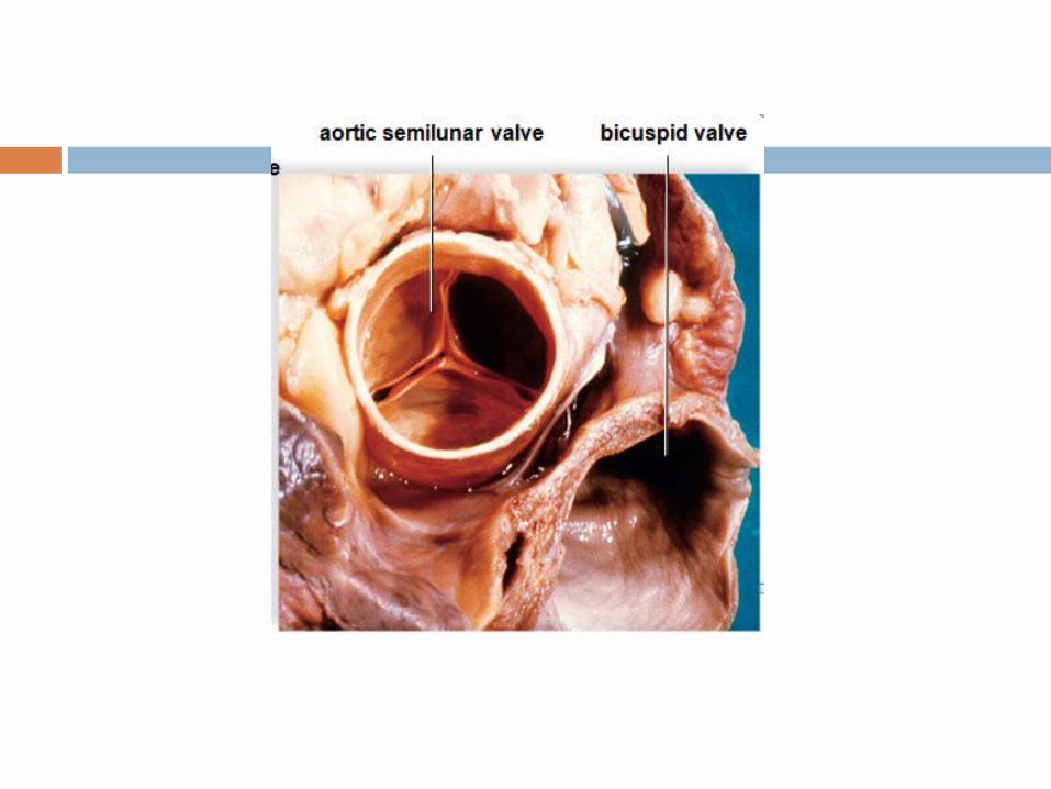
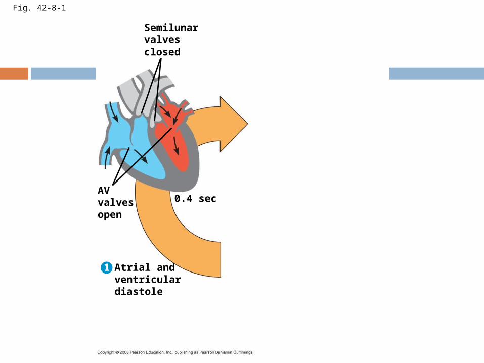
Fig. 42-8-1
Semilunarvalvesclosed
0.4 secAVvalvesopen
Atrial andventriculardiastole
1
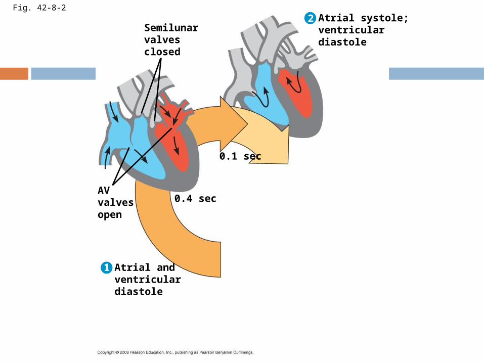
Fig. 42-8-2
Semilunarvalvesclosed
0.4 secAVvalvesopen
Atrial andventriculardiastole
1
2
0.1 sec
Atrial systole;ventriculardiastole
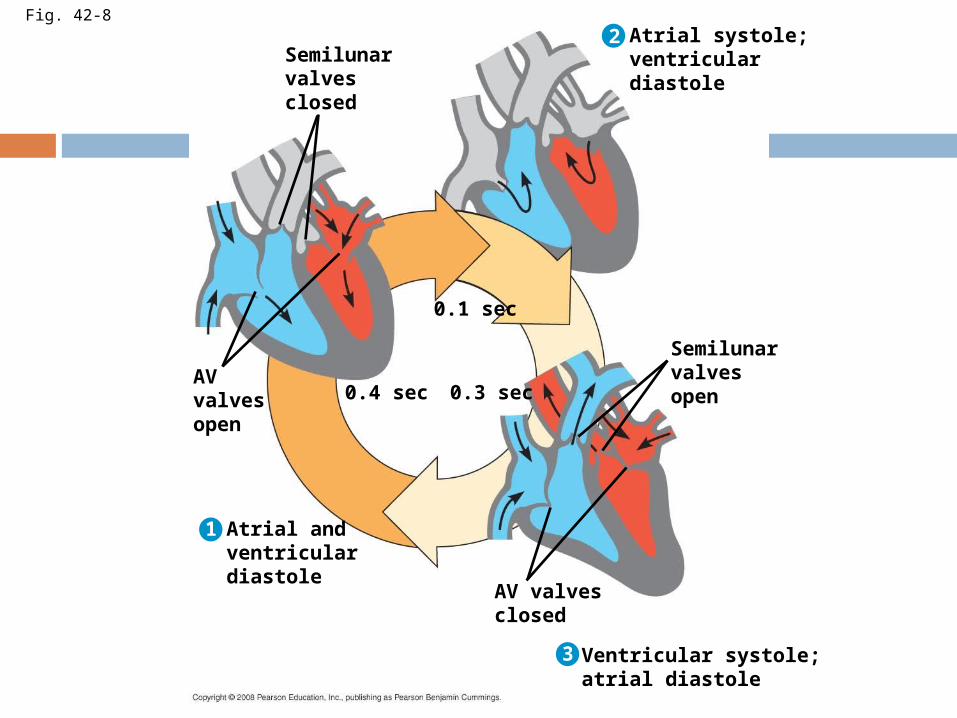
Fig. 42-8
Semilunarvalvesclosed
0.4 secAVvalvesopen
Atrial andventriculardiastole
1
2
0.1 sec
Atrial systole;ventriculardiastole
3
0.3 sec
Semilunarvalvesopen
AV valvesclosed
Ventricular systole;atrial diastole
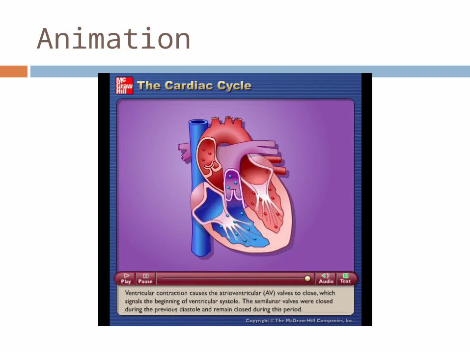
Animation
The heart rate, also called the pulse, is the number of beats per minute
The stroke volume is the amount of blood pumped in a single contraction
The cardiac output is the volume of blood pumped into the systemic circulation per minute and depends on both the heart rate and stroke volume
Four valves prevent backflow of blood in the heart
The atrioventricular (AV) valves separate each atrium and ventricle
The semilunar valves control blood flow to the aorta and the pulmonary artery
The “lub-dup” sound of a heart beat is caused by the recoil of blood against the AV valves (lub) then against the semilunar (dup) valves
Backflow of blood through a defective valve causes a heart murmur
Maintaining the Heart’s Rhythmic Beat
Some cardiac muscle cells are self-excitable, meaning they contract without any signal from the nervous system
The sinoatrial (SA) node, or pacemaker, sets the rate and timing at which cardiac muscle cells contract
Impulses from the SA node travel to the atrioventricular (AV) node
At the AV node, the impulses are delayed and then travel to the Purkinje fibers that make the ventricles contract
Impulses that travel during the cardiac cycle can be recorded as an electrocardiogram (ECG or EKG)
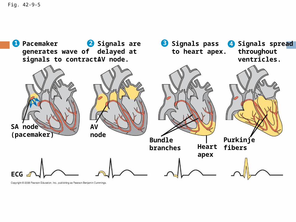
Fig. 42-9-5
Signals spreadthroughoutventricles.
4
Purkinjefibers
Pacemakergenerates wave ofsignals to contract.
1
SA node(pacemaker)
ECG
Signals aredelayed atAV node.
2
AVnode
Signals passto heart apex.
3
Bundlebranches Heart
apex
Blood Vessel Structure and Function
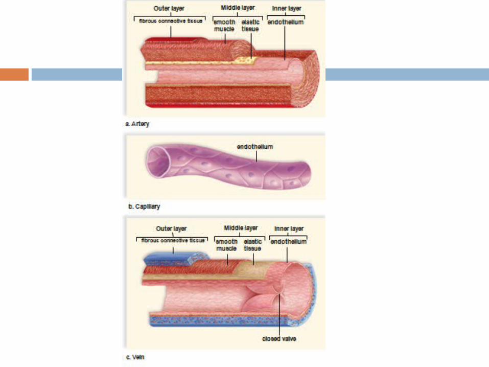
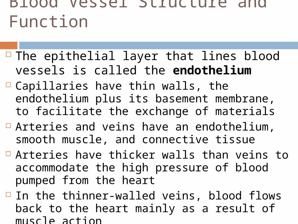
The epithelial layer that lines blood vessels is called the endothelium
Capillaries have thin walls, the endothelium plus its basement membrane, to facilitate the exchange of materials
Arteries and veins have an endothelium, smooth muscle, and connective tissue
Arteries have thicker walls than veins to accommodate the high pressure of blood pumped from the heart
In the thinner-walled veins, blood flows back to the heart mainly as a result of muscle action
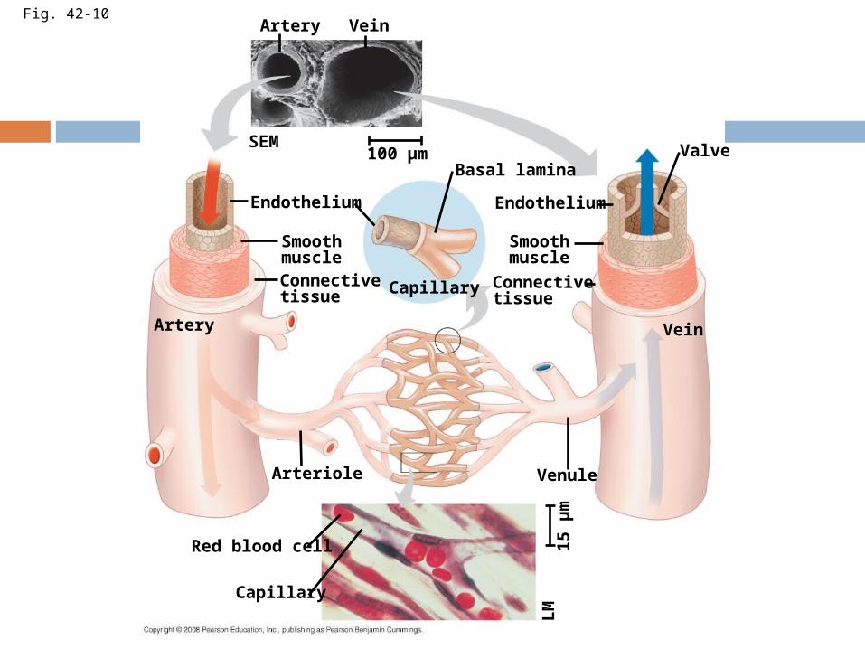
Fig. 42-10Artery Vein
SEM100 µm
Endothelium
Artery
SmoothmuscleConnectivetissue Capillary
Basal lamina
Endothelium
Smoothmuscle
Connectivetissue
Valve
Vein
Arteriole Venule
Red blood cell
Capillary
15 µ
mLM
Blood Flow Velocity
Physical laws governing movement of fluids through pipes affect blood flow and blood pressure
Velocity of blood flow is slowest in the capillary beds, as a result of the high resistance and large total cross-sectional area
Blood flow in capillaries is necessarily slow for exchange of materials
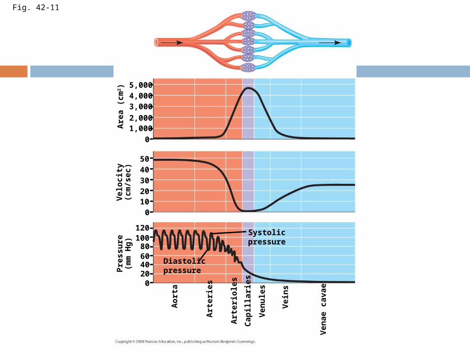
Fig. 42-11
5,0004,0003,000
2,0001,000
0
0
5040302010
120
80100
6040200
Are
a (
cm
2)
Velo
cit
y(c
m/s
ec)
Pre
ssu
re(m
m H
g)
Aort
a
Art
eri
es
Art
eri
ole
s
Cap
illa
ries
Ven
ule
s
Vein
s
Ven
ae c
avae
Diastolicpressure
Systolicpressure
Blood Pressure
Blood pressure is the hydrostatic pressure that blood exerts against the wall of a vessel
In rigid vessels blood pressure is maintained; less rigid vessels deform and blood pressure is lost
Changes in Blood Pressure During the Cardiac Cycle
Systolic pressure is the pressure in the arteries during ventricular systole; it is the highest pressure in the arteries
Diastolic pressure is the pressure in the arteries during diastole; it is lower than systolic pressure
A pulse is the rhythmic bulging of artery walls with each heartbeat
Regulation of Blood Pressure
Blood pressure is determined by cardiac output and peripheral resistance due to constriction of arterioles
Vasoconstriction is the contraction of smooth muscle in arteriole walls; it increases blood pressure
Vasodilation is the relaxation of smooth muscles in the arterioles; it causes blood pressure to fall
Vasoconstriction and vasodilation help maintain adequate blood flow as the body’s demands change
The peptide endothelin is an important inducer of vasoconstriction
Blood Pressure and Gravity
Blood pressure is generally measured for an artery in the arm at the same height as the heart
Blood pressure for a healthy 20 year old at rest is 120 mm Hg at systole and 70 mm Hg at diastole
Fainting is caused by inadequate blood flow to the head
Animals with longer necks require a higher systolic pressure to pump blood a greater distance against gravity
Blood is moved through veins by smooth muscle contraction, skeletal muscle contraction, and expansion of the vena cava with inhalation
One-way valves in veins prevent backflow of blood
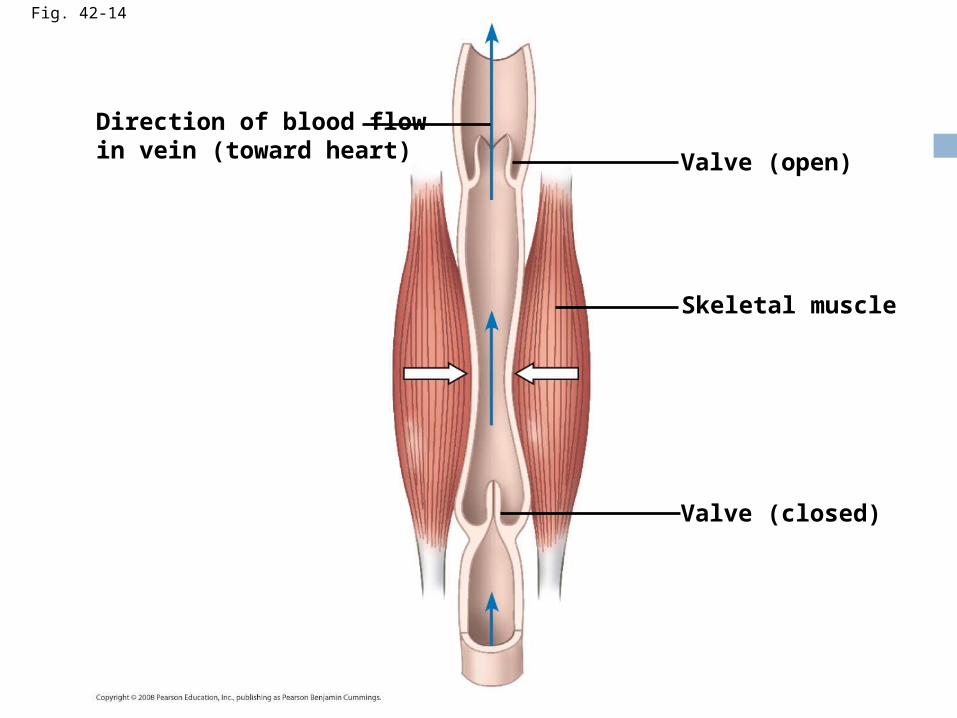
Fig. 42-14
Direction of blood flowin vein (toward heart) Valve (open)
Skeletal muscle
Valve (closed)
Capillary Function
Capillaries in major organs are usually filled to capacity
Blood supply varies in many other sites Two mechanisms regulate distribution of
blood in capillary beds: Contraction of the smooth muscle layer in the
wall of an arteriole constricts the vessel Precapillary sphincters control flow of blood
between arterioles and venules
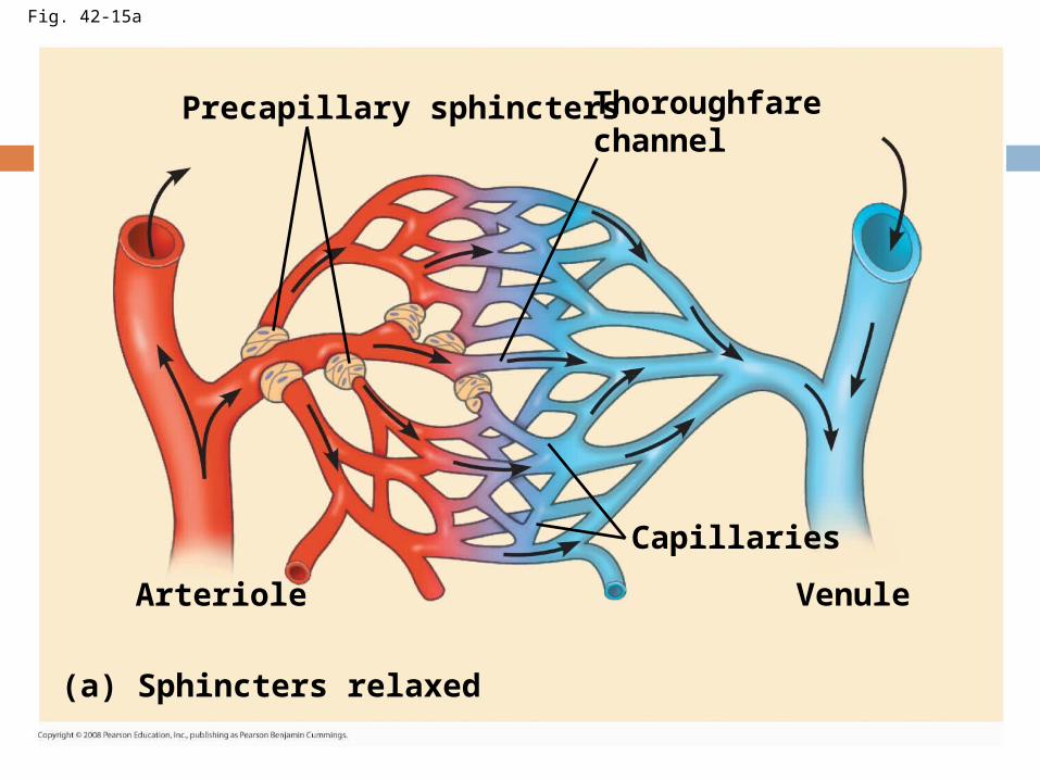
Fig. 42-15a
Precapillary sphinctersThoroughfarechannel
Arteriole
Capillaries
Venule
(a) Sphincters relaxed
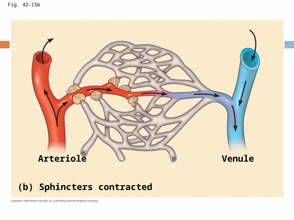
Fig. 42-15b
(b) Sphincters contracted
Arteriole Venule
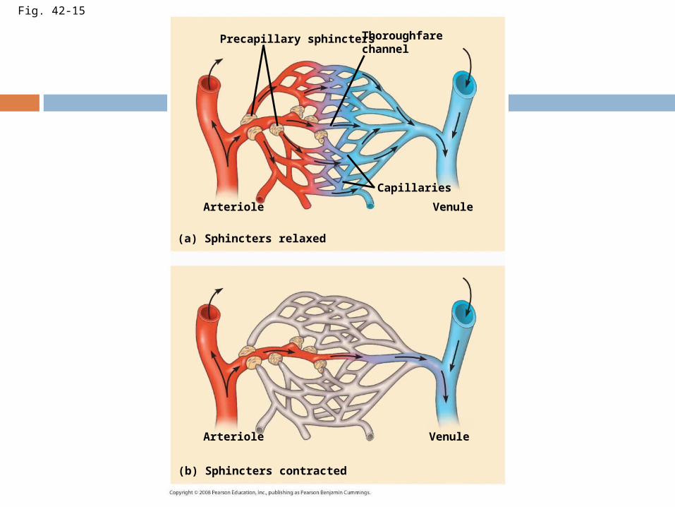
Fig. 42-15
Precapillary sphinctersThoroughfarechannel
Arteriole
Capillaries
Venule
(a) Sphincters relaxed
(b) Sphincters contracted
Arteriole Venule
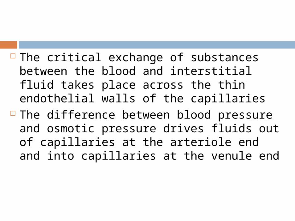
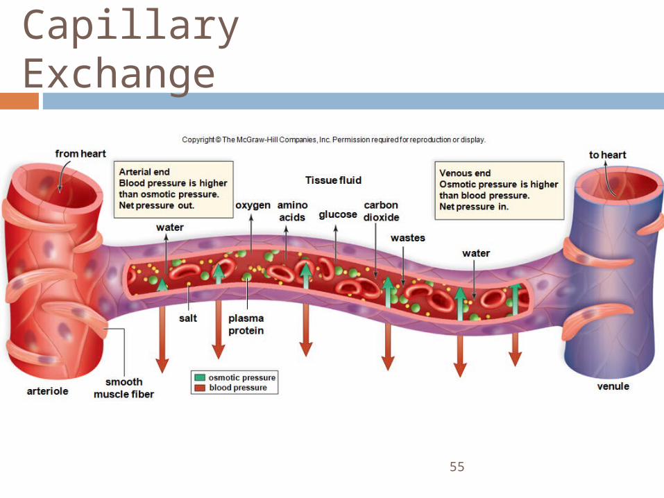
The critical exchange of substances between the blood and interstitial fluid takes place across the thin endothelial walls of the capillaries
The difference between blood pressure and osmotic pressure drives fluids out of capillaries at the arteriole end and into capillaries at the venule end
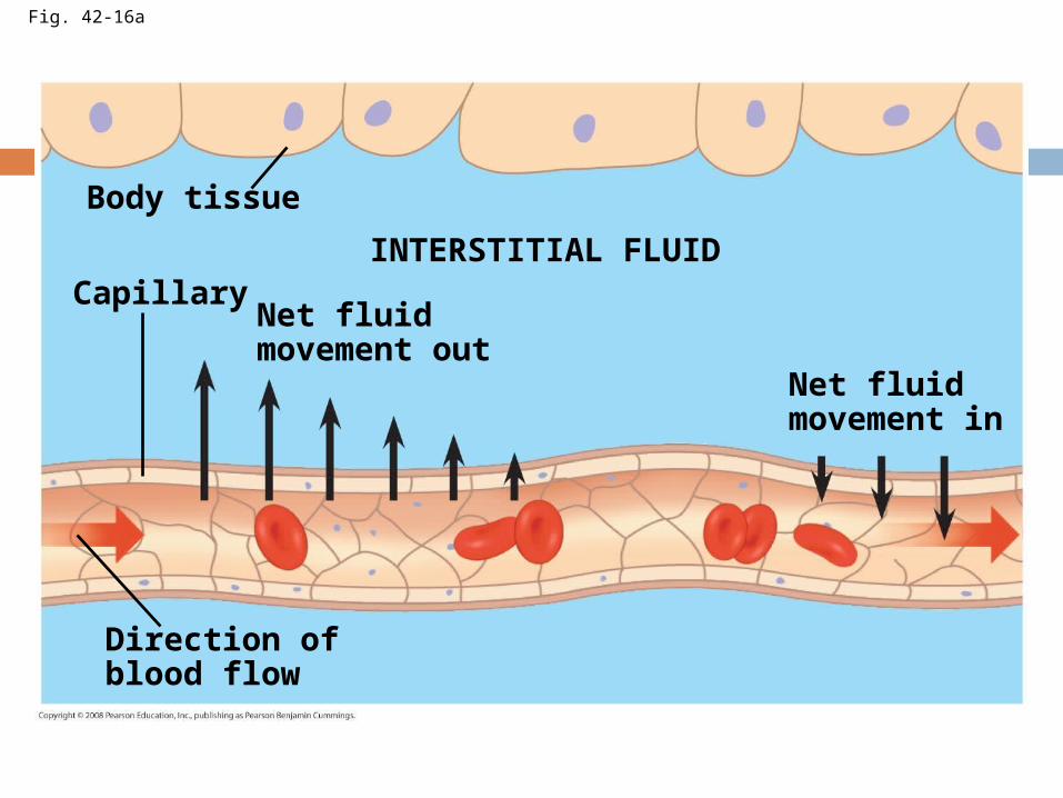
Fig. 42-16a
Body tissue
CapillaryNet fluidmovement out
INTERSTITIAL FLUID
Net fluidmovement in
Direction ofblood flow
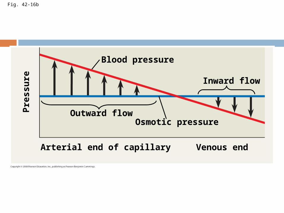
Fig. 42-16b
Blood pressure
Inward flow
Outward flowOsmotic pressure
Arterial end of capillary Venous end
Pre
ssu
re
55
Capillary Exchange
Copyright © The McGraw-Hill Companies, Inc. Permission required for reproduction or display.
venulearteriole
water
oxygenglucose
salt
water
wastes
osmotic pressureblood pressure
to heartfrom heart
Arterial endBlood pressure is higherthan osmotic pressure.Net pressure out. amino
acidscarbondioxide
Venous endOsmotic pressure is higherthan blood pressure.Net pressure in.
plasmaprotein
smoothmuscle fiber
Tissue fluid
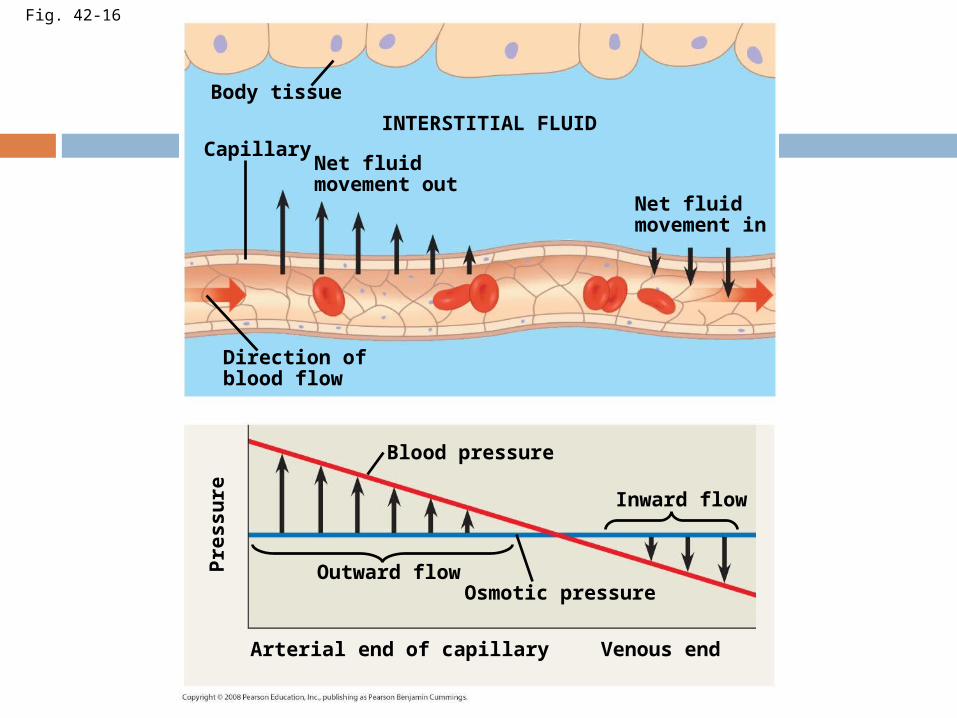
Fig. 42-16
Body tissue
CapillaryINTERSTITIAL FLUID
Net fluidmovement out
Direction ofblood flow
Net fluidmovement in
Blood pressure
Inward flow
Outward flowOsmotic pressure
Arterial end of capillary Venous end
Pre
ssu
re
Fluid Return by the Lymphatic System
The lymphatic system returns fluid that leaks out in the capillary beds
This system aids in body defense Fluid, called lymph, reenters the circulation
directly at the venous end of the capillary bed and indirectly through the lymphatic system
The lymphatic system drains into veins in the neck
Lymph nodes are organs that filter lymph and play an important role in the body’s defense
Edema is swelling caused by disruptions in the flow of lymph
Blood Composition and Function
In invertebrates with open circulation, blood (hemolymph) is not different from interstitial fluid
Blood in the circulatory systems of vertebrates is a specialized connective tissue
Blood consists of several kinds of cells suspended in a liquid matrix called plasma
The cellular elements occupy about 45% of the volume of blood
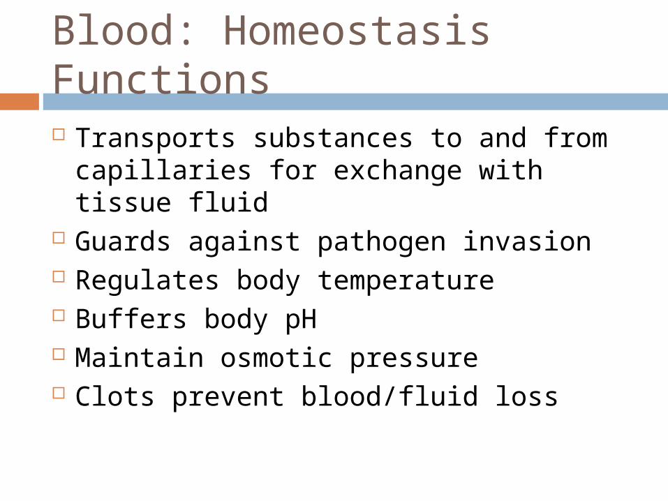
Blood: Homeostasis Functions Transports substances to and from
capillaries for exchange with tissue fluid Guards against pathogen invasion Regulates body temperature Buffers body pH Maintain osmotic pressure Clots prevent blood/fluid loss
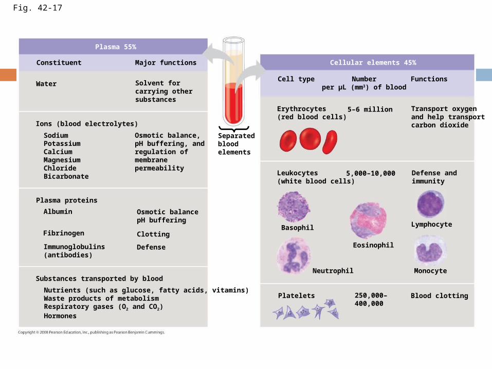
Fig. 42-17
Plasma 55%
Constituent Major functions
Water Solvent forcarrying othersubstances
Ions (blood electrolytes)
Osmotic balance,pH buffering, andregulation ofmembranepermeability
SodiumPotassiumCalciumMagnesiumChlorideBicarbonate
Osmotic balancepH buffering
Clotting
Defense
Plasma proteins
Albumin
Fibrinogen
Immunoglobulins(antibodies)
Substances transported by blood
Nutrients (such as glucose, fatty acids, vitamins)Waste products of metabolismRespiratory gases (O2 and CO2)Hormones
Separatedbloodelements
Cellular elements 45%
Cell type FunctionsNumberper µL (mm3) of blood
Erythrocytes(red blood cells)
5–6 million Transport oxygenand help transportcarbon dioxide
Leukocytes(white blood cells)
5,000–10,000 Defense andimmunity
Basophil
Neutrophil
Eosinophil
Lymphocyte
Monocyte
Platelets Blood clotting250,000–400,000
61
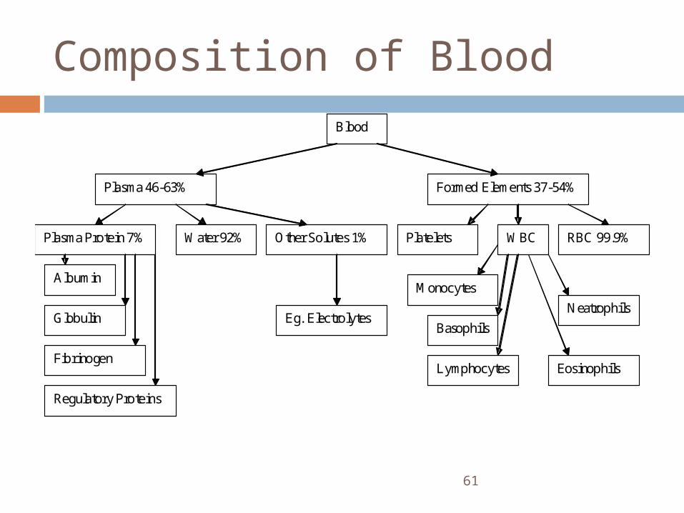
Composition of Blood
Blood
Plasma 46-63% Formed Elements 37-54%
Plasma Protein 7% Water 92% Other Solutes 1% Platelets RBC 99.9% WBC
Albumin
Fibrinogen
Globulin
Regulatory Proteins
Eg. Electrolytes
Monocytes
Basophils
Eosinophils
Neatrophils
Lymphocytes
Plasma
Blood plasma is about 90% water Among its solutes are inorganic salts in
the form of dissolved ions, sometimes called electrolytes
Another important class of solutes is the plasma proteins, which influence blood pH, osmotic pressure, and viscosity
Various plasma proteins function in lipid transport, immunity, and blood clotting

Cellular Elements
Suspended in blood plasma are two types of cells: Red blood cells (erythrocytes) transport
oxygen White blood cells (leukocytes) function in
defense Platelets, a third cellular element, are
fragments of cells that are involved in clotting
Red blood cells, or erythrocytes, are by far the most numerous blood cells
They transport oxygen throughout the body
They contain hemoglobin, the iron-containing protein that transports oxygen
Erythrocytes
Leukocytes There are five major types of white blood
cells, or leukocytes: monocytes, neutrophils, basophils, eosinophils, and lymphocytes
They function in defense by phagocytizing bacteria and debris or by producing antibodies
They are found both in and outside of the circulatory system
Platelets Platelets are fragments of cells and
function in blood clotting
Blood Clotting
When the endothelium of a blood vessel is damaged, the clotting mechanism begins
A cascade of complex reactions converts fibrinogen to fibrin, forming a clot
A blood clot formed within a blood vessel is called a thrombus and can block blood flow
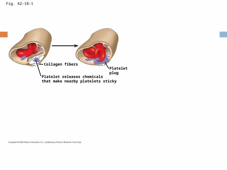
Fig. 42-18-1
Collagen fibersPlateletplug
Platelet releases chemicalsthat make nearby platelets sticky
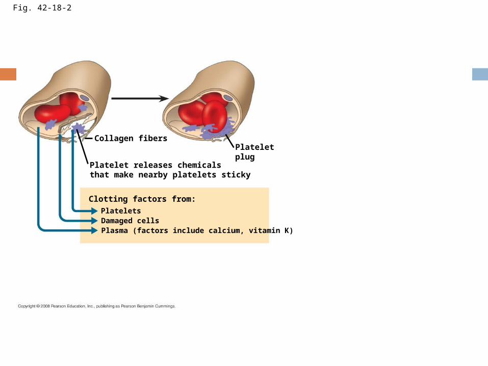
Fig. 42-18-2
Collagen fibersPlateletplug
Platelet releases chemicalsthat make nearby platelets sticky
Clotting factors from:PlateletsDamaged cellsPlasma (factors include calcium, vitamin K)
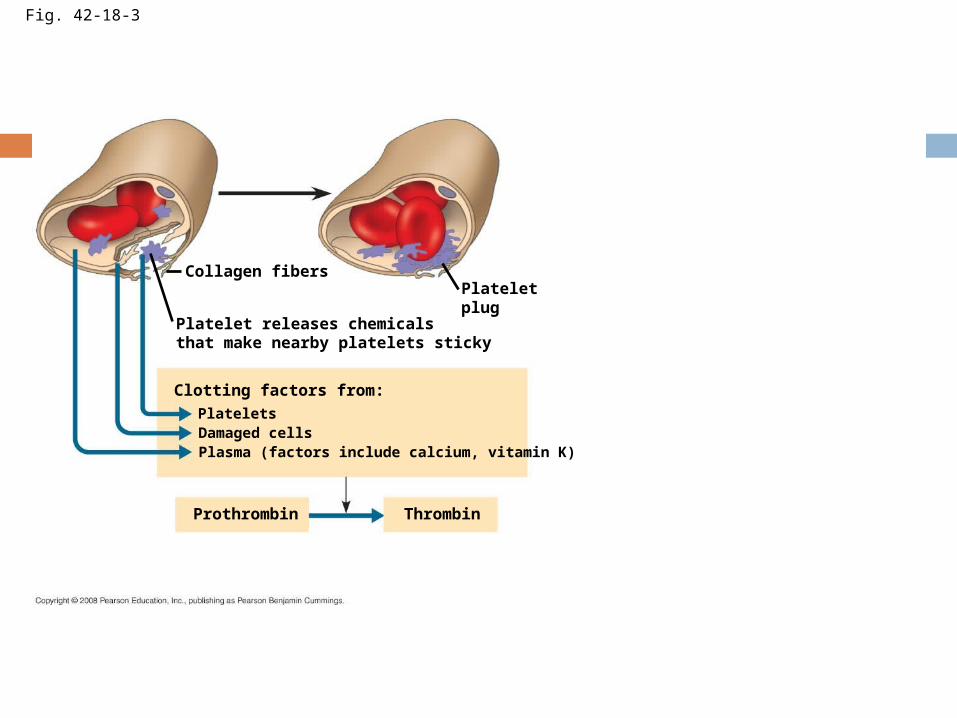
Fig. 42-18-3
Collagen fibersPlateletplug
Platelet releases chemicalsthat make nearby platelets sticky
Clotting factors from:PlateletsDamaged cellsPlasma (factors include calcium, vitamin K)
Prothrombin Thrombin
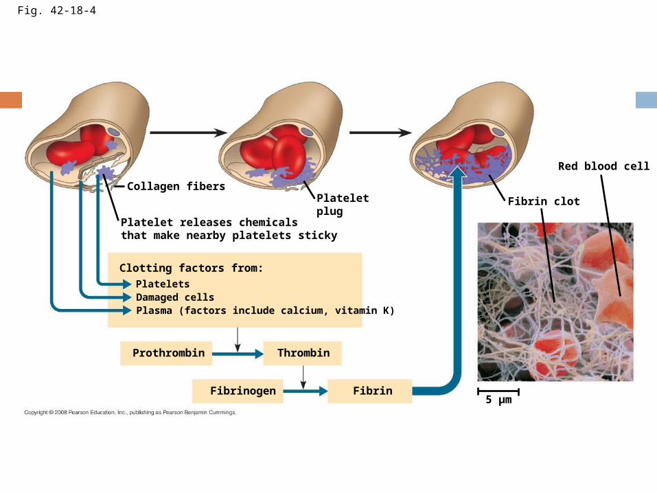
Collagen fibersPlateletplug
Platelet releases chemicalsthat make nearby platelets sticky
Clotting factors from:PlateletsDamaged cellsPlasma (factors include calcium, vitamin K)
Prothrombin Thrombin
Fibrinogen Fibrin5 µm
Fibrin clot
Red blood cell
Fig. 42-18-4
Stem Cells and the Replacement of Cellular Elements The cellular elements of blood wear out
and are replaced constantly throughout a person’s life
Erythrocytes, leukocytes, and platelets all develop from a common source of stem cells in the red marrow of bones
The hormone erythropoietin (EPO) stimulates erythrocyte production when oxygen delivery is low
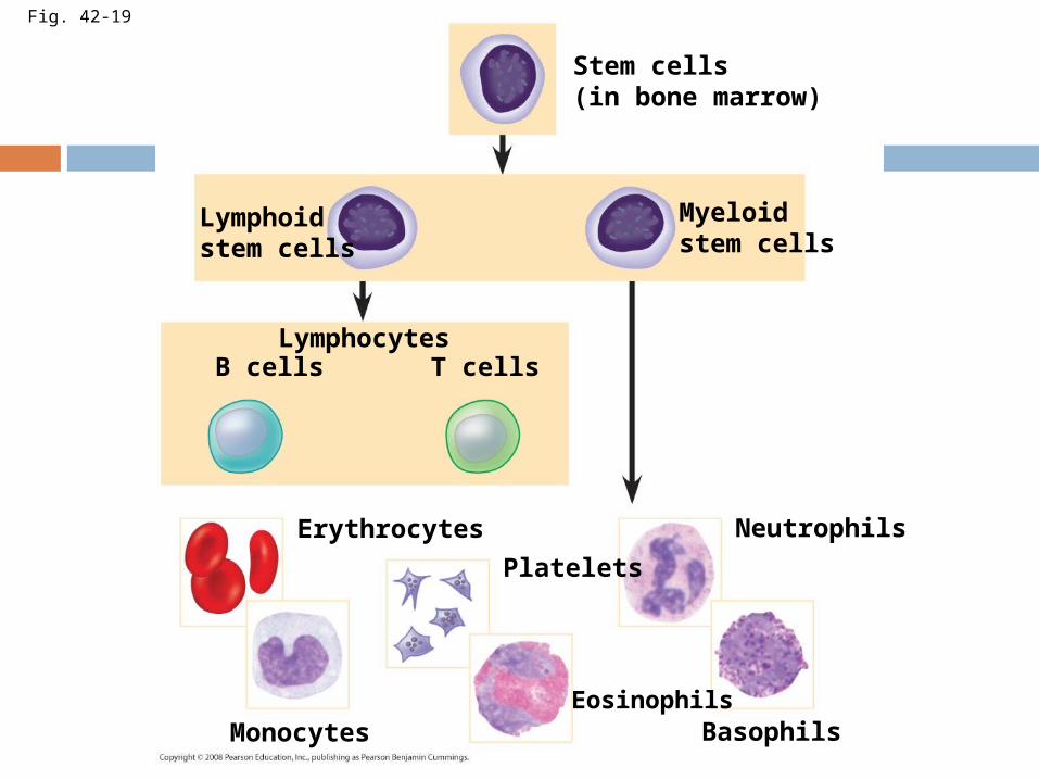
Fig. 42-19
Stem cells(in bone marrow)
Myeloidstem cells
Lymphoidstem cells
LymphocytesB cells T cells
Erythrocytes
Platelets
Neutrophils
BasophilsEosinophils
Monocytes
73
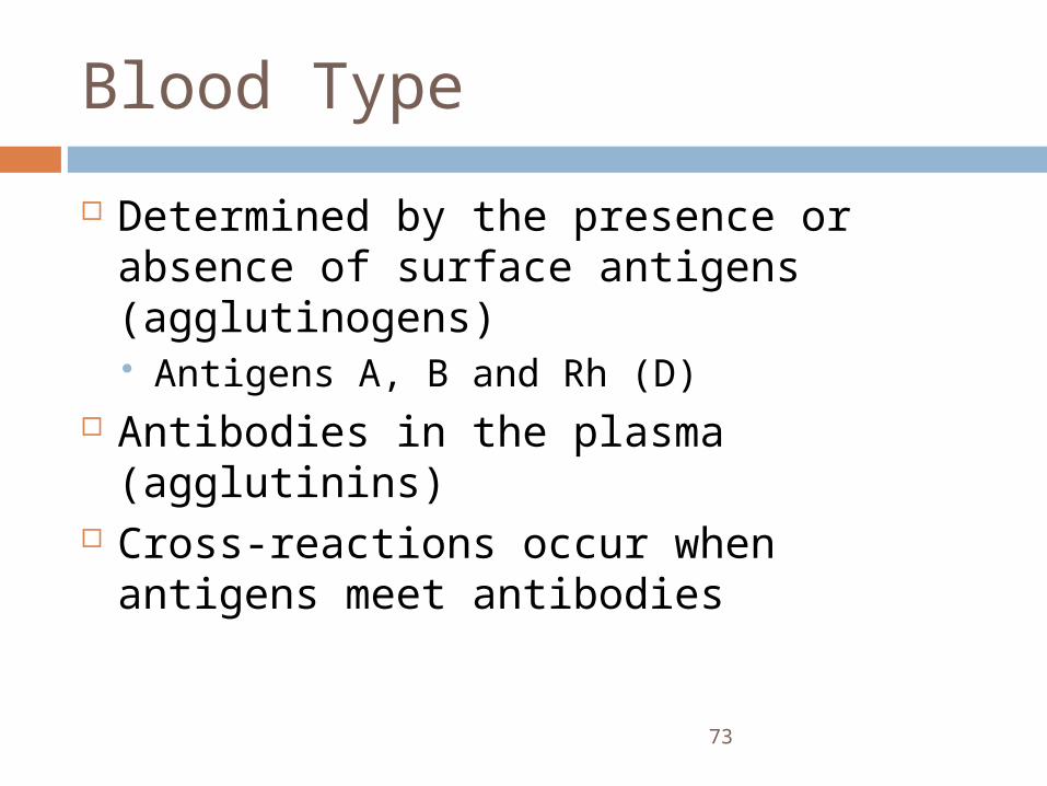
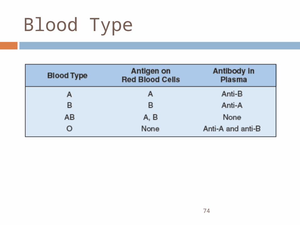
Blood Type
Determined by the presence or absence of surface antigens (agglutinogens) Antigens A, B and Rh (D)
Antibodies in the plasma (agglutinins) Cross-reactions occur when antigens
meet antibodies
74
Blood Type
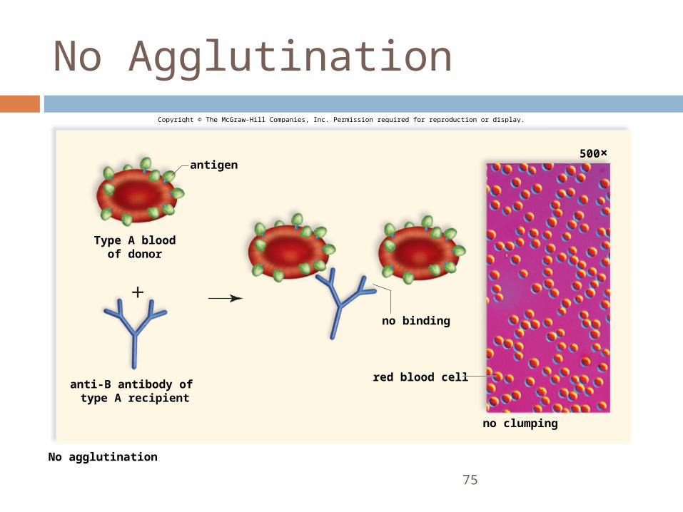
75
No AgglutinationCopyright © The McGraw-Hill Companies, Inc. Permission required for reproduction or display.
No agglutination
no binding
500×
red blood cell
antigen
no clumping
anti-B antibody of type A recipient
Type A bloodof donor
76
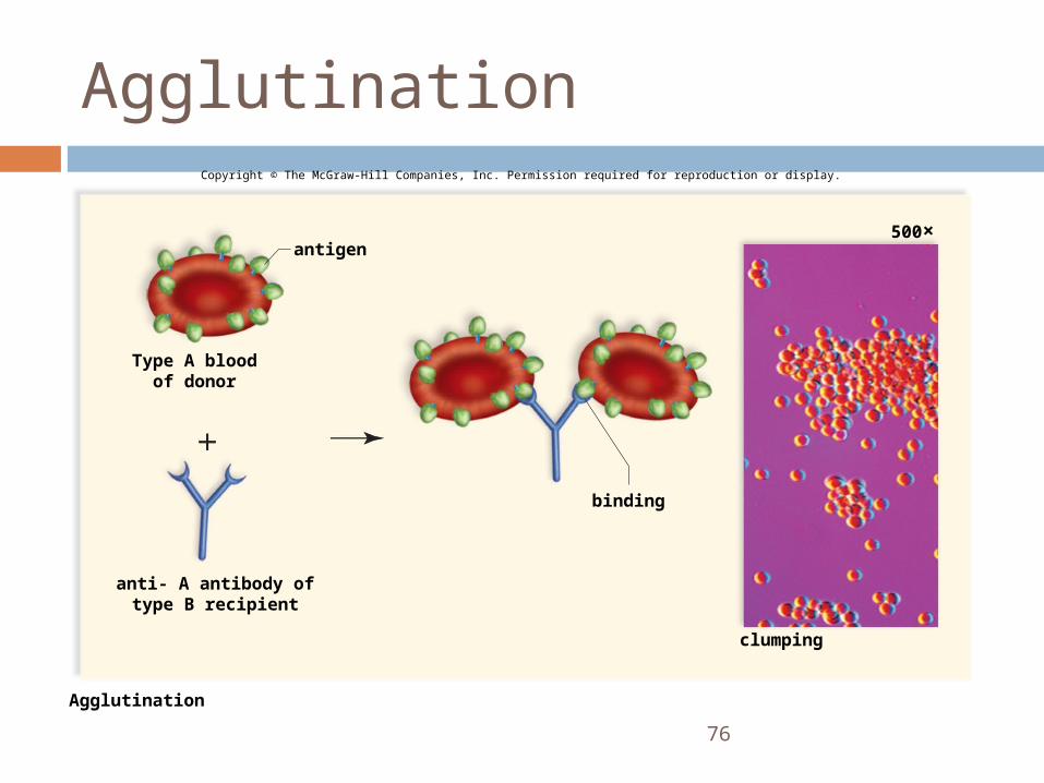
AgglutinationCopyright © The McGraw-Hill Companies, Inc. Permission required for reproduction or display.
Agglutination
binding
500×
clumping
antigen
Type A bloodof donor
anti- A antibody oftype B recipient
77
Blood Type
During pregnancy, if the mother is Rh negative and the father is Rh positive, the child may be Rh positive. Rh-positive red blood cells may leak across
the placenta The mother will produce anti-Rh antibodies. Antibodies may attack the embryo in a
subsequent pregnancy
Cardiovascular Disease
Cardiovascular diseases are disorders of the heart and the blood vessels
They account for more than half the deaths in the United States
One type of cardiovascular disease, atherosclerosis, is caused by the buildup of plaque deposits within arteries
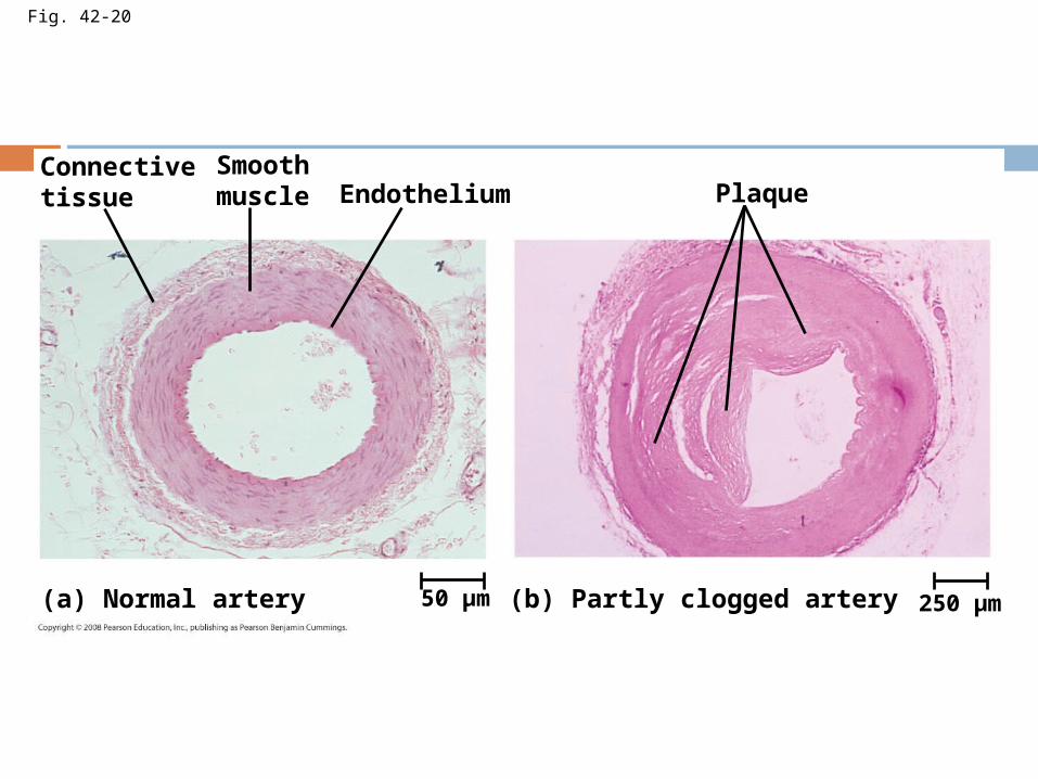
Fig. 42-20
Connectivetissue
Smoothmuscle Endothelium Plaque
(a) Normal artery (b) Partly clogged artery50 µm 250 µm
Heart Attacks and Stroke
A heart attack is the death of cardiac muscle tissue resulting from blockage of one or more coronary arteries
A stroke is the death of nervous tissue in the brain, usually resulting from rupture or blockage of arteries in the head
Treatment and Diagnosis of Cardiovascular Disease Cholesterol is a major contributor to
atherosclerosis
Low-density lipoproteins (LDLs) are associated with plaque formation; these are “bad cholesterol”
High-density lipoproteins (HDLs) reduce the deposition of cholesterol; these are “good cholesterol”
The proportion of LDL relative to HDL can be decreased by exercise, not smoking, and avoiding foods with trans fats
Hypertension, or high blood pressure, promotes atherosclerosis and increases the risk of heart attack and stroke
Hypertension can be reduced by dietary changes, exercise, and/or medication
You should now be able to:
1. Compare and contrast open and closed circulatory systems
2. Compare and contrast the circulatory systems of fish, amphibians, non-bird reptiles, and mammals or birds
3. Distinguish between pulmonary and systemic circuits and explain the function of each
4. Trace the path of a red blood cell through the human heart, pulmonary circuit, and systemic circuit
5. Define cardiac cycle and explain the role of the sinoatrial node
6. Relate the structures of capillaries, arteries, and veins to their function
7. Define blood pressure and cardiac output and describe two factors that influence each
8. Explain how osmotic pressure and hydrostatic pressure regulate the exchange of fluid and solutes across the capillary walls
9. Describe the role played by the lymphatic system in relation to the circulatory system
10. Describe the function of erythrocytes, leukocytes, platelets, fibrin
11. Distinguish between a heart attack and stroke
Copyright © 2008 Pearson Education, Inc., publishing as Pearson Benjamin Cummings.
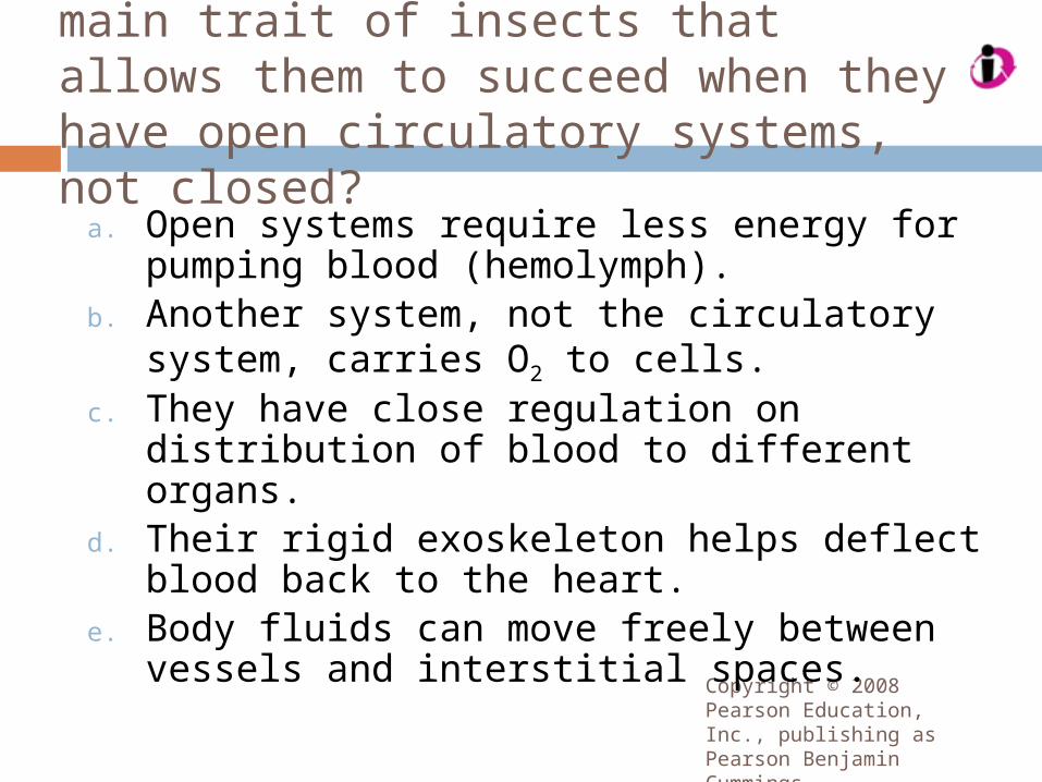
Which of the following is the main trait of insects that allows them to succeed when they have open circulatory systems, not closed?
a. Open systems require less energy for pumping blood (hemolymph).
b. Another system, not the circulatory system, carries O2 to cells.
c. They have close regulation on distribution of blood to different organs.
d. Their rigid exoskeleton helps deflect blood back to the heart.
e. Body fluids can move freely between vessels and interstitial spaces.
Copyright © 2008 Pearson Education, Inc., publishing as Pearson Benjamin Cummings.
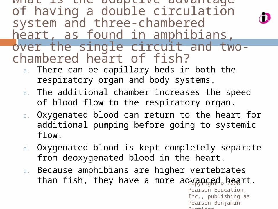
What is the adaptive advantage of having a double circulation system and three-chambered heart, as found in amphibians, over the single circuit and two-chambered heart of fish?
a. There can be capillary beds in both the respiratory organ and body systems.
b. The additional chamber increases the speed of blood flow to the respiratory organ.
c. Oxygenated blood can return to the heart for additional pumping before going to systemic flow.
d. Oxygenated blood is kept completely separate from deoxygenated blood in the heart.
e. Because amphibians are higher vertebrates than fish, they have a more advanced heart.
Copyright © 2008 Pearson Education, Inc., publishing as Pearson Benjamin Cummings.
What attributes of specialized atria cells account for two specialized heart features: their spontaneous contraction and the contraction of the two atria in unison?
a. Autorhythmic pacemaker cells account for both features.
b. Gap junctions account for both features.c. External input accounts for both features.d. Autorhythmic pacemaker cells account for
the first and gap junctions for the second.e. Gap junctions account for the first and
autorhythmic pacemaker cells for the second.
Copyright © 2008 Pearson Education, Inc., publishing as Pearson Benjamin Cummings.
In Edgar Allan Poe’s short story “The Tell-Tale Heart,” a murder victim’s heart continued to beat after it was removed from the body. What feature of the heartbeat is the fact behind this fiction?
a. Heart pacemaker cells contract spontaneously, requiring no input.
b. Nerves controlling heartbeat fire spontaneously, requiring no input.
c. Hormones controlling heartbeat are released spontaneously.
d. Powerful ventricle contractions are spontaneous, requiring no input.
e. Pulsing of blood in the heart chambers is spontaneous, maintaining the heartbeat.
Copyright © 2008 Pearson Education, Inc., publishing as Pearson Benjamin Cummings.
Which combination explains the fluid exchange between blood and interstitial fluid as blood is entering and leaving a capillary?
a. Fluid pressure forces fluid out of the vessel and into interstitial fluid when blood enters a capillary, then pulls it back into the blood at the venous end.
b. Osmotic pressure forces fluid out of the vessel and into interstitial fluid when blood enters a capillary, then pulls it back at the venous end.
c. Fluid pressure forces fluid out of the vessel and into interstitial fluid when blood enters a capillary, and osmotic pressure pulls it back in at the venous end of the capillary.
d. Osmotic pressure forces fluid out of the vessel and into interstitial fluid when blood enters a capillary, and fluid pressure pulls it back in at the venous end of the capillary.
Copyright © 2008 Pearson Education, Inc., publishing as Pearson Benjamin Cummings.
In response to a period of low O2 in circulating blood, the kidney secretes the hormone erythropoietin (EPO), which stimulates erythrocyte production. This system involves a negative feedback control because
a. when the EPO level rises above a certain point, the kidney stops producing it.
b. when the EPO level rises above a certain point, the kidney makes much more of it.
c. when the O2 level in tissues falls to a normal level, the kidney stops producing EPO.
d. when the O2 level in tissues rises to a normal level, the kidney stops producing EPO.
e. when the O2 level in tissues rises to a normal level, the kidney makes much more EPO.
Copyright © 2008 Pearson Education, Inc., publishing as Pearson Benjamin Cummings.
Which of the following best describes the advantage of a capillary system with counter-current flow over a system with concurrent flow?a. More diffusion occurs at the beginning of
capillary flow than midway through the capillary.
b. More diffusion occurs at the end of capillary flow than midway through the capillary.
c. At each point in the capillary is a concentration gradient that promotes diffusion.
d. At each point in the capillary, the walls are thin to promote diffusion.
e. Counter-current systems provide greater surface area for diffusion.
Reiber C. L. & McGaw I. J. (2009). "A Review of the "Open" and "Closed" Circulatory Systems: New Terminology for Complex Invertebrate Circulatory Systems in Light of Current Findings". International Journal of Zoology 2009: 8 pages. doi:10.1155/2009/301284.Patwardhan K. The history of the discovery of blood circulation: unrecognized contributions of Ayurveda masters. Adv Physiol Educ. 2012 Jun;36(2):77–82.Michael Servetus Research Study on the Manuscript of Paris by Servetus (1546 description of the Pulmonary Circulation)