Embed Size (px)
Citation preview
Thyroid gland
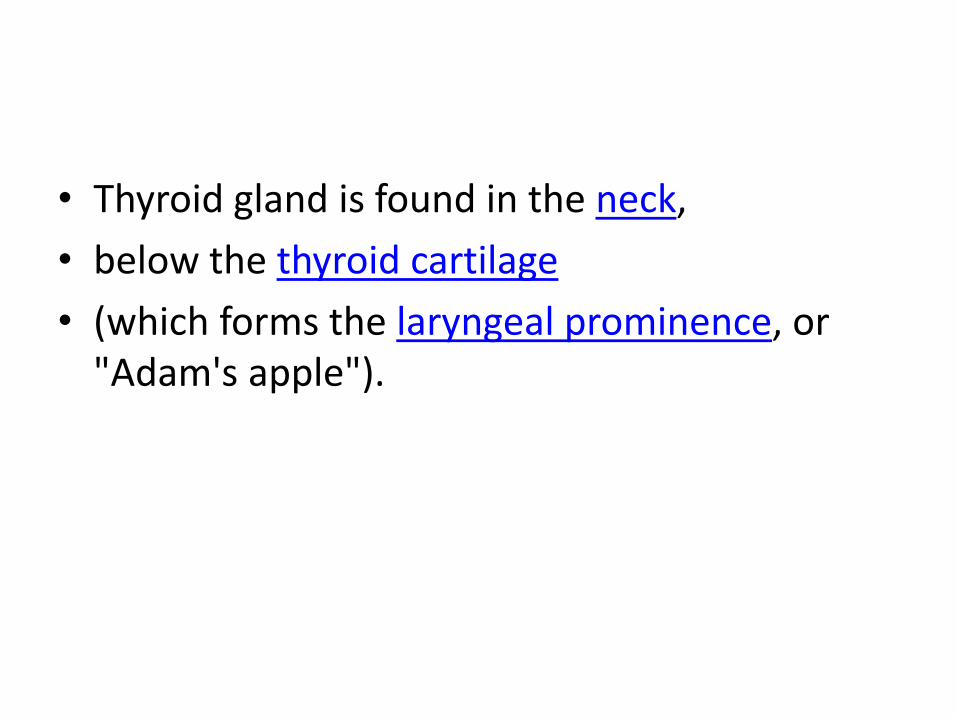
• Thyroid gland is found in the neck,
• below the thyroid cartilage
• (which forms the laryngeal prominence, or "Adam's apple").

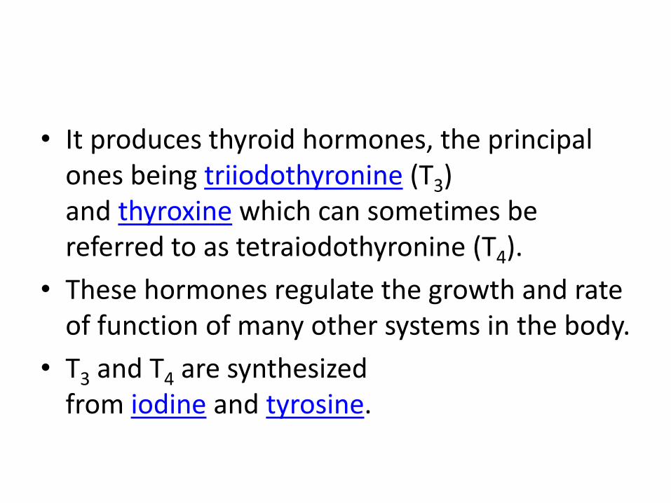
• It produces thyroid hormones, the principal ones being triiodothyronine (T3) and thyroxine which can sometimes be referred to as tetraiodothyronine (T4).
• These hormones regulate the growth and rate of function of many other systems in the body.
• T3 and T4 are synthesized from iodine and tyrosine.
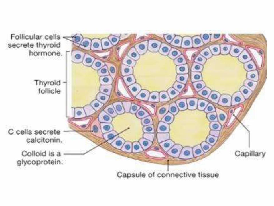
• The thyroid also produces calcitonin, which plays a role in calcium homeostasis.
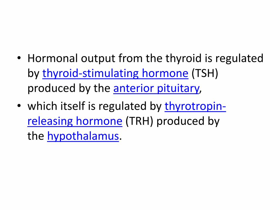
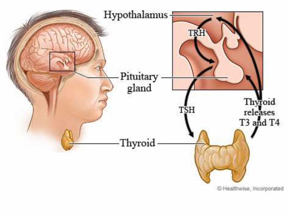
• Hormonal output from the thyroid is regulated by thyroid-stimulating hormone (TSH) produced by the anterior pituitary,
• which itself is regulated by thyrotropin-releasing hormone (TRH) produced by the hypothalamus.
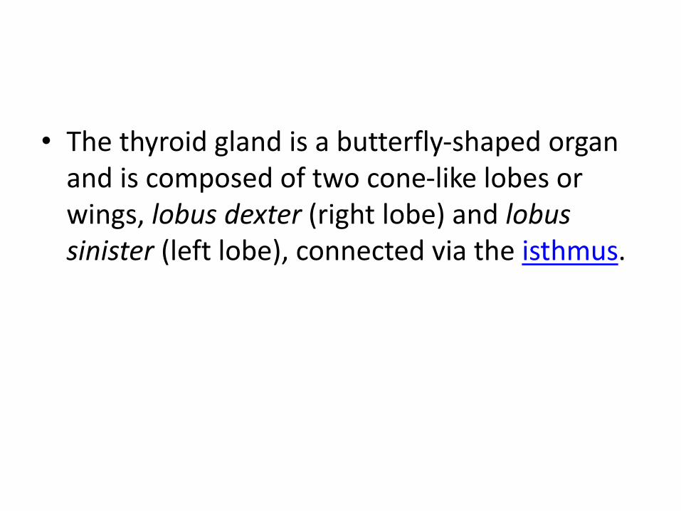
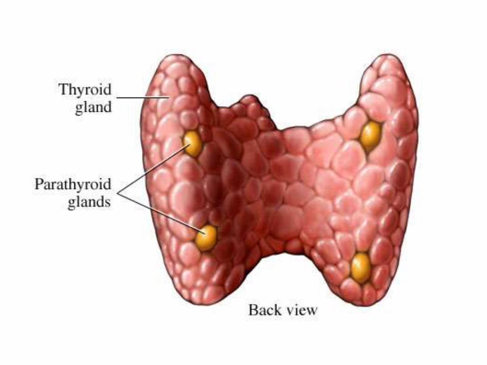
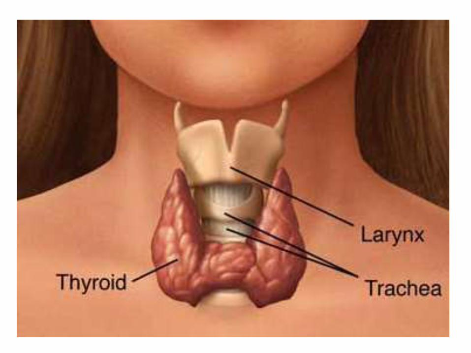
• The thyroid gland is a butterfly-shaped organ and is composed of two cone-like lobes or wings, lobus dexter (right lobe) and lobussinister (left lobe), connected via the isthmus.
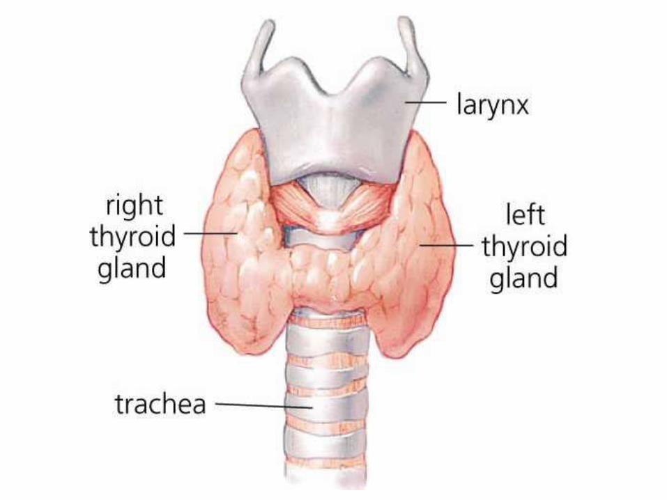
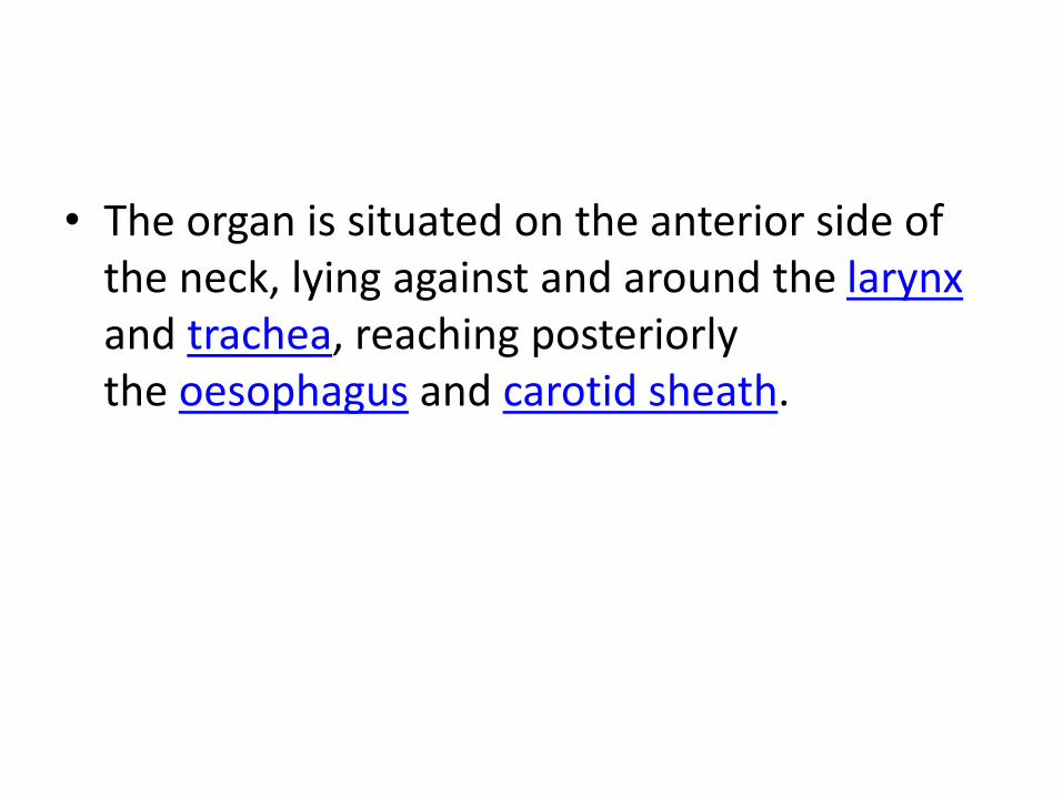
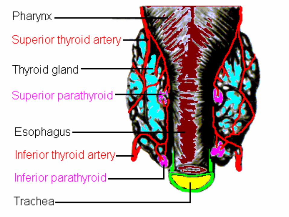
• The organ is situated on the anterior side of the neck, lying against and around the larynxand trachea, reaching posteriorly the oesophagus and carotid sheath.
• It starts cranially at the oblique line on the thyroid cartilage (just below the laryngeal prominence, or 'Adam's Apple'), and extends inferiorly to approximately the fifth or sixth tracheal ring.
• It is difficult to demarcate the gland's upper and lower border with vertebral levels because it moves position in relation to these during swallowing.
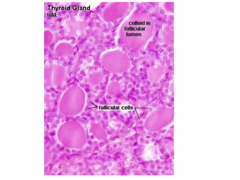
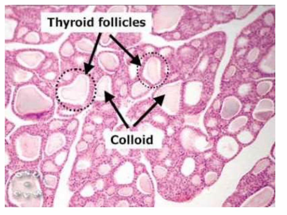
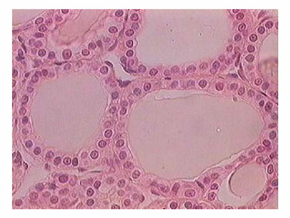
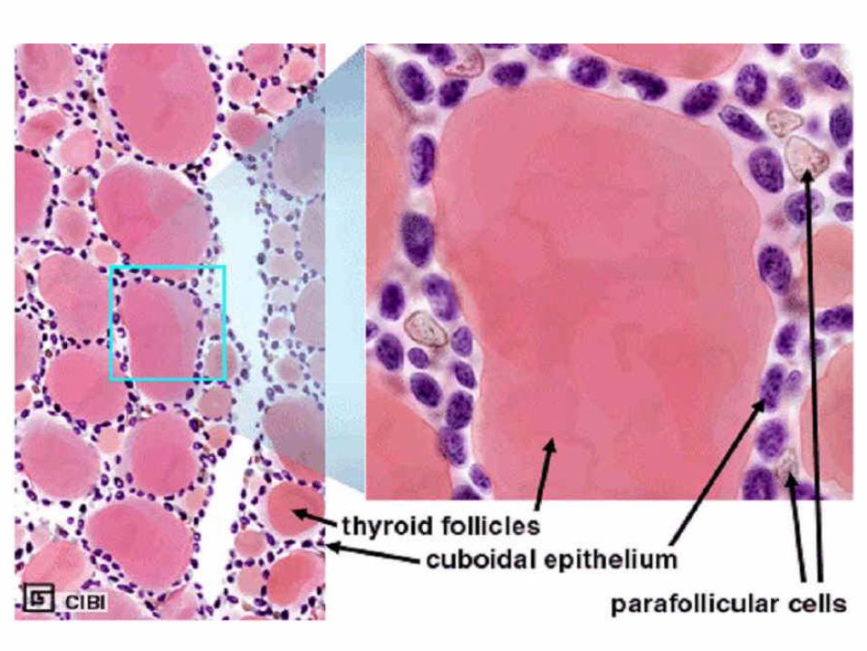
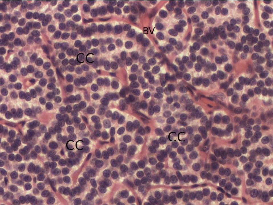
Histology
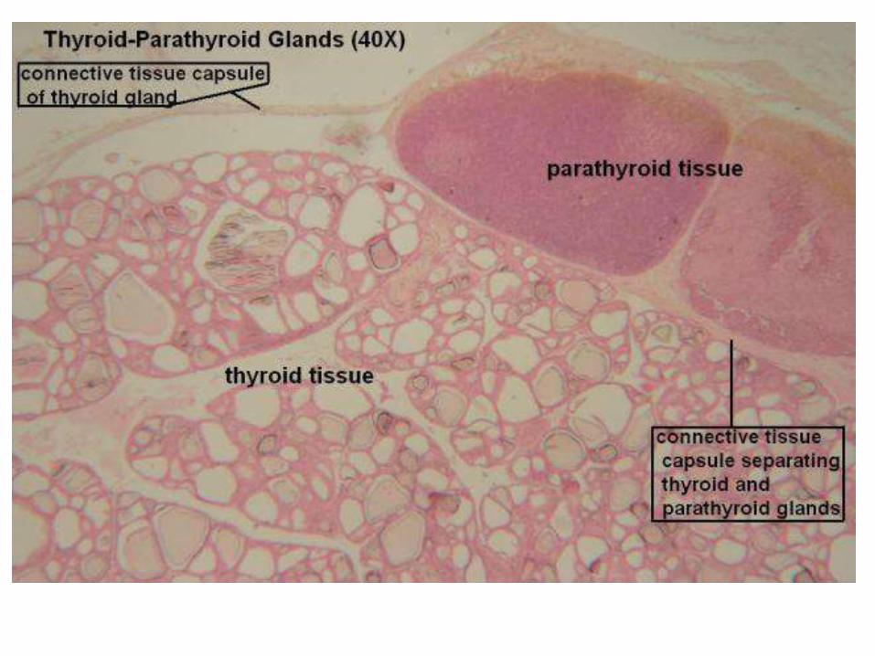
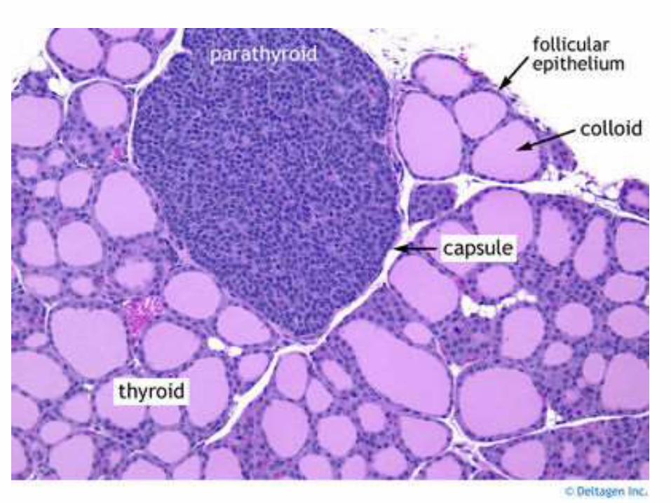
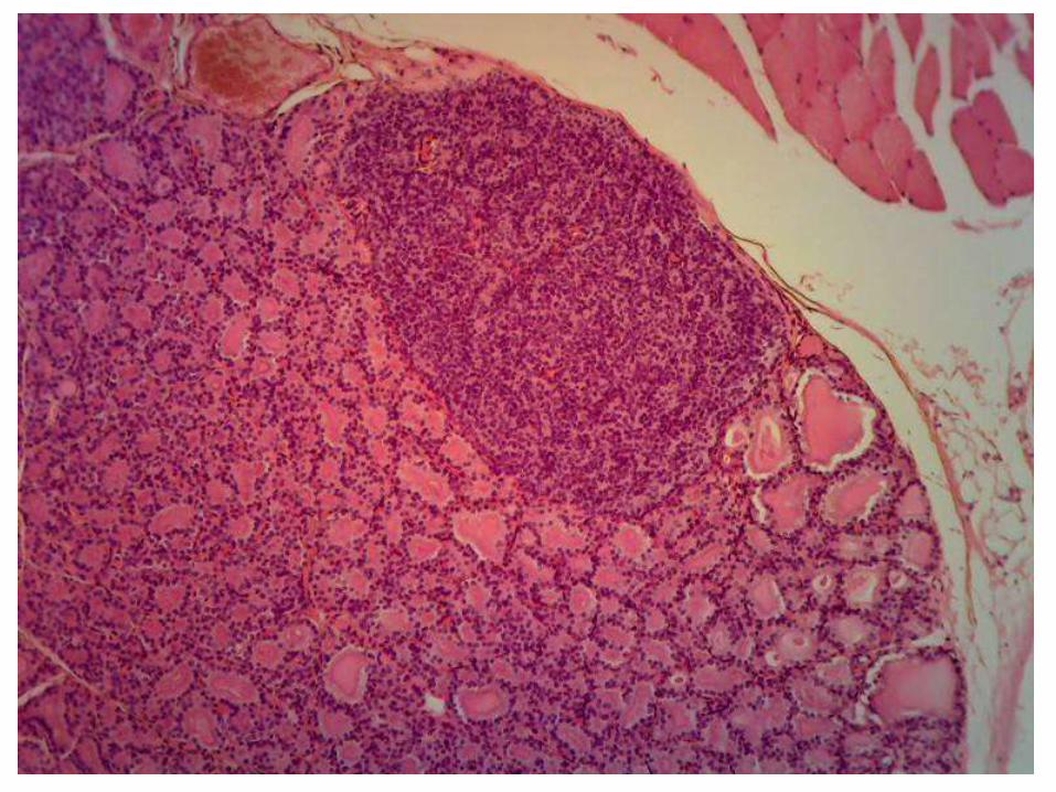
• The gland consists of many indistinct lobules containing follicles and many blood vessels enmeshed in fine connective tissue
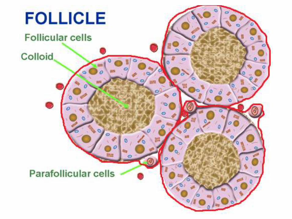
• The follicles contain a homogeneous, acidophilic material called colloid
• The follicles are lined by simple cuboidal epithelium but the epithelium may be columnar or squamous depending upon the function activity of the gland
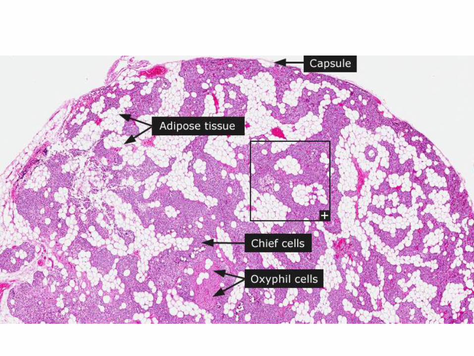
Parathyroid gland
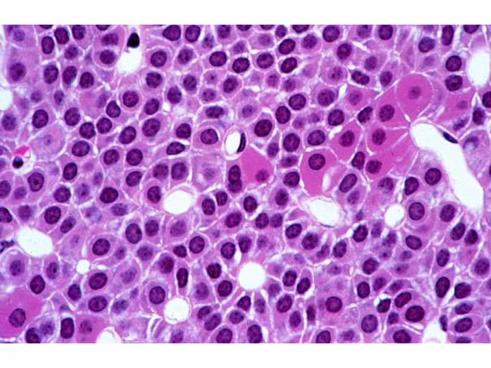
• The parenchyma of the gland consists of irregular masses of cords of cells, separated by blood vessels embedded in loose connective tissue containing many fat cells
• The cords of cells contain two types of cells Chief or principal cells are most common and a few oxyphil cells. The chief cells have prominent nuclei and relatively little cytoplasm. The oxyphil cells are larger and more darker stained cytoplasm.
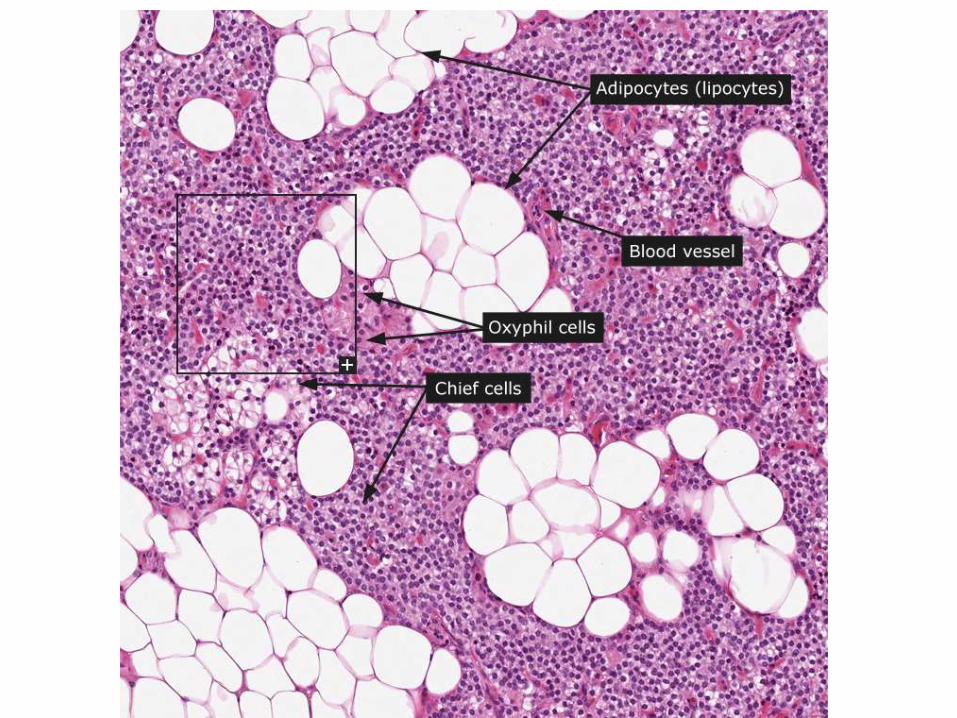
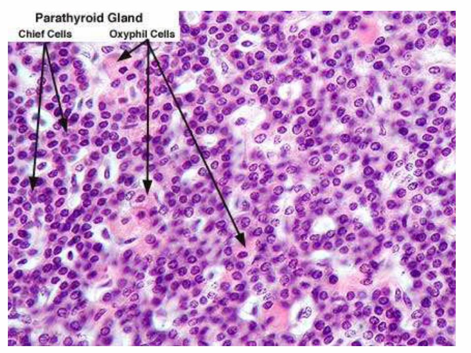
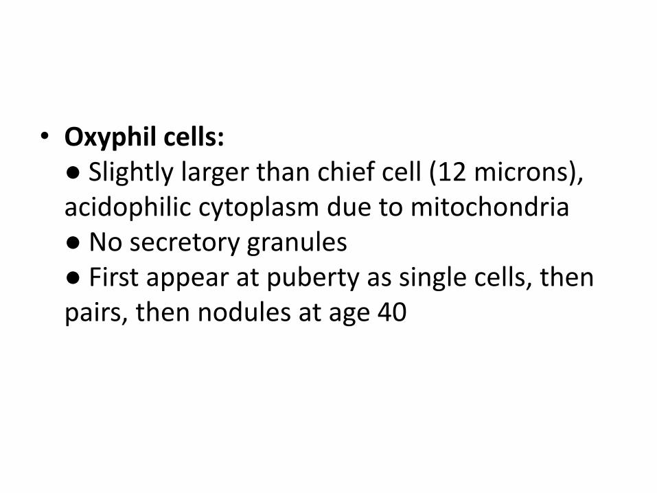
• Oxyphil cells:● Slightly larger than chief cell (12 microns), acidophilic cytoplasm due to mitochondria● No secretory granules● First appear at puberty as single cells, then pairs, then nodules at age 40
Development
• The thyroid gland is the first endocrine gland to develop in the embryo.
• It begins to form about 24 days after fertilization.
• It appears as a median endodermal thickening in the floor of the primordial pharynx between tuberculum impar and copula.
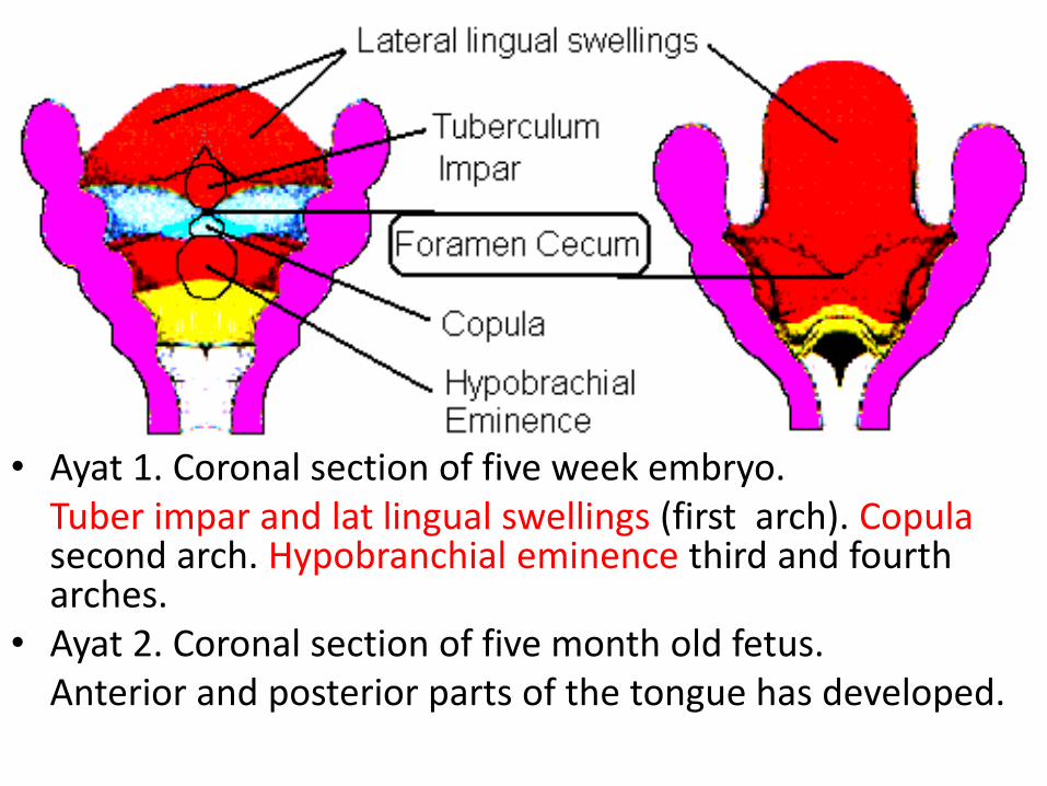
• Ayat 1. Coronal section of five week embryo. Tuber impar and lat lingual swellings (first arch). Copulasecond arch. Hypobranchial eminence third and fourth arches.
• Ayat 2. Coronal section of five month old fetus. Anterior and posterior parts of the tongue has developed.
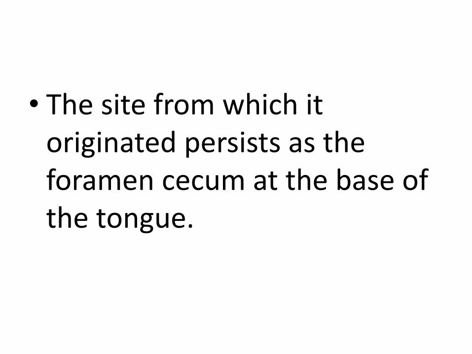
• The site from which it originated persists as the foramen cecum at the base of the tongue.
• This thickening soon forms a small out pouching downward –the thyroid diverticulum.
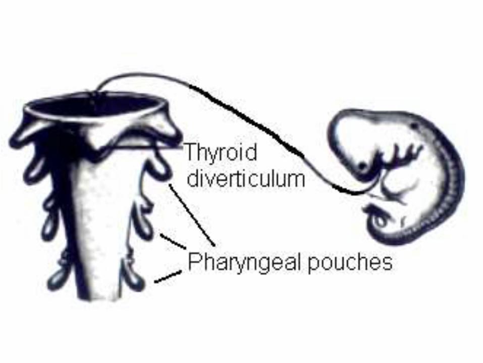
• Diagram showing the developing thyroid diverticulum.
• On the right the lateral view of the developing embryo during 4th week is shown.
• On left the developing pharynx has been shown during 4th week.
• 5 week 5 month
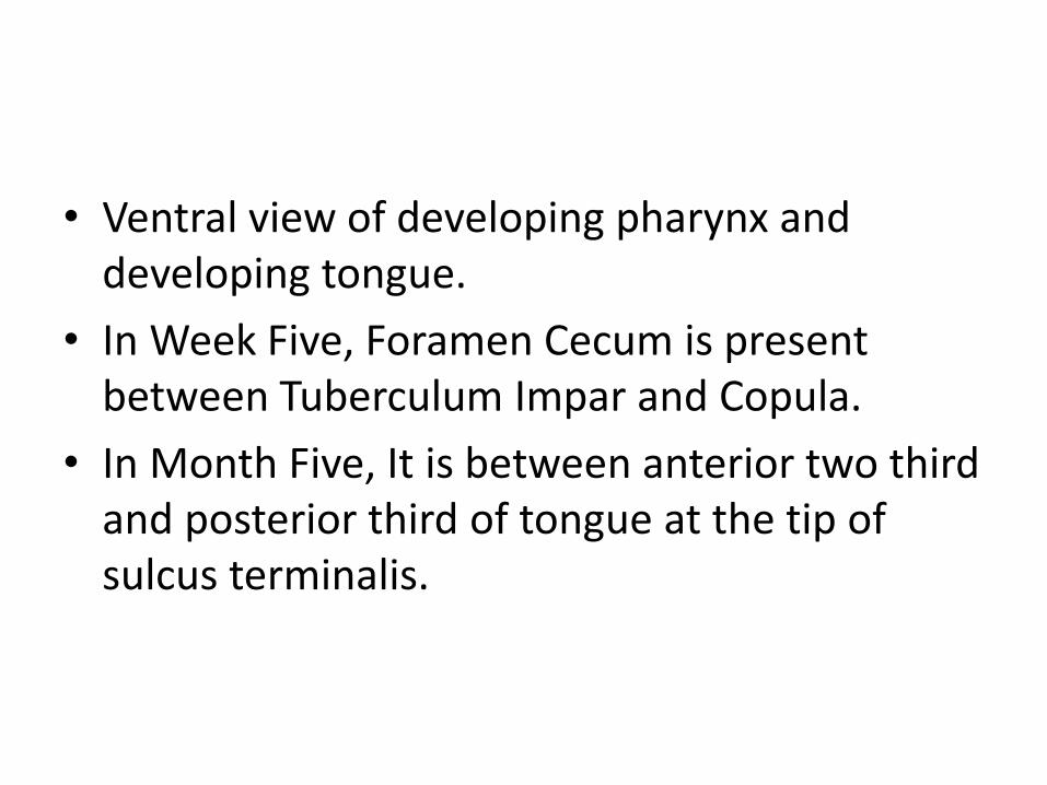
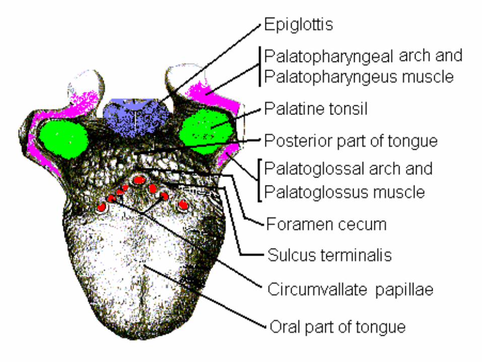
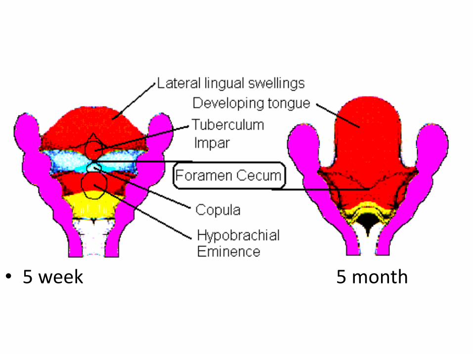
• Ventral view of developing pharynx and developing tongue.
• In Week Five, Foramen Cecum is present between Tuberculum Impar and Copula.
• In Month Five, It is between anterior two third and posterior third of tongue at the tip of sulcus terminalis.
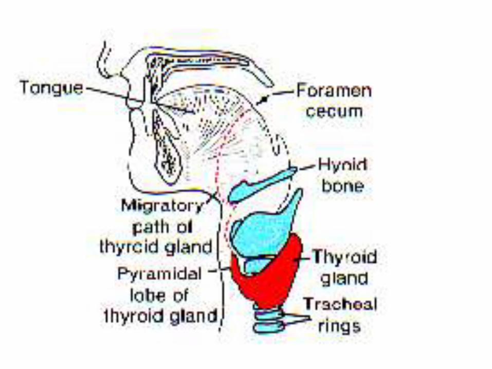
• As the embryo and tongue grow, the developing thyroid gland descends in the neck in front of pharyngeal gut, passing ventral to the developing hyoid bone and laryngeal cartilages.
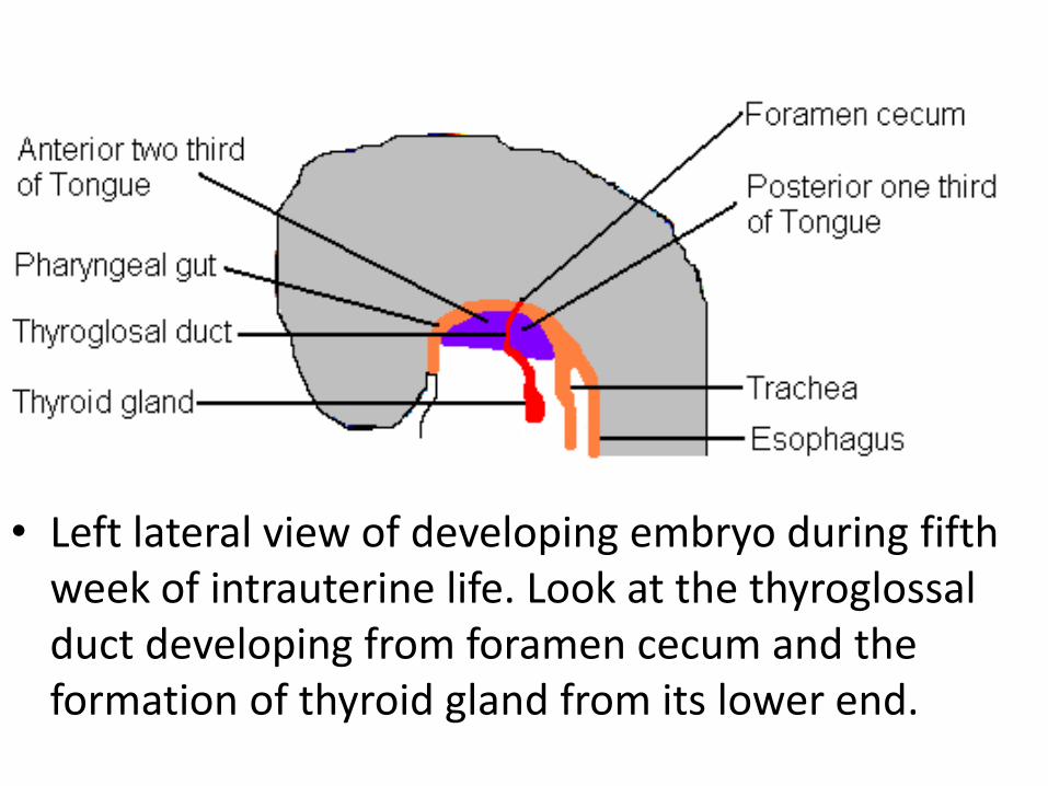
• Left lateral view of developing embryo during fifth week of intrauterine life. Look at the thyroglossal duct developing from foramen cecum and the formation of thyroid gland from its lower end.
• During this migration for a short time the developing thyroid gland is connected to the tongue by a narrow tube, the thyroglossal duct.
• This duct later disappears.
• At first the thyroid diverticulum is hollow but it soon becomes solid and divides into right and left branches.
• It reaches its final position in front of trachea, just below the cricoid cartilage, during 7th
week.
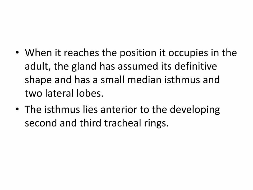
• When it reaches the position it occupies in the adult, the gland has assumed its definitive shape and has a small median isthmus and two lateral lobes.
• The isthmus lies anterior to the developing second and third tracheal rings.
• By this time the thyroglossal duct has normally degenerated and disappeared.
• The proximal opening of the thyroglossal duct persists as a small blind pit, the foramen cecum of the tongue.
• The proximal opening of the thyroglossal duct persists as a small blind pit, the foramen cecum of the tongue.
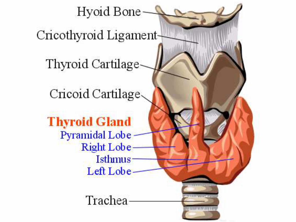
• In about 50% of people thyroglossal duct persists as a pyramidal lobe extending superiorly from the isthmus.
Histogenesis
• Thyroid primordium consists of a solid mass of endodermal cells.
• This cellular aggression breaks up into a network of epithelial cords because of the invasion of the surrounding vascular mesenchyme.
• By tenth week cords have formed small cellular groups.
• A lumen soon forms in each cell cluster and the cells become arranged in a single layer around a lumen.
• These are thyroid follicles.
• During eleventh week colloid begins to appear in thyroid follicles.

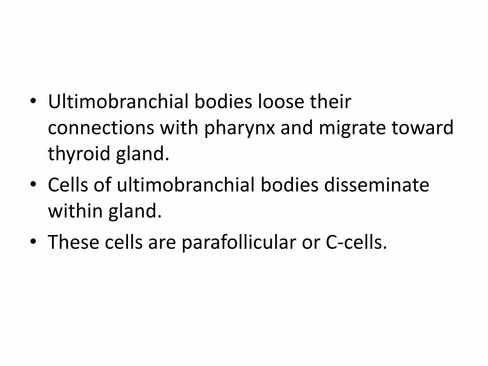
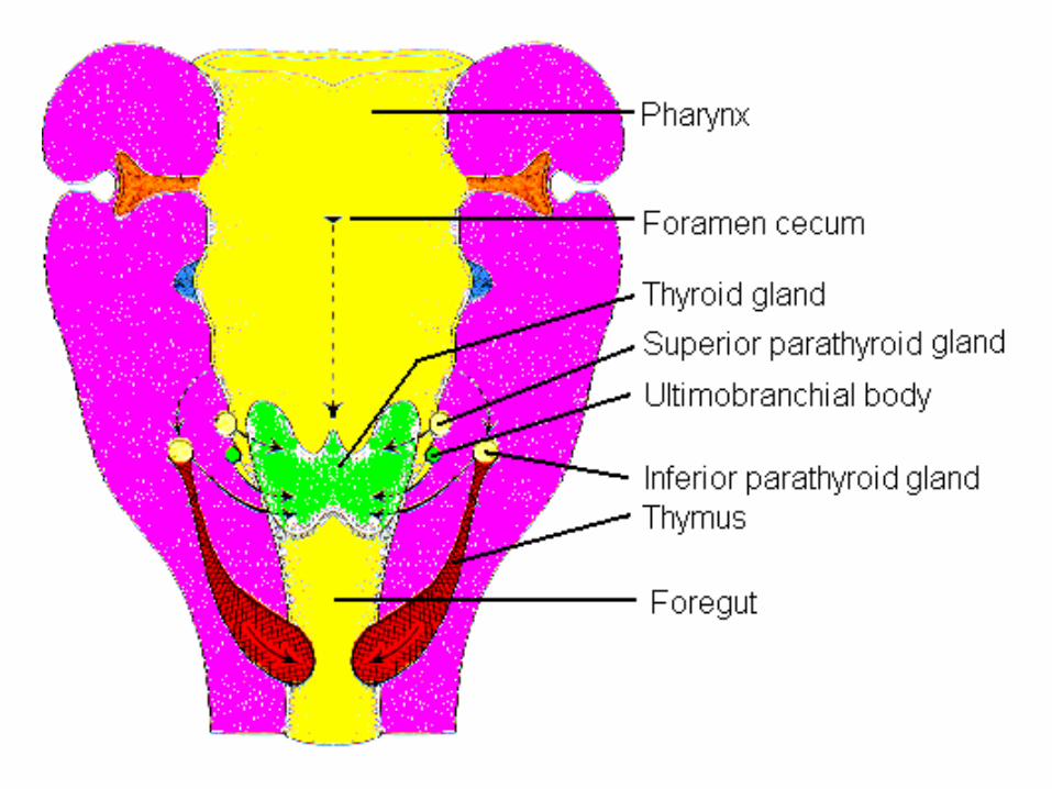
• Ultimobranchial bodies loose their connections with pharynx and migrate toward thyroid gland.
• Cells of ultimobranchial bodies disseminate within gland.
• These cells are parafollicular or C-cells.
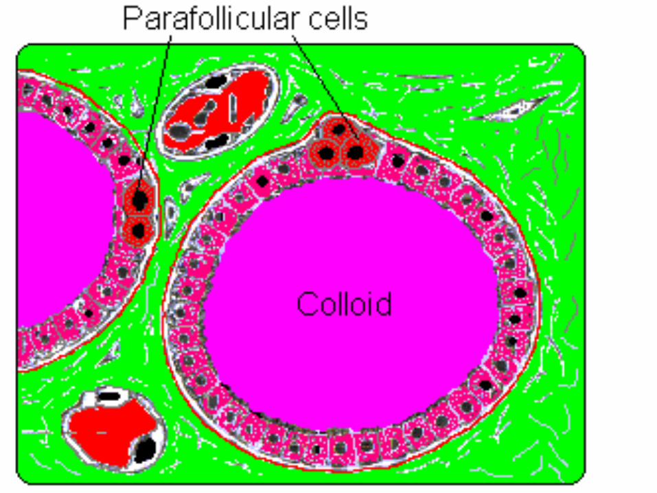
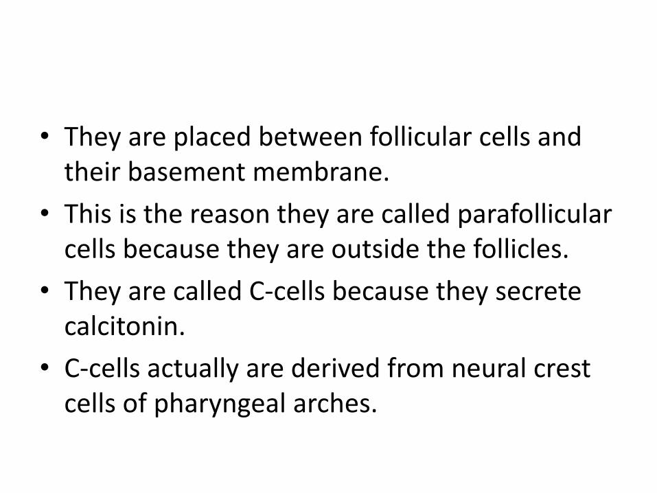
• They are placed between follicular cells and their basement membrane.
• This is the reason they are called parafollicular cells because they are outside the follicles.
• They are called C-cells because they secrete calcitonin.
• C-cells actually are derived from neural crest cells of pharyngeal arches.
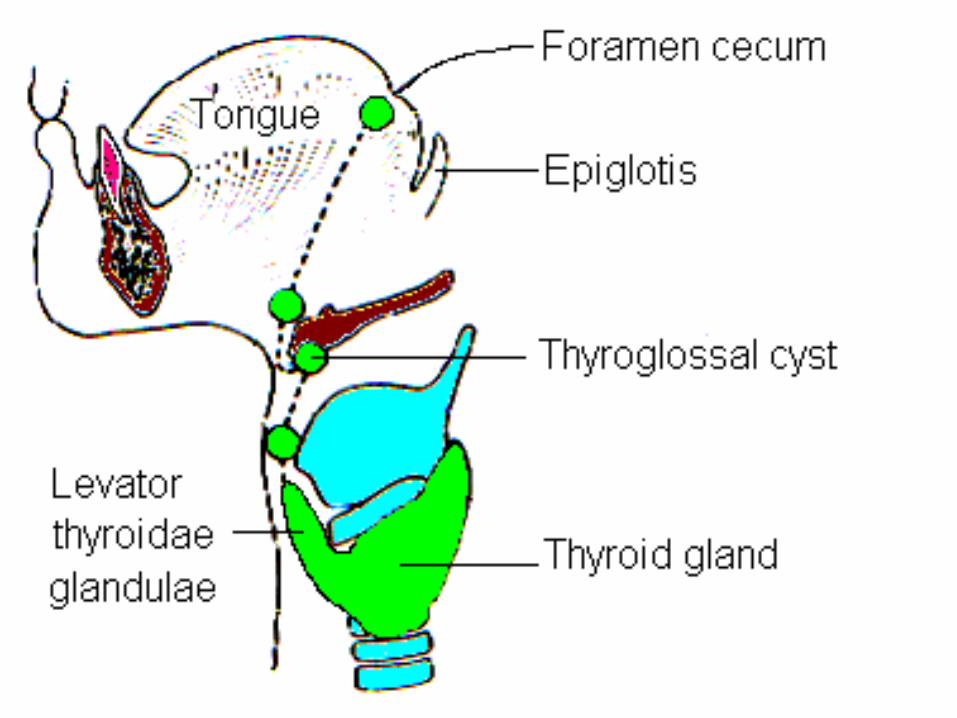
Thyroglossal Cyst
• A thyroglossal cyst may be found at any point along the migratory path followed by thyroid gland in the midline of neck.
• A thyroglossal cyst is a cystic remnant of thyroglossal duct.
• In 50 per cent cases such cyst is located close to hyoid bone.
• It may also be found at the base of tongue or close to thyroid cartilage.

Thyroglossal Fistula
• Sometimes the cyst is connected to outside by a fistulous canal.
• Then it is called thyroglossal fistula.
• The fistula may be primary when it is present at birth. It may be secondary thyroglossal fistula when a cyst ruptures and communicate outside at later stage.
Aberrant Thyroid Tissue
• Thyroglossal tissue may be found anywhere along the path of the descent of thyroid gland.
• It is most commonly found in the base of tongue, just behind foramen cecum.
• It is subject to the same diseases as thyroid gland itself.