Embed Size (px)
Citation preview

RHEUMATOID ARTHRITIS
• Rheumatoid arthritis (RA) is one of the most common chronic inflammatory conditions, affecting the population aetiology affecting both articular tissue and extra-articular organs.
• There is a wide range of extra-articular features. The disease is progressive and results in pain, stiffness and swelling of joints which can lead to significant morbidity and increased mortality

EXTRA-ARTICULAR FEATURES OF RHEUMATOID ARTHIRITS
COMMON UNCOMMON
Anaemia Nodules (subcutaneous)Muscle wastingDry eyes Depression Osteoporosis EpiscleritisCarpal tunnel syndrome Leg ulcers LymphadenopathyNailfold vasculitis
Pleural and pericardial effusionsFibrosing alveolitis PericarditisScleritisSystemic vasculitisMitral value and conduction defectsNodules (lungs, eyes, heart)Felty’s syndrome (seropositive rheumatoid arthritis, splenomegaly and neutropenia, incidence <1%)

EPIDEMOLOGY
• Approximately 1% of the adult population worldwide is affected by rheumatoid arthritis.
• The prevalence of rheumatoid arthritis increases with age in both sexes with nearly 5% of women and 2% of men over 55 years of age affected.
• The peak age of incidence is around 55-64 years in women and 65-75 years in men.

AETIOLOGY
• The cause of rheumatoid arthritis remains unclear. It is postulated that a genetically susceptible host is exposed to an unknown pathogen (antigen) and this interaction gives rise to a persistent immunological response.
• Whether the initiating agent is an infection, a self-antigen or an environmental factor remains unproven.
• This model makes the assumption that the host’s immune human leucocytes antigen (HLA) response genes, immunoglobulin genes and T-cell receptor genes have an impact later in the chain of pathological events during stage II of the disease. The initial stage I, in this complex model, involves synovial tissue injury.
• The most definite genetic association with rheumatoid arthritis is with HLA alleles. The HLA-DR4 allele is associated with development and severity of rheumatoid arthritis.
• In American whites, 60-70% of rheumatoid arthritis patients are positive for HLA-DR4. This seems to be particularly important in severe forms of the disease. The frequency of HLA-DR4 among Dutch patients with severe extra-articular disease is greater than 90%.

• Epstein-Barr virus has been lined to rheumatoid arthritis for many years. • Of patients with rheumatoid arthritis, 80% have a circulating antibody
directed against antigens specific for Epstein-Barr virus, and the autoantibody response in rheumatoid arthritis enhances the response to these antigens.
• Parvoviruses (small DNA viruses that cause disease in many species), particularly B19, have been linked to rheumatoid arthritis. Mycobacteria have also been linked to rheumatoid arthritis because these bacteria express heat shock proteins (HSPs).
• A potential hypothesis is that antibodies and T-cell exist that recognize epitopes shared by the HSPs of both the infectious agents and host cells. This would facilitate cross reactivity of lymphocytes with host cells, triggering an immunological reaction

pathophysiology
• Chronic inflammation of the synovial tissue lining the joint capsule results in the proliferation of this tissue.
• The inflamed, proliferating synovium characteristic of rheumatoid arthritis is called pannus.
• This pannus invades the cartilage and eventually the bone surface, producing erosions of bone and cartilage and leading to destruction of the joint.
• The factors that initiate the inflammatory process are unknown.

The proinflammatory cytokines tumor necrosis factor (TNF), interleukin (IL)-1 and IL-6 are key substances in the initiation
and continuance of rheumatoid inflammation
• The processed antigen is recognized by major histocompatibility complex proteins on the lymphocyte, which activates it to stimulate the production of T and B cells.
• Lymphocytes may be either B cells (derived from bone marrow) or T cells (derived from thymus tissue).
• T cells may be either CD4+ (T-helper) or CD8+ (cytotoxic or killer) T cells. There are two subtypes of T-helper cells, TH1, which promote inflammation by producing interferon-γ, tumor necrosis factor, and interleukin-2, and TH2, which produce the antiinflammatory cytokines IL-4, IL-5 and IL-10.
• CD8+ killer T cells have a regulatory effect on the immune process by suppressing activity of CD4+ cells through release of antiinflammatory cytokines and promoting apoptosis (cell death).
• Activated T cells produce cytotoxins, which are directly toxic to tissues, and cytokines, which stimulate further activation of inflammatory processes and attract cells to areas of inflammation. Macrophages are stimulated to release prostaglandins and cytotoxins.
• The released inflammatory mediators causes the erosion of synovial tissue and cartilage destruction.

CLINICAL MANIFESTATIONS
CRITERIA FOR DIAGNOSIS OF RHEUMATOID ARTHRITIS CRITERIA COMMENT
1.Morning stiffness During lasting >1hr for >6 weeks
2. Arthritis of at least three joint areas Soft tissue swelling or exudation for >6 weeks
3.Arthritis of hand joints Swelling in wrist, metacarpophalangeal joints or proximal interphalangeal joints lasting >6 weeks
4.Symmetrical arthritis Symmetrical involvement of same joint areas on both sides of body lasting >6 weeks
5.Rheumatoid nodules Subcutaneous nodules as observed by physician
6.Serum rheumatoid factor Abnormal level of serum rheumatoid factor assessed by a method positive in less than 5% of control subjects
7.Radiographic changes Typical changes seen an anteroposterior films of wrist and hands
Presence of four or more of the above criteria indicates that the patient has rheumatoid arthritis.


Sc nodules

Investigations
• The diagnosis of rheumatoid arthritis is made on presenting signs, symptoms and some biochemical investigations.
• The most useful of these are the inflammatory markers, for example erythrocyte sedimentation rare (ESR), C-reactive protein (CRP) and plasma viscosity (PV), rheumatoid factor (RF) and antinuclear antibodies (ANA).
• Note: A raised inflammatory marker simply confirms the presence of an inflammatory condition and occurs in many disease states. A normal inflammatory marker, however, does not preclude active disease.
• Although these markers are not specific to rheumatoid arthritis they may be used to assess response to drug treatment as they are usually raised when the disease is active.

• Rheumatoid factors are autoantibodies are mostly IgM type which is present in75-80% of patients with rheumatoid arthritis (seropositive disease) and 5% of normal subjects.
• Extra-articular features of rheumatoid arthritis are much more common in the patients with a high titre for RF.
• Antinuclear antibodies are investigated to rule out the possibility of other connective tissue disorders such as systemic lupus erythematosus (SLE). Antinuclear antibodies are raised in 80% of patients with SLE, and about 20% of patients with rheumatoid arthritis.

• Radiographs, mainly of the hands and feet, have been used to establish the diagnosis of rheumatoid arthritis and to follow its progression.
• Erosions can be seen at the joint margins and loss of joint space due to erosion of cartilage and bone may be identified.
• In severe long standing disease the dominant features include magnetic resonance imaging (MRI) and ultrasound (US) is being increasingly used to detect inflammatory activity.
• Erosions traditionally detected on X-ray will not be apparent in the early stages of the disease. Early diagnosis and treatment of inflammation are important to limit joint damage and so MRI and US are used to detect early changes in rheumatoid arthritis patients (Keen & Emey 2005).

GOALS OF TREATMENT
• Decrease pain and inflammation• Prevent joint destruction• Improve joint function• Maintain normal life style

Non-pharmacological treatment
• Physiotherapy• Heat, cold and electrotherapy• Occupational therapy• Surgical treatment(knee replacement/ hip
replacement)

Pharmacological treatment
• NSAIDS• DMARDS• Corticosteroids• Cytokine inhibitors

• FLOW CHART IN DIPIRO

Non-Steroidal anti-inflammatories (NSAIDS) / Coxibs for symptom control
NSAIDS should seldom be used as monotherapy for RA because
1) Only Reduce pain and swelling by inhibiting COX
2) Do not alter course of the disease.
Note: they should be used in combination with DMARDS

CHOICE OF NSAIDS
• Based on 12 controlled epidemiological studiesIbuprofen has the lower risk of gastrointestinal complications and azapropazone highest• Drug relative risk• Ibuprofen 1.0• Diclofenac 2.3• Naproxen 7.0• Indomethacin 8.0• Piroxicam 9.0• Azapropazone 11.7

• In patients with high risk of gastrointestinal toxicity, first line choice of analgesic would be paracetamol.
• If anti-inflammatory effect is required, then either ibuprofen or diclofenac is preferred In addition to PPI, or misoprostol.
• Patient response:• Patient response to NSAIDS is highly variable• It is estimated that 60% patients will respond to any one NSAID.• If patient does not respond it may be necessary to try several
other NSADIS before the most appropriate agent is found.• It is recommended that drug should be changed after one week of
non response if analgesic action is required or 3 weeks if anti infammatoryaction is required.

Adverse effects
• Gastrointestinal complications like abdominal pain, diarrhoea (minor effects), duodenal and gastric uclers( major effects).
• Renal impairment,• Angio-edema,• Hepaptic dysfunction,• Heamatological abnoramalities• And bronchospasm.

Drug interactionsAffected drug Drug causing
effectEffect produced
Oral anticoagulants NSAIDS Aspirin enhances hypothrombinaemic effectAll NSAIDS increase risk of GI bleeding, anti platelet action
Hypotensive agents NSAIDS Decreased hypotensive effect
Diuretics NSAIDS Decreased diuretic effect
Potassium- sparing drugs, e.g. ACE inhibitors, Potassium- sparing diuretics
Indomethacin
hyperkalaemia
Lithium Most NSAIDS Increased lithium toxicity
Methotrexate NSAIDS Increased methotrexate toxicity
Most NSAIDS Probenicid Incresaed NSAID concentration

Notes:• Use of cox-2 selective inhibitors with PPI or misoprostol has
no use.• cox-2 selective inhibitors are less used evethough they have
lower GI complications. Because they are contrindicated in IHD, CVD, PAD, and CHF.
• But non-selective inhibitors are not known to cause increased cariovascular toxicity

Dosage Regimens for Nonsteroidal Antiinflammatory Drugs
Drug Dosage Dosing schedule
Aspirin 2.6-5.6g Four times daily
diclofenac 150-200mg Three times to four times daily
Ibuprofen 1.2-3.2g Three times to four times daily
Indomethacin 50-200mg Two times to four times daily

Disease Modifying Anti-rheumatic Agents
• Drugs that actually alter the disease course .
• Should be used as soon as diagnosis is made.
• Appearance of benefit delayed for weeks to months. Initial benefit can take 4-16 weeks, with response to treatment usually expected within 4-6 months.
• NSAIDS must be continued with them until true remission is achieved.

How to select DMARDS
• The selection is based on the adverse effects profile and efficacy profile.
• In practice, initial treatment of RA is generally with a single agent. If satisfactory response is not achieved after a 3-6 month trial with monotherapy, then combination treatment s usually given.
• The popularly used combination is methotrexate and sulfasalazine(5 years continuous rate)

DMARDs

Popular combinations
• Methotrexate + sulfasalazine• Methotrexate + hydroxychloroquine• Methotrexate + leflunomide• Ciclosporine + hydroxychloroquine• Ciclosporine + Methotrexate • Note: if any of this combination is not
working, then cytokine inhibitors are given

• Sulfasalazine:• Has high continuous rate and low rate of serious
adverse effects• Used in mild to moderate RA• Monitoring requirements are less required• Dose: initially 500 mg OD increasing in weekly steps of 500mg to 3g dailyAE: Reversible male infertility, bone marrow suppression, hepatitisMONITORING PARAMETERS: FBC AND LFT

Methotrexate:It has high continuous rate and low incidence of AE.Is used in moderate to severe diseaseHas rapid onset of action of 4-6 weeksIs easy to administer as a single weekly dose and response rate is 40-50%.Dose: 5-25mg once weekly(on the same day each week)Dose should be reduced in renal impairment patients.DI: alcohol AE: bone marrow suppression, hepatitis, stomatitis, pneumonitis.MONITORING PARAMETERS: FBC AND LFTCI: pregnancy, peptic ulcer disease, liver diseaseNote: nausea and stomatitis can be managed by the addition of folic acid which should be omitted on the day methotrexate is administered.

Sodium aurothiomalate(gold inj):Is very agent in the treatment of RAHowever its is limited due to side effect profile.Precaution:Patient must be checked for proteinurea. If significant protein is detected, the gold therapy should be withheld.Auranofin(oral gold):Less toxic and less effective.It is now seldom used.Adverse effects are similar to gold inj.Diarrhoea caused by oral gold can be managed by high fiber diet.

Cyclosporin:• Patients who have failed to respond to conventional therapy, may
respond to cyclosporin therapy.• Patient should have normal BP and creatinin level before this drug
therapy is given because it causes HTN and nephrotoxicity• Other side effects are hirsutism, tremor and gum hyperplasia.Hydrroxychloroquine:• Least toxic and least effectiveDose: 400 to 800mg/dayAE: GI toxicity, retinopathyLeflunomide:Has both anti-inflammatory and immunomodulatory propertiesHas rapid onset of action of 4 weeks.MOA: inhibits the synthesis of DNA and RNA in immune response cells, particularly activated T-cells.it also inhibits the production of proinflammatory cytokines TNF-α and IL-1.

Dosage, side effects and monitoring guidelines for DMARDS
Drug Dose schedule Averse effetcs Monitoring
Sulfasalazine initially 500 mg OD increasing in weekly steps of 500mg to 1g Bd
Reversible male infertility, bone marrow suppression, hepatitis
FBC and LFTs fortnightly for 2 months then monthly for 4 months then 3 monthly
Methotrexate 5-25mg once weekly bone marrow suppression, hepatitis, stomatitis, pneumonitis
FBC and LFTs fortnightly for 2 months then monthly for 4 months then 3 monthly
Ciclosporine 2.5 mg/ kg per day HTN, renal impairment, hirsutism, tremor and gum hyperplasia.
FBC, LFT BP, CREATININE LEVELS
Hydroxychlotoquine 200mg twice daily up to 3 months, then OD
Skin pigmentation, retinopathy, psychosis
Fundoscopy, perimetry yearly
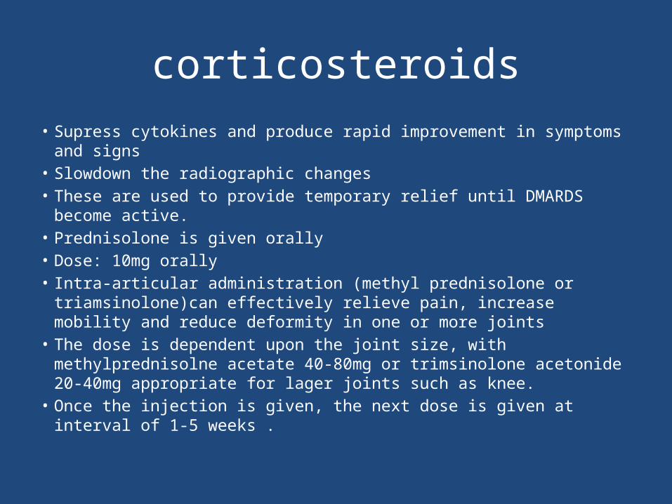
corticosteroids• Supress cytokines and produce rapid improvement in symptoms and signs• Slowdown the radiographic changes• These are used to provide temporary relief until DMARDS become active.• Prednisolone is given orally• Dose: 10mg orally• Intra-articular administration (methyl prednisolone or triamsinolone)can
effectively relieve pain, increase mobility and reduce deformity in one or more joints
• The dose is dependent upon the joint size, with methylprednisolne acetate 40-80mg or trimsinolone acetonide 20-40mg appropriate for lager joints such as knee.
• Once the injection is given, the next dose is given at interval of 1-5 weeks .

BIOLOGICS IN RA
• Cytokines such as TNF-α ,IL-1,IL-10 etc. are key mediators of immune function in RA and have been major targets of therapeutic manipulations in RA.
• Of the various cytokines,TNF-α has attaracted maximum attention.
• Various biologicals approved in RA are:-1) Anti TNF agents : Infliximab Etanercept Adalimumab 2) IL-1 receptor antagonist : Anakinra3) IL-6 receptor antagonist : Tocilizumab4) Anti CD20 antibody : Rituximab5) T cell costimulatory inhibitor : Abatacept

25 mg s/c twice a wkMay be given with MTX or as monotherapy.
Injection site reaction,URTI , reactivation of TB,development of ANA,exacerbation of demyelenating disease.

Agent Usual dose/route
Side effects
.
Anakinra
100 mg s/c once dailyMay be given with MTX or as monotherapy.
Injection site pain,infections,neutropenia
Active infections
Contraindications
(Anti-IL-1)

Lifestyle Modifications1. Quit smoking: According to Dr. Edward Skol, a rheumatologist with Scripps Health,
environmental factors such as smoking can sometimes trigger the development of rheumatoid arthritis.
2. Use dietary supplements: Fish oils and omega-3a can help control the inflammatory process of the disease, said Dr. Gregg Middleton, an associate clinical professor of rheumatology and orthopedics at the University of California, San Diego.
3. Lose weight: Dr. Natasha Conley, director of osteopathy at Kaiser Permanente, said it’s common sense that less pressure on the weight-bearing joints will ease discomfort for those with rheumatoid arthritis.
4. Perform low-impact exercises: Swimming, yoga, aerobics and other exercises that are easy on the joints can help keep the body properly stretched while not placing too much pressure on the body, said Skol.
5. Follow an anti-inflammatory diet: “A healthy diet is a good thing, even though the relationship between specific foods (and rheumatoid arthritis) are not linked,” Conley said. She stated that such foods would include fruits, vegetables and fish.