Embed Size (px)
Citation preview
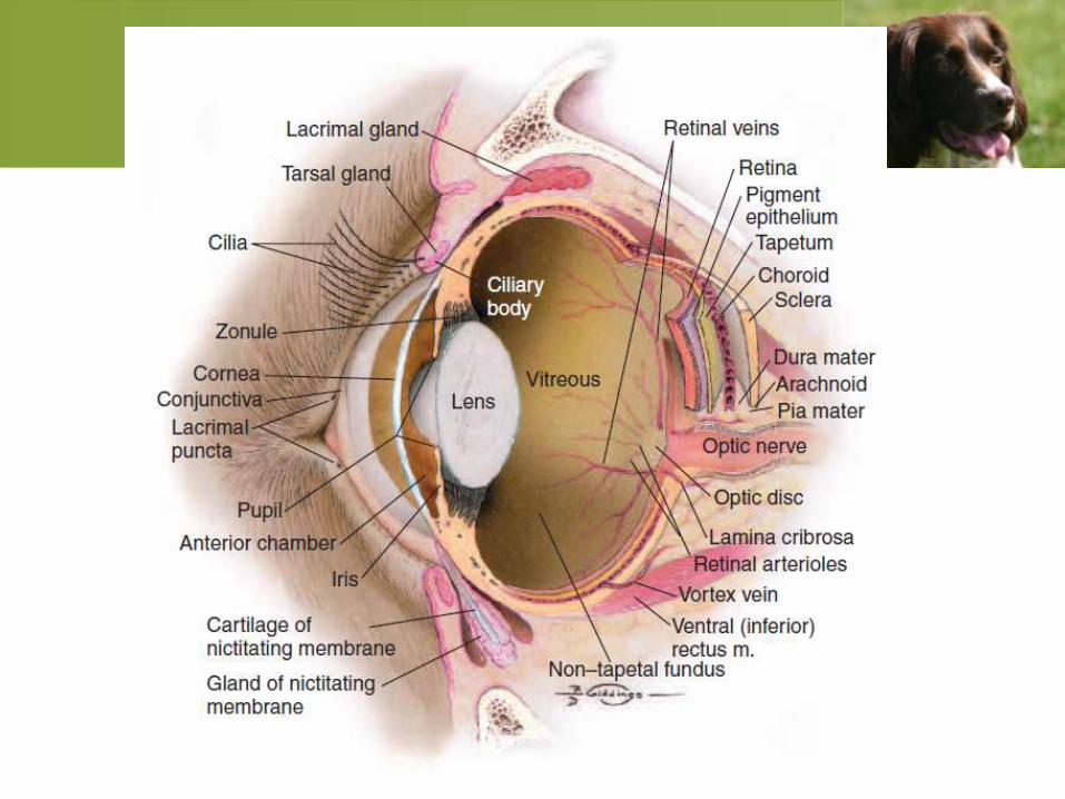
Ocular Emergencies
Dr Amit SinglaSurgery & Radiology,DGCN COVAS, CSKHPKV, PalampurHimachal Pradesh (INDIA)
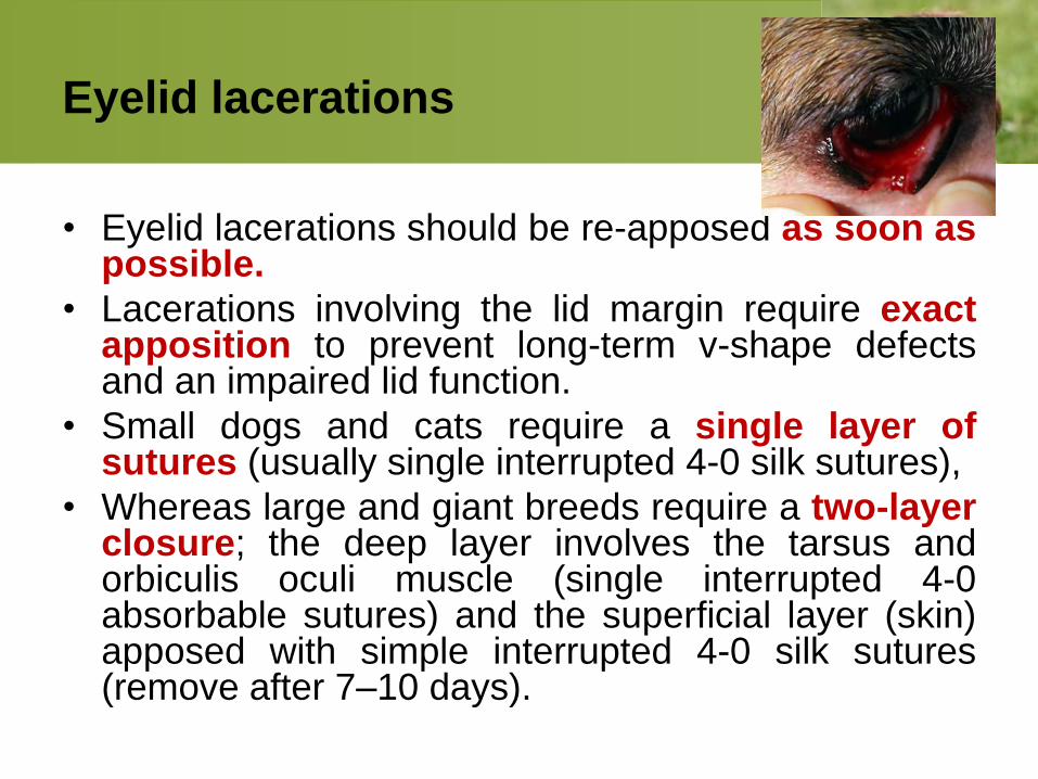
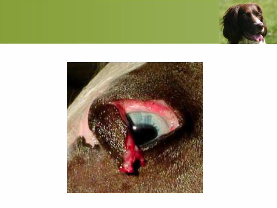
Eyelid lacerations
• Eyelid lacerations should be re-apposed as soon aspossible.
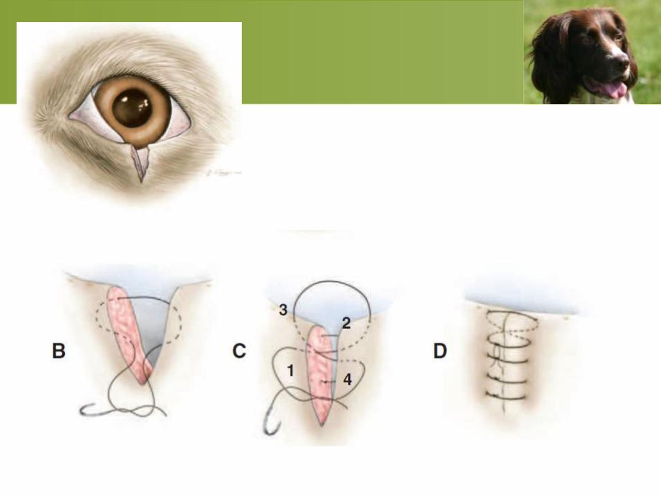
• Lacerations involving the lid margin require exactapposition to prevent long-term v-shape defectsand an impaired lid function.
• Small dogs and cats require a single layer ofsutures (usually single interrupted 4-0 silk sutures),
• Whereas large and giant breeds require a two-layerclosure; the deep layer involves the tarsus andorbiculis oculi muscle (single interrupted 4-0absorbable sutures) and the superficial layer (skin)apposed with simple interrupted 4-0 silk sutures(remove after 7–10 days).
• Horses require double-layer closure.
• When skin sutures are in place, the lid must be
protected from self-trauma by either an
Elizabethan collar (dogs and cats) or hard eye
cup (horses).
• Because the blink response is often impaired
by the swollen lid, a temporary tarsorrhaphy is
necessary to protect the cornea.
• Post-operative therapy often includes topical
antibiotics and corticosteroids, as well as
systemic antibiotics and NSAIDs.
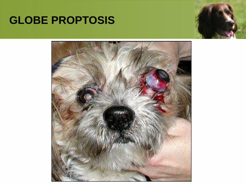
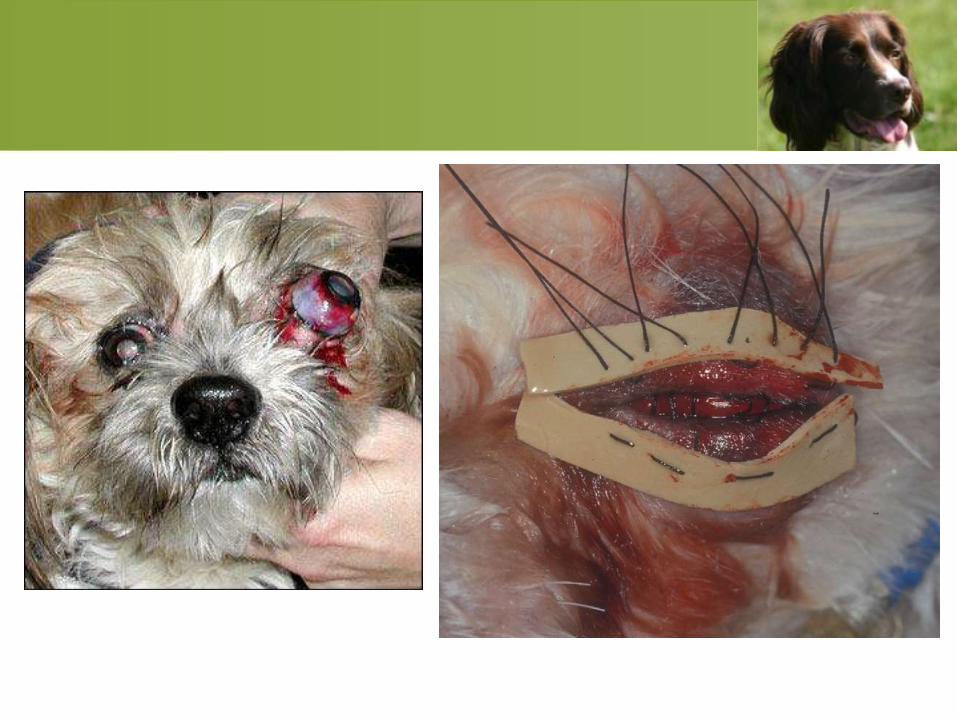
GLOBE PROPTOSIS
GLOBE PROPTOSIS
• Proptosis of the globe is common in
brachycephalic breeds with often surprisingly
little trauma causing the event.
• Non-brachycephalic breeds with a proptosis
have generally suffered severe head trauma.
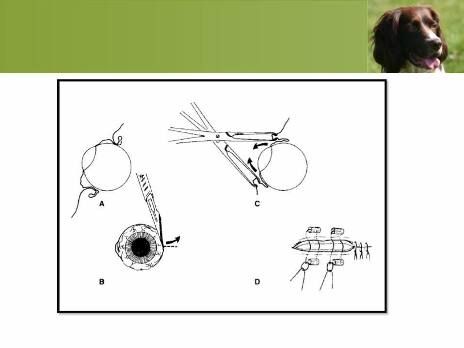
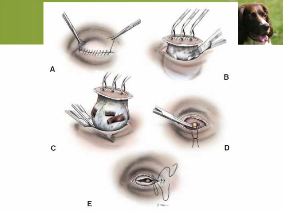
Repair
• General Anesthesia
• Site Prepration
• Perform a lateral canthotomy to improve globeaccess.
• Gentle traction on the eyelids. Use a flatinstrument such as a scalpel handlehorizontally across the cornea. Apply gentlepressure against the cornea to move globeposteriorly as the assistant pulls the eyelidscranially in front of globe.
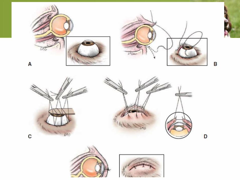
Precautions
• The globe will often not fully reduce due toswelling and hemorrhage of retrobulbar tissues.
• Close lateral canthotomy.
• Conjunctival surface (rubbing cornea) Shouldnot included in suture .
• Place 2-4 horizontal mattress sutures (temporarytarsorrhaphy) half depth through the eyelids (so asnot to have suture on the Be sure to use stints (cutpieces of IV tubing work well).
• Leave enough space at the medial canthus fortopical medications to be used.
Postoperative Care
• E collar;
• Topical antibiotic solution (neomycin tobramycin)
three times daily;
• NSAID;
• Antibiotic such as amoxicillin or cephalexin.
• Recheck in 5 days to assess tarsorrhaphy
sutures and any ocular discharge – if doing well,
leave sutures in place for 2 weeks.
CORNEAL ULCERS
• Broadly defined, a corneal ulcer is any
keratopathy in which there is loss of epithelium.
Ulcerative keratitis is an equivalent term
because there is always some inflammation
associated with corneal ulceration.
• Should be assumed to be infected with bacteria
regardless of appearance.
• Most corneal stromal ulcers will have a purulent
cellular infiltrate and associated uveitis.
• Very aggressive medical therapy is required to
treat deep corneal ulcers.
• Once ulcers become descemetoceles, or if they
fail to respond to medical therapy within a
couple of days, surgery with a conjunctival
graft will be needed to prevent corneal
perforation.
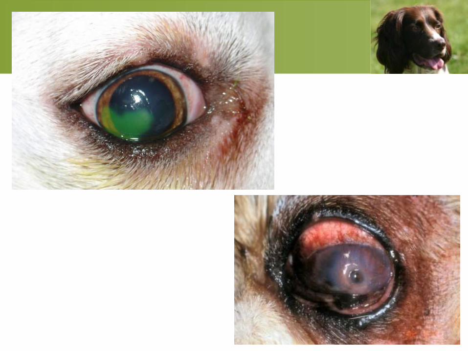
Clinical Signs of Deep Corneal
Ulceration
• Pain,
• Tearing (unless dry eye is present),
• Conjunctival hyperemia,
• Corneal edema and yellowish cellular infiltrate
around the ulcer,
• Variable corneal vascularization arising from the
limbus
• Miosis due to associated uveitis.
The basic diagnostic approach to
corneal ulceration should consist of
the following evaluations:
•Schirmer tear test
•Assessment of corneal and palpebral reflex
•Thorough examination of lid and conjunctival
anatomy and function, including the posterior
face of the third eyelid
•Microbiologic assessment if the ulcer is believed
to be infected
• Fluorescein staining
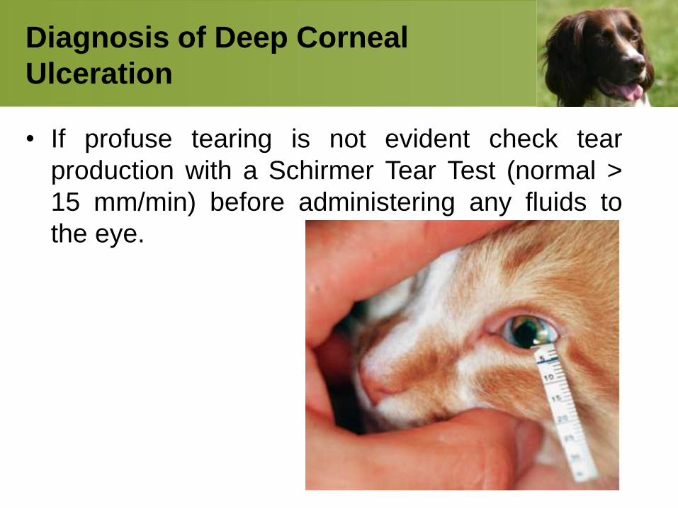
Diagnosis of Deep Corneal
Ulceration
• If profuse tearing is not evident check tear
production with a Schirmer Tear Test (normal >
15 mm/min) before administering any fluids to
the eye.
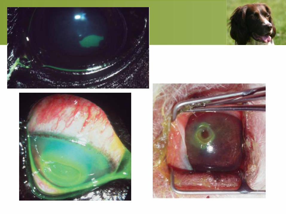
Fluorescein staining
• A stromal ulcer will take up fluorescein dye;
• A desmetocele will not absorb the dye on
Descemet’s membrane).
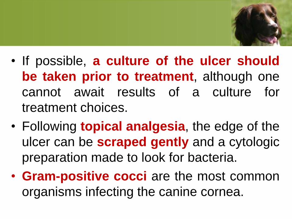
• If possible, a culture of the ulcer should
be taken prior to treatment, although one
cannot await results of a culture for
treatment choices.
• Following topical analgesia, the edge of the
ulcer can be scraped gently and a cytologic
preparation made to look for bacteria.
• Gram-positive cocci are the most common
organisms infecting the canine cornea.
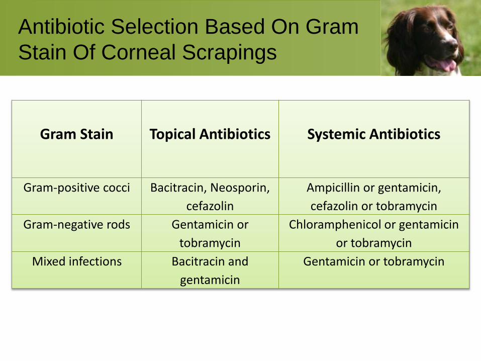
Antibiotic Selection Based On Gram
Stain Of Corneal Scrapings
Gram Stain Topical Antibiotics Systemic Antibiotics
Gram-positive cocci Bacitracin, Neosporin,
cefazolin
Ampicillin or gentamicin,
cefazolin or tobramycin
Gram-negative rods Gentamicin or
tobramycin
Chloramphenicol or gentamicin
or tobramycin
Mixed infections Bacitracin and
gentamicin
Gentamicin or tobramycin
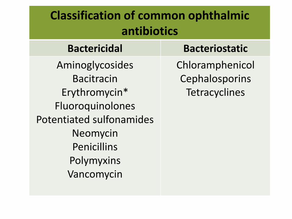
Classification of common ophthalmic antibiotics
Bactericidal Bacteriostatic
AminoglycosidesBacitracin
Erythromycin*Fluoroquinolones
Potentiated sulfonamidesNeomycinPenicillins
PolymyxinsVancomycin
ChloramphenicolCephalosporins
Tetracyclines
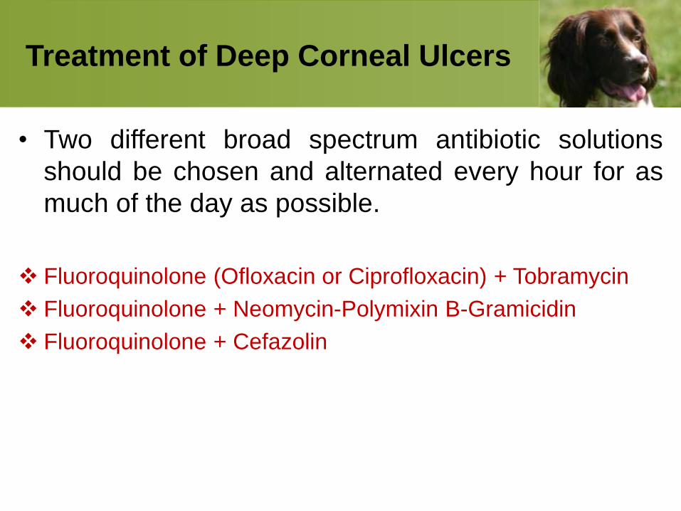
Treatment of Deep Corneal Ulcers
• Two different broad spectrum antibiotic solutions
should be chosen and alternated every hour for as
much of the day as possible.
Fluoroquinolone (Ofloxacin or Ciprofloxacin) + Tobramycin
Fluoroquinolone + Neomycin-Polymixin B-Gramicidin
Fluoroquinolone + Cefazolin
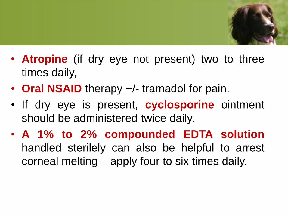
• Atropine (if dry eye not present) two to three
times daily,
• Oral NSAID therapy +/- tramadol for pain.
• If dry eye is present, cyclosporine ointment
should be administered twice daily.
• A 1% to 2% compounded EDTA solution
handled sterilely can also be helpful to arrest
corneal melting – apply four to six times daily.
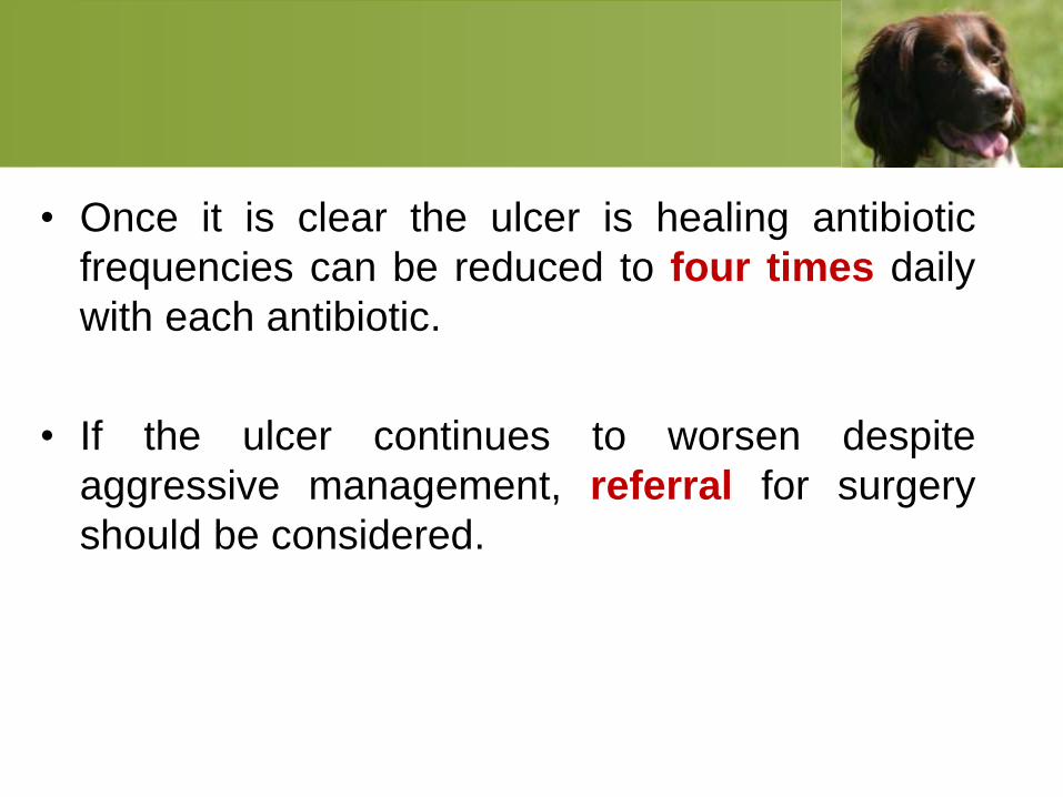
• Once it is clear the ulcer is healing antibiotic
frequencies can be reduced to four times daily
with each antibiotic.
• If the ulcer continues to worsen despite
aggressive management, referral for surgery
should be considered.
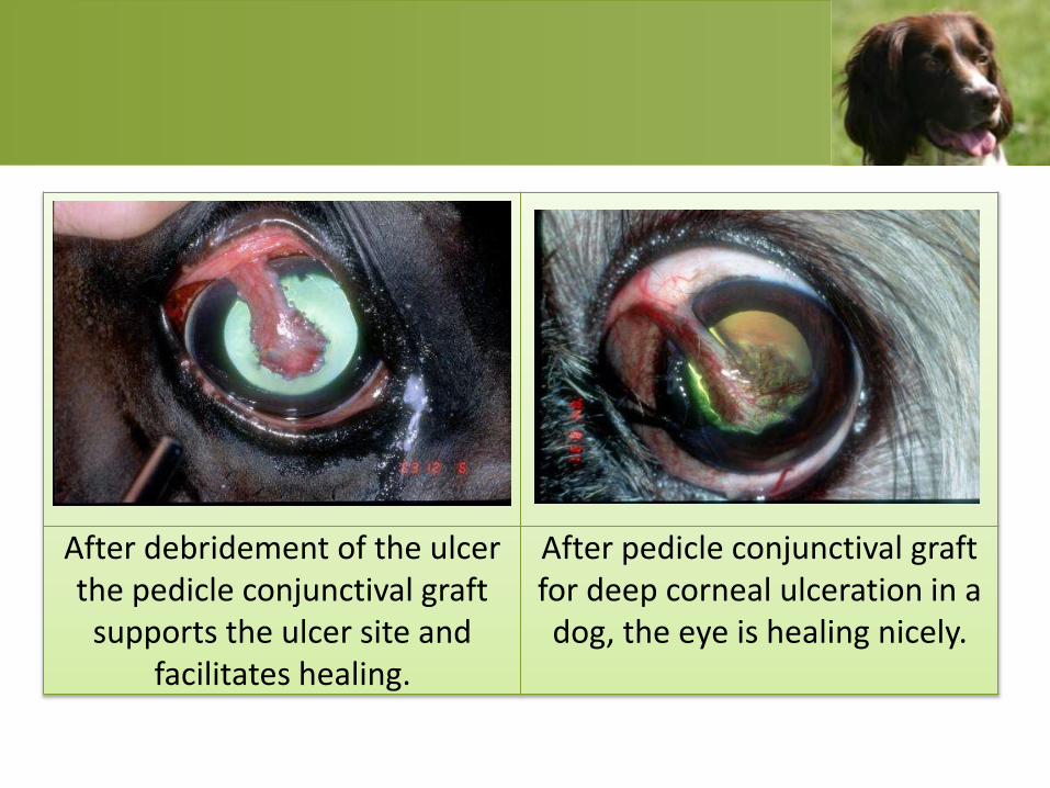
After debridement of the ulcer the pedicle conjunctival graft
supports the ulcer site and facilitates healing.
After pedicle conjunctival graft for deep corneal ulceration in a dog, the eye is healing nicely.
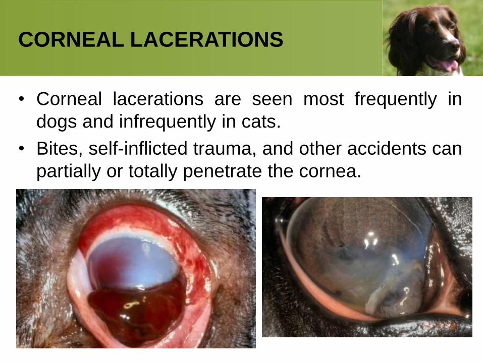
CORNEAL LACERATIONS
• Corneal lacerations are seen most frequently in
dogs and infrequently in cats.
• Bites, self-inflicted trauma, and other accidents can
partially or totally penetrate the cornea.
Partial-thickness corneal
lacerations
• Are usually highly painful and require apposition
with simple interrupted absorbable sutures to the
healthy cornea.
• Excision of the lacerated section is not
recommended.
For full-thickness corneal
lacerations
• Signs usually include pain, blepharospasm,
tearing, a corneal defect, and variable iris
prolapse.
• Marked aqueous flare, hyphema, miosis, and
distortion of the pupil are common.
• Often, the size of the iris prolapse is much
larger than the underlying corneal laceration.
• Small (< 4 mm) beveled lacerations near the
limbus often do not require suturing, especially
in younger patients.
• If suturing is required, it is better to wait 24 to 48
hours to evaluate the lens (the pupil is usually
dilated by this time) provided the anterior
chamber is formed. Slow aqueous leakage is
tolerated well for a few days, but if the chamber
is collapsed and does not reform in 3 to 5 hours,
then suturing is best.
IF THERE IS IRIS PROLAPSE:
• Iris prolapse helps seals an acute corneal
laceration and often results in reformation of the
anterior chamber and improved patient comfort.
But, the corneal can not heal with an iris
prolapse present. Therefore, lacerations with iris
prolapse (above the corneal surface) will require
surgical repair. However, they too can often wait
24 to 48 hours provided anterior chamber is
sealed.
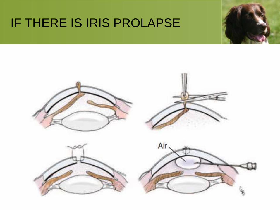
IF THERE IS IRIS PROLAPSE
The key to suturing a cornea is
the correct NEEDLE
• A spatula needle is required. 9-0 vicryl works
well, but 8-0 or 7-0 vicryl is acceptable and
with simple interrupted pattern.
• Remember to NOT penetrate into the anterior
chamber or epithelial down growth into the eye
can occur.
• To provide additional protection and support, the
sutured laceration may be covered with a third
eyelid flap, bulbar conjunctival graft, or
partial temporary tarsorrhaphy.
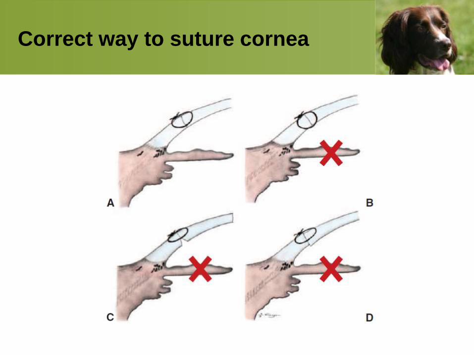
Correct way to suture cornea
• Once the laceration is bridged with fibroblasts
(and usually blood vessels) then topical
corticosteroid therapy to minimize scarring is
beneficial. The incision is often bridged in 10 to
12 days
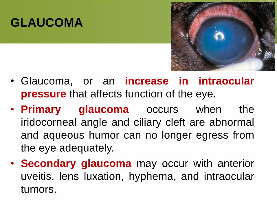
GLAUCOMA
• Glaucoma, or an increase in intraocular
pressure that affects function of the eye.
• Primary glaucoma occurs when the
iridocorneal angle and ciliary cleft are abnormal
and aqueous humor can no longer egress from
the eye adequately.
• Secondary glaucoma may occur with anterior
uveitis, lens luxation, hyphema, and intraocular
tumors.
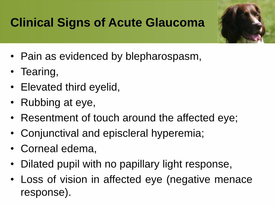
Clinical Signs of Acute Glaucoma
• Pain as evidenced by blepharospasm,
• Tearing,
• Elevated third eyelid,
• Rubbing at eye,
• Resentment of touch around the affected eye;
• Conjunctival and episcleral hyperemia;
• Corneal edema,
• Dilated pupil with no papillary light response,
• Loss of vision in affected eye (negative menace
response).
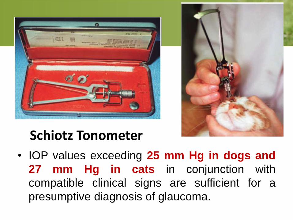
• IOP values exceeding 25 mm Hg in dogs and
27 mm Hg in cats in conjunction with
compatible clinical signs are sufficient for a
presumptive diagnosis of glaucoma.
Schiotz Tonometer
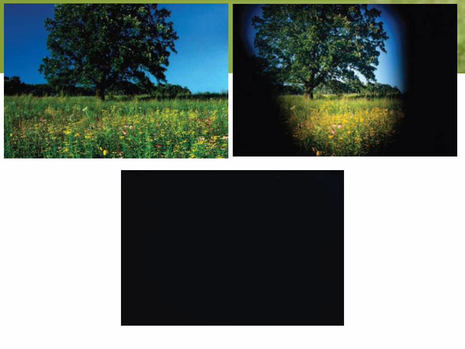
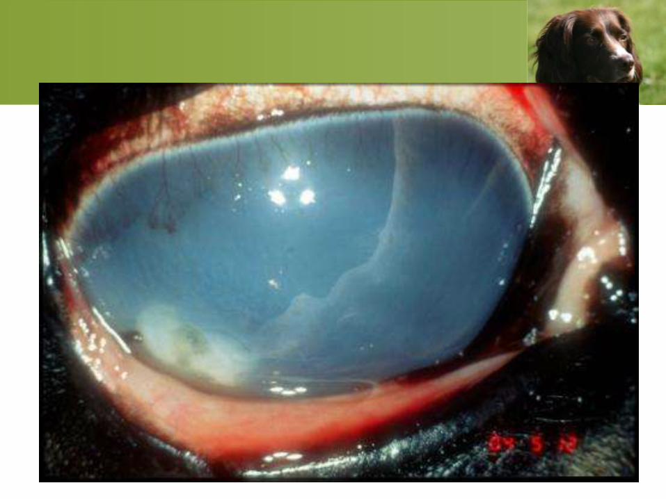
• In acute glaucoma the globe will be normal
sized. When the eye becomes buphthalmic
(enlarges) this is a sign of chronic glaucoma.
• Dogs also tend to be less demonstrative of
pain when glaucoma becomes chronic than
when it is acute, although their pain should not
be discounted.
• Most dogs are irreversibly blind with chronic
glaucoma.
Diagnosis of Glaucoma
• Measurement of intraocular pressure (IOP) is
imperative.
• Normal IOP in the dog is typically between 12
and 20 mmHg.
• In veterinary medicine the applanation and
rebound tonometers are recommended.
Treatment of Acute Glaucoma
• Reducing IOP as quickly as possible should be
the goal of treatment.
• A variety of topical glaucoma drugs are
available, some very good at reducing IOP.
• Drugs working by different mechanisms can be
administered together, waiting approximately 10
minutes between drops.
Carbonic Anhydrase Inhibitors
(CAI):
• These drugs decrease aqueous humor
production. (CAIs) by up to 50%.
• The most common drug in this category used in
dogs is dorzolamide.
• CAIs inhibit aqueous humor production and
have their onset of action within a few hours.
These agents should be used every 8 hours.
Beta Blocking Agents:
• These agents decrease aqueous humor
production.
• The most common one used in the dog is 0.5%
timolol maleate and is recommended not as a
sole agent but for use with other types of drugs.
• Timolol should be administered every 12 hours.
At this frequency heart rates are not typically
affected in the dog.
• Other beta blockers available are betaxolol and
levobunolol. These latter two have not been well
studied in the dog.
Hyperosmotic Agents:
• If combinations of the above topically applied
agents are unsuccessful at reducing IOP within
2 to 4 hours, mannitol can be administered.
• The dose is 1 to 2 g/kg given intravenously, over
about 30 to 45 minutes. Water should be
withheld for about 6 hours.
• Topical glaucoma drugs should be continued as
the effects of mannitol will last no longer than 24
hours.
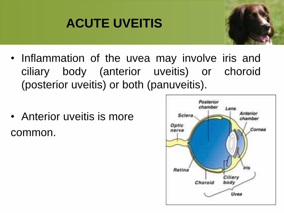
ACUTE UVEITIS
• Inflammation of the uvea may involve iris and
ciliary body (anterior uveitis) or choroid
(posterior uveitis) or both (panuveitis).
• Anterior uveitis is more
common.
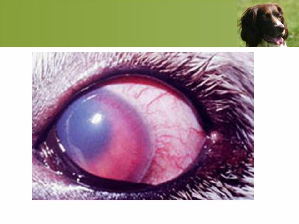
Clinical Signs
• Clinical signs include pain, conjunctival and
episcleral hyperemia, corneal edema, miosis,
decreased intraocular pressure, aqueous
flare, hyperemic iris, fibrin or blood
(hyphema) or white blood cells (hypopyon) in
the anterior chamber, and blindness in
affected eye (especially if posterior uveitis is
present as well).
Diagnosis
• Visualization of some or all of the above clinical
signs.
• Rule out glaucoma through measurement of
IOP.
• Stain the cornea with fluorescein dye to check
for ulceration.
Treatment
• Treatment should be very aggressive to manage
ocular inflammation and pain.
• Topical corticosteroid therapy should be instituted
(as long as no corneal ulceration is present) with
either 1% predinisolone acetate (shake very well) or
0.1% dexamethasone, four to six times daily.
• Atropine for iridocycloplegia should be instituted at
twice daily as long as IOP is low.
• If IOP is normal in the face of uveitis, glaucoma may
already be developing and atropine should be
avoided.
• Systemic anti-inflammatory agents such as
NSAIDs or in severe uveitis, oral prednisolone at
1 to 2 mg/kg/day, should also be started.
• If posterior uveitis exists, only systemically
administered agents will reach these tissues and
oral prednisolone becomes imperative.
• If pain is severe, oral narcotic agents such as
tramadol should also be administered.
• Dogs with acute uveitis should be re-examined
in about 2 to 3 days.
• Therapies should be only gradually decreased
as improvement is seen. IOP should be
measured at every visit.
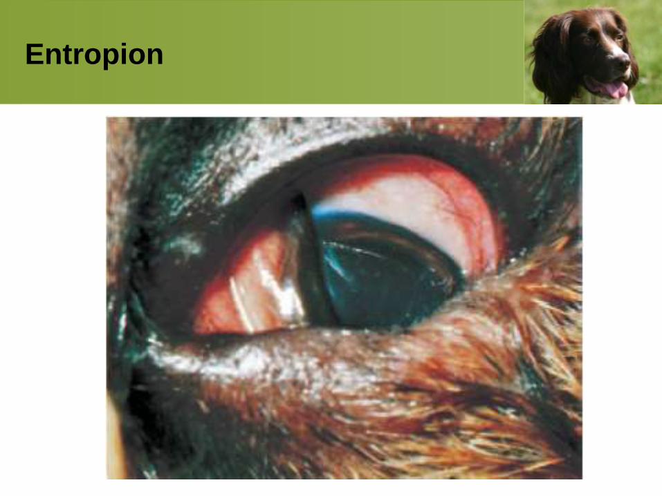
Entropion
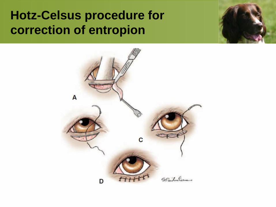
Hotz-Celsus procedure for
correction of entropion
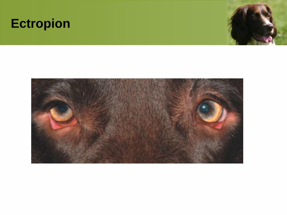
Ectropion
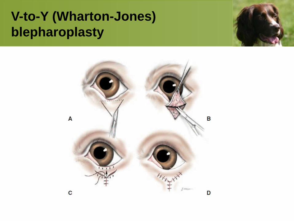
V-to-Y (Wharton-Jones)
blepharoplasty
Transpalpebral enucleation/exenteration
INDICATIONS
• Intraocular neoplasia
• Severe perforating ocular trauma
• Uncontrollable endophthalmitis or panophthalmitis
• Intractable ocular pain, especially in glaucomatous
eyes
• Owner inability or unwillingness to give long-term
treatment to a blind eye to keep it comfortable

Thanks


![ocular emergencies-1.ppt [Read-Only] - ocw.usu.ac.idocw.usu.ac.id/.../emd166_slide_ocular_emergencies.pdf · Objectives of presentation Review ocular anatomy Understand basic ophthalmic](https://img.pdfslide.us/doc/110x75/5b5334587f8b9a575f8b6a7a/ocular-emergencies-1ppt-read-only-ocwusuacidocwusuacidemd166slideocular.jpg)