Embed Size (px)
Citation preview

Shrivardhan DheemanAsst. Professor (Adhoc)
Department of Botany and MicrobiologyGurukul Kangri University, Haridwar
(INDIA)
www.sites.google.com/site/shrivardhandheeman

SD

Environment MonitoringDefinition:
“Monitoring of viable contamination within clean environments.”
Type of Environmental Monitoring:Physical Test
Microbiological Test
SD
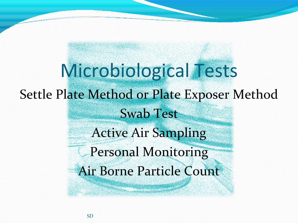
Microbiological TestsSettle Plate Method or Plate Exposer Method
Swab TestActive Air SamplingPersonal Monitoring
Air Borne Particle Count
SD

Settle Plate Method or Plate Exposer Method
Test done under following points:Prepare SCDA plate and incubate for 24 hrs. at 32.5ºC.
Observe all incubated plate for contamination.Select plate and put into stain less steel container.
Go for samplingLeave all plate front of raiser with marking date, time and operator name
and respective sampling position on plate.Collect all plate after on air change cycle time (4 hrs.)
Incubate all plate for 3 days at 22.5ºC and later by 2 days at 32.5ºC.
SD
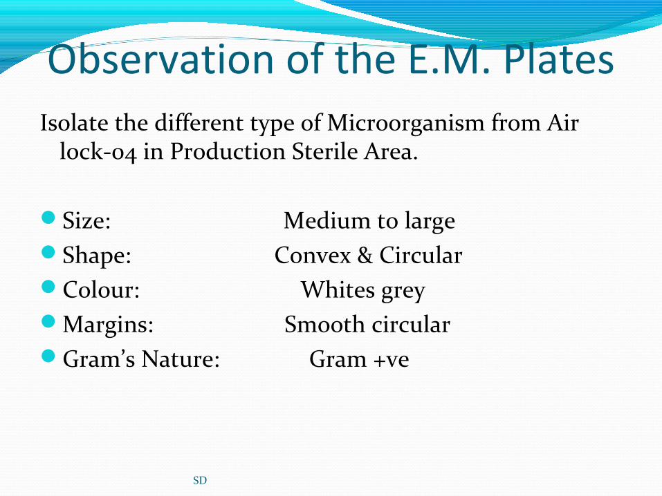
Observation of the E.M. PlatesIsolate the different type of Microorganism from Air
lock-04 in Production Sterile Area.
Size: Medium to largeShape: Convex & CircularColour: Whites greyMargins: Smooth circularGram’s Nature: Gram +ve
SD

Growth on Selective Agar For E.coli
MacConkey Broth: Turbidity found
MacConkey Agar: Growth observed on Agar Plates but E.coli character not observed on MacConkey Agar.
EMB Agar: Growth observed on EMB Agar Plates but
E.coli character not observed on EMB Agar.
SD

Growth on Selective Agar For Salmonella
Rappaport Vasiladis Salmonella Enrichment Broth: Turbidity found
XLDA Agar: Growth observed on Agar Plates but Salmonella character not observed on XLDA Plates.
BGA Agar: Growth observed on Agar Plates but Salmonella character not observed on BGA Agar
Plates.
SD

Growth on Selective Agar For Pseudomonas
Cetrimide Agar: Growth observed on Agar Plates but Pseudomonas character not observed on
Cetrimide Agar. Pseudomonas Agar: Growth observed on EMB Agar
Plates but Pseudomonas character not observed on Pseudomonas Agar.
SD

Growth on Selective Agar For Staphylococcus
Mannitol Salt Agar: Growth observed on Agar PlatesGrowth characteristic: Yellow colonies with yellow
zone
Vogel Johnson Agar: Growth observed on VJA Agar Plates
Growth characteristic : Black colonies with yellow zone
SD

IDENTIFICATION BY BIOCHEMICAL
0.5 ml of Mammalian plasma & transfer the selective colonies incubate on water bath at 37ºC and observed
after 3hr coagulation in the test tube.It means Staphylococcus are confirm.
GENETICALLY CONFIRMATION BY MTCC CHANDIGARH
ENVIRONMENT ISOLATE-IStaphylococcus hominis
SD

Swab TestTest done under following points:• Prepare swab or use pre-sterile cotton swab.
• Take 0.9% sterile saline solution• Take aluminum foil centrally holed of 5x5 cm.
• Put all requirement into stain less steel container.• Go for sampling in production area/microbiology lab.• Select sampling location as per testing schedule.
SD

Swab Test• Fix the aluminum foil at surface where test will be done.• Open the swab and rotate the swab horizontal and vertical
position across 5x5 hole on aluminum foil.• Take swab inside its tube and marked with date, time, operator
name and location identity.• Take them all in SS Container and bring to lab.
• Swab it on media containing petri plate.• Incubation petri plate for 3 days at 22.5ºC and later by 2 days at
32.5ºC.
SD
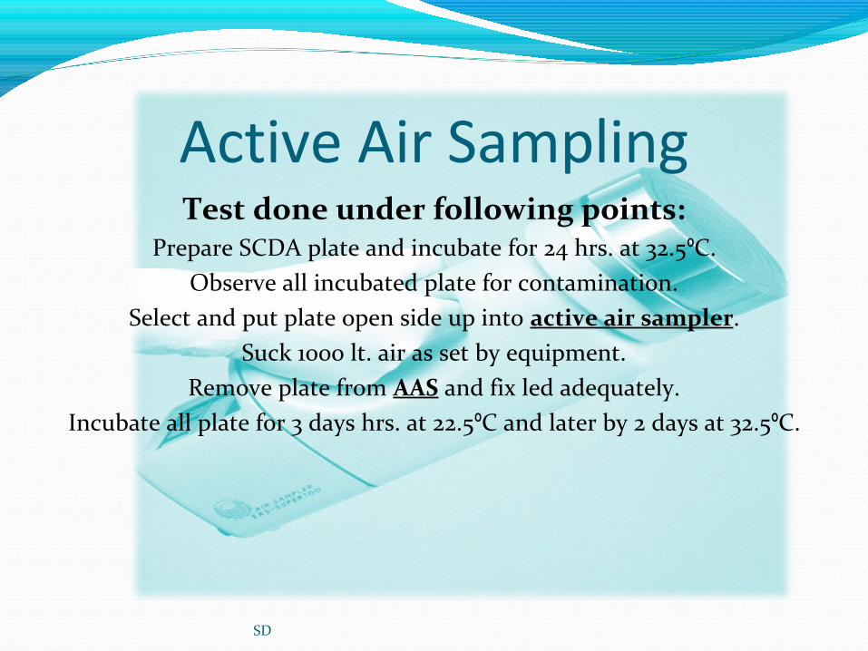
Active Air SamplingTest done under following points:
Prepare SCDA plate and incubate for 24 hrs. at 32.5ºC.Observe all incubated plate for contamination.
Select and put plate open side up into active air sampler.Suck 1000 lt. air as set by equipment.
Remove plate from AAS and fix led adequately.Incubate all plate for 3 days hrs. at 22.5ºC and later by 2 days at 32.5ºC.
SD
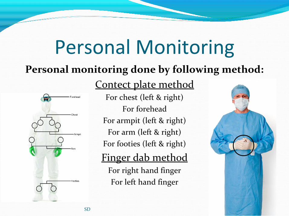
Personal MonitoringPersonal monitoring done by following method:
Contect plate methodFor chest (left & right)
For foreheadFor armpit (left & right)
For arm (left & right)For footies (left & right)
Finger dab methodFor right hand fingerFor left hand finger
SD

Air Borne Particle CountThis test is totally eqipment based test.
Equipment count the 0.5 to 5.0 µ size particle in the clean room. Operating
procedure are as following:Switch on the equipment.
Select the location to monitor.Press run/ok on main screen.
Observe the reading.Collect the printout.
SD

SD

Sterility TestingObjective:
To measure the sterility of finished parenteral and pediatric formulations administrated I.V./I.M/inf. This test is
employed to ascertain sterility of product, that it is sterile or not.
Definition of sterility:“Sterility is phase of product when product is consider sterile”. It whether terminally sterilize or the processed
under sterilize condition and handling.
SD

Sterility TestingPrinciple:
This test is based upon the principle that “if bacteria and fungi are placed in a medium which provides nutritive
material and water, and kept at favorable temperature, the organisms will grow and their presence can be indicated by
turbidity in the originally clear medium.”
Type of Sterility Testing:Filtration method
Direct inoculation method(Filtration method is widely accepted)
SD

Filtration MethodRequirements:
Filtration assemblyLaminar air flow hood
Filter paperBOD Incubator
IPA 70%Tissue paper role
ForcepsBunsen burner
FTGM & SCDM brothSterile blade
Vacuum pumpSD

Filtration MethodProcedure:
Open the filtration assembly.Dissolve injection into 100 ml peptone solution aseptically.
Put the filter paper into assembly.Filter the solution through filtration assembly.
Wait until all solution filter.Open filtration assembly and cut the filter into two.
One insert into SCDM broth and second in FTGM broth.Mark each with date, time, injection name, batch no.,etc.
Incubate all the tubes at 32.5ºC for 14 days
SD

SD

Microbial Limit TestThis test is designed to perform:
Total Viable Count (TVC) of bacteria and fungi (Quantitative estimation).
There is two test carried for quantitative estimation:TBC (Total Bacterial Count)
TFC (Total Fungal count)
Four Method are employed for this test:Filtration methodPour pate method
Spread plate methodSerial Dilution Method
SD

Microbial Limit TestEnrichment for qualitative estimation:
Identification of microorganism by cultivating on selective media comparing ATCC/MTCC culture of pathogen (Qualitative
estimation).
In qualitative estimation the four more pathogenic bacteria are detecting under this test, which are following:
Escherichia coliPseudomonas aeruginosa
Staphylococcus aureusSalmonella abony
SD

Pathogen Detection: Escherichia ColiPrimery test
Transfer 1 ml enrichment broth into Mac Conkey broth and incubate for 48 hrs. at 42ºC.Observation:
Turbidity shows E. coli may be presentClear broth shows E. coli may be absent.
Secondry testStreak one loop full culture on EMB/MCA media and incubate for 48 hrs. at 32ºC.
Observation:Metallic sheen on EMB and brick red colonies on MCA shows E. coli present
No growth shows E. coli absent.Confirmatory test
Inoculate a loop full colony in trypton broth and incubate for 48 hrs. at 32ºC.After incubation add 1 ml kovac’s reagent.
Observation:Red ring on broth surface shows E. coli confirmed
No ring formation shows E. coli absent.
SD
Primery testTransfer 0.1 ml enrichment broth into Rappaport Visiliadis Salmonella Enrichment broth and
incubate for 48 hrs. at 32.5ºC.Observation:
Color change from blue to pale shows Salmonella spp. may be presentNo color change in broth shows Salmonella spp. may be absent.
Secondry testStreak one loop full culture on XLDA/BGA media and incubate for 48 hrs. at 32.5ºC.
Observation:Colorless/pink-white colony on XLDA and Black centered red on BGA shows Salmonella spp. present
No growth shows Salmonella spp. absent.Confirmatory test
Subculture in TSI butt and incubate for 48 hrs. at 32.5ºC.Observation:
Pink color with yellow butt shows Salmonella spp. confirmedNo change in butt shows Salmonella spp. absent.
SD
Pathogen Detection: Salmonella abony

Primery testTransfer 1 ml enrichment broth into 100 ml Citrimide broth and incubate for 48 hrs. at 32.5ºC.
Observation:Turbidity shows Pseudomonas aeruginosa may be present
Clear broth shows Pseudomonas aeruginosa may be absent.Secondry test
Streak one loop full culture on CtA/PA media and incubate for 48 hrs. at 32.5ºC.Observation:
Greenish colony on CtA and green-blue on PA shows Pseudomonas aeruginosa presentNo growth shows Pseudomonas aeruginosa absent.
Confirmatory testReplica colony on whatman filter paper and impragment N, N, N, N,-tetra-methyl, 4 phenyl
adenine.Observation:
Pink color shows Pseudomonas aeruginosa confirmedNo change shows Pseudomonas aeruginosa absent.
SD
Pathogen Detection: Pseudomonas aeruginosa

Primery testTransfer 1 ml enrichment broth into 100 ml MSB and incubate for 48 hrs. at 32.5ºC.
Observation:Turbidity shows Staphylococcus aureus may be present
Clear broth shows Staphylococcus aureus may be absent.Secondry test
Streak one loop full culture on MSA/VJA media and incubate for 48 hrs. at 32.5ºC.Observation:
Yellow colony on MSA and yellow surrounded black on VJA shows Staphylococcus aureus presentNo growth shows Staphylococcus aureus absent.
Confirmatory testSubculture in MSB and add 0.5 ml mammalian/ horse plasma and incubate in water bath at 37ºC
for 3 hrs.Observation:
Coagulation shows Staphylococcus aureus confirmedNo change shows Staphylococcus aureus absent.
SD
Pathogen Detection: Staphylococcus aureus

Result of Pathogen Detection
SD
Medium Description of colonyMCA Brick red colonies with a surrounding zone of ppt. bileEMB Metallic sheen under reflected light and blue black appearance
under transmitted light.
Medium Description of colony MSA Yellow color with yellow zone
VJA Black, surrounded by yellow zone
Sample plate have no colony
Escherichia coli
Staphylococcus aureus
Sample plate have no colony

Result of Pathogen Detection
SD
Medium Cetrimide Agar Medium
Pseudomonas agar medium for detection of fluorescein
Pseudomonas agar medium for detection of pyocyanin
Characteristic colonial
morphology
Generally greenish Generally colorless to yellowish
Generally greenish
Florescence in UV light
Greenish Yellowish Blue
Medium Description of colony BGA Pinkish white colony
XLDA Red colonies with or without black center.
Pseudomonas aeruginosa
Sample plate have no colony
Sample plate have no colony
Salmonella abony

SD