Embed Size (px)
Citation preview

• Mastication.• Chewing Cycles• Oral Reflexes
• Dr: Mu’taz alshawabekah
Definition
• Mastication: is process whereby ingested food is cut or
crushed into small pieces mixed with saliva and formed into bolus in preparation for swallowing.
Function of Mastication • Function of Mastication:1. Enables the food bolus to be easily swallowed2. Enhances the digestibility of food by:a. Decreasing the size of particles to increase the surface area for
enzyme activityb. Reflexively stimulating the secretion of digestive Juices (saliva and
Gastric Juice)3.Mixes the food with saliva, initiating digestion by the activity of
salivary amylase4.Prevent irritation of the GI system be large food masses5.Enusres healthy Growth and development of the oral tissues.6. Increase in Digestive efficiency, the Primary purpose of mastication

How does it occur?
• Mastication occurs by the convergent movements of max. & man. Teeth.
• Most foods are first crushed by vertical movements of the mandible before being sheared by lateral to medial movements of the mandible to make a bolus.
• The initial crushing of the food does not require full occlusion of the teeth. Indeed, it is often only after the food has been well softened that the maxillary and mandibular teeth eventually contact.
• Once the cusp can interdigitate, the ridges on the slopes of the cusp shear the food as the mandibular teeth move across the maxillary teeth.

Cont’d
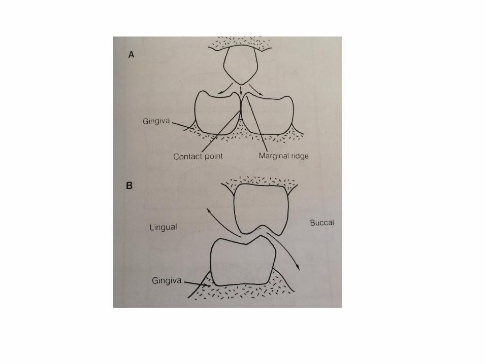
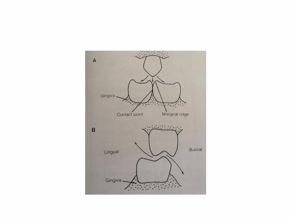
• PIC FIG 6.1 pg 91 Several features provide protection for the adjacent gingiva
during chewing.1)The marginal ridges bounding the interproximal edges of
the occlusal surfaces of the tooth are the important protective features. These ridges deflect most of the food, potentially driven between adjacent teeth by their opponents, onto the occlusal surfaces.
2)The Contact points beneath the marginal ridges should abut firmly to prevent food being wedged between the teeth and above the interdental papillae.
Cont’d
• The buccal cusps of the mandibular teeth bite between the buccal and palatal cusps of the maxillary teeth, with the result that food trapped between them is forced up over the palatal sides of the maxillary teeth and down over the buccal sides of the mandibular teeth.
Fig 6.1 page 91 B
Cont’d
• Mastication is not simply a result of rhythmically closing teeth of a particular form on a piece of food. Also it includes the placement of food between the occluding surfaces of the teeth by the tongue and the selection by the tongue of those pieces of food in the mouth.

Structural features associated with mastication:• TMJ articulation • Serous salivary gland• Prismatic Enamel• 2nd Palate• Significant muscle development associated with lips
,cheeks, tongue and muscles of mastication• Diphyodonty• Gomphosis type of tooth attachment
Bite Force
• Mastication is dependent upon a complex chain of events, that produce rhythmic opening and closing movements of the jaws and correlated tongue movements.
• The forces that are exerted on the teeth and jaws are very large and physiologically significant.
• The bite Force exerted on the food during mastication is of the order of 5-15kg. It varies according to the texture of the food
• The bite pressure is measured with a gnathodynamometer, maximum pressures of the order of 50 kg can be recorded
• 64 N in denture Wearer.
Fig 6.2 pg 91
• Rhythmic jaw movements are generated by a centre within the brainstem. This is referred to as an Oral Rhythm/Pattern generator and is activated both by drive from the higher centers and peripheral sensory input. And the pattern of activity is distributed to the motor neuron pools which receive excitatory or inhibitory sensory inputs from a variety of peripheral structures
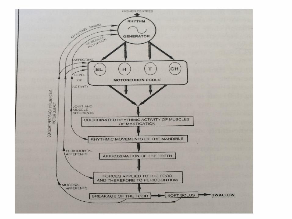
• Sensory input generated by closing on hard food generation of rhythmic jaw activity closing on a softened bolus
Tongue movement and food transport swallow terminate the rhythmic jaw activity.
Sensory Receptors in masticatroy Muscles
• Control of Muscle Movement• Unevenly distributed in the muscle of mastication.Muscle Spindles:• Many in elevators and tongue muscles• Few in the depressor musclesGolgi tendon organs:• Not known if they exist in Elevators• Protect against overdevelopment of muscle tension• Performed by PDL receptors= it limits the force applied
in the mastication.
Control of mastication
Amount of chewing before swallowing is :Characteristic of the individual.Influenced by nature of food. Number of strokes before swallowing:In men>womenwomen> childrenNot markedly influenced by state of Dentition
Chewing/Chewing Cycle
• Is highly complex process.• 2 methods of chewing have been distinguished depending
upon the texture of the food:1) puncture/crushing :hard food is first crushed and pierced
between the teeth without direct tooth-to-tooth contact. This results in wear of the teeth, especially at the tips of the cusps.
2) Shearing stroke. This method involves tooth contacts that take place only after the food has been reduced. This type of movement produces attrition facet with characteristic directional scratch lines on the faces of the cusps
• .
• The mean of the vertical dimension of the chewing cycle are between 16 and 20 mm and between 3 and 5 for lateral movements
• The duration of the cycle varies between 0.6 and 1second depending on the type of food
• The speed of masticatory movement varies within each cycle according to types of food and among individuals
• Speed , duration and form of the chewing cycle vary with the type of occlusion , kind of food and presence of dysfunction
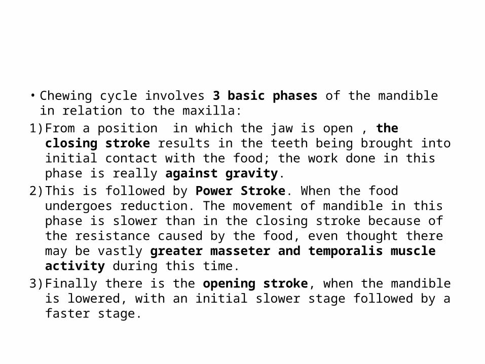
• Chewing cycle involves 3 basic phases of the mandible in relation to the maxilla:
1) From a position in which the jaw is open , the closing stroke results in the teeth being brought into initial contact with the food; the work done in this phase is really against gravity.
2) This is followed by Power Stroke. When the food undergoes reduction. The movement of mandible in this phase is slower than in the closing stroke because of the resistance caused by the food, even thought there may be vastly greater masseter and temporalis muscle activity during this time.
3) Finally there is the opening stroke, when the mandible is lowered, with an initial slower stage followed by a faster stage.
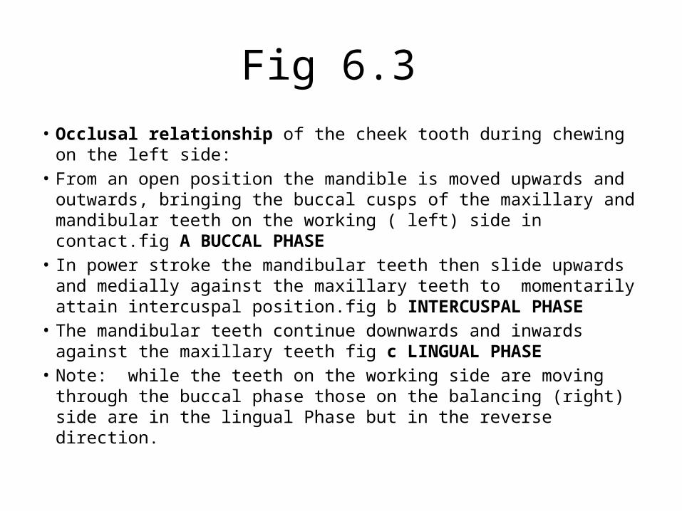
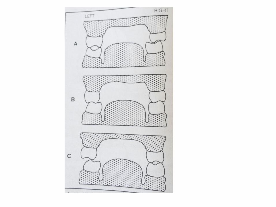
Fig 6.3 • Occlusal relationship of the cheek tooth during chewing on the left
side:• From an open position the mandible is moved upwards and
outwards, bringing the buccal cusps of the maxillary and mandibular teeth on the working ( left) side in contact.fig A BUCCAL PHASE
• In power stroke the mandibular teeth then slide upwards and medially against the maxillary teeth to momentarily attain intercuspal position.fig b INTERCUSPAL PHASE
• The mandibular teeth continue downwards and inwards against the maxillary teeth fig c LINGUAL PHASE
• Note: while the teeth on the working side are moving through the buccal phase those on the balancing (right) side are in the lingual Phase but in the reverse direction.
The envelope of Motion fig 6.5
• The pathway followed by the mandible during chewing is termed “ the envelope of Motion”
• It demonstrates the symmetrical mandibular movements produced during opening and closing of the jaw.
• The envelope of motion is the volume of space within which all movements of a specified point on the mandible occur.
• The envelope is limited by the anatomical considerations such as ligaments and tooth contacts
• Most natural movements occur within the “envelope” .
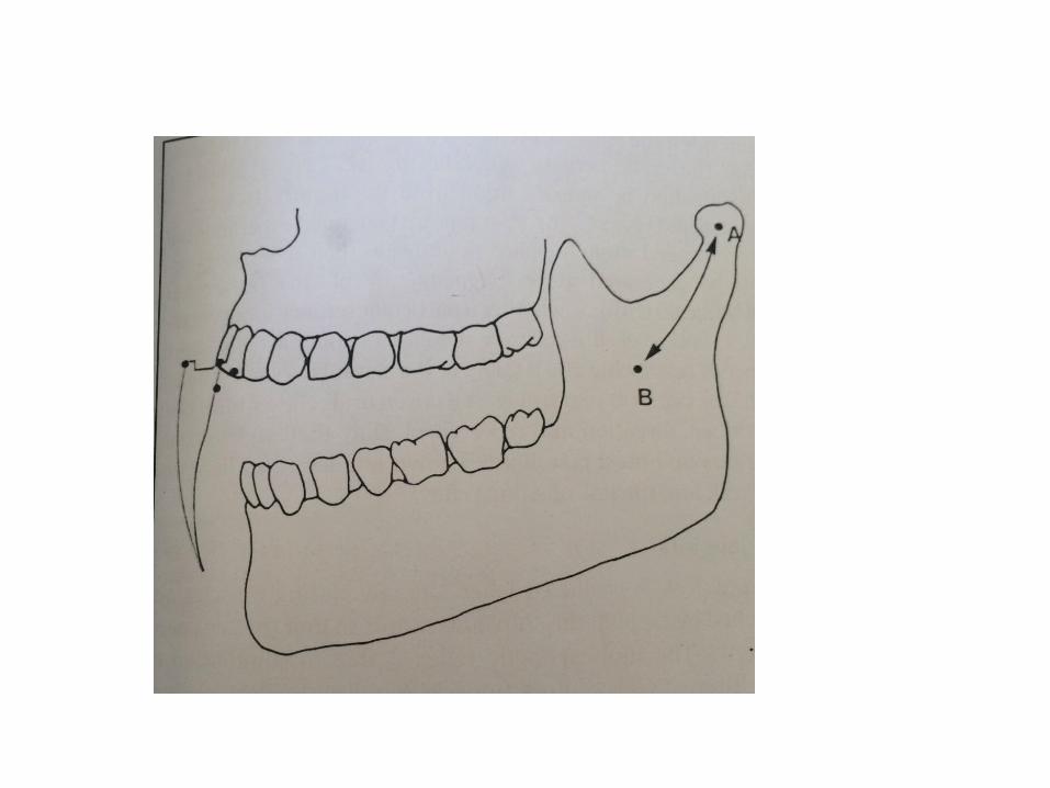
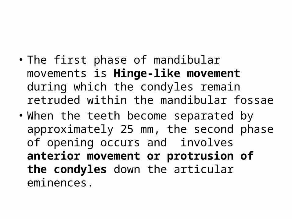
• The first phase of mandibular movements is Hinge-like movement during which the condyles remain retruded within the mandibular fossae
• When the teeth become separated by approximately 25 mm, the second phase of opening occurs and involves anterior movement or protrusion of the condyles down the articular eminences.
• Fig 6.5 • Point A is the fulcrum associated with simple hinge
movements.• The path described between points A and B represents
the shift of the centre of rotation of the mandible , this shift occurs because of the transition from a pure hinge movement at the condyle to protrusion and rotation during opening ( with reverse during closing).
• Point B has been described as representing the point of rotation around the attachment of Sphenomandibular ligament at the lingula.

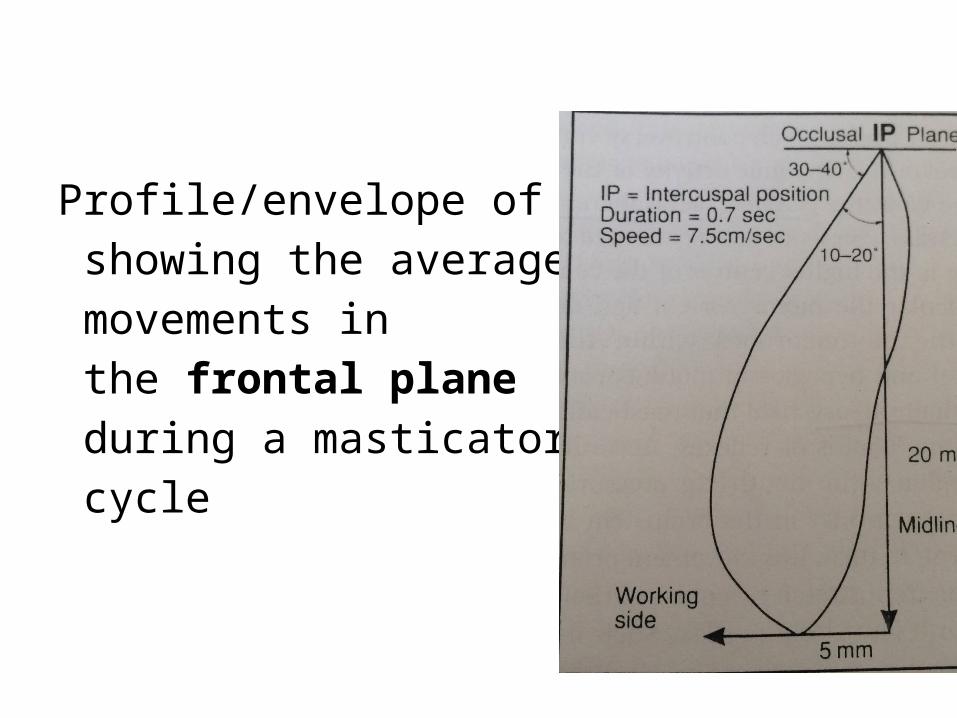
Profile/envelope of motion showing the average incisal movements in the frontal plane during a masticatory cycle
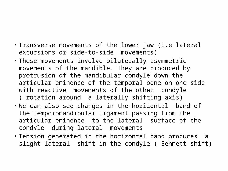
• Transverse movements of the lower jaw (i.e lateral excursions or side-to-side movements)
• These movements involve bilaterally asymmetric movements of the mandible. They are produced by protrusion of the mandibular condyle down the articular eminence of the temporal bone on one side with reactive movements of the other condyle ( rotation around a laterally shifting axis)
• We can also see changes in the horizontal band of the temporomandibular ligament passing from the articular eminence to the lateral surface of the condyle during lateral movements
• Tension generated in the horizontal band produces a slight lateral shift in the condyle ( Bennett shift)
The Control of Mastication
• In the past, there has been much controversy concerning the origin and control of the rhythmic activity of the jaws during mastication.
• The Cerebral Hemispheres Theory: • Mastication was a conscious act, a patterned set of
instructions originating in the higher centers of the CNS ( particular the motor cortex) and descending to directly drive the motorneurons within the brainstem ( trigeminal, facial , hypoglossal motorneurons)
Cont’d
• The reflex Chain Theory • Mastication involved a series of interacting chains of reflexes,
accordingly sensory input from the region of the mouth (e.g. pressure on the teeth) triggered the motorneurons in the brainstem to elicit a jaw opening movement. In turn, this movement produced another sensory input(e.g. from stretch receptors in the jaw muscles), which resulted in a jaw closing reflex, such a theory could explain the rhythmic jaw movements seen in decerbrate animals.
• Objections to this theory: mastication involves prolonged bursts of muscle activity and not the brief and abrupt behaviour usually associated with reflex activation of muscle.
Cont’d
• Rhythm (pattern)generation theory:• Most accepted theory
• Is based upon the proposition that there are central pattern generators(CPGS) within the brainstem ,which, on being stimulated from either higher centers or sensory input in the region of the mouth, are driven on rhythmic activity.
Cont’d
• The activity of this generators depends upon excitation descending in the pathways from cerebral cortex and upon excitation deriving from peripheral stimulation
• The rhythmic activity can be generated by conscious drive and/or by the presence of the food in the mouth.
Cont’d
• During mastication the cycles of jaw movement differ depending upon the consistency of the food initially ingested and upon the stage of breakdown of that food
• This indicates that the cyclic activity generated by the CPG is subjected to modification by sensory from the mouth.
• Oral reflexes : • The Jaw Jerk.• Jaw Opening Jerk.
• Jaw jerk:• Is produced when the jaw closing muscles are stretched by
tapping the chin downwards so that the jaw open suddenly.• It is monosynaptic Reflex• Is due to stimulation stretch-sensitive receptors (muscle
spindle ) in the masseter and temporalis muscles.• The stretch produces a burst of impulses in the sensory
nerves that is conveyed back to the motor neurons of those muscles
• The muscles are consequently activated briefly to produce a short-lived contraction

• Jaw opening reflex• Polysynaptic• Produced by applying mechanical or electrical stimuli to oral
mucosa, PDL or teeth• The stimuli do not have to be painful to elicit the reflex but
stronger stimuli do produce correspondingly more vigorous responses
• Characterized by a brief period of inhibition of activity in the motor neurons of the jaw closing muscles, however in other mammals there is in addition a simultaneous activation of the jaw opening muscles (digastrics and infrahyoid muscles )