Embed Size (px)
Citation preview

MANAGEMENT OF NECK METASTASIS Dr DISHA SHARMA JR ENT-HNS IGMC ,SHIMLA
Introduction When cancer cell spreads to lymph nodes in the neck or around clavicle,it is called “neck metastasis”.
Most important prognostic factor in head & neck cancers Presence or absence ,level and size of metastasis .
Most common squamous cell carcinoma of head & neck Non squamous cell carcinoma of head & neck includeMalignant melanoma, cancer of salivary gland origin,Cancer of thyroid and lymphomas…
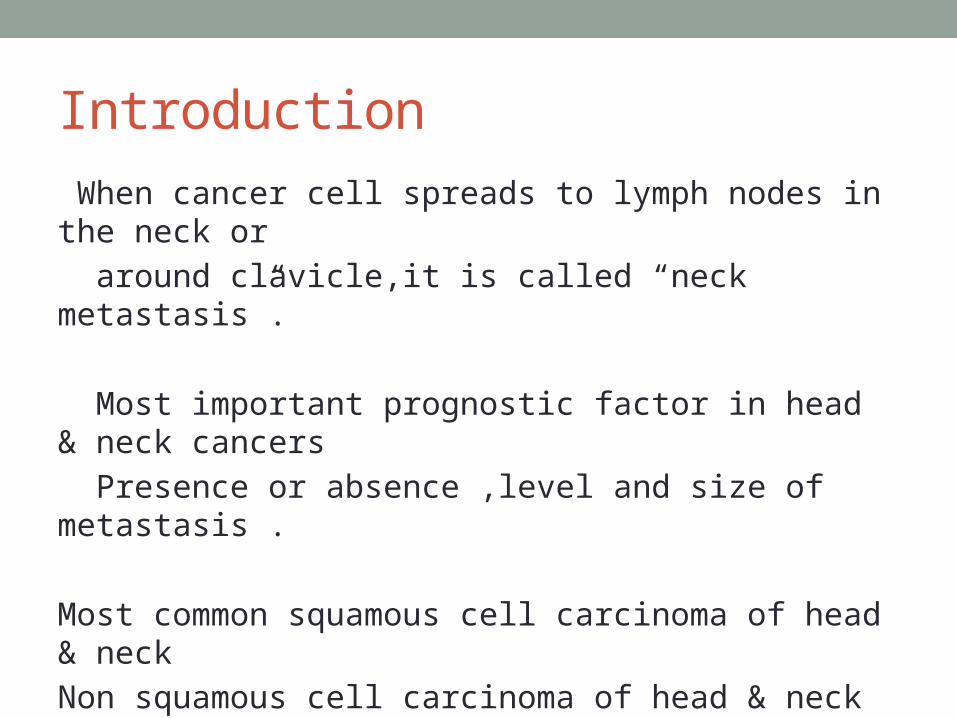
Behaviour of disease within cervical lymph nodes.
• Spread of disease from primary tumour to regional lymph nodes by passive transport within lymph.
• Progresses from superior to inferior• Some situations lymph node group bypassed in normal
lymphogram• Tumour cells proliferate,die, remain dormant or enter
blood circulation through blood vessels in nodes
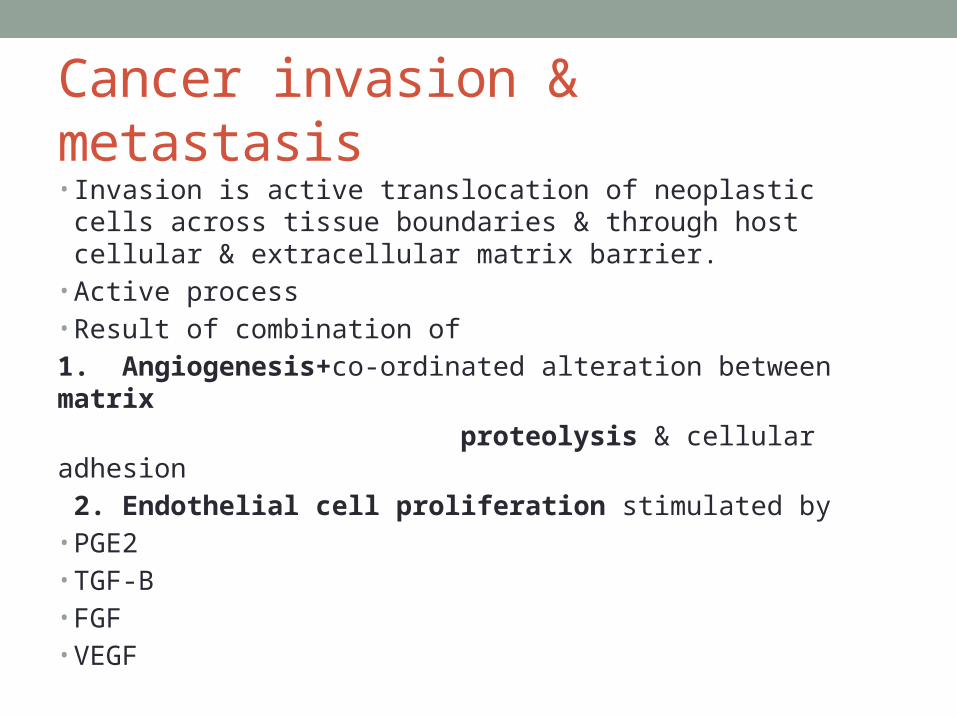
Cancer invasion & metastasis• Invasion is active translocation of neoplastic cells across
tissue boundaries & through host cellular & extracellular matrix barrier.
• Active process• Result of combination of1. Angiogenesis+co-ordinated alteration between matrix proteolysis & cellular adhesion 2. Endothelial cell proliferation stimulated by• PGE2• TGF-B• FGF• VEGF

High Degree of vascularization provide better access to circulation for metastatic spread.3.Degradation of extracellular material- by MMP(metalloproteinase)produced by tumour cell- lead to proteolysis- disruption of extracellular matrix-local tumour invasion- penetration of blood vessels and lymphatics- Leading to tumour dissemination and metastasis.

ASSESMENT OF NECK NODES

LYMPH NODE LEVELS/REGIONS

Subgroups• Ia Submental• Ib Submandibular
• IIa Upper jugular (Anterior to XI)• IIb Upper jugular (Posterior to XI)
• III Middle jugular
• IVa Lower jugular (Clavicular)• IVb Lower jugular (Sternal)
• Va Posterior triangle (XI)• Vb Posterior triangle (Transverse
cervical)
• VI Central compartment


PATTERN OF NODAL DRAINAGE

Oral cavity drainage• Extend from level I to IV • Median lower Lip,floor of mouth,ventral tongueIa• Upper lip, lateral lower lip, buccal mucosa- Ib• Post portion of oral cavity -II
• OROPHARYNGEAL LYMPHATIC DRAINAGE
Soft palate,palatine,lingual tonsils,post pharyngeal wall.Level II.level III primarily

Tongue lymphatic drainage• Ventral tongue follows drainage pattern of floor of mouth• To level IA anteromedially IB laterally.• Dorsal oral tongue- level I medially level II laterally.• Tongue base –level II and level III.• Level IV –involved in advanced disease or via alternative
drainage pattern.• Midline lesion- bilateral drainage pattern.

Laryngeal drainageUpper and lower system;division at true vocal cord level.
Supraglottis level II,level IIILower system level III,level IV
Vocal cords : watershed area few lymphatics; uncommon metastasisSubglottis : level III. Level IV
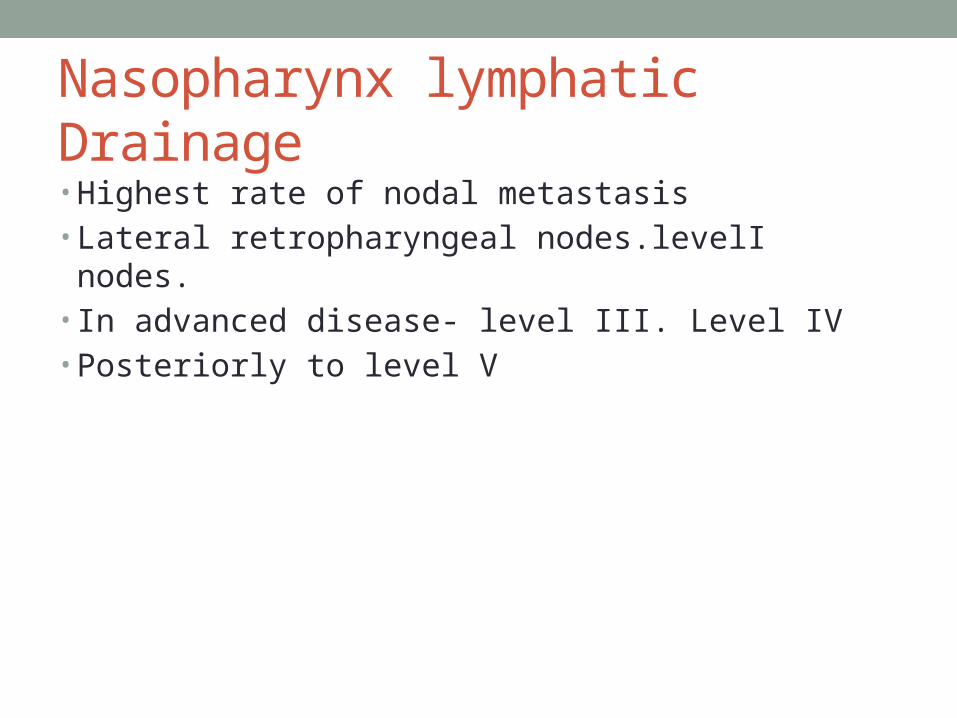
Nasopharynx lymphatic Drainage• Highest rate of nodal metastasis• Lateral retropharyngeal nodes.levelI nodes.• In advanced disease- level III. Level IV• Posteriorly to level V

lymphatic drainage to paratracheal lymph nodes
• Cervical oesophagus, thyroid,and parathyroid glands• Some subglottic location shows paratracheal drainage.
level VI and level VII
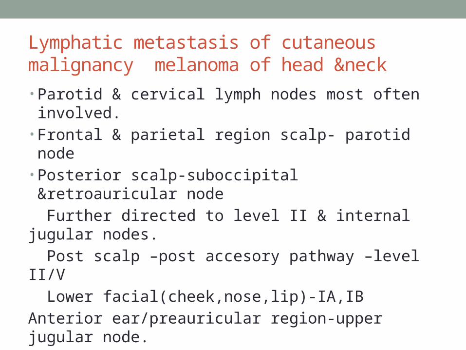
Lymphatic metastasis of cutaneous malignancy melanoma of head &neck• Parotid & cervical lymph nodes most often involved.• Frontal & parietal region scalp- parotid node• Posterior scalp-suboccipital &retroauricular node Further directed to level II & internal jugular nodes. Post scalp –post accesory pathway –level II/V Lower facial(cheek,nose,lip)-IA,IBAnterior ear/preauricular region-upper jugular node.Helix/lateral aspect of auricular- retroauricular,suboccipitalNeck skin-nodes adjacent to neck levelSkin in midline anterior neck- pretracheal level VI

ECHELON GROUPS• Oral Cavity :level I,II, III
• Larynx and pharynx :level II,III,IV
• Thyroid :level VI,VII,IV
• Parotid gland :preauricular,periparotid and intraparotid lymph nodes
• Submandibular and Sublingual gland :level I,II,III
CLINICAL ASSESMENT

History taking
Lump in neckSorethroatHoarsenessDysphagiaEarache

Clinical examination• Neck Exposed to the level of clavicles and manubrium• Palpate the neck while standing behind the patient• Following characteristics are noted- anatomic
location,size,shape,consistency,mobility and tenderness• False positive rate between 20-30%• False negative 30-40%• Examination of oral cavity, hypopharynx, larynx, • Digital palpation of the tonsillar fossa and base of tongue.• Nasolaryngoscopy- for assesment of nasal cavity,
nasopharynx

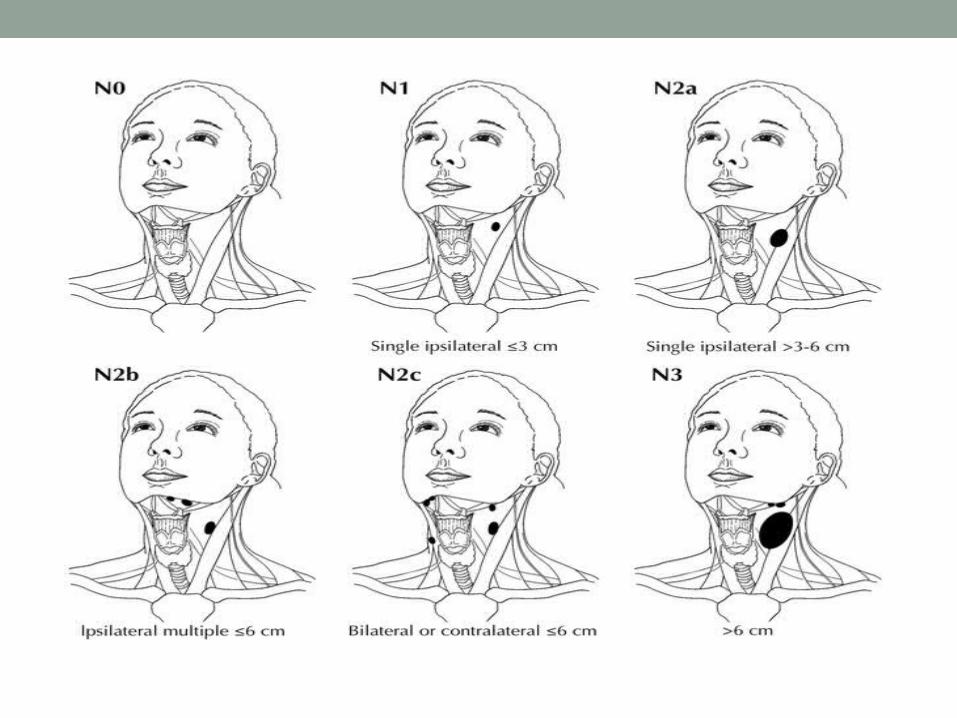
Staging of the neck
• “N” classification – AJCC • Consistent for all mucosal sites except the nasopharynx• Thyroid and nasopharynx have different staging based on
tumor behaviour and prognosis
Developed by Memorial Sloan-Kettering Cancer Center Ease and uniformity in describing regional nodal
involvement in cancer of the head and neck
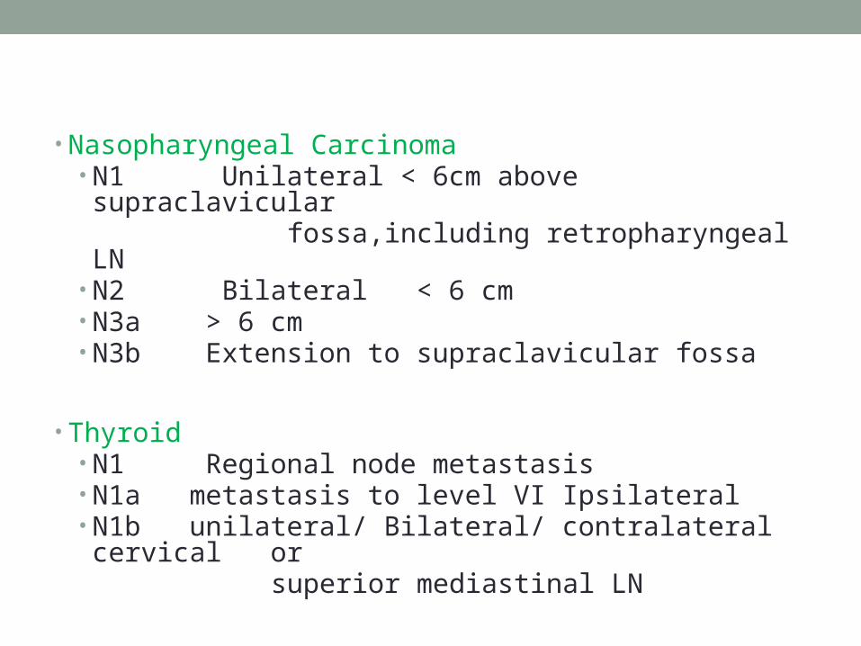
• Nasopharyngeal Carcinoma
• N1 Unilateral < 6cm above supraclavicular fossa,including retropharyngeal LN• N2 Bilateral < 6 cm• N3a > 6 cm• N3b Extension to supraclavicular fossa
• Thyroid• N1 Regional node metastasis• N1a metastasis to level VI Ipsilateral• N1b unilateral/ Bilateral/ contralateral cervical or superior mediastinal LN

INVESTIGATIONS

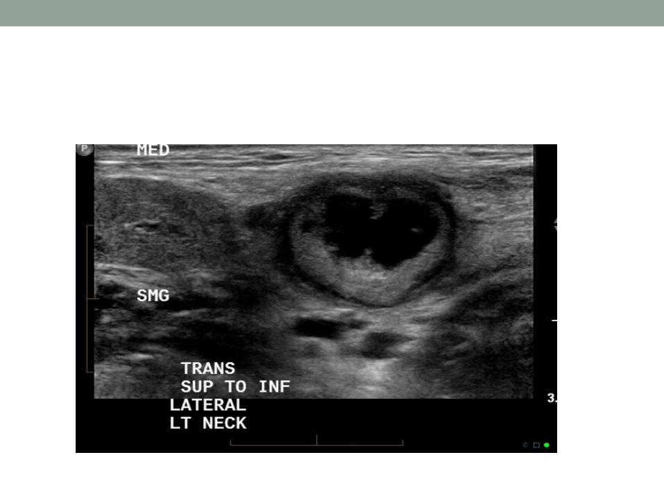
UltrasonographyoGrey scale and doppler usg- high frequency 13MHz probeoNormal nodes- echogenicoPathological nodes – decreased echogenicityoPower doppler differentiate-Benign and malignant nodes SIZE >1.5cm (jugulodigastric) > 1 cm for other nodes > 6-8mm retropharyngeal nodeoShape: ovalbenign roundmalignant• L/S RATIO: long to short axis ratio >2 in benign,<2 in malignant

• Vascularity(doppler usg):hilar in benign :peripherial in malignant.• Borders :sharp in malignancy due to intranodal tumour infilteration Necrosis Extracapsular spreadSenstivity- 75% to 92%Specificity- 63% to 91%Some studies say that measurement criteria for sonography considered > 6 to 7mm dia for level I & level II> 5mm level III & IV is abnormal.

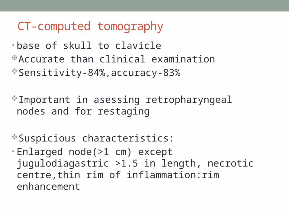
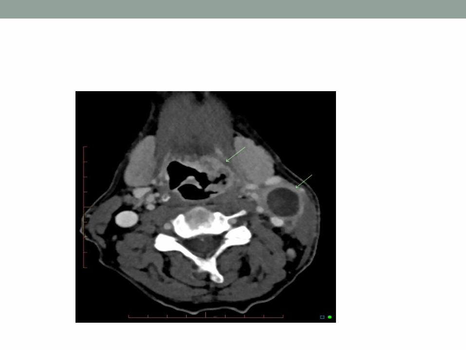
CT-computed tomography• base of skull to clavicleAccurate than clinical examinationSensitivity-84%,accuracy-83%
Important in asessing retropharyngeal nodes and for restaging
Suspicious characteristics:• Enlarged node(>1 cm) except jugulodiagastric >1.5 in
length, necrotic centre,thin rim of inflammation:rim enhancement
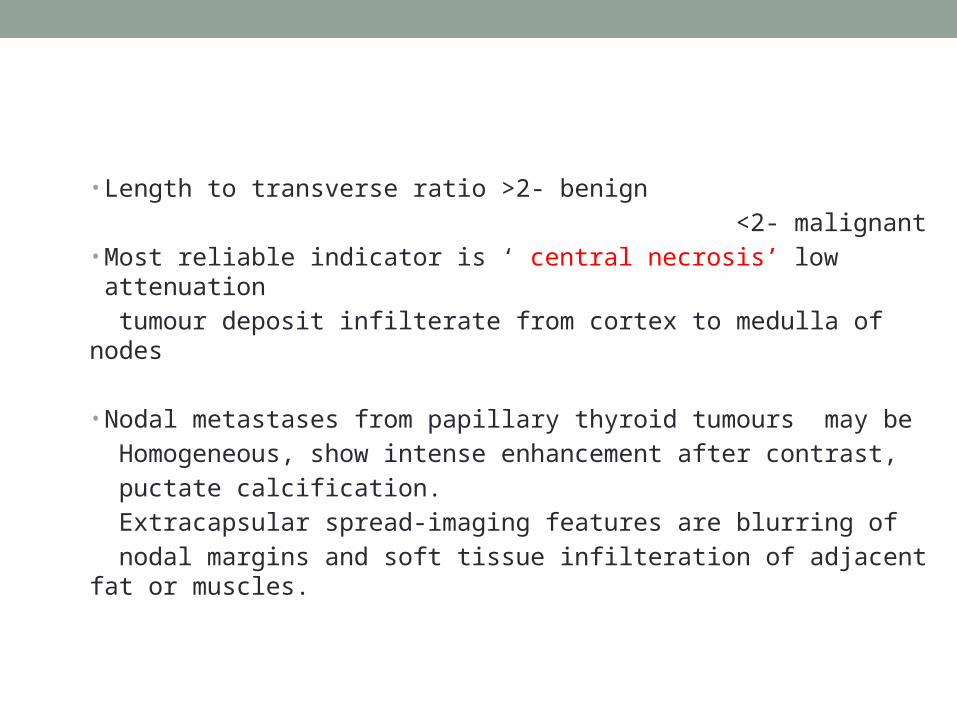
• Length to transverse ratio >2- benign <2- malignant• Most reliable indicator is ‘ central necrosis’ low attenuation tumour deposit infilterate from cortex to medulla of nodes
• Nodal metastases from papillary thyroid tumours may be Homogeneous, show intense enhancement after contrast, puctate calcification. Extracapsular spread-imaging features are blurring of nodal margins and soft tissue infilteration of adjacent fat or muscles.

Indications
1. Primary tumour assessment2. High chances of occult disease3. Staging prior to nonsurgical treatment4. Restaging5. To know the presence of deep fixation or contralateral
spread ,so as to decide management of the diseaseNote: if < 180 degree of vessel circumference involved – direct vascular invasion unlikelyIf > 270 degree of encasement present- arterial invasion possible
Magnetic resonance imaging Reactive nodes- homogeneous low signal on T1W high signal on T2W
Malignant nodes –mixed nodes so heterogeneous signal on both T1 andT2Central low signal with peripheral enhancement

• SPIO- superparamagnetic iron oxide- lymphoangiographic agent in MRI
• Iron oxide injected I/V taken up by reticuloendothelial system
• Signal drop not seen in metastatic nodes
Positron emission tomography• Measures biochemical pysiological process with 3D images of functional process of body.• Picks up metabolic signals of actively growing cancer cell in the body.• Used to stage primary squamous cell carcinoma
• Nodal involvement,distant metastasis, recurrent tumour.
• Tracer element use fluorine-18 fluorodeoxyglucose.

• PET detect primary that goes unnoticed by other modalities in 25% cases
• PET detect systemic metastatic disease in additional 27% of cases missed by other modalities
• FDG-PET low specificity and 40% false positive rate in tonsil
• PET performed prior to tests such as to avoid false positives.

Single –photon emission computed tomography (SPECT)• Thallium -201 SPECT useful for detection of occult head
and neck cancer and for assessing recurrences.• SPECT images, obtained 60min after administration of
150MBq201thallium –chloride.• 95% versus 88% for CT/MRI in confirming malignancy.

Fine needle aspirate cytology Useful in palpable node in unknown primaryNature of histology may help in search of primaryAccuracy is 90%

Pathology • Histology reporting following neck dissection prepare specimen on board Report number of nodes Report nodes levels Is there any extracapsular spread?

Sentinel lymph node• Initial lymph node metastasis is most likely to occur at first
node in lymphatic drainage pathway.• Accurate identification & evaluation of sentinel lymph
node for presence of micrometastasis crucial component for evaluate & treatment of cutaneous malignant melanoma.
• Tumour thickness >1mm with ulceration & increase mitosis more risk of metastasis.
• if metastatic node evaluated the patients stage increases to stage III- neck dissection required

Sentinal node mapping • Three techniques 1. Radioisotope scan imaging2. Injection of blue dye3. Use of handheld isotope tracer probe for localization technitium 99m labeled sulfur colloid – 0.05mci injected in 4 quadrants around primary lesion Visual images 3min and 15 min and 1hour. 1st lymph node identified is considered sentinal lymph node

Open biopsy
• Course of lymphatic spread is altered and previous scar tissue may pose difficulty in future surgery
• Only indicated when repeated FNAC is inconclusive or suggest lymphoma or anaplastic carcinoma

Risk for regional metastasis• 1-for tumors of larynx ,pharynx risk increases with
progression from center of upper aerodigestive tract to periphery.
• 2<15% T1 15%-30% T2 30-50% T3 75% for T4 head and neck sq cell ca.• 3-endophytic tumour have high risk than exophytic.• 4-poorly differentiated > well differentiated• 5-Tm 4mm thickness > thinner lesion• 6-Risk low in salivary and cutaneous malignancies

Molecular diagnosis • Polymerase chain reaction-LOH (loss of heterozygosity) • Of specific markers in biopsy specimen of tissue prior to
development of invasive malignancy.• Overexpression of telomerase & mutant P53• Detection of microsatellite & methylated DNA- marker• For inactivated tumour suppressor gene assayed in
blood.• Overexpressed proteins in blood- such as cytokines &
angiogenesis factor predicting behaviour & reoccurance.

TREATMENT

• Adverse effect on survival
• Only 50% with neck metastasis survive 5 Years
• Untreated neck:predictable pattern of spreadfirst echelon groupconcept of selective neck dissection.

No neckElective surgeryElective radiotherapyElective neck investigations-CT/MRIWait and watch policy

Indication for elective neck surgeryMore than 20-25%chance of occult diseaseVigilant follow up is not possibleLimited neck dissection has low morbidity and mortalityClinical evaluation of neck is difficultSurgery being done on neck for reconstructionImaging suggests possibility of occult nodal metastasisIf neck is being entered to remove primary its better to
perform resection
If metastasis is not identified on examination or imaging, treatment of cervical lymph nodes becomes elective decision based on risk of metastasic disease.If there is >15% risk of metastasis disease to regional lymph nodes then nodes should be electively treated with elective neck dissection or elective radiation.
Radiotherapy for neck nodes
- External beam radiation approx 40-50 gy to N0 neck will control metastasis in upto 90-95% cases
-If primary tumour is being treated by radiotherapy then elective treatment Given to first echelon group of nodes or whole of neck
- Bilateral radiotherapy when midline extension occurs
Treatment – N1• Most patientsmetastatic disease -All five levels can be involved -Minimum operation recommended is MODIFIED
RADICAL NECK DISSECTION• ROLE OF RADIOTHERAPY• -Less efficient than surgery• -Less preferred option unless primary is also being treated
with RT alone(nasopharyngeal carcinoma)

Treatment –N2• N2a and N2b Advanced disease Radical Surgery advocated for treatment Post operative radiotherapy N2c 5% of head and neck cancer Common primary site:base of tongue and supraglottis Prognosissize,number,of nodes,extracapsular spread

Radical surgery if primary site is operable
Post operative radiotherapy: MUST

INTERVAL CONTRALATERAL LYMPHADENOPATHY• Node appear on contralateral side after neck dissection
on one side
• Can be salvaged surgically
• Modified radical or radical neck dissection- 30% of 5 year survival

Treatment N3• Uncommon
• 5%N3 nodes• Nodes fixed to skin or underlying structure
• Most:incurable,surgery in certain instances
• Decision depends on stage of disease,presence or absence of fixation and structure to which node is fixed
• Radiological imaging :mandatory for assesment.
• Fixation to mandible,SCM,midline muscles ,prevertebral fascia :treatable
• Extended radical neck dissection may be helpful
• Fixation to brachial plexus or skull base: contraindication to surgery
• Arterial invasion,if present,careful assessment.
•Contraindication of neck dissection• Unresectable/uncontrolled primary• Unfit for surgery ;risk of anaesthesia• Inoperable neck disease• Distant metastasis
RECURRENCE AND SALVAGE SURGERYBad prognosisCareful evaluation and staging with radiological imaging.Surgery:wide resection followed by Post operative RTIf post op RT already received ,further radiotherapy may
be given in form of LOCAL BRACYTHERAPY.

Lymphomas of neck• 75% of lymphomas that occur in head &neck are nodal• Hodgkin lymphomas – young adult, painless
lymphadenopathy, rubbery ,wax or wanes in size in cervical (60-70%)/supraclavicular region( 75-90%), ‘B’
• symptoms• Non hodgkin’s lymphoma 70% of primary head &neck
cases- small lymphocytic lymphomas involves tonsil,adenoid,tongue base,nasopharynx.
• Common presentation : dysphagia,neck mass ,airway obstruction .
Management • Open biopsy-gold standard• Chemotherapy- ABVD (doxorubicn,bleomycin,vinblastin, dacarbazine ) followed by 20Gy involved field RTBEACOPP- ( bleomycin,,etoposide,doxorubicin, cyclophosphamide.,,vincristin,procarbazine and prednisone For advance stages)

Clinical applications
• Possible future therapeutic strategiesVirus directed enzyme prodrug therapy- adenovirus
mediated p53 gene transfer in patients with advanced recurrent head & neck squamous cell carcinoma reduces tumour growth.
Tissue inhibitors of matrix metalloprotienases (TIMP) prevent degradation of matrix.Antiangiogenesis agents. – bevacizumab, Erlotinib
(EGFR) epidermal growth factor inhibitor.

NECK DISSECTION
CLASSIFICATIONThe radical neck dissection is classified according to the Academy’s committee for head & neck surgery & oncology into four major type:Radical Neck Dissection (RND)Modified Radical Neck Dissection (MRND)Selective Neck DissectionSupraomohyoidPosterolateralLateralAnterior Extended Radical Neck Dissection

MEDINA classification(1989)• Comprehensive neck dissection-Radical neck dissection-Modified radical neck dissection -type I(XI preserved) -type II(XI,IJV preserved) -type III(XI,IJV,SCM )
• Selective neck dissection

Radical neck dissection• All lymph nodes from level I to V including spinal
accessory nerve,IJV,SCM along with submandibular gland and tail of parotid
• Indication :extensive cervical involvement or matted lymph nodes with gross extracapsular spread and invasion into SAN,IJV,SCM.

Modified radical neck dissection• Definition: excision of same node bearing regions as RND
with preservation of one or more nonlymphatic structures(SAN,IJV,SCM)
• 3 types• ADVANTAGES:-Shoulder syndrome incidence reduced-Improve cosmetic outcome-Reduce likelihood of bilateral IJV resection
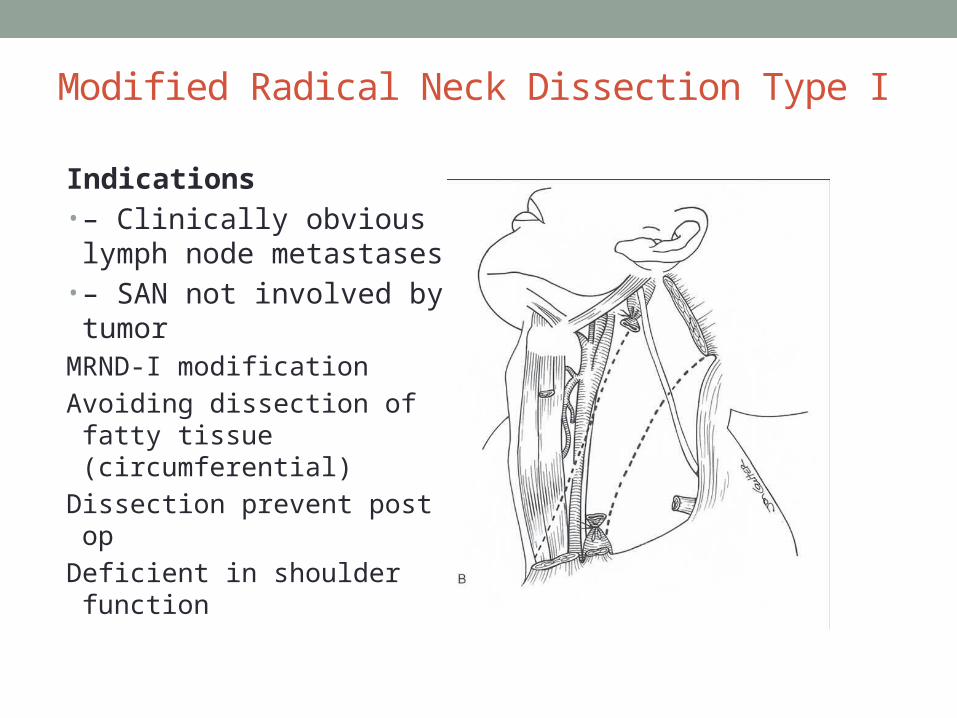
Modified Radical Neck Dissection Type I
Indications• – Clinically obvious lymph node metastases
• – SAN not involved by tumor
MRND-I modification Avoiding dissection of fatty
tissue (circumferential) Dissection prevent post op Deficient in shoulder function

Modified Radical Neck DisectionType II
Indications
• -it preserves SCM and SAN & sacrifies IJV
- Differentiated thyroid cancer

MRND Type III
Neck dissection of choice for N0 neck
Preserving SCM,SAN,IJV Metastatic lymph nodes
from differentiated carcinoma thyroid gland
Skin tumour :melanoma,merkels carcinoma, squamous cell carcinoma


(a)Supraomohyoid ( b)Extended Supraomohyoid

Primary carcinoma of lateral border of the oral tongue have small risk of having skip metastasis to level IV of ipsilateral neck.In addition to standard supraomohyoid neck dissection extended operation being undertaken to include level IV
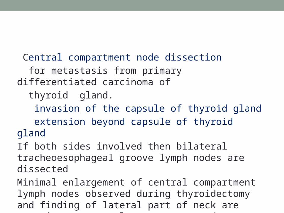
Central compartment node dissection for metastasis from primary differentiated carcinoma of thyroid gland. invasion of the capsule of thyroid gland extension beyond capsule of thyroid gland If both sides involved then bilateral tracheoesophageal groove lymph nodes are dissectedMinimal enlargement of central compartment lymph nodes observed during thyroidectomy and finding of lateral part of neck are negative, a central compartment node dissection is adequate.


EXTENDED RADICAL NECK DISSECTION
Removal of one or more additional lymph node groups or nonlymphatic structures,or both, not encompassed by the RADICAL NECK DISSECTION such as parapharyngeal,superior mediastinal,and paratracheal.

Super Selective Neck Dissection• Limited to 1 or 2 contigous neck levels.• Most common applied- supraglottic cancers.• Tumour has high propensity to metastatise to level IIA & III.• Role in treatment of residual disease followed by chemoradiation that is confined to single level.

INCISIONS


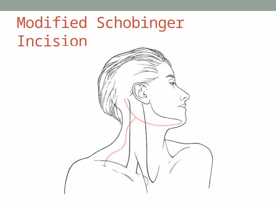
Modified Schobinger Incision


Apron Incision
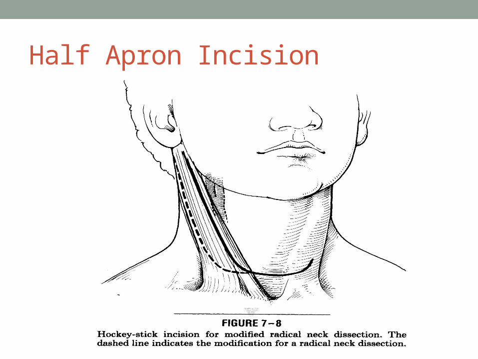
Half Apron Incision

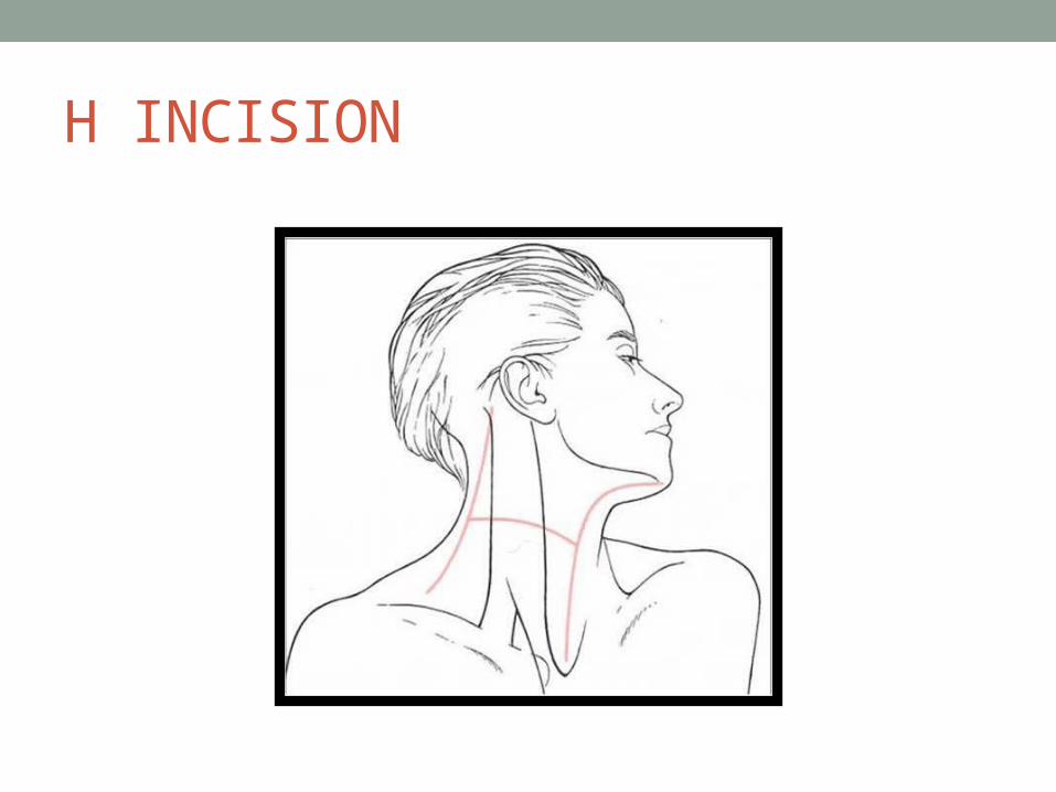
H INCISION

Hetters incision

Utility incision

LATERAL UTILITY
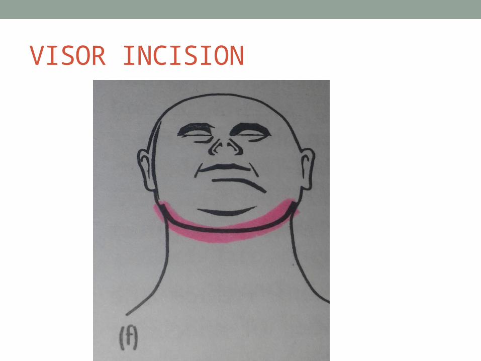
VISOR INCISION

EXTENDED THYROID INCISION

STEPS OF RND• 4 MAJOR AND MINOR POINTS OF CONSTERATION• MAJOR:-Lower end of internal jugular vein(IJV)-Junction of clavicle with trapezius border-Upper end of IJV-Submandibular triangle

• Minor -lymph nodes -retrophayngeal lymph nodes -chaissaignac triangle*Chaissaignac triangle is angle made by longus
colli,scalene anterior,base by subclavian artery and apex by tubercle of C6 vertebrae(chaissaigac tubercle
*Content:-thoracic duct, vertebral vein and thyrocervical trunk.

• Marginal Mandibular and cervical branch of facial nerve.
Lower end of Internal Jugular Vein• Main vein draining the primary tumour being removed
divided first.• Reduce no of systemic metastasis due to release of small
tumour emboli released by manipulation of tumour• Injury to IJV: risk of AIR EMBOLISM• SUTURE may slip:put finger on hole ,tilt the patient down
and stitch the hole with non absorbable suture


• Houseman‘ s Suture• Transfixion Stitch at the Lower end.
Thoracic duct
• Passes medial to jugular vein,then posterior to it • Finally curve around to enter the junction of IJV and
subclavian vein.• Ideally- to be tied off • Any chyle leak :repair there and then.• VALSALVA manouver

Supraclavicular dissection• Omohyoid muscle retracted upwards.• Should not bleed if cut through tendon• Fat lateral to IJV removed ,prevertebral fascia identified.• Phrenic nerve is identified (runs over scaleneus anterior .• Not to breech the prevertebral fascia.• Chaissagnaic triangle :dissection of scalene node
Dissection of posterior triangle• Imp: accessory nerve (in roof)• Chance of injury high early in course of dissection• Identification :A)Erb`s point: 1 cm above point where greater auricular
nerve winds around the SCM
B)Leaves post border of SCM at upper third and lower two third.

Division of upper end of Internal Jugular Vein
• Identified by palpating he transverse process of C2 vertebrae
• Retract the posterior belly of digastric upwards.• Vagus and hypoglossal nerve should be identified and
preserved• Venous tributaries to venae nervi hypoglossi should be
ligated

• Hypoglossal tunnel• Occipital artery crosses posterior part of IJV ligated • PRESSURE -4cm of water- bleeding controlled by
packingligation.

Dissection of submandibular triangle• Fourth corner of consternation.• Anterior belly and posterior belly of digastric visualized• Mylohyoid muscle is retracted forward to identify the
submandibular duct,lingual nerve is pulled down in a curve
• Submandibular duct is identified and ligated. And submandibular gland is removed


Closure
Wound is irrigated with normal saline and bleeding point ,if any,secured.
• Large drain put• Drain should never cross the carotid sheath• Final check for any chyle leak,any bleeding• Wound is closed in two layers with absorbable suture and
skin closed with non-absorbable.• Any three point junction should be away from carotid
artery.

Complications of neck dissection1)Anaesthetic complication-• Post operative atelectasis• Urinary retention/pneumonia• Deep vein thrombosis

Local complicationsHaemorrhageWound infectionCarotid artery ruptureChylous fistulaPneumothoraxNerve injuriesCerebral oedema

Haemorrhage
Injury to IJV-pressure with finger -ligation of veinInjury to carotid arterial system- more tumour invading the artery,attempts to dissect cancer off a vessel
Wound infection• Contamination of surgical field• Postoperative hematoma that later get infected..• Flap necrosis • Wound breakdown

Carotid artery rupture• Necrosis in arterial wall due to infection• Common in preoperative radiotherapy (post op salivary
fistula with loss of skin)-surgical debridement with systemic antibiotics
• Removal of adventitiaincreased risk• Rupture of artery:ligation; mortality 38% and morbidity
hemiplegia rate 50%

Chylous fistula• <100 ml leak/dayconservative management Pressure dressing and parental feeding.• 300ml leak/dayreexploration identifying and repairing
the source of leak.• Loss of proteins and electroytes• Lateral thoracotomy- suture between the oesophagus and
descending aorta in posterior and inferior mediastinum

Pneumothorax• Injury to apical pleura• Air leak at time of surgery• To be repaired.

Nerve injury
• Accessory nerve• Branches of cervical plexuses-lesser occipital,greater
auricular,transverse cutaneous,supraclavicular nerve• Descendens hypoglossi

Cerebral odema• Complication of B/L neck dissection U/L:three fold increase B/L: 5 fold increase

Laryngeal oedema• If planning on doing B/L Neck Dissection- laryngeal
oedema –elective tracheostomy.
NECK METASTASIS FROM UNKNOWN PRIMARY

Unknown or occult primary carcinoma
• Pesentation of metastatic neck lymph adenopathy without the development of a primary lesion within a subsequent five year period.
Failure to identify occult primary-Spontaneous regression of primary tumourAutoimmune destruction Accelerated tumour progression
• Metastatic carcinoma with no evidence of primary site after history,physical examination and radiological imaging.
• Secondary of neck<10% of all unknown primary
• Most likely head and neck primary site: tonsil(45%),base of tongue(40%) and piriform fossa (10%).

Diagnostic steps• History
• Physical examination:• complete head and neck examination ipsilateral otalgia with normal otoscopy• direct attention to tongue base, tonsil, supraglottis and
hypopharynx.• Unilateral serous otitis media• Nasopharyngeal examination

Diagnostic work up of patient with unknown primary
• History• Physical examination(including skin and scalp)• Careful examination of neck and supraclavicular lymph
nodes• Examination of oral cavity,pharynx and larynx

Radiological Investigations
• Chest X-ray• Ultrasonography• CT scan and MRI of head and neck cancer• FDF- PET scan of whole body if lower neck node.• Radionuclide scanning

Direct laryngoscopy and directed biopsiesNasopharynx,tonsils,base of tongue,pyriform fossa and
any suspicious or abnormal mucosal areassFNAC –fine needle aspiration cytology(accuracy-90%)Open biopsy or core needle biopsy

FNAC • Adenocarcinoma
upper neck- imaging for salivary gland• primary found- treat primary and neck• no primary found- treat neck
Lower neck- rule out thyroid gland malignancy +ve - thyroidectomy and neck dissection -ve - metastatic workup

Possible histology• MOST:sq cell ca or poorly differentiated carcinoma• Adenocarcinoma in neck:rule out salivary gland,thyroid or
parathyroid primary tumours• Other possibilities:lymphoma,tuberculoma• Rarely: sarcoma

Role of PET IN unknown primary• Recommended for detecting primary disease and staging • Define suspicious regions and lymph nodes to biopsy in
head and neck region as well identify metastasis• FDG-PET is 88.3% sensitive• 74.9% specific• 78.8% accurate in detecting unknown primary

Role of ipsilateral tonsillectomy• Ipsilateral tonsillectomy suggested for patients with
submandibular ,subdigastric and mid-jugular nodes as this may be site of primary disease in 25%-35% of patients.
Pan-endoscopy• If sq cell carcinoma on FNACpanendoscopy
• Direct visualization of nasopharynx,oropharynx,hypopharynx,larynx,trachea,
bronchial airway and esophagus. • Directed biopsy
• - all suspicious mucosal lesions• -areas of concern on CT or MRI• - nasopharynx, tonsil, base of tongue, pyriform fossa,retromolar trigone

• Sites of primary tumours are tonsillar fossae and base of tongue in 82% of cases

Treatment of squamous cell ca of unknown primary head and neck cancer
• Multidisplinary team approach: surgery,radiation and chemotherapy
• Treatment modality : • limited neck disease without extracapsular
extentionNeck Dissection or ipsilateral neck radiation• Nodal disease with extracapsular spread:post op
radiation.

Pan mucosal irradiation• Radiotherapy to the neck and naso-oro-hypopharynx
except when there is a strong suspicion that nasopharynx is a primary site in which case the hypopharynx may be spared
• Nasopharynx may be omitted when disease is limited to lower nodes
• Treatment of both sides of neck
MANAGEMENT OF PATIENT WITH METASTATIC NECK NODE FROM AN UNKNOWN PRIMARY
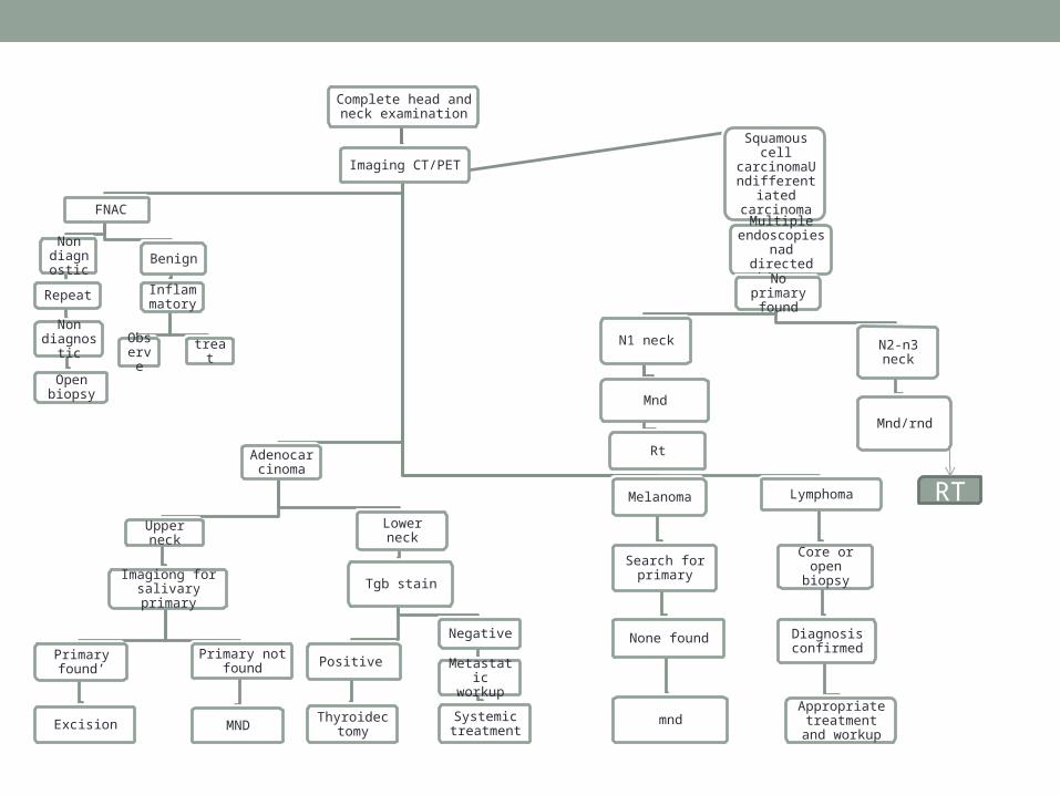
Complete head and neck examination
Imaging CT/PET
FNAC
Non diagno
stic
Repeat
Non diagnosti
c
Open biopsy
Benign
Inflammatory
Observe treat
Adenocarcinoma
Upper neck
Imagiong for salivary primary
Primary found’
Excision
Primary not found
MND
Lower neck
Tgb stain
Positive
Thyroidectomy
Negative
Metastatic workup
Systemic treatment
Melanoma
Search for primary
None found
mnd
Lymphoma
Core or open biopsy
Diagnosis confirmed
Appropriate treatment and
workup
Squamous cell
carcinomaUndifferentiated
carcinoma
Multiple endoscopies nad directed
biopsyNo primary
found
N1 neck
Mnd
Rt
N2-n3 neck
Mnd/rnd
RT
EVALUATION AND MANAGEMENT OF NECK OF PATIENT WHO HAD CHEMORADIATION

Completion of CTRT
Clinical assessment at 6 weeks
CT/MRI PET/CT+-CT with contrast at 10-12 weeks
Neck dissection
PET +ve or CT +veHigh SUV or large volume abnormality
PET –ve /CT –ve Low volume abnormality
PET +VE/CT –ve Low SUV
PET –ve/CT-ve
Neck dissection
Observe or Neck dissection
Selective neck dissection
Excisional biopsy and Neck dissection
Observe


![Current Concepts in Surgical Management of Neck Metastases ... · Current Concepts in Surgical Management of Neck Metastases from Head and Neck Cancer Review Article [1] | May 31,](https://img.pdfslide.us/doc/110x75/5f0333b37e708231d4080a9a/current-concepts-in-surgical-management-of-neck-metastases-current-concepts.jpg)