Embed Size (px)
Citation preview

Life Cycle of Pythium, Albugo, Erysiphe,
Claviceps, Ustilao and Puccinia Fungi
DR. RAJBIR SINGH
Assistant Professor
Department of Plant Pathology
Gochar Mahavidyalaya (Post Graduate College)
Rampur Maiharan, Saharanpur (UP), India
Affiliated to: CCS University, Meerut (UP), India
Email: [email protected]
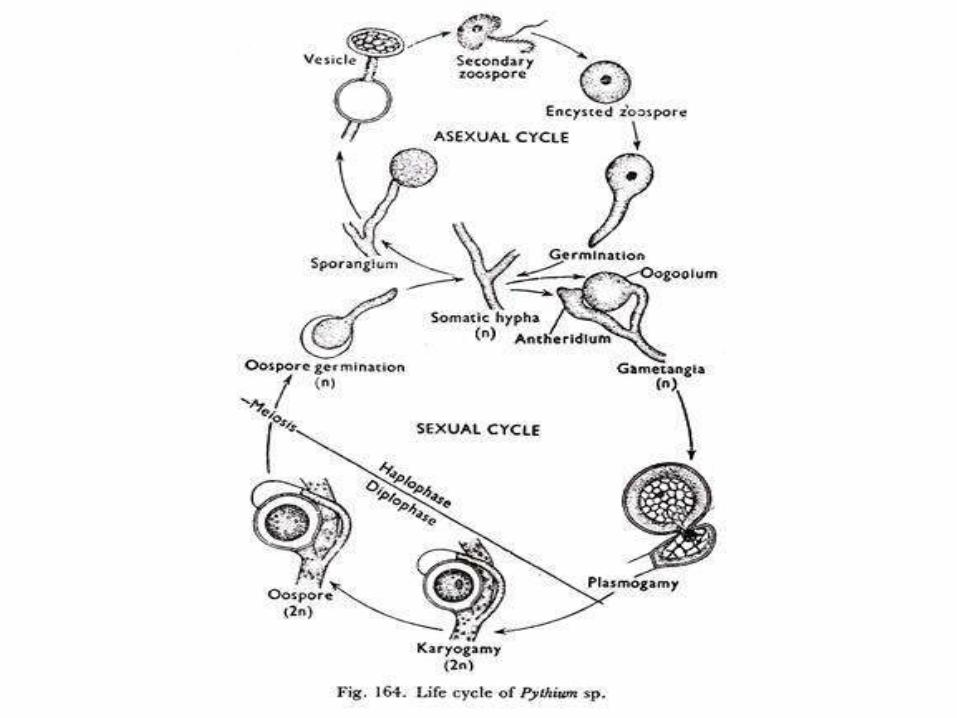
Life Cycle of Pythium
• Pythium spp. cause “Damping off” diseases in plants
• Mycelium of this fungus grow fast and is branched, thin, white in color,non-septate inter or intracellular.
• Asexual reproduction by sporangia which are produced terminal andintercalary on mycelium.
• Sporangia are ovoid or spherical, filamentous or indefinite shape.
• Sporangia detached from mycelium and spread by air or water fromone place to other place.
• These sporangia germinate directly by germ tube or mycelium producea vesicle.
• Protoplasm of sporangia move towards vesicle and form zoospores.
• After bursting of vesicle zoospores released.
• After release zoospore rest and lose their flagella and Encyst.
• Encysted zoospore later germinate and infect to host.
• Asexual reproduction also take place by Chlamydospores.
• Some hypha of mycelium swollen, separated by a septum and formspherical structures.
• These chlamydospores germinate by germ tube and infect host.
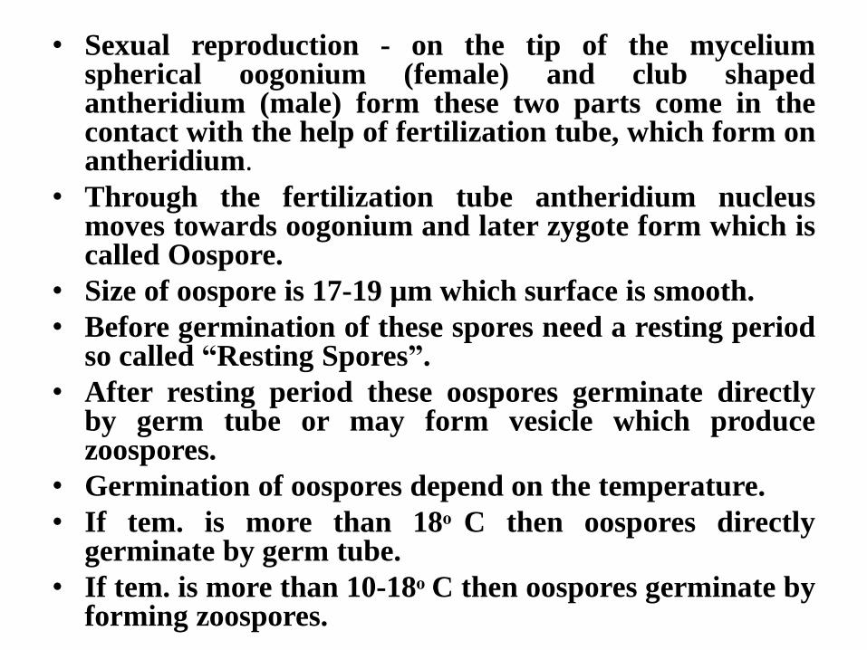
• Sexual reproduction - on the tip of the myceliumspherical oogonium (female) and club shapedantheridium (male) form these two parts come in thecontact with the help of fertilization tube, which form onantheridium.
• Through the fertilization tube antheridium nucleusmoves towards oogonium and later zygote form which iscalled Oospore.
• Size of oospore is 17-19 µm which surface is smooth.
• Before germination of these spores need a resting periodso called “Resting Spores”.
• After resting period these oospores germinate directlyby germ tube or may form vesicle which producezoospores.
• Germination of oospores depend on the temperature.
• If tem. is more than 18ͦ C then oospores directlygerminate by germ tube.
• If tem. is more than 10-18ͦ C then oospores germinate byforming zoospores.
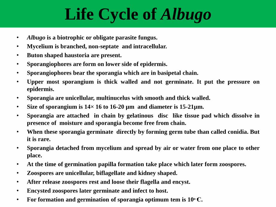
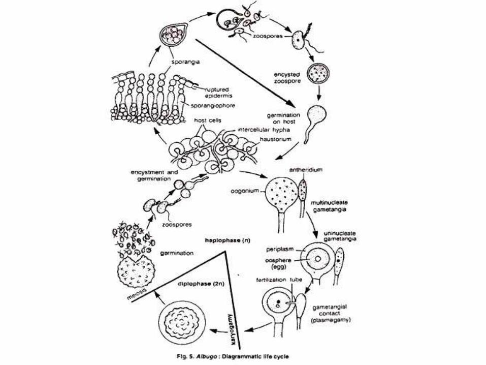
Life Cycle of Albugo
• Albugo is a biotrophic or obligate parasite fungus.
• Mycelium is branched, non-septate and intracellular.
• Buton shaped haustoria are present.
• Sporangiophores are form on lower side of epidermis.
• Sporangiophores bear the sporangia which are in basipetal chain.
• Upper most sporangium is thick walled and not germinate. It put the pressure on
epidermis.
• Sporangia are unicellular, multinucelus with smooth and thick walled.
• Size of sporangium is 14× 16 to 16-20 µm and diameter is 15-21µm.
• Sporangia are attached in chain by gelatinous disc like tissue pad which dissolve in
presence of moisture and sporangia become free from chain.
• When these sporangia germinate directly by forming germ tube than called conidia. But
it is rare.
• Sporangia detached from mycelium and spread by air or water from one place to other
place.
• At the time of germination papilla formation take place which later form zoospores.
• Zoospores are unicellular, biflagellate and kidney shaped.
• After release zoospores rest and loose their flagella and encyst.
• Encysted zoospores later germinate and infect to host.
• For formation and germination of sporangia optimum tem is 10ͦ Cͦ.
• Sexual reproduction by Oogamy.
• Oogonium (female) is spherical, terminal or intercalary and
have about 200 nucleus. Its protoplasm is divided in Periplasm
and Ooplasm.
• Antheridium (male) is club shaped and have 6-12 nucleus.
• Oogonium and Antheridium come in the contact by a papilla
which disappear shortly. At the point of contact of Oogonium
cell wall become thin and form papilla.
• Through the contact of both sex organs Antheridium nucleus
moves towards Oogonium and later Zygote form which is called
Oospore.
• Oospores are spherical, yellow to dark brown in colour and
diameter is 40-55 µm which cell wall is thick and tuberculate .
• Before germination of these spores need a resting period so
called “Resting Spores”.
• After resting period these oospores germinate by forming sessile
vesicle which produce zoospores.
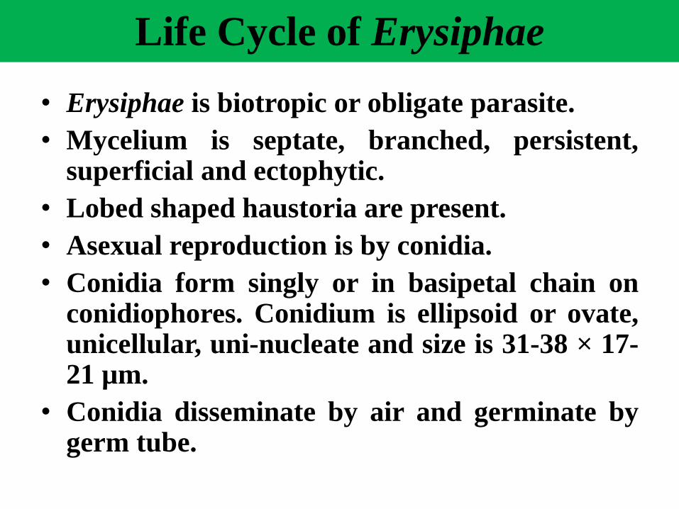
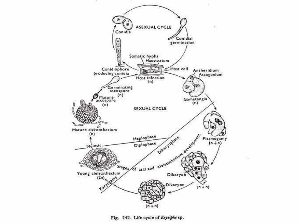
Life Cycle of Erysiphae
• Erysiphae is biotropic or obligate parasite.
• Mycelium is septate, branched, persistent,superficial and ectophytic.
• Lobed shaped haustoria are present.
• Asexual reproduction is by conidia.
• Conidia form singly or in basipetal chain onconidiophores. Conidium is ellipsoid or ovate,unicellular, uni-nucleate and size is 31-38 × 17-21 µm.
• Conidia disseminate by air and germinate bygerm tube.

•Sexual reproduction occurs at the end of summer, when conidial
production slows down and eventually ceases.
•The white powdery appearance of the host surface now changes
to greyish or brown shade, and the hyphae prepare to form
ascocarps which are cleistothecia. The cleistothecia are large
enough to be visible to be naked eye as black dots on the infected
host surface.
•Asci and ascospores are developed within the cleistothecium.
•The cleistothecium contains one to several asci which arise in
one or more tufts from the base.
•The asci are globose to ovoid, and may have short’ stalk.
•Members of the Erysiphaceae survive the winter as ascospores in
the asci developed in the cleistothecium, and in the spring both
cleistothecium and asci absorb water and swell.
•The cleistothecium cracks open, and the asci discharge the
ascospores which on germination on a suitable host produce new
mycelia.

Life Cycle of Claviceps
• The species of Claviceps are parasitic on grasses. It causes ergotdisease. The fungus attacks only the inflorescence of its host. Grainsare replaced by blunt trilateral horn-shaped black sclerotia known bythe name of ergots.
• These sclerotia are resting stages. In the mature state they arecomposed of a dirty-white medullary tissue surrounded by a violet-brown rind. The medulla or the central core consists of a prosenchymaformed of looser cylindric prismatic cells of pale-brown colour whichare about one to four times as long as breadth.
• Sclerotia after surviving in winter germinate in spring by formingperithecium.
• Ascus form in perithecium which release ascospores. These ascosporesdisseminate by air to stigma of flower and like pollen tube reach toovary of flower and resulting in Honey Dew formation.
• Yellow-white, septate, branched mycelium develop which produceconidia on conidiophores.
• Earlier macro conidia and in later stage micro conidia form. Conidiaare infective.
• After Honey Dew Stage sclerotia form which are known as ergot.

Life Cycle of Claviceps sp.
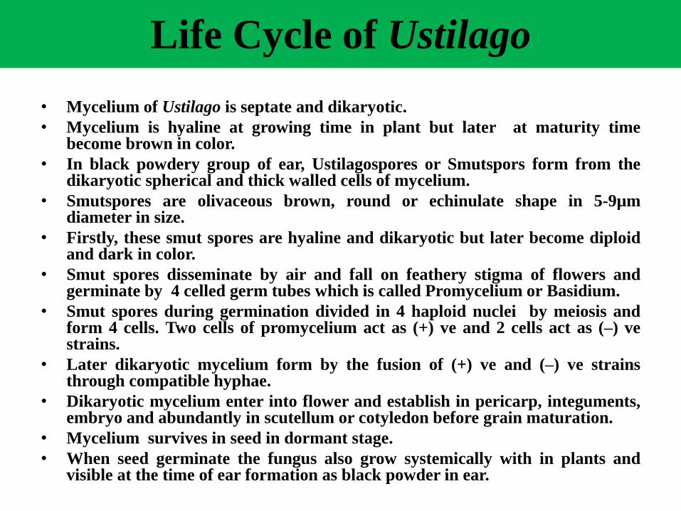
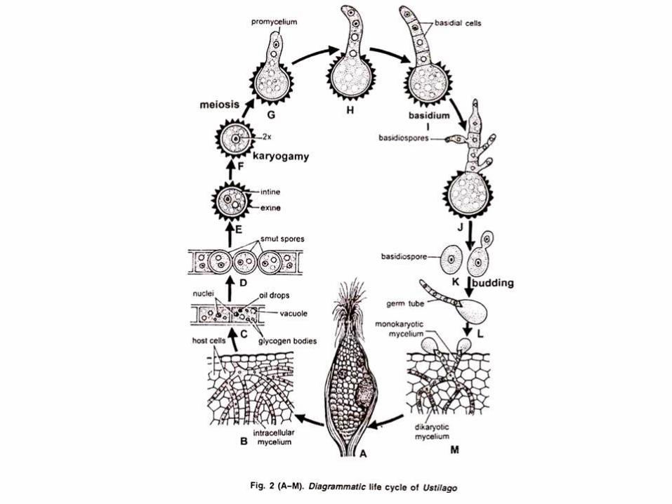
Life Cycle of Ustilago
• Mycelium of Ustilago is septate and dikaryotic.
• Mycelium is hyaline at growing time in plant but later at maturity timebecome brown in color.
• In black powdery group of ear, Ustilagospores or Smutspors form from thedikaryotic spherical and thick walled cells of mycelium.
• Smutspores are olivaceous brown, round or echinulate shape in 5-9µmdiameter in size.
• Firstly, these smut spores are hyaline and dikaryotic but later become diploidand dark in color.
• Smut spores disseminate by air and fall on feathery stigma of flowers andgerminate by 4 celled germ tubes which is called Promycelium or Basidium.
• Smut spores during germination divided in 4 haploid nuclei by meiosis andform 4 cells. Two cells of promycelium act as (+) ve and 2 cells act as (–) vestrains.
• Later dikaryotic mycelium form by the fusion of (+) ve and (–) ve strainsthrough compatible hyphae.
• Dikaryotic mycelium enter into flower and establish in pericarp, integuments,embryo and abundantly in scutellum or cotyledon before grain maturation.
• Mycelium survives in seed in dormant stage.
• When seed germinate the fungus also grow systemically with in plants andvisible at the time of ear formation as black powder in ear.
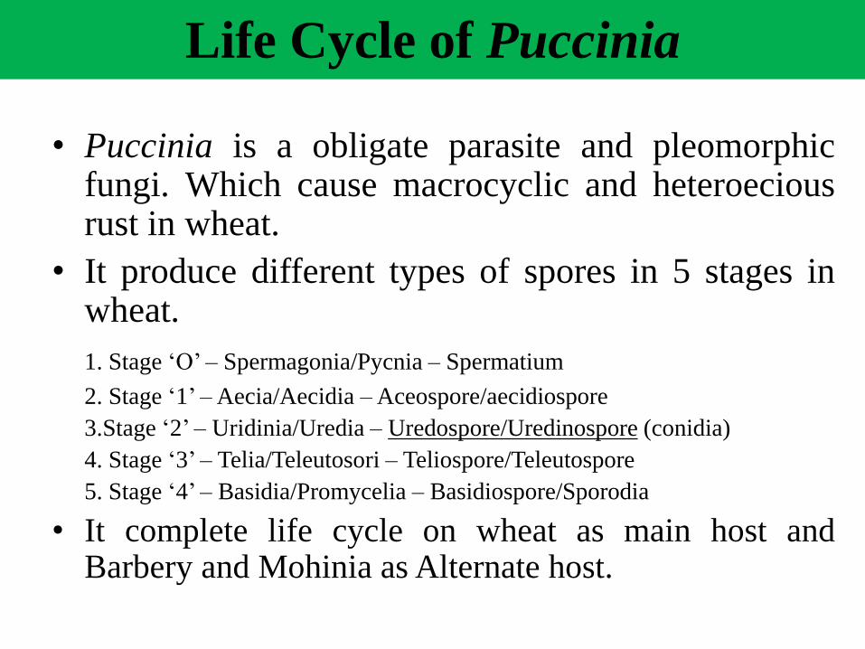
Life Cycle of Puccinia
• Puccinia is a obligate parasite and pleomorphicfungi. Which cause macrocyclic and heteroeciousrust in wheat.
• It produce different types of spores in 5 stages inwheat.
1. Stage ‘O’ – Spermagonia/Pycnia – Spermatium
2. Stage ‘1’ –Aecia/Aecidia –Aceospore/aecidiospore
3.Stage ‘2’ – Uridinia/Uredia – Uredospore/Uredinospore (conidia)
4. Stage ‘3’ – Telia/Teleutosori – Teliospore/Teleutospore
5. Stage ‘4’ – Basidia/Promycelia – Basidiospore/Sporodia
• It complete life cycle on wheat as main host andBarbery and Mohinia as Alternate host.
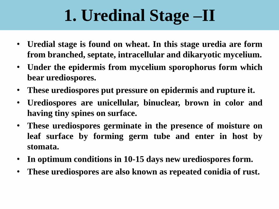
1. Uredinal Stage –II
• Uredial stage is found on wheat. In this stage uredia are form
from branched, septate, intracellular and dikaryotic mycelium.
• Under the epidermis from mycelium sporophorus form which
bear urediospores.
• These urediospores put pressure on epidermis and rupture it.
• Urediospores are unicellular, binuclear, brown in color and
having tiny spines on surface.
• These urediospores germinate in the presence of moisture on
leaf surface by forming germ tube and enter in host by
stomata.
• In optimum conditions in 10-15 days new urediospores form.
• These urediospores are also known as repeated conidia of rust.


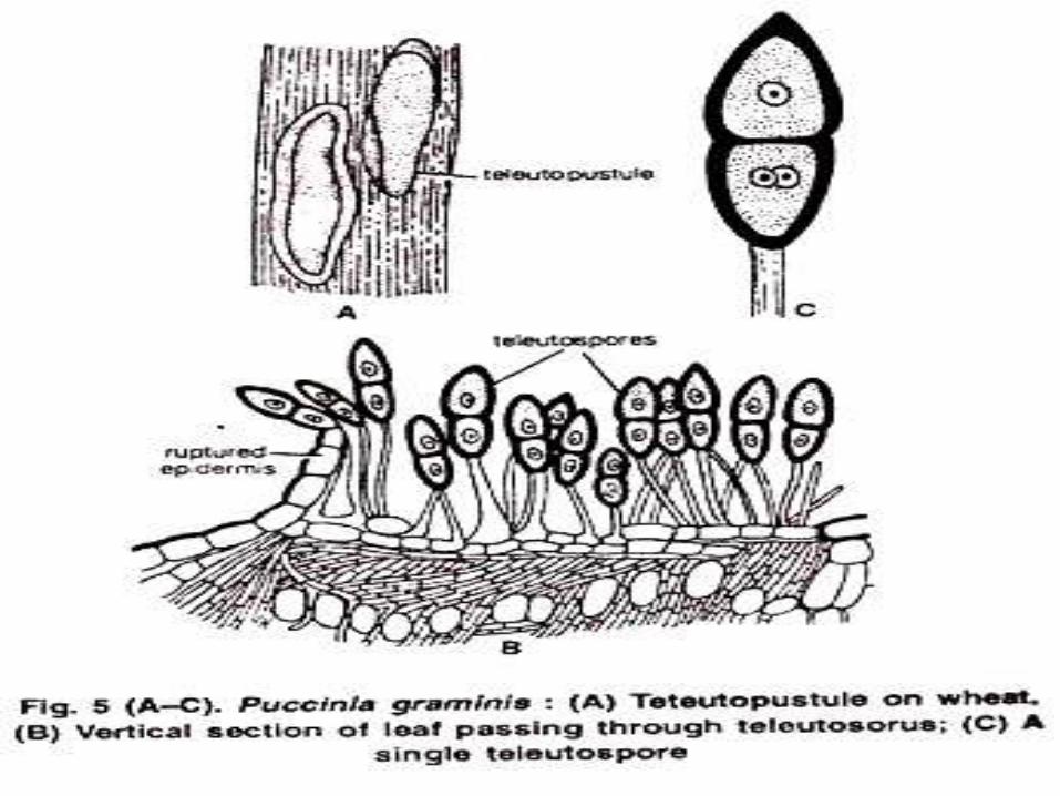
Telial Stage –III
• In the late of season at the time of crop maturity
teliospores form in place of urediospores.
• Teliospores are stalked, bicellular, diploid, spindle
shaped, thick wall celled and color is chestunt
brown.
• Size of teliospore is 40-50 × 15-20µm.
• Teliospores not germinate like urediospores.
• In hilly areas teliospores remain in dormant stage
on wheat residue but in plain area of India due to
high temperature teliospore not survive.

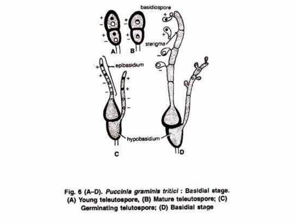
Basidial Stage –IV:
• After dormancy period each cell of teliospore
germinate by forming promycelium or basidium.
• Before germination of teliospore diploid nucleus by
meiosis divided in four haploid nucleus.
• Four basidiospores from 4 haploid nucleus form 4
basidiospores in which 2 act as + ve and 2 act as – ve
factor.
• These spores are unicellular, monokaryotic and
haploid
• These basidiospores by air reach on Barberry
(Alternate host).
• These spores can not infect to wheat.
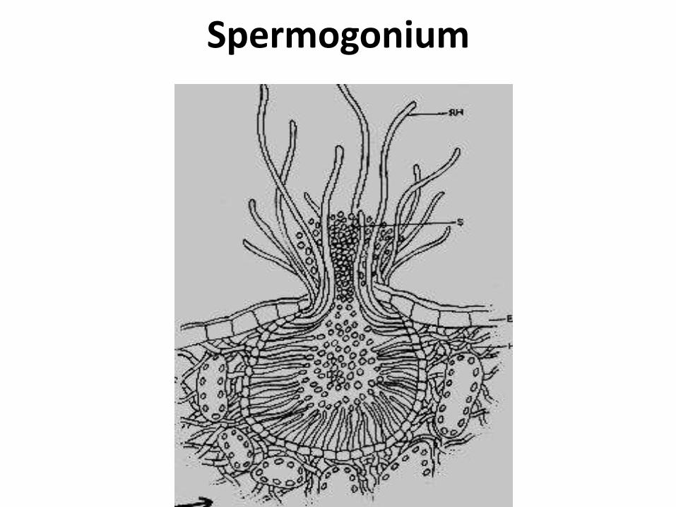
Spermogonial or Pycnial Stage –O
• Basidiospores fall on alternate host (Barberry), in presence of
moisture, germinate by germ tube and after entering into host
mycelium grow intercellularly.
• After few days pycnia form which put pressure on epidermis
and rupture it and open by ostiole.
• Flexuous receptive hyphae come out from ostiole.
• Pycnospores form in pycnia and come out from pycnia.
• Sex of pycniospore depend upon the type of mycelium from
which they are form (+ve or –ve factor)
• Due to color, smell of sticky liquid and sweetness of pycnidia
insects attract towards pycnidia and transfer the pycnospores to
other receptive hypha.
• When one opposite sex factor pycnospre reach to other sex
factor Dikaryotization take place resulting in dikaryotic
mycelium form.
Spermogonium
Aecial Stage –I
• After dikaryotization in pycnial stage this dikaryotic mycelium
grow downside in leaves and form cup shaped aecia on lower
side of barberry.
• Aeciospores form in these aecia.
• Aeciospores are unicellular, dikaryotic, spherical or hexagonal
with 14-26 µm diameter.
• Aeciospores put pressure on epidermis and rupture it resulting
in release of aeciospores in the air.
• These spores disseminate by air and reach on wheat leaves and
in presence of moisture germinate by germ tube.
• Germ tube enter into plant through stomata and form
dikaryotic, septate, intercellular mycelium which form
urediospores.

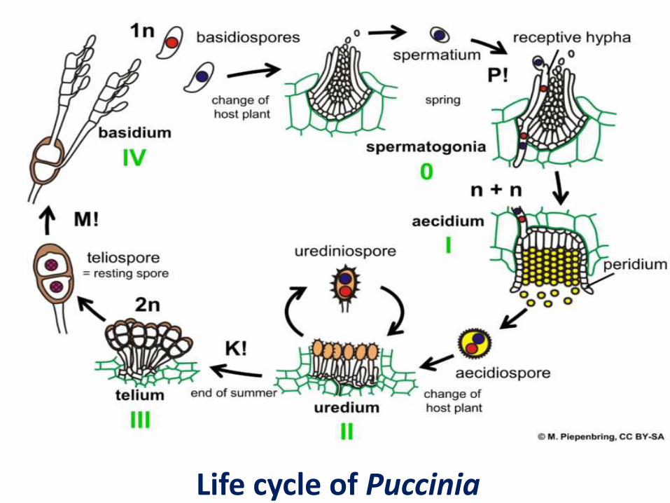
Life cycle of Puccinia

Thanks