Embed Size (px)
Citation preview

Adopted from Nilsen and cox – Lehninger principles of biochemistry (sixth edition)
Chapter 3 : Amino Acids, Peptides and Proteins

Learning Objectives
• To know the structure and naming of all 20 protein amino acids
• To know the structure and properties of peptides and the particularly the structure of the peptide bond.
• Ionization behavior of amino acids and peptides at different pH’s.
• To know the general pKa’s of amino acids: their carboxyl's, aminos, the R-group weak acids.

Some Function of proteins

Amino Acids: Building Blocks of Protein
• Proteins are linear heteropolymers of -amino acids
• Amino acids have properties that are well-suited to carry out a variety of biological functions
– Capacity to polymerize– Useful acid-base properties– Varied physical properties– Varied chemical functionality

Amino acids share many features, differing only at the R substituent

L and D forms

Carbon Numbering System
Athe carboxyl carbon of an amino acid would be C-1 and the
alpha carbon would be C-2.

Amino Acids: Classification
Common amino acids can be placed in five basic groups depending on their R substituents:
• Nonpolar, aliphatic R groups (7)
• Aromatic R groups (3)
• Polar, uncharged R groups (5)
• Positively charged R groups (3)
• Negatively charged R groups (2)

Invented the One Letter Amino Acid Code.


Spectrophotometry

UV light Absorption by Proteins – due to 2 Amino Acids

Cysteine can form Disulfide Bonds

Uncommon Amino Acids
Extra functional groups added by modification reactions are shown in red.
The four carbon backbones are shaded in yellow

Amino acids in Proteins Can be Reversibly Modified
A
•Reversible amino acid modifications involved in regulation of protein activity.•Phosphorylation is the most common type of regulatory modification.

Non Protein Amino Acids

Toxic Amino Acids
A search for compounds producing Yunnan Sudden Unexplained Deaths found related to eating a mushroom.
Trogia venenata Zhu LHalford, B. C+E News Feb 13, 2012
Which Form Occurs in Water ?
A zwitterion can act as either an acid (proton donor)
A zwitterion can act a base (proton acceptor)

Glycine Acid/Base Titration
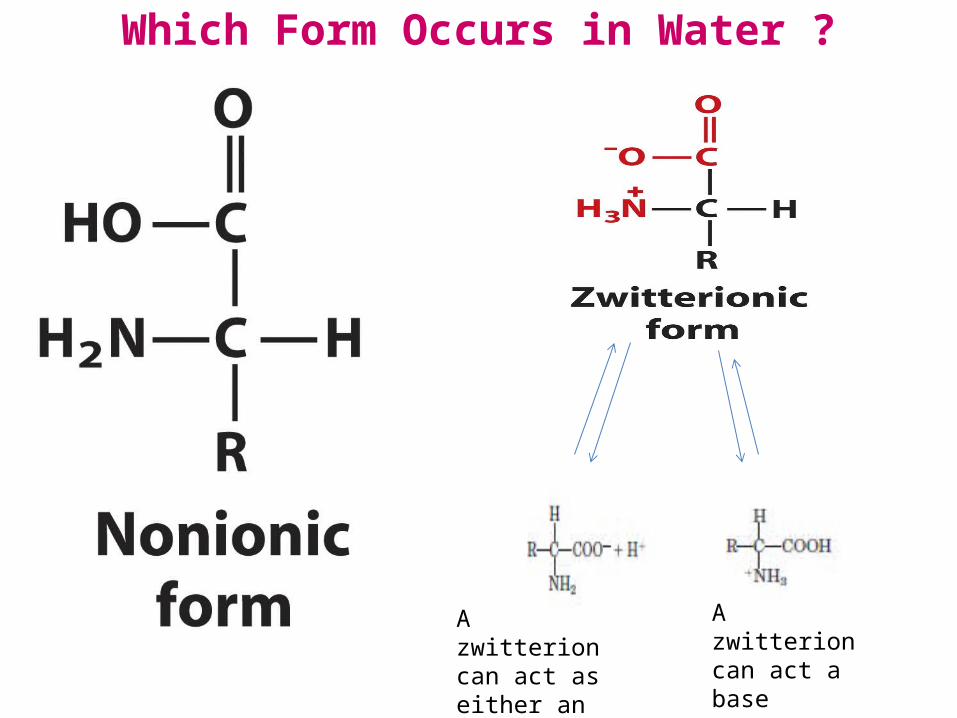
Compare Amino Acids to Simple Carboxylic Acids and Amines
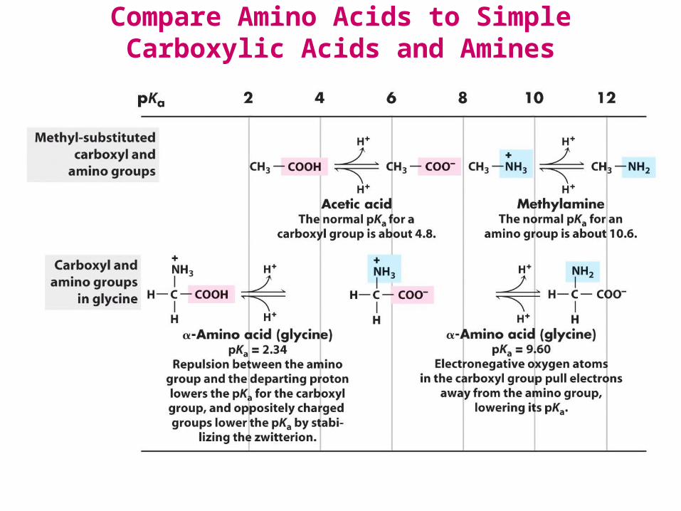
Glutamate has 3 pKa’s

Histidine has 3 pKa’s
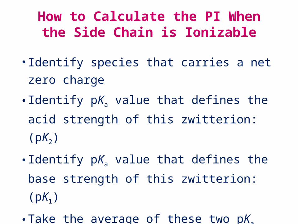
How to Calculate the PI When the Side Chain is Ionizable
• Identify species that carries a net zero charge
• Identify pKa value that defines the acid strength of this
zwitterion: (pK2)
• Identify pKa value that defines the base strength of this
zwitterion: (pK1)
• Take the average of these two pKa values
What is the pI of histidine?

Peptide Bond Formation by condensation
Where does this occur? Where does this occur?
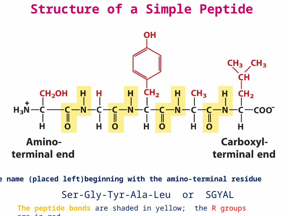
Structure of a Simple Peptide
Ser-Gly-Tyr-Ala-Leu or SGYALPeptide name (placed left)beginning with the amino-terminal residue
The peptide bonds are shaded in yellow; the R groups are in red.

Naming peptides: start at the N-terminus
• Using full amino acid names– Serylglycyltyrosylalanylleucine
• Using the three-letter code abbreviation– Ser-Gly-Tyr-Ala-Leu
• For longer peptides (like proteins) the one- letter code can be used– SGYAL
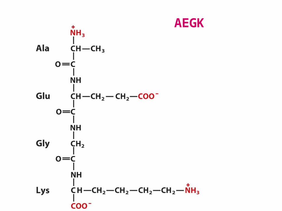
AEGK
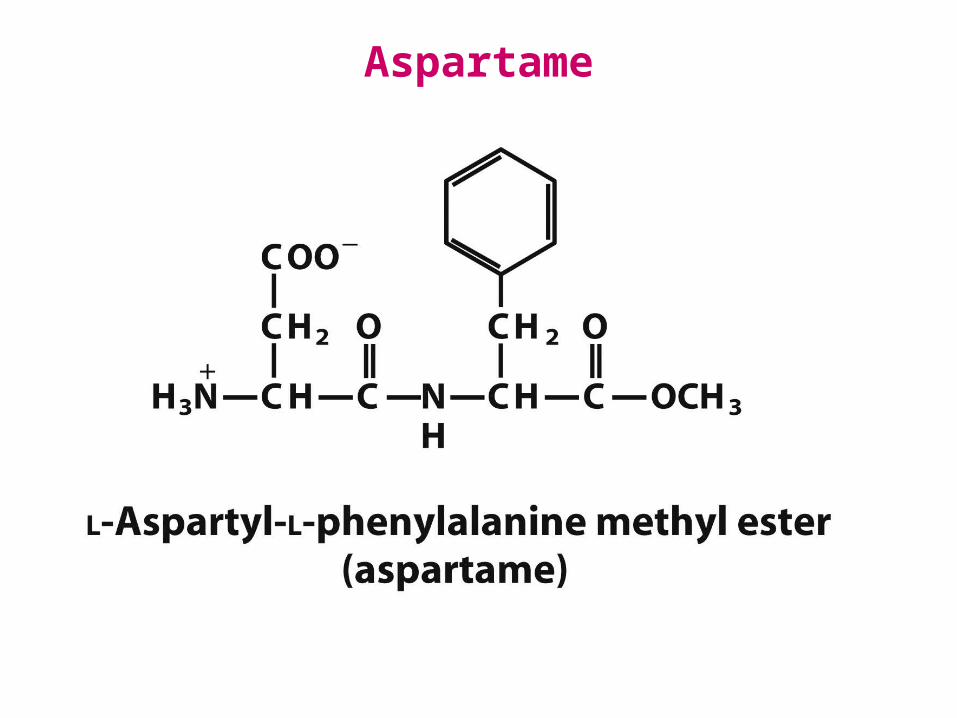
Aspartame
A

Peptides: A Variety of Functions
• Hormones and pheromones– insulin (think sugar)– oxytocin (think childbirth)– sex-peptide (think fruit fly mating)
• Neuropeptides– substance P (pain mediator)
• Antibiotics– polymyxin B (for Gram – bacteria)– bacitracin (for Gram + bacteria)
• Protection, e.g., toxins– amanitin (mushrooms)– conotoxin (cone snails)– chlorotoxin (scorpions)

Proteins are:
• Polypeptides (covalently linked -amino acids) + possibly: • cofactors
functional non-amino acid component metal ions or organic molecules
• coenzymes organic cofactors NAD+ in lactate dehydrogenase
• prosthetic groups covalently attached cofactors heme in myoglobin
• other modifications

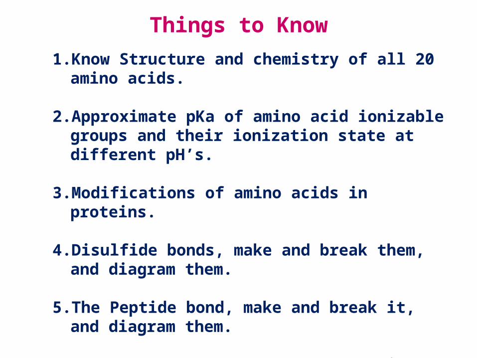
Things to Know1. Know Structure and chemistry of all 20 amino acids.
2. Approximate pKa of amino acid ionizable groups and their ionization state at different pH’s.
3. Modifications of amino acids in proteins.
4. Disulfide bonds, make and break them, and diagram them.
5. The Peptide bond, make and break it, and diagram them.
6. EOC Problems 1, 2, 3a, 4-7: we will have problems to solve (clicker questions) in class like these. Please practice these well before class.

Protein Purification

Learning Objectives
1. Know how each classical method of protein purification works.
2. Know how to measure protein and then calculate total activity and specific activity in each protein purification step.
3. Know how to evaluate protein purity.
4. Know how 2D PAGE gels work.
5. Know how molecular methods of protein purification work.
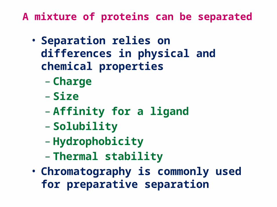
A mixture of proteins can be separated
• Separation relies on differences in physical and chemical properties– Charge– Size– Affinity for a ligand– Solubility– Hydrophobicity– Thermal stability
• Chromatography is commonly used for preparative separation

Solubility of Some Proteins in Ammonium Sulfate

Classical Protein Purification MethodsThe Oldest: Ammonium Sulfate Fractionation
Clear Cell Free Extract Cloudy
Clear Supernate
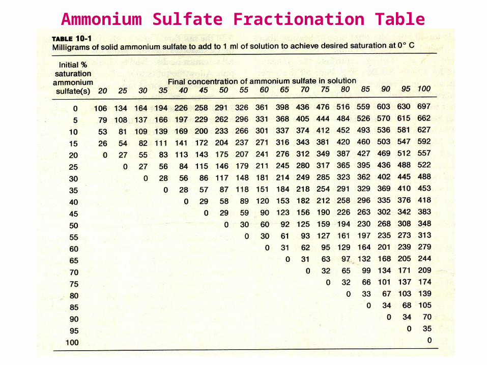
Ammonium Sulfate Fractionation Table
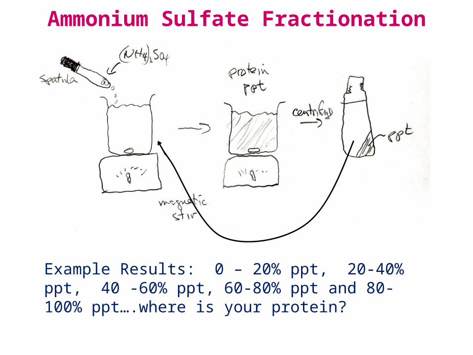
Ammonium Sulfate Fractionation
Example Results: 0 – 20% ppt, 20-40% ppt, 40 -60% ppt, 60-80% ppt and 80-100% ppt….where is your protein?

These Fractions Need to be Assayed
For:1. Total Protein can be done by
a. Absorbance at 280 nmb. Colorimetric tests for
proteinLowry Assay, Bradford
Assay
2. Your Specific Proteina. specific enzyme assay, orb. specific binding assay, orc. unique spectral property.

Example Results – each Cut in 100 ml of Buffer(NH4)2SO4 Cut Total Protein Your EnzymeCrude Extract 1.73 g 3,895 mM/sec 0 – 20% 452 mg 0.1 mM/sec20 – 40% 323 mg 3,560 mM/sec40 – 60% 541 mg 12 mM/sec60 – 80% 329 mg 0.1 mM/sec80 – 100% 78 mg 0.01 mM/sec
Total Enzyme Activity Specific ActivityCrude: 3.9 x 10³ mM/sec 2.25 (mM/sec)/mg protein20-40 Cut 3.56 x 10³ mM/sec 11.0 (mM/sec)/mg protein (91% orig. activity) 4.9X fold purified

Example Results – each Cut in 100 ml of Buffer(NH4)2SO4 Cut Total Protein Your EnzymeCell Free Extract 1.73 g 3,895 mM/sec 0 – 20% 452 mg 0.1 mM/sec20 – 40% 323 mg 3,560 mM/sec40 – 60% 541 mg 12 mM/sec60 – 80% 329 mg 0.1 mM/sec80 – 100% 78 mg 0.01 mM/sec
How did you get rid of the high conc of (NH4)2SO4 ?
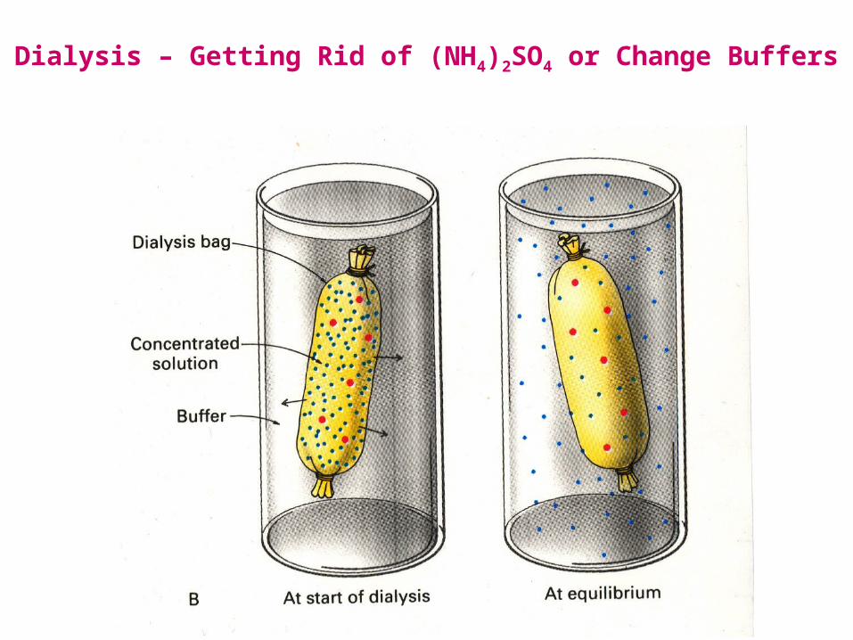
Dialysis – Getting Rid of (NH4)2SO4 or Change Buffers

Column Chromatography Principle

Columns Connected to Fraction CollectorsTube from column attached here

Separation by Charge: Ion Exchange
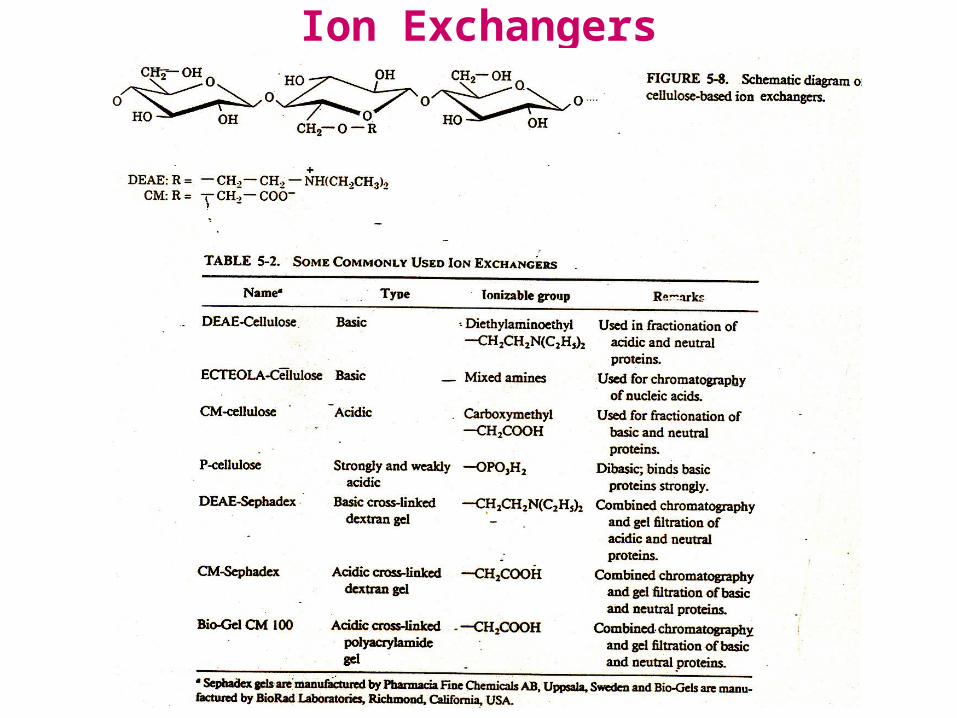
Ion Exchangers
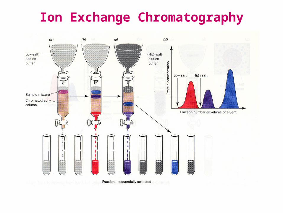
Ion Exchange Chromatography
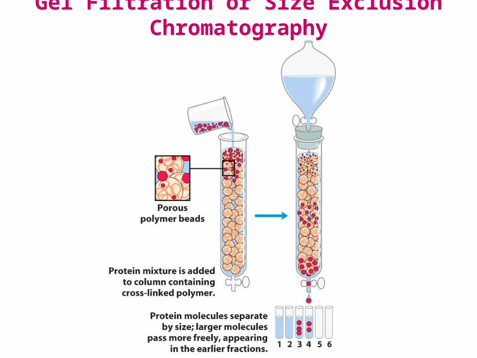
Gel Filtration or Size Exclusion Chromatography

Separation by Size: Gel or Size Exclusion

Gel Filtration Media

Gel Filtration or Exclusion Chromatography
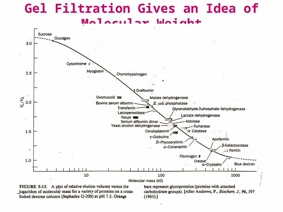
Gel Filtration Gives an Idea of Molecular Weight

Separation by Binding Affinity
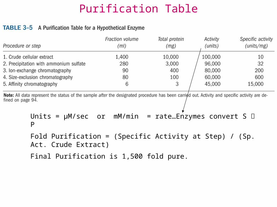
Units = μM/sec or mM/min = rate…Enzymes convert S P
Fold Purification = (Specific Activity at Step) / (Sp. Act. Crude Extract)
Final Purification is 1,500 fold pure.
Purification Table

Electrophoresis for Protein Analysis
Separation in analytical scale is commonly done by electrophoresis
– Electric field pulls proteins according to their charge
– Gel matrix hinders mobility of proteins according to their size and shape

Polyacrylamide Gel Electrophoresis

Gel Showing Steps in
Purification
Purification of RecA from E. coli.

SDS PAGE: Molecular Weight• SDS – sodium dodecyl sulfate – a detergent
• SDS micelles bind to and partially unfold all the proteins– SDS gives all proteins a uniformly negative charge– The native shape of proteins does not matter– Rate of movement will only depend on size: small proteins will move faster

SDS-PAGE can be used to calculate the molecular weight of a protein

Isoelectric focusing can be used to determine the pI of a protein

A

2-D Gels Start with Isoelectric Focusing

2-D Gels End with SDS PAGE
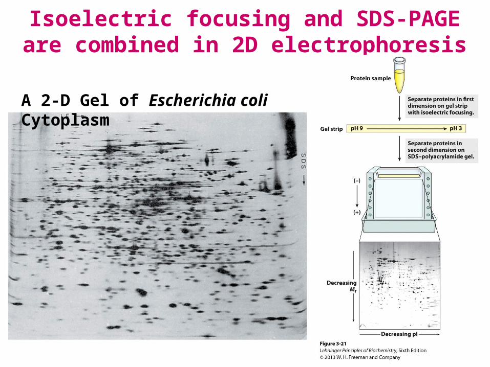
Isoelectric focusing and SDS-PAGE are combined in 2D electrophoresis
A 2-D Gel of Escherichia coli Cytoplasm

Molecular Methods of Protein Purification1. Isolate the gene…restriction enzymes, separation DNA on
agarose gels, insert gene into a plasmid behind an active promoter (turns on gene), and with a “tag” (such has six-histidines) or Maltose Binding Protein or other tag. The tag makes this a fusion protein:
Protein-his-his-his-his-his-his or Protein-MBP
2. Insert the plasmid into a bacterium (usually E. coli) and turn-on the promoter to express the fusion-protein in large quantities (the protein can be 10-30% cell volume!).
3. Lyse cells, fusion-protein binds affinity column which after binding and washing provides the fusion protein essentially pure.
4. Cleave off the his tag, dialyze pure protein.
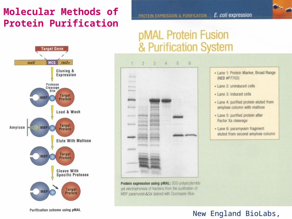
Molecular Methods of Protein Purification
New England BioLabs, Inc
Expression Purification
Time after induction (hours) 0 0.5 1.0 1.5 2.0 2.5 3.0
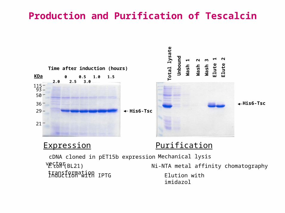
Production and Purification of Tescalcin
Tota
l lys
ate
Unb
ound
Was
h 2
Was
h 3
Was
h 1
Elut
e 2
Elut
e 1
1159350
3629
21
KDa
His6-TscHis6-Tsc
cDNA cloned in pET15b expression vector
E. coli (BL21) transformation
Induction with IPTG
Mechanical lysis
Ni-NTA metal affinity chomatography
Elution with imidazol
by Erasmo Perera, FIU student

Things to Know and Do Before Class
1. Know each method used Purify Proteins and How they Work.
2. Calculation of Total protein and Specific Activity in the steps of protein purification.
3. Testing for protein purity.
4. How to do 2D PAGE gels.
5. Molecular Methods of Protein Purification.
6. Be able to do EOC Problems: 8-11, 13, 15 (protein purification), 16.