Embed Size (px)
Citation preview
Keratosis and related disorder of the oral mucosa
By:Dr. Mo’ad A. Albdour
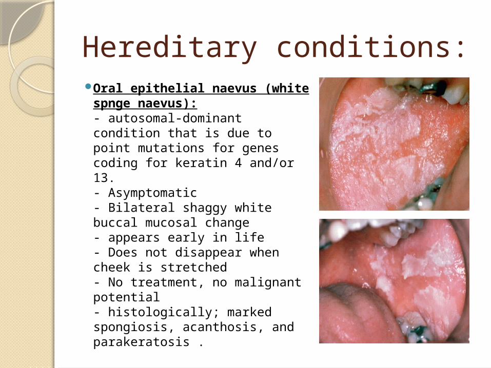
Hereditary conditions: Oral epithelial naevus
(white spnge naevus):- autosomal-dominant condition that is due to point mutations for genes coding for keratin 4 and/or 13. - Asymptomatic- Bilateral shaggy white buccal mucosal change- appears early in life- Does not disappear when cheek is stretched- No treatment, no malignant potential- histologically; marked spongiosis, acanthosis, and parakeratosis .
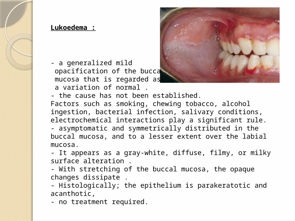
Lukoedema :
- a generalized mild opacification of the buccal mucosa that is regarded as a variation of normal .- the cause has not been established. Factors such as smoking, chewing tobacco, alcohol ingestion, bacterial infection, salivary conditions, electrochemical interactions play a significant rule.- asymptomatic and symmetrically distributed in the buccal mucosa, and to a lesser extent over the labial mucosa. - It appears as a gray-white, diffuse, filmy, or milky surface alteration .- With stretching of the buccal mucosa, the opaque changes dissipate .- Histologically; the epithelium is parakeratotic and acanthotic, - no treatment required.
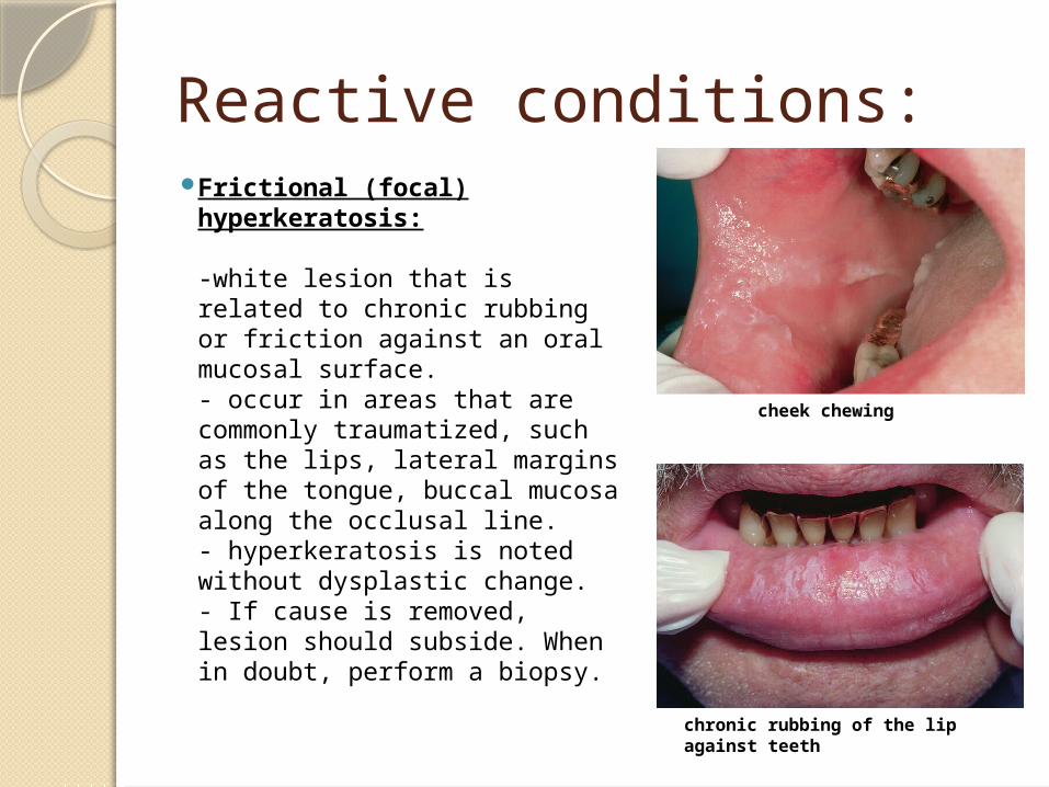
Reactive conditions: Frictional (focal)
hyperkeratosis:
-white lesion that is related to chronic rubbing or friction against an oral mucosal surface.- occur in areas that are commonly traumatized, such as the lips, lateral margins of the tongue, buccal mucosa along the occlusal line.- hyperkeratosis is noted without dysplastic change.- If cause is removed, lesion should subside. When in doubt, perform a biopsy.
chronic rubbing of the lip against teeth
cheek chewing
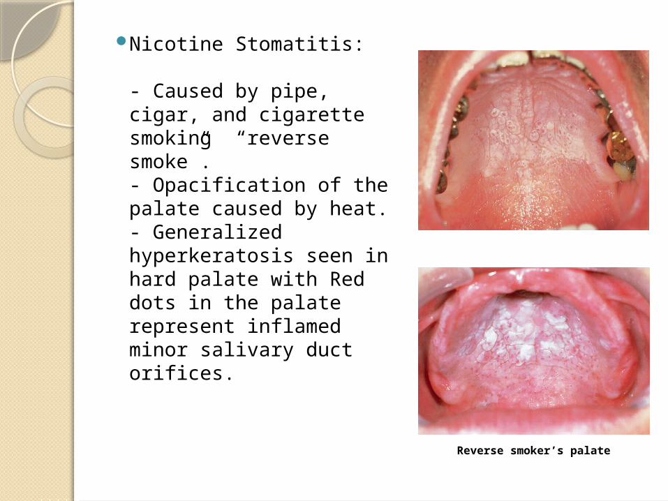
Nicotine Stomatitis: - Caused by pipe, cigar, and cigarette smoking “reverse smoke”.- Opacification of the palate caused by heat.- Generalized hyperkeratosis seen in hard palate with Red dots in the palate represent inflamed minor salivary duct orifices.
Reverse smoker’s palate
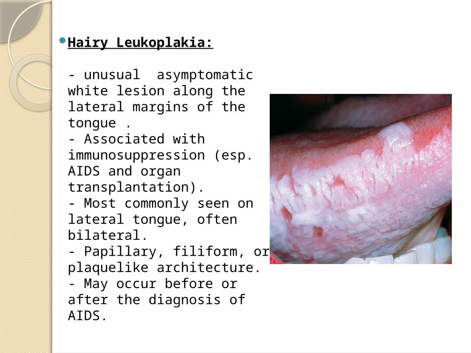
Hairy Leukoplakia:
- unusual asymptomatic white lesion along the lateral margins of the tongue .- Associated with immunosuppression (esp. AIDS and organ transplantation).- Most commonly seen on lateral tongue, often bilateral.- Papillary, filiform, or plaquelike architecture.- May occur before or after the diagnosis of AIDS.
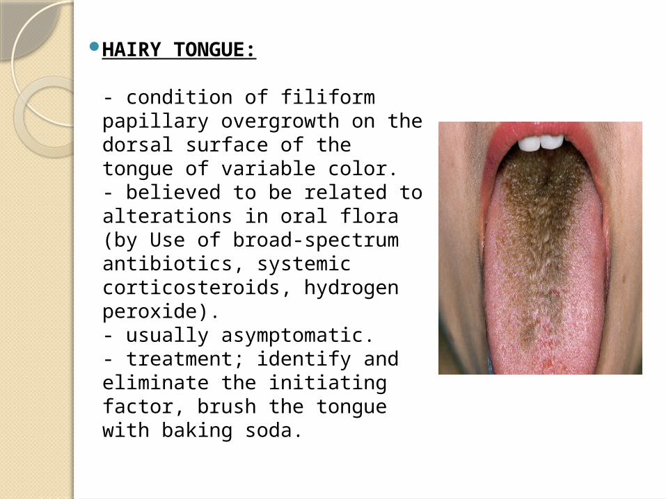
HAIRY TONGUE:
- condition of filiform papillary overgrowth on the dorsal surface of the tongue of variable color.- believed to be related to alterations in oral flora (by Use of broad-spectrum antibiotics, systemic corticosteroids, hydrogen peroxide).- usually asymptomatic. - treatment; identify and eliminate the initiating factor, brush the tongue with baking soda.
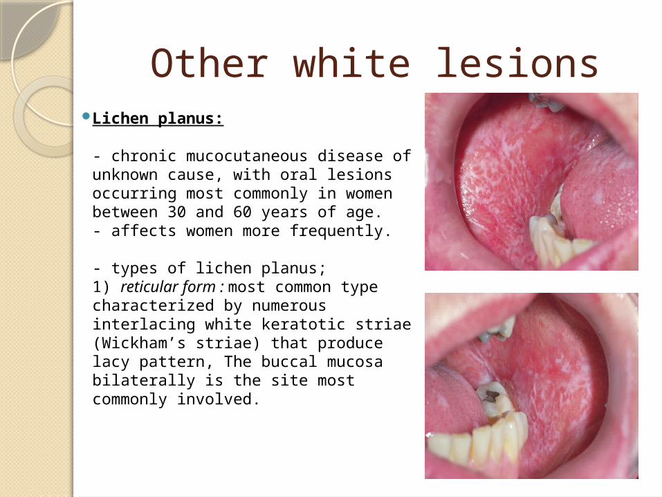
Other white lesions Lichen planus:
- chronic mucocutaneous disease of unknown cause, with oral lesions occurring most commonly in women between 30 and 60 years of age.- affects women more frequently.
- types of lichen planus; 1) reticular form : most common type
characterized by numerous interlacing white keratotic striae (Wickham’s striae) that produce lacy pattern, The buccal mucosa bilaterally is the site most commonly involved.
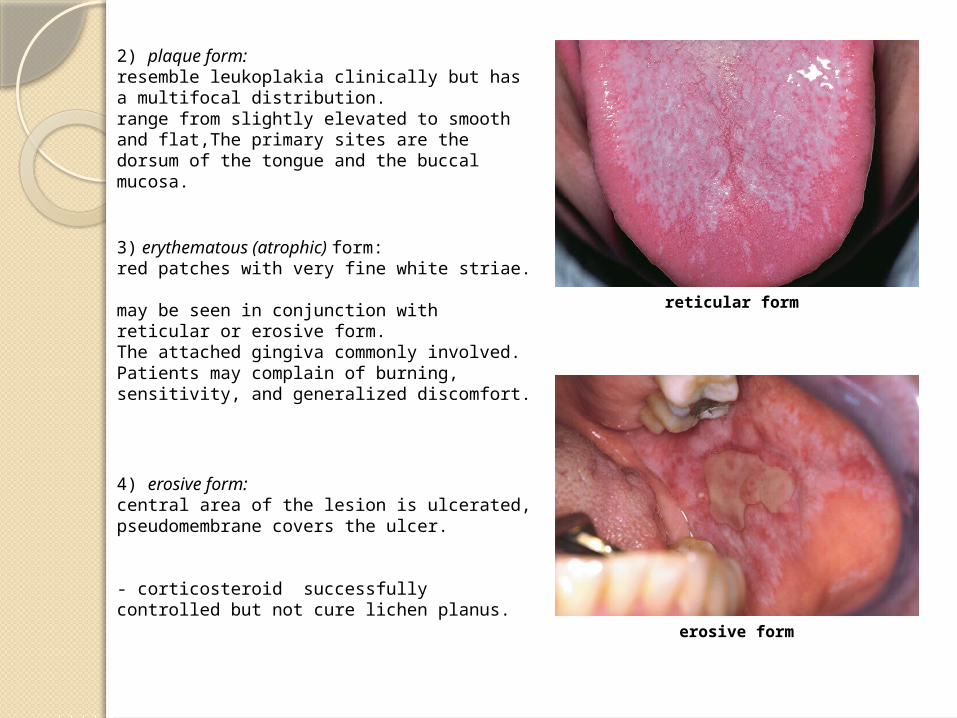
2) plaque form:resemble leukoplakia clinically but has a multifocal distribution.range from slightly elevated to smooth and flat,The primary sites are the dorsum of the tongue and the buccal mucosa.
3) erythematous (atrophic) form:red patches with very fine white striae. may be seen in conjunction with reticular or erosive form.The attached gingiva commonly involved.Patients may complain of burning, sensitivity, and generalized discomfort.
4) erosive form:central area of the lesion is ulcerated, pseudomembrane covers the ulcer.
- corticosteroid successfully controlled but not cure lichen planus. erosive form
reticular form