Embed Size (px)
Citation preview

MUSADIQ KHAN DURRANI 1
MUSADIQ KHAN Durrani
UCMD
University of Lahore

It is longest bone of upper limb and has upper end,lower end & shaft
Introduction
MUSADIQ KHAN DURRANI 2
MUSADIQ KHAN DURRANI 3
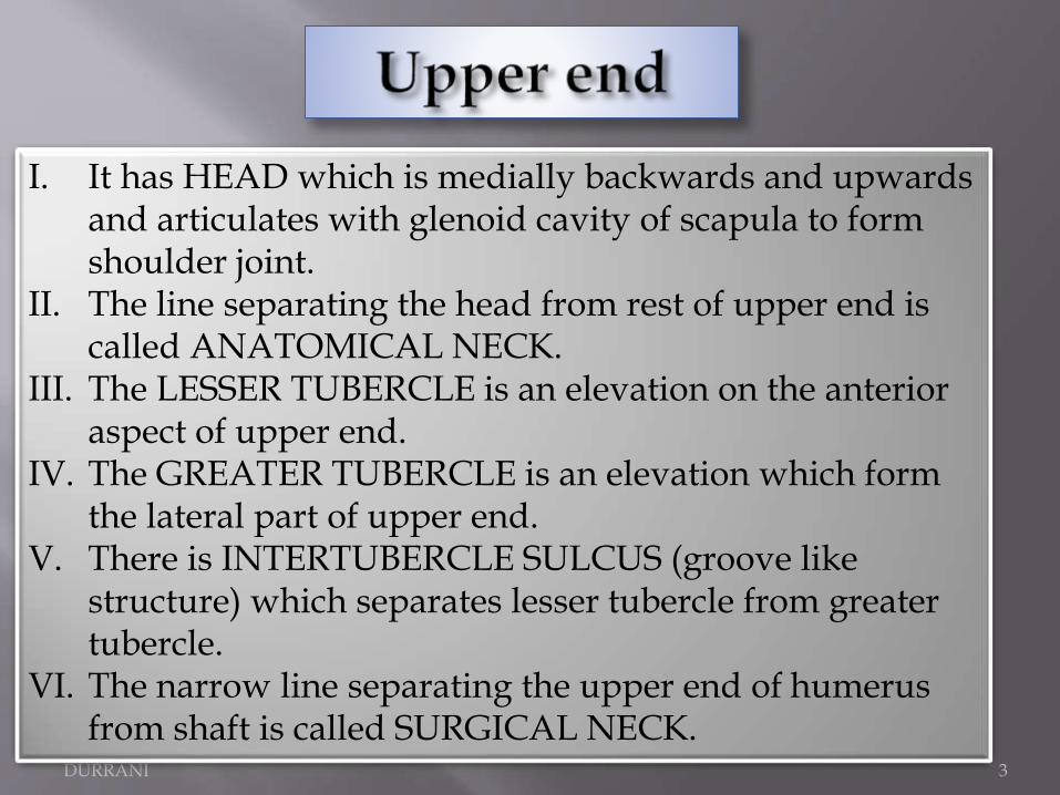
I. It has HEAD which is medially backwards and upwards and articulates with glenoid cavity of scapula to form shoulder joint.
II. The line separating the head from rest of upper end is called ANATOMICAL NECK.
III. The LESSER TUBERCLE is an elevation on the anterior aspect of upper end.
IV. The GREATER TUBERCLE is an elevation which form the lateral part of upper end.
V. There is INTERTUBERCLE SULCUS (groove like structure) which separates lesser tubercle from greater tubercle.
VI. The narrow line separating the upper end of humerusfrom shaft is called SURGICAL NECK.

MUSADIQ KHAN DURRANI 4

MUSADIQ KHAN DURRANI 5
Bony Features
Articular Part:
•Capitulum: A rounded projection
which articulates with the head of the radius.
•Trochlea: Pulley shaped surface.
Articulates with the trochlear notch of ulna. Medial edge of projects 6mm downwards to form the carrying angle.

MUSADIQ KHAN DURRANI 6
•Medial Epicondyle: Prominent bony projection on the lower side medially.
Subcutaneous & easily felt.
•Lateral Epicondyle: smaller than medial epicondyle & has a muscular
impression.
•Lateral Supracondylar ridge: A small lateral margin just above the
lower end.
•Medial Supracondylar ridge: Small medial ridge on the medial side.
•Coronoid Fossa: Depression just above the anterior aspect of trochlea.
Accommodates with the coronoid process of ulna when Elbow is Flexed.
•Radial Fossa: Depression just above the anterior aspect of Capitulum.
Accommodates with the head of radius when elbow in Flexed.
•Olecranon Fossa: Depression just above the posterior aspect of Trochlea.
Accommodates with olecranon process of ulna when elbow is Extended.

MUSADIQ KHAN DURRANI 7

MUSADIQ KHAN DURRANI 8
•Rounded in the upper half & triangular in the lower half. Contains three borders & three surfaces.
Borders:
o Anterior Border: Upper one third forms the Lateral lip of the
intertubercular sulcus. Middle part forms the anterior region of Deltoid tuberosity. Lower half is smooth & rounded.
o Lateral Border: Prominent only in the lower region where it forms
lateral supracondylar ridge. In the middle it is interrupted by Radial groove.
o Medial Border: Forms the medial lip of the intertubercular sulcus.
Rough through its middle & continuous below with the medial supracondylar ridge.

MUSADIQ KHAN DURRANI 9
Surfaces:
o Anterolateral Surface(b/w anterior & lateral borders): Upper half is covered by the deltoid. A little above it is marked by a V shaped
deltoid tuberosity. Radial groove runs downwards & forwards across the surface.
o Anteromedial Surface(b/w anterior & medial border): Upper end is narrow & forms
floor of the intertubercular sulcus. Nutrient foramen is seen near the medial border.
o Posterior Surface(b/w medial & lateral borders): Upper part is marked by an oblique ridge
& lower end is crossed by a radial groove.

MUSADIQ KHAN DURRANI 10
•Upper end: Rounded.• Lower end: Expanded from side to side & flattened from before backwards.• Head: Directed posterior & medially upwards.• Lesser Tubercle: Projects from the front of the upper end & is limited laterally by the intertubercular sulcus.

MUSADIQ KHAN DURRANI 11

MUSADIQ KHAN DURRANI 12
MUSCLE ATTACHMENTS
Lesser Tubercle: Insertion of The Multipennate SUBSCAPULARIS.
Greater Tubercle(uppermost impression): Insertion of the SUPRASPINATUS.
Greater Tubercle(middle impression): Insertion of the INFRASPINATUS.
Greater Tubercle(lower impression): Insertion of TERES MINOR.
Intertubercular Sulcus(lateral lip): Insertion of PECTORALIS MAJOR.
Intertubercular Sulcus(floor): Insertion of LATISSMUS DORSI.
Intertubercular Sulcus(medial lip): Insertion of TERES MAJOR.Deltoid Tuberosity: Insertion of DELTOID.
Medial Border(rough area): Insertion of CHORACOBRACHIALLIS.
Shaft: BRACHIALIS arises from the lower halves of the anteromedial & anterolateral surfaces of the shaft.

Attachments of Upper END
MUSADIQ KHAN DURRANI 13

MUSADIQ KHAN DURRANI 14

MUSADIQ KHAN DURRANI 15

MUSADIQ KHAN DURRANI 16
Lower end: Lateral Supracondylar Ridge: BRACHIORADIALUS arises from the upper two thirds. EXTENSOR CARPI RADIALIS LONGUS arises from the lower one third. Medial Supracondylar Ridge: Humeral head of PRONATOR TERES arises from lower one third. Lateral Epicondyle: ANCONEUS arises from the posterior surface of lateral epicondyle. Radial Groove: Lateral head of TRICEPS BRACHII arises from the oblique ridge on the upper part of posterior surface above the radial groove, while its Medial head arises from the posterior surface below radial groove. Medial Epicondyle(anterior aspect): Superficial FLEXOR muscles arises from the common origin of the anterior aspect. Lateral Epicondyle: Superficial EXTENSOR muscles have a common origin from the lateral epicondyle.

MUSADIQ KHAN DURRANI 17
CAPSULAR LIGAMENT of shoulder jointis attached to the anatomical neck except on the medial side where the line of attachment is down by 2cm to include a
small area of shaft. TENDON Of the LONG HEAD OF BICEPS BRACHII leaves the joint
cavity through the aperture provided by Intertubercularsulcus.
CAPSULAR LIGAMENT of the Elbow joint is attached to the lower end along the line & reaches
the upper limits of coronoid fossae & radial fossa anteriorly & of then olecranon fossa posterioirly.

MUSADIQ KHAN DURRANI 18
Nerves• Three Nerves are directly related to
the Humerus.
• AXILLARY Nerve at the Surgical neck.
• RADIAL Nerve at the Radial groove.
• ULNAR Nerve behind the medial epicondyle.

MUSADIQ KHAN DURRANI 19
THANKS