Embed Size (px)
Citation preview
My boyfriend has 20/20 vision after lasik surgery
but he is not pleased!
WHY???
He thought I was prettier
before…
That makes sense.
Prepared By, Ms Wong Fui Yen 2012Prepared By, Ms Wong Fui Yen 2012

The EyeThe Eye

Lesson ObjectivesLesson Objectives Describe the gross structure of the eye as seen Describe the gross structure of the eye as seen
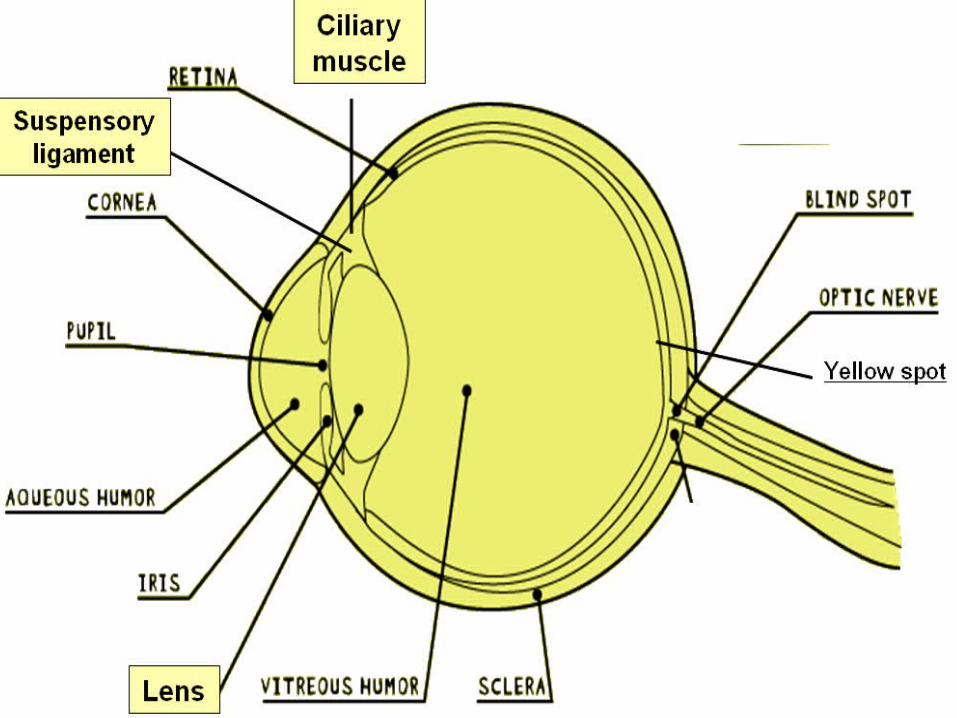
in:in:- front view; and- front view; and- horizontal section.- horizontal section.
State the principal functions of the component State the principal functions of the component parts of the eye in producing a focused image parts of the eye in producing a focused image of:of:- near objects; and- near objects; and- distant objects on the retina. - distant objects on the retina.
Describe the pupil reflex in response to:Describe the pupil reflex in response to:- bright light; and- bright light; and- dim light.- dim light.
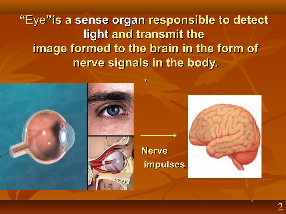
““EyeEye”is a ”is a sensesense organorgan responsible to detect responsible to detect lightlight and transmit the and transmit the
image formed to the brain image formed to the brain in the form of in the form of nerve signals in the body.nerve signals in the body.
..
NerveNerve
impulsesimpulses
2
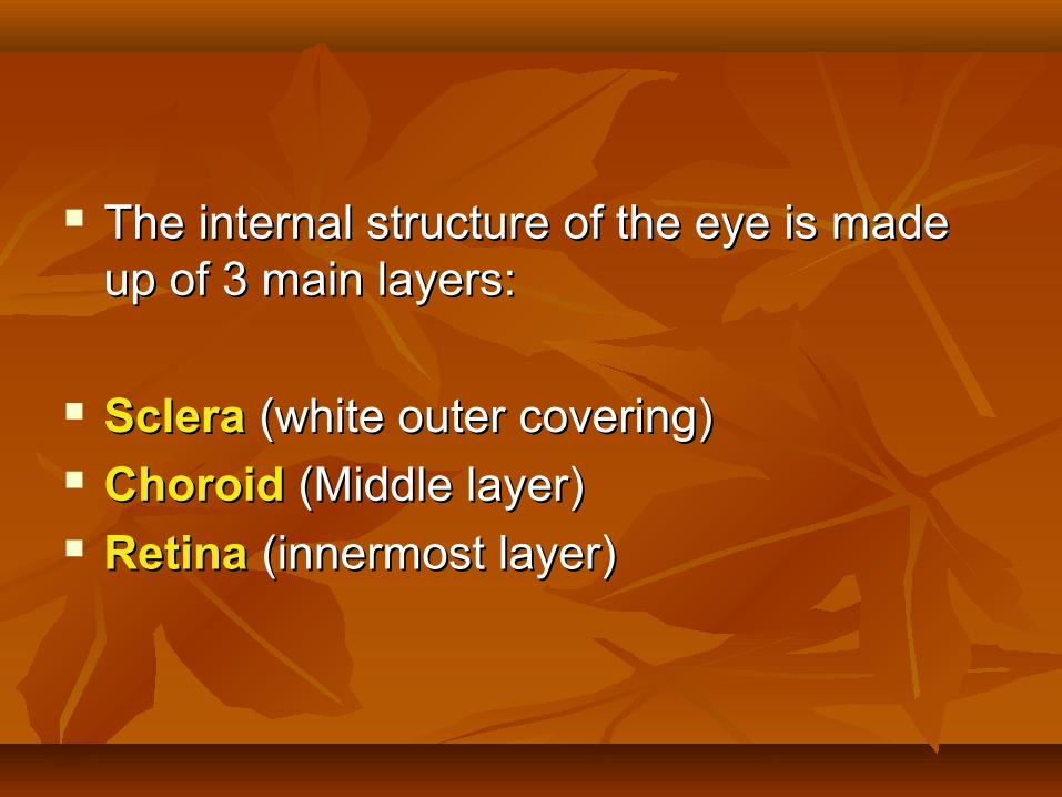
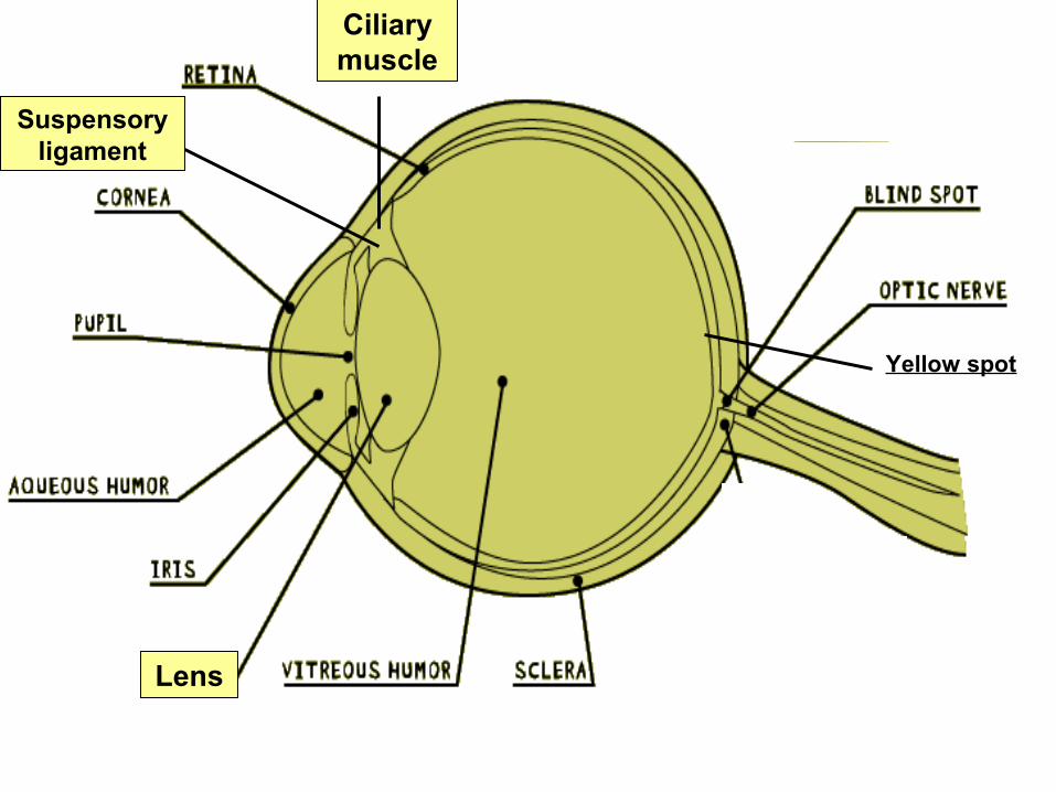
The internal structure of the eye is made The internal structure of the eye is made up of 3 main layers:up of 3 main layers:
ScleraSclera (white outer covering) (white outer covering) Choroid Choroid (Middle layer)(Middle layer) RetinaRetina (innermost layer) (innermost layer)

Class ActivityClass Activity : : Look at your friend eyeball to eyeball. Look at your friend eyeball to eyeball. Which part of the eye you could see?Which part of the eye you could see?
9
Conjunctiva (covers sclera)
Lower eyelid
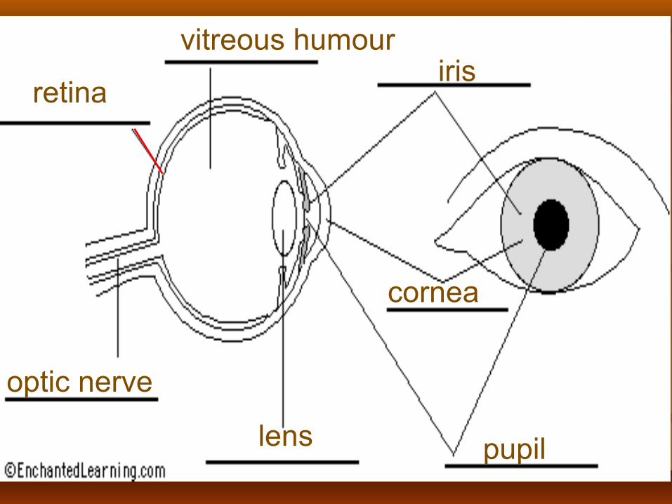
retina
vitreous humouriris
optic nerve
lens
cornea
pupil
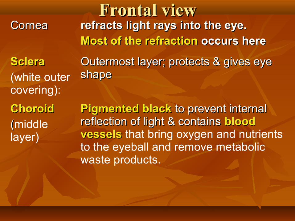
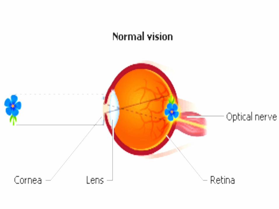
Frontal viewFrontal viewCorneaCornea refracts light rays into the eye.refracts light rays into the eye.
Most of the refraction Most of the refraction occurs hereoccurs here
Sclera Sclera (white outer covering):
Outermost layer; protects & gives eye Outermost layer; protects & gives eye shapeshape
Choroid Choroid
((middle layer)
Pigmented blackPigmented black to prevent internal to prevent internal reflection of light & contains reflection of light & contains blood blood vesselsvessels that bring oxygen and nutrients to the eyeball and remove metabolic waste products.

Ciliary BodyCiliary Body Contains ciliary muscles which control Contains ciliary muscles which control thicknessthickness of the lens. of the lens.
Suspensory Suspensory LigamentsLigaments
AttachesAttaches the edge of the lens to the the edge of the lens to the ciliary bodyciliary body
LensLens Refracts/focuses light Refracts/focuses light onto retina by onto retina by changing its shape or thicknesschanging its shape or thickness
IrisIris Controls the Controls the amount of lightamount of light entering the entering the eye using its 2 sets of muscles (circular eye using its 2 sets of muscles (circular and radial muscles)and radial muscles)
PupilPupil A hole in the centre of the iris that A hole in the centre of the iris that allows light to enter the eyeallows light to enter the eye

Aqueous Chamber Aqueous Chamber containing containing aqueous aqueous humourhumour
Keeps eyeball firm. Keeps eyeball firm.
Refracts light onto the lensRefracts light onto the lens
Vitreous Chamber Vitreous Chamber containing containing vitreous vitreous humourhumour
Refracts light onto retinaRefracts light onto retina
RetinaRetina - light sensitive layer light sensitive layer - high concentration of high concentration of photoreceptorsphotoreceptors (rods and (rods and cones);cones); - allows image to be - allows image to be formed and color to be seenformed and color to be seen
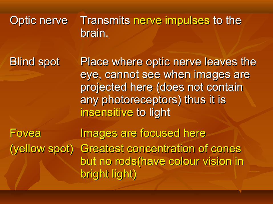
Optic nerveOptic nerve Transmits Transmits nerve impulses nerve impulses to the to the brain. brain.
Blind spotBlind spot Place where optic nerve leaves the Place where optic nerve leaves the eye, cannot see when images are eye, cannot see when images are projected here (does not contain projected here (does not contain any photoreceptors) thus it is any photoreceptors) thus it is insensitiveinsensitive to light to light
FoveaFovea
(yellow spot)(yellow spot)Images are focused hereImages are focused here
Greatest concentration of cones Greatest concentration of cones but no rods(have colour vision in but no rods(have colour vision in bright light)bright light)

How The Eye WorksHow The Eye Works
Light rays are reflected from an object.Light rays are reflected from an object.
Light rays are Light rays are refractedrefracted as they pass through as they pass through cornea and lens. cornea and lens.
The lens are involved in The lens are involved in focusingfocusing ( or ( or accommodation) so that clear images of objects accommodation) so that clear images of objects at at different distancesdifferent distances can be formed on retina. can be formed on retina.
Focusing for vision involves the Focusing for vision involves the ciliary musclesciliary muscles and and suspensory ligamentssuspensory ligaments to help adjust the to help adjust the lens.lens.
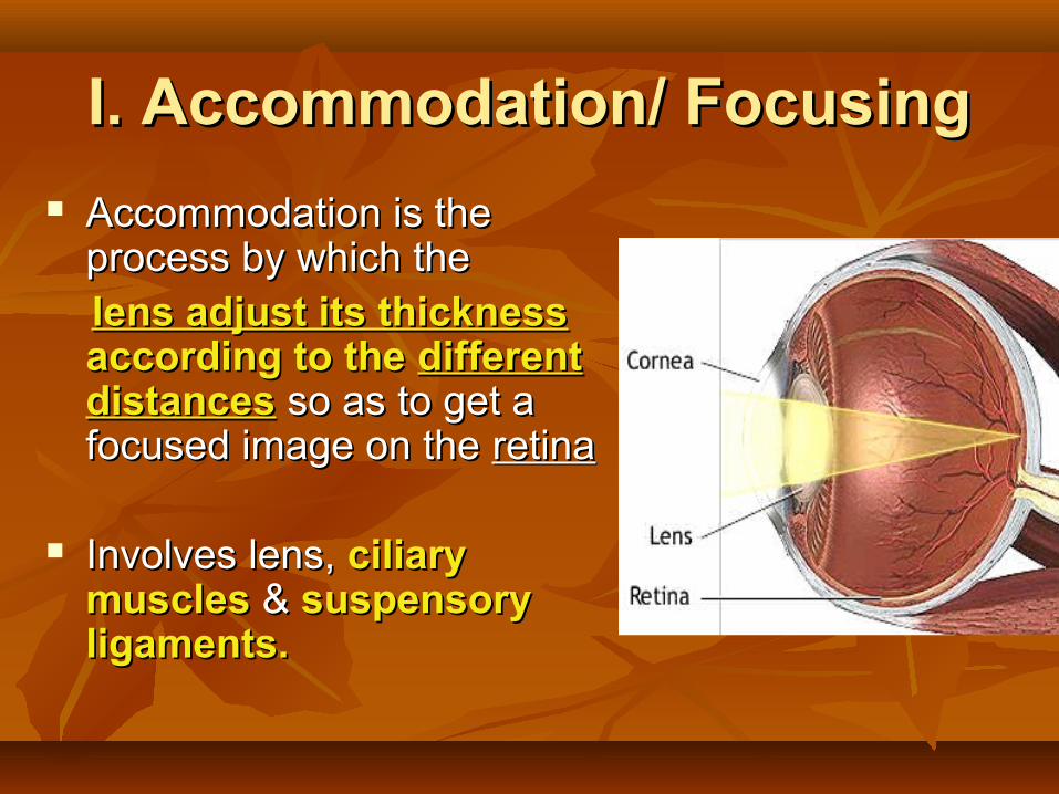
I. Accommodation/ FocusingI. Accommodation/ Focusing
Accommodation is the Accommodation is the process by which the process by which the
lens adjust its thicknesslens adjust its thickness according to the according to the different different distancesdistances so as to get a so as to get a focused image on the focused image on the retinaretina
Involves lens, Involves lens, ciliary ciliary musclesmuscles & & suspensory suspensory ligaments.ligaments.
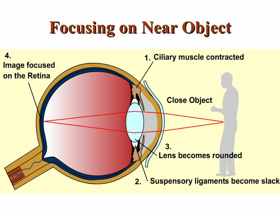
Focusing on Near ObjectFocusing on Near Object
1.
2.
3.
4.
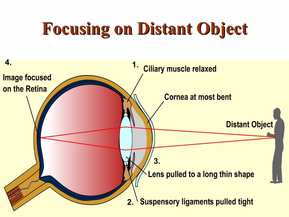
Focusing on Distant ObjectFocusing on Distant Object
1.
2.
3.
4.
Ciliary muscle
Suspensory ligament
Lens
Yellow spot

Ciliary muscle
Suspensory ligament
Lens
Yellow spot
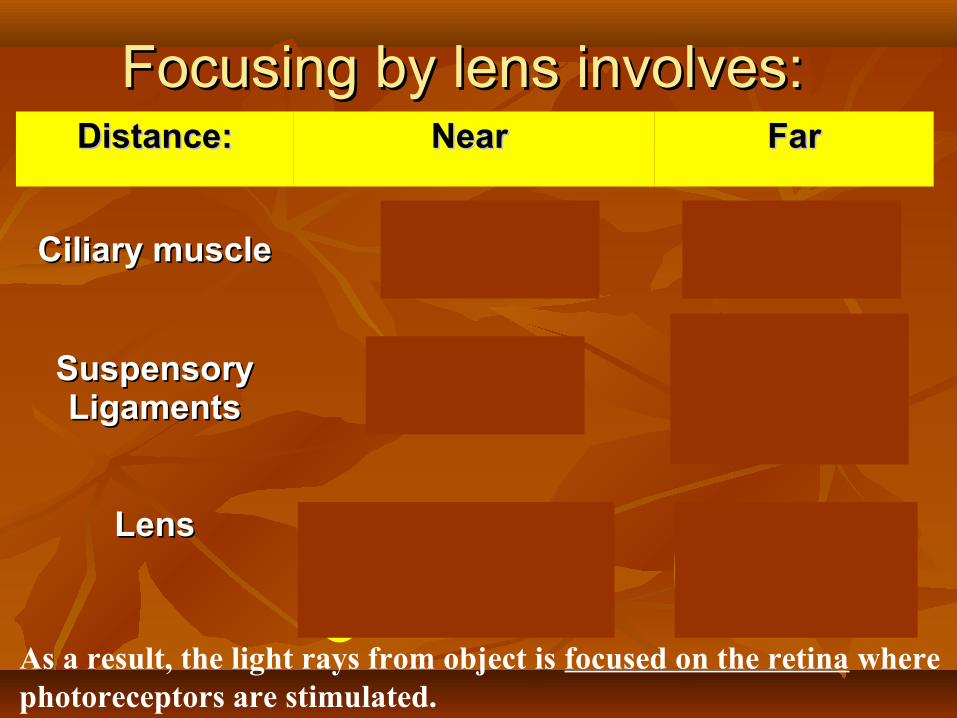
Distance:Distance: Near Near FarFar
Ciliary muscleCiliary muscle ContractContract RelaxRelax
Suspensory Suspensory LigamentsLigaments
BecomeBecomeslackenedslackened
Are pulled Are pulled tight tight
(become taut)(become taut)
LensLens ThickerThickerand and
more convexmore convex
ThinnerThinnerand and
less convexless convex
Focusing by lens involves:Focusing by lens involves:
As a result, the light rays from object is focused on the retina where photoreceptors are stimulated.
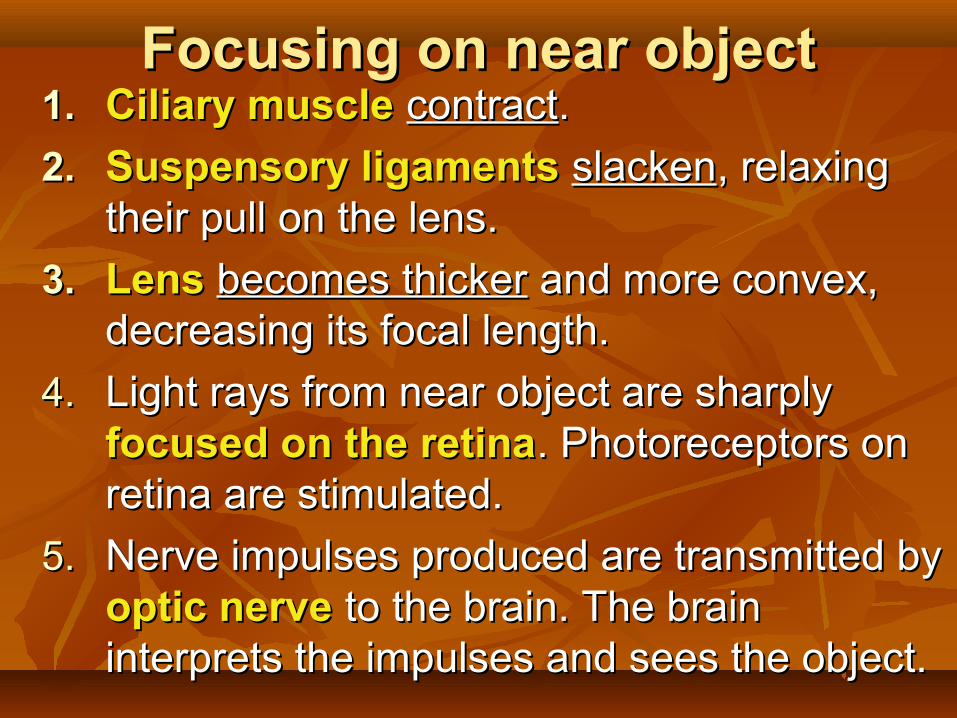
Focusing on near objectFocusing on near object1.1. Ciliary muscleCiliary muscle contractcontract..
2.2. Suspensory ligamentsSuspensory ligaments slackenslacken, relaxing , relaxing their pull on the lens.their pull on the lens.
3.3. Lens Lens becomes thickerbecomes thicker and more convex, and more convex, decreasing its focal length.decreasing its focal length.
4.4. Light rays from near object are sharply Light rays from near object are sharply focused on the retinafocused on the retina. Photoreceptors on . Photoreceptors on retina are stimulated.retina are stimulated.
5.5. Nerve impulses produced are transmitted by Nerve impulses produced are transmitted by optic nerveoptic nerve to the brain. The brain to the brain. The brain interprets the impulses and sees the object.interprets the impulses and sees the object.

Bob
What kind of help does Bob need?
I am seated right at the back of the class and I
can’t see well.
Mr Ng always scold me for not copying down
notes.
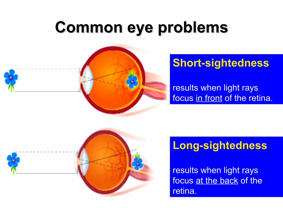
Common eye problemsCommon eye problems
Short-sightedness
results when light rays focus in front of the retina.
Long-sightedness
results when light rays focus at the back of the retina.

Short-sightedness
Correction using lens

Implanted tooth helps blind woman recovers sight
http://sg.news.yahoo.com/afp/20090917/tts-health-research-us-eye-972e412.html


The size of the pupil is controlled by two sets of The size of the pupil is controlled by two sets of muscles in the iris.muscles in the iris.
1.1. Circular Circular musclesmuscles
2.2. Radial Radial musclesmuscles
II. Controlling amount of lightII. Controlling amount of light

• Pupil changes size as a result of changes in light intensity.
• Pupil becomes larger when the surrounding light intensity is low, and smaller when the light intensity is high.
Stimulus (change in light intensity)
Receptor (retina)Sensory neurone in
optic nerve
BrainMotor neuroneEffector (iris)
The Reflex Arc involved in the pupil reflex: The Reflex Arc involved in the pupil reflex:
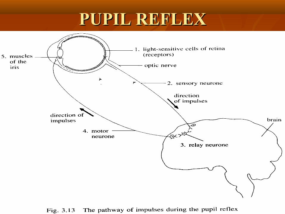
PUPIL REFLEXPUPIL REFLEX
radial muscles
circular muscles
pupil
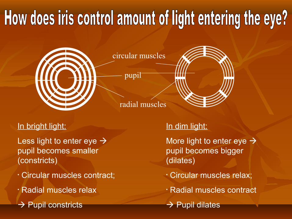
In bright light:
Less light to enter eye pupil becomes smaller (constricts)
• Circular muscles contract;
• Radial muscles relax
Pupil constricts
In dim light:
More light to enter eye pupil becomes bigger (dilates)
• Circular muscles relax;
• Radial muscles contract
Pupil dilates
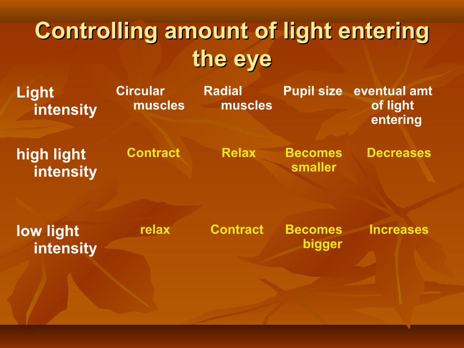
Controlling amount of light entering Controlling amount of light entering the eyethe eye
Light intensity
Circular muscles
Radial muscles
Pupil size eventual amt of light entering
high light intensity
Contract Relax Becomessmaller
Decreases
low light intensity
relax Contract Becomes bigger
Increases


What happen to the pupil when you leave the dark halls of the cinema?
The circular muscles will contract while the radial muscles of iris will relax.
Hence, it reduces the size of the pupil, thus decreasing the amount of light entering the eye in the light.

Why we cannot see colour at night?
•The cones, which allow detection of colour do not function under low light intensities.
•Since only the rods function, no colour is seen.
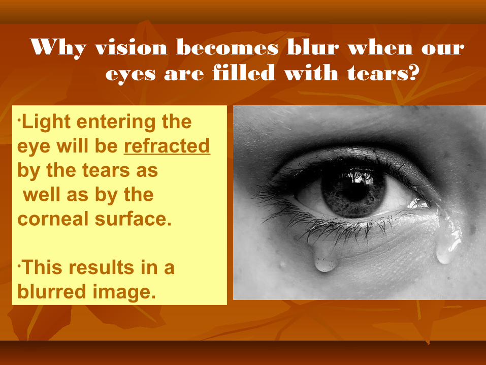
Why vision becomes blur when our eyes are filled with tears?
•Light entering the eye will be refracted by the tears as well as by the corneal surface.
•This results in a blurred image.
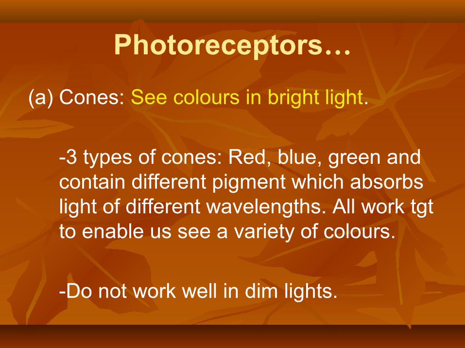
Photoreceptors…
(a) Cones: See colours in bright light.
-3 types of cones: Red, blue, green and contain different pigment which absorbs light of different wavelengths. All work tgt to enable us see a variety of colours.
-Do not work well in dim lights.

(b) Rod : More sensitive to light than cones.
Enable us to see things in black and white in dim light because they are sensitive to low intensity light. (Reason: Contain Visual purple pigment)
Nerve impulses cannot be send to brain from rods hence person cannot see anything.
Visual purple must be formed again before the person can see in the dark.

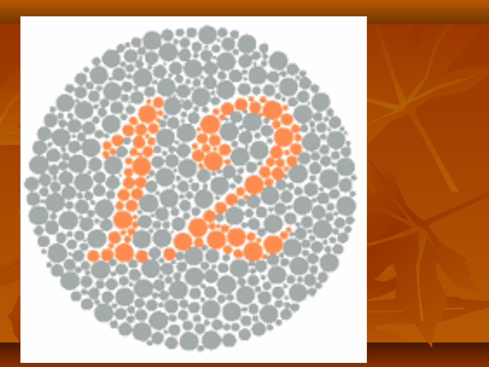
Colour blindness
People that are effected by colour blindness, have less numbers of particular cones than normal, so they get colours confused.
They may be able to see a bright green coloured object out side, but when viewed in artificial light, the same object may appear brown in colour.
Or if the object was a dull green, it could even appear red.

People with normal colour vision should see the number 8. Those with red-green colour vision deficiencies should see the number 3.
Total colour blindness should not be able to read any numeral.
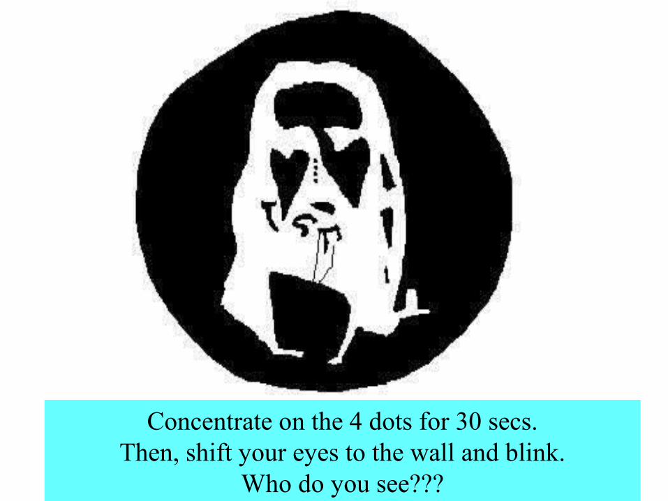
Concentrate on the 4 dots for 30 secs.Then, shift your eyes to the wall and blink.
Who do you see???
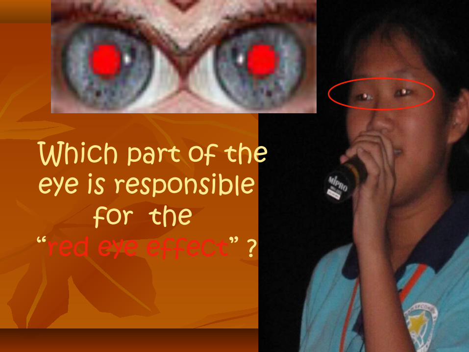
Which part of the eye is responsible
for the “red eye effect” ?

How cataracts affect vision?
http://www.allaboutvision.com/conditions/cataracts.htm

Online Online ResourcesResources
http://www.1800contacts.com/vision101/frames.htmlhttp://www.1800contacts.com/vision101/frames.html
http://www.horton.ednet.ns.ca/staff/selig/Activities/nervous/eye1.htmhttp://www.horton.ednet.ns.ca/staff/selig/Activities/nervous/eye1.htm
http://www.med.uwo.ca/physpharm/courses/sensesweb/L1Eye/l1eye.swfhttp://www.med.uwo.ca/physpharm/courses/sensesweb/L1Eye/l1eye.swf
http://www.richmondeye.com/simulation.htmhttp://www.richmondeye.com/simulation.htm