Embed Size (px)
Citation preview
ISSN 2320-7078
Volume 1 Issue 3
Online Available at www.entomoljournal.com
Journal of Entomology and Zoology Studies
Vol. 1 No. 3 2013 www.entomoljournal.com Page | 22
Histophysiology and Histochemical Analysis of the Saccus Vasculosus of Butter Catfish Ompok bimacualtus
(Bloch, 1794) Saroj Kumar Ghosh 1, Padmanabha Chakrabarti 1*
1. Fisheries Laboratory, Department of Zoology, The University of Burdwan, Burdwan 713 104, West Bengal, India. [E-mail: [email protected]; Telephone: +91-342-2634798, Fax No.: +91-342-2657938]
Cellular details including histochemical nature and functional aspects of saccus vasculosus in Ompok bimaculatus (Bloch, 1794) were studied using histological and histochemical techniques. The oval protruded saccus vasculosus situated on the ventral side of the diencephalon behind the pituitary. This circumventricular organ contained a typical large central lumen lined by single stratified epithelium with characteristic coronet cells and supporting cells. Silver stain was adopted for the occurrence and distribution of neurons if any in the epithelium and various regions of the saccus vasculosus. Intensity of glycogen was found to be associated with the coronet cells and blood vessels. Histochemical localization of basic protein and bound lipid were recorded in the cells of saccus epithelial lining as well as in the blood vessels. Coronet cells perhaps involved in secretory as well as sensory function of the fish concerned. Keyword: Histoarchitecture, Histochemistry, Function, Saccus vasculosus, Ompok bimaculatus 1. Introduction The saccus vasculosus is highly vascular and sac like swelling, occupies a centrocaudal portion of the diencephalic infundibulum just behind the hypophysial complex. The wall of this circumventricular organ may be single or multiple layers thick and composed of chiefly coronet cells and supporting cells with interspersed liquor contacting neurons. This specialized ependymal organ is mainly concerned with a secretory function [1] and works for an umbilical conjoin between cerebrospinal fluid and the blood vascular system [2]. The cellular organization of the saccus vasculosus in teleosts have been investigated by many researchers using light and electron microscope [3, 4, 5, 6, 7, 8]. However, the histochemical approaches of the saccus vasculosus in fishes have been examined
by few workers [9, 10, 11, 12, 13] to evaluate the chemical content. Still lacunae exist in detailed cellular architecture and chemical constitution of various cells to correlate with the physiology of this ependymo-vascular organ. The aim of the present work is to perform a histological study and histochemical detection of neuron, glycogen, protein and lipid in the epithelium of the saccus vasculosus of freshwater teleost Ompok bimaculatus (Siluriformes: Siluridae). 2. Materials and Methods Live mature fishes of O. bimaculatus (16-18 cm in length) were procured from the river Ganga near Samudragar, Burdwan, West Bengal, India. Fishes were anaesthetized under benzocaine (4 mg/L) and sacrificed following the guidelines

Journal of Entomology and Zoology Studies
Vol. 1 No. 3 2013 www.entomoljournal.com Page | 23
given by the Institutional Ethical Committee. The brain together with the saccus vasculosus was disclosed from the ventral side of the head and initially kept in 10% neutral formalin. After few minute the saccus vasculosus beside with the rest of the brain was carefully eliminated from the cranium and instantly processed for histological and histochemical studies.
Fig. 1: Oval shaped saccus vasculosus (SV) surrounded by connective tissue (solid arrow) situated beyond the pituitary (PT) posteriorly. Note the presence of wide lumen (LU) and
vascular channels (arrow heads). Broken arrow denotes third ventricle of the brain (B) (MT) 50 X.
Fig 2: Showing conspicuous nucleus (N) of coronet cells (CC) scattered with supporting cells (SC) (arrow heads).
Note the presence of nerve fibres (broken arrows) and blood vessels (BV) below the epithelium. Solid arrows
mark luminar protrusions of CC (HE) 400 X. 2.1 Histological study The saccus vasculosus was fixed in aqueous Bouin’s fluid for 16-18 hour and subsequently dehydrated properly through an ascending series
of ethyl alcohol. The tissues were cleared with xylene, embedded in paraffin wax (56-58 ˚C) and cut serially at 4 µm thickness. The dewaxed sections were stained with Delafield’s Haematoxylin-Eosin (HE) and Mallory’s triple (MT) stain.
Fig 3: Higher magnification of saccus epithelium shows tall columnar CC with prominent N and few SC (arrow
heads). Note the nerve fibre (broken arrow) from CC and BV below the epithelium. Solid arrows indicate globular
protrusion of CC towards the LU (HE) 1000 X.
2.2 Histochemical study Intact tissues were immersed in 10% neutral formalin for a period of 18 h for proper fixation. Subsequent to dehydration the tissues were embedded in paraffin wax (52-54˚C) in vacuum embedding bath. Sagittal sections were cut at 8 m thickness and subjected to the following histochemical test to evaluate the chemical nature of cells lining the saccus vasculosus: Silver Impregnation Method (SIM) for detection of neurons [14], Best's Carmine (BC) method for detection of glycogen[15], Mercury-Bromphenol Blue (MBPB) method for detection of basic protein [16] and Sudan Black B (SB) method for detection of bound lipid [17]. 3. Results 3.1 Histology The saccus vasculosus of O. bimaculatus is well developed and oval sac like texture, located on the ventral side of the diencephalon near the third ventricle of the brain (Fig 1). It is situated postero-dorsal to the pituitary and enclosed by loose connective tissue. The saccus is
Journal of Entomology and Zoology Studies
Vol. 1 No. 3 2013 www.entomoljournal.com Page | 24
encapsulated vascular organ and is precisely separated from the pituitary. The blood vessels are existed interior to the capsule of saccus vasculosus in the form of vascular channels, exhibit intense red colour (Fig 1).
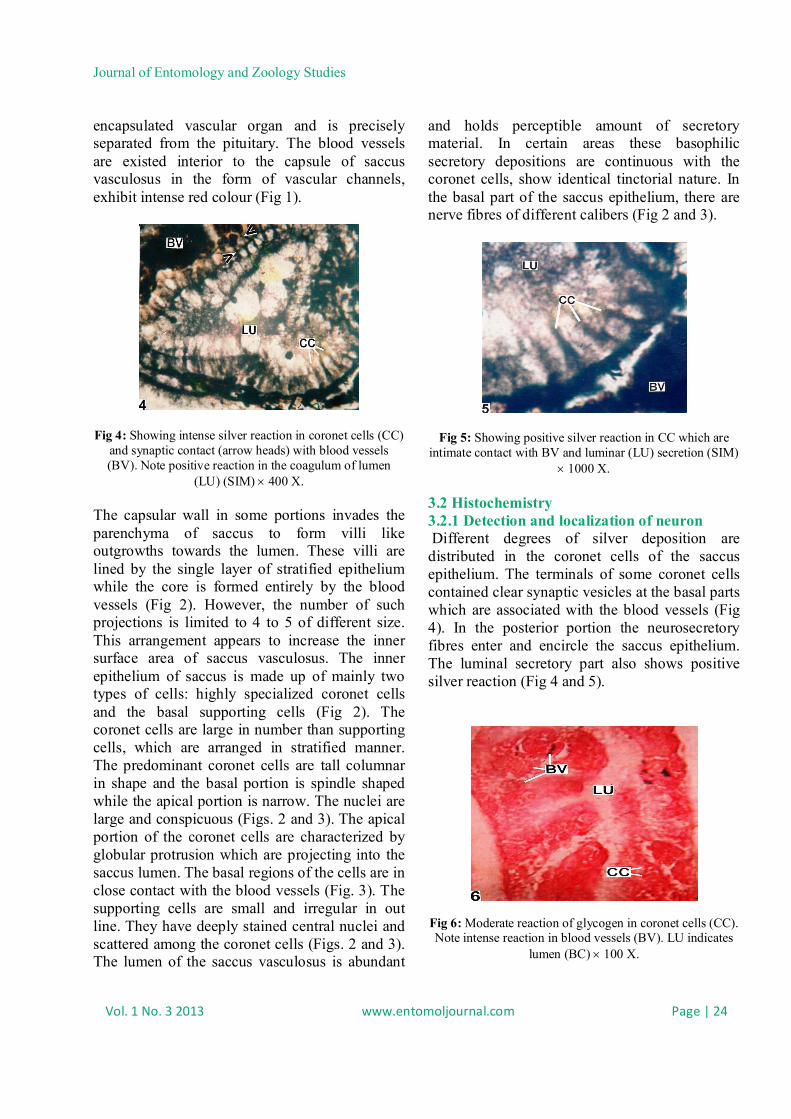
Fig 4: Showing intense silver reaction in coronet cells (CC) and synaptic contact (arrow heads) with blood vessels (BV). Note positive reaction in the coagulum of lumen
(LU) (SIM) 400 X. The capsular wall in some portions invades the parenchyma of saccus to form villi like outgrowths towards the lumen. These villi are lined by the single layer of stratified epithelium while the core is formed entirely by the blood vessels (Fig 2). However, the number of such projections is limited to 4 to 5 of different size. This arrangement appears to increase the inner surface area of saccus vasculosus. The inner epithelium of saccus is made up of mainly two types of cells: highly specialized coronet cells and the basal supporting cells (Fig 2). The coronet cells are large in number than supporting cells, which are arranged in stratified manner. The predominant coronet cells are tall columnar in shape and the basal portion is spindle shaped while the apical portion is narrow. The nuclei are large and conspicuous (Figs. 2 and 3). The apical portion of the coronet cells are characterized by globular protrusion which are projecting into the saccus lumen. The basal regions of the cells are in close contact with the blood vessels (Fig. 3). The supporting cells are small and irregular in out line. They have deeply stained central nuclei and scattered among the coronet cells (Figs. 2 and 3). The lumen of the saccus vasculosus is abundant
and holds perceptible amount of secretory material. In certain areas these basophilic secretory depositions are continuous with the coronet cells, show identical tinctorial nature. In the basal part of the saccus epithelium, there are nerve fibres of different calibers (Fig 2 and 3).
Fig 5: Showing positive silver reaction in CC which are intimate contact with BV and luminar (LU) secretion (SIM)
1000 X.
3.2 Histochemistry 3.2.1 Detection and localization of neuron Different degrees of silver deposition are distributed in the coronet cells of the saccus epithelium. The terminals of some coronet cells contained clear synaptic vesicles at the basal parts which are associated with the blood vessels (Fig 4). In the posterior portion the neurosecretory fibres enter and encircle the saccus epithelium. The luminal secretory part also shows positive silver reaction (Fig 4 and 5).
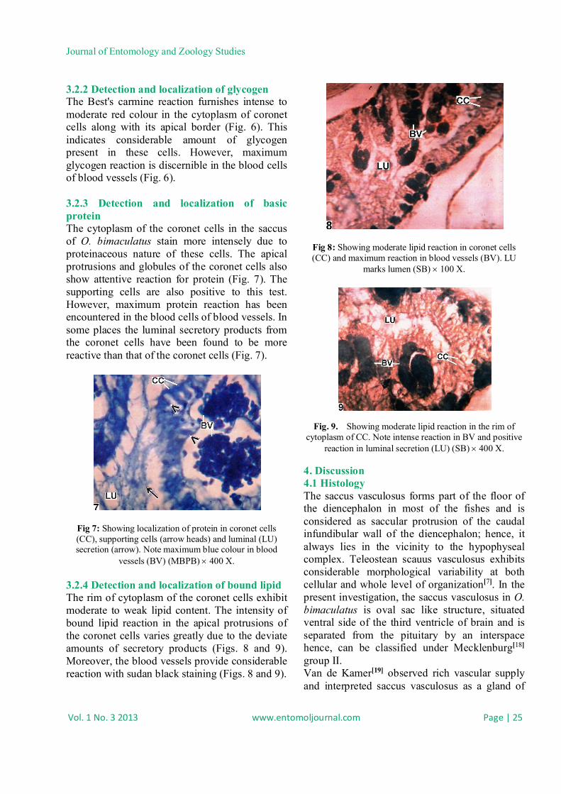
Fig 6: Moderate reaction of glycogen in coronet cells (CC). Note intense reaction in blood vessels (BV). LU indicates
lumen (BC) 100 X.
Journal of Entomology and Zoology Studies
Vol. 1 No. 3 2013 www.entomoljournal.com Page | 25
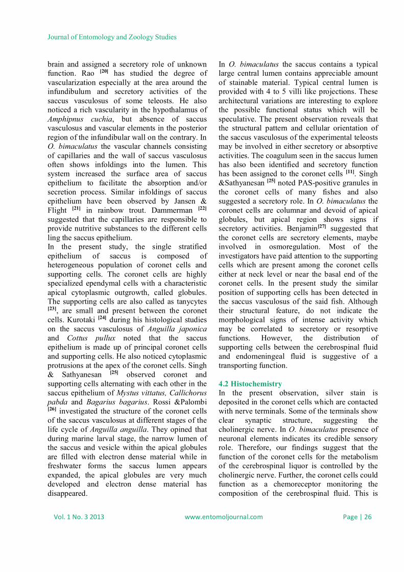
3.2.2 Detection and localization of glycogen The Best's carmine reaction furnishes intense to moderate red colour in the cytoplasm of coronet cells along with its apical border (Fig. 6). This indicates considerable amount of glycogen present in these cells. However, maximum glycogen reaction is discernible in the blood cells of blood vessels (Fig. 6). 3.2.3 Detection and localization of basic protein The cytoplasm of the coronet cells in the saccus of O. bimaculatus stain more intensely due to proteinaceous nature of these cells. The apical protrusions and globules of the coronet cells also show attentive reaction for protein (Fig. 7). The supporting cells are also positive to this test. However, maximum protein reaction has been encountered in the blood cells of blood vessels. In some places the luminal secretory products from the coronet cells have been found to be more reactive than that of the coronet cells (Fig. 7).
Fig 7: Showing localization of protein in coronet cells (CC), supporting cells (arrow heads) and luminal (LU) secretion (arrow). Note maximum blue colour in blood
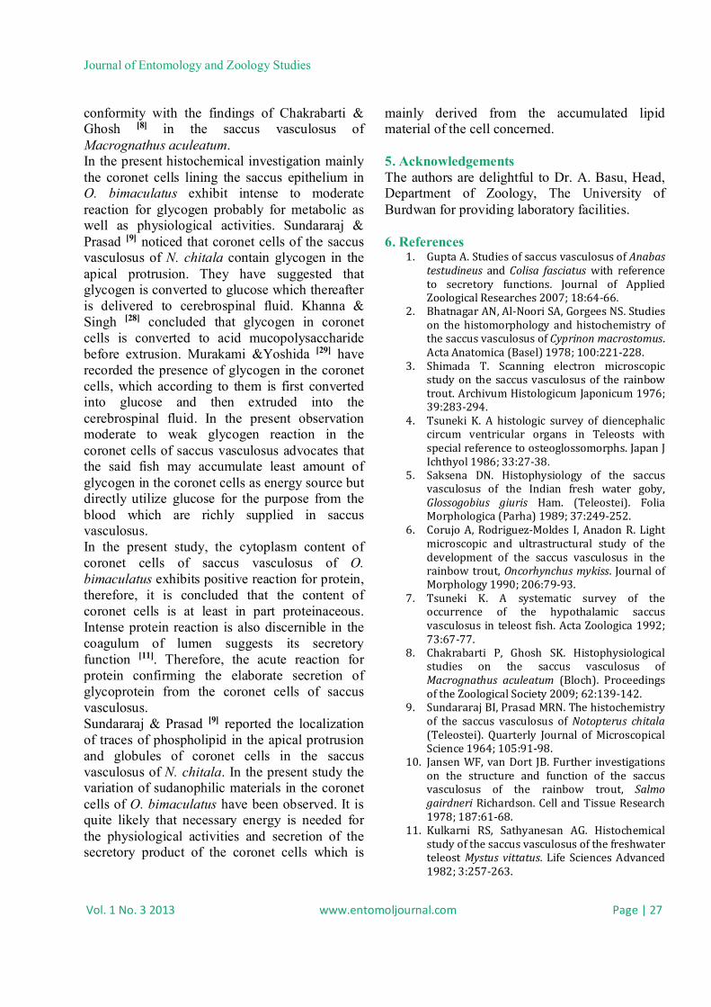
vessels (BV) (MBPB) 400 X. 3.2.4 Detection and localization of bound lipid The rim of cytoplasm of the coronet cells exhibit moderate to weak lipid content. The intensity of bound lipid reaction in the apical protrusions of the coronet cells varies greatly due to the deviate amounts of secretory products (Figs. 8 and 9). Moreover, the blood vessels provide considerable reaction with sudan black staining (Figs. 8 and 9).
Fig 8: Showing moderate lipid reaction in coronet cells (CC) and maximum reaction in blood vessels (BV). LU
marks lumen (SB) 100 X.
Fig. 9. Showing moderate lipid reaction in the rim of cytoplasm of CC. Note intense reaction in BV and positive
reaction in luminal secretion (LU) (SB) 400 X.
4. Discussion 4.1 Histology The saccus vasculosus forms part of the floor of the diencephalon in most of the fishes and is considered as saccular protrusion of the caudal infundibular wall of the diencephalon; hence, it always lies in the vicinity to the hypophyseal complex. Teleostean scauus vasculosus exhibits considerable morphological variability at both cellular and whole level of organization[7]. In the present investigation, the saccus vasculosus in O. bimaculatus is oval sac like structure, situated ventral side of the third ventricle of brain and is separated from the pituitary by an interspace hence, can be classified under Mecklenburg[18] group II. Van de Kamer[19] observed rich vascular supply and interpreted saccus vasculosus as a gland of
Journal of Entomology and Zoology Studies
Vol. 1 No. 3 2013 www.entomoljournal.com Page | 26
brain and assigned a secretory role of unknown function. Rao [20] has studied the degree of vascularization especially at the area around the infundibulum and secretory activities of the saccus vasculosus of some teleosts. He also noticed a rich vascularity in the hypothalamus of Amphipnus cuchia, but absence of saccus vasculosus and vascular elements in the posterior region of the infundibular wall on the contrary. In O. bimaculatus the vascular channels consisting of capillaries and the wall of saccus vasculosus often shows infoldings into the lumen. This system increased the surface area of saccus epithelium to facilitate the absorption and/or secretion process. Similar infoldings of saccus epithelium have been observed by Jansen & Flight [21] in rainbow trout. Dammerman [22] suggested that the capillaries are responsible to provide nutritive substances to the different cells ling the saccus epithelium. In the present study, the single stratified epithelium of saccus is composed of heterogeneous population of coronet cells and supporting cells. The coronet cells are highly specialized ependymal cells with a characteristic apical cytoplasmic outgrowth, called globules. The supporting cells are also called as tanycytes [23], are small and present between the coronet cells. Kurotaki [24] during his histological studies on the saccus vasculosus of Anguilla japonica and Cottus pullux noted that the saccus epithelium is made up of principal coronet cells and supporting cells. He also noticed cytoplasmic protrusions at the apex of the coronet cells. Singh & Sathyanesan [25] observed coronet and supporting cells alternating with each other in the saccus epithelium of Mystus vittatus, Callichorus pabda and Bagarius bagarius. Rossi &Palombi [26] investigated the structure of the coronet cells of the saccus vasculosus at different stages of the life cycle of Anguilla anguilla. They opined that during marine larval stage, the narrow lumen of the saccus and vesicle within the apical globules are filled with electron dense material while in freshwater forms the saccus lumen appears expanded, the apical globules are very much developed and electron dense material has disappeared.
In O. bimaculatus the saccus contains a typical large central lumen contains appreciable amount of stainable material. Typical central lumen is provided with 4 to 5 villi like projections. These architectural variations are interesting to explore the possible functional status which will be speculative. The present observation reveals that the structural pattern and cellular orientation of the saccus vasculosus of the experimental teleosts may be involved in either secretory or absorptive activities. The coagulum seen in the saccus lumen has also been identified and secretory function has been assigned to the coronet cells [11]. Singh &Sathyanesan [25] noted PAS-positive granules in the coronet cells of many fishes and also suggested a secretory role. In O. bimaculatus the coronet cells are columnar and devoid of apical globules, but apical region shows signs if secretory activities. Benjamin[27] suggested that the coronet cells are secretory elements, maybe involved in osmoregulation. Most of the investigators have paid attention to the supporting cells which are present among the coronet cells either at neck level or near the basal end of the coronet cells. In the present study the similar position of supporting cells has been detected in the saccus vasculosus of the said fish. Although their structural feature, do not indicate the morphological signs of intense activity which may be correlated to secretory or resorptive functions. However, the distribution of supporting cells between the cerebrospinal fluid and endomeningeal fluid is suggestive of a transporting function. 4.2 Histochemistry In the present observation, silver stain is deposited in the coronet cells which are contacted with nerve terminals. Some of the terminals show clear synaptic structure, suggesting the cholinergic nerve. In O. bimaculatus presence of neuronal elements indicates its credible sensory role. Therefore, our findings suggest that the function of the coronet cells for the metabolism of the cerebrospinal liquor is controlled by the cholinergic nerve. Further, the coronet cells could function as a chemoreceptor monitoring the composition of the cerebrospinal fluid. This is
Journal of Entomology and Zoology Studies
Vol. 1 No. 3 2013 www.entomoljournal.com Page | 27
conformity with the findings of Chakrabarti & Ghosh [8] in the saccus vasculosus of Macrognathus aculeatum. In the present histochemical investigation mainly the coronet cells lining the saccus epithelium in O. bimaculatus exhibit intense to moderate reaction for glycogen probably for metabolic as well as physiological activities. Sundararaj & Prasad [9] noticed that coronet cells of the saccus vasculosus of N. chitala contain glycogen in the apical protrusion. They have suggested that glycogen is converted to glucose which thereafter is delivered to cerebrospinal fluid. Khanna & Singh [28] concluded that glycogen in coronet cells is converted to acid mucopolysaccharide before extrusion. Murakami &Yoshida [29] have recorded the presence of glycogen in the coronet cells, which according to them is first converted into glucose and then extruded into the cerebrospinal fluid. In the present observation moderate to weak glycogen reaction in the coronet cells of saccus vasculosus advocates that the said fish may accumulate least amount of glycogen in the coronet cells as energy source but directly utilize glucose for the purpose from the blood which are richly supplied in saccus vasculosus. In the present study, the cytoplasm content of coronet cells of saccus vasculosus of O. bimaculatus exhibits positive reaction for protein, therefore, it is concluded that the content of coronet cells is at least in part proteinaceous. Intense protein reaction is also discernible in the coagulum of lumen suggests its secretory function [11]. Therefore, the acute reaction for protein confirming the elaborate secretion of glycoprotein from the coronet cells of saccus vasculosus. Sundararaj & Prasad [9] reported the localization of traces of phospholipid in the apical protrusion and globules of coronet cells in the saccus vasculosus of N. chitala. In the present study the variation of sudanophilic materials in the coronet cells of O. bimaculatus have been observed. It is quite likely that necessary energy is needed for the physiological activities and secretion of the secretory product of the coronet cells which is
mainly derived from the accumulated lipid material of the cell concerned. 5. Acknowledgements The authors are delightful to Dr. A. Basu, Head, Department of Zoology, The University of Burdwan for providing laboratory facilities. 6. References
1. Gupta A. Studies of saccus vasculosus of Anabas testudineus and Colisa fasciatus with reference to secretory functions. Journal of Applied Zoological Researches 2007; 18:64-66.
2. Bhatnagar AN, Al-Noori SA, Gorgees NS. Studies on the histomorphology and histochemistry of the saccus vasculosus of Cyprinon macrostomus. Acta Anatomica (Basel) 1978; 100:221-228.
3. Shimada T. Scanning electron microscopic study on the saccus vasculosus of the rainbow trout. Archivum Histologicum Japonicum 1976; 39:283-294.
4. Tsuneki K. A histologic survey of diencephalic circum ventricular organs in Teleosts with special reference to osteoglossomorphs. Japan J Ichthyol 1986; 33:27-38.
5. Saksena DN. Histophysiology of the saccus vasculosus of the Indian fresh water goby, Glossogobius giuris Ham. (Teleostei). Folia Morphologica (Parha) 1989; 37:249-252.
6. Corujo A, Rodriguez-Moldes I, Anadon R. Light microscopic and ultrastructural study of the development of the saccus vasculosus in the rainbow trout, Oncorhynchus mykiss. Journal of Morphology 1990; 206:79-93.
7. Tsuneki K. A systematic survey of the occurrence of the hypothalamic saccus vasculosus in teleost fish. Acta Zoologica 1992; 73:67-77.
8. Chakrabarti P, Ghosh SK. Histophysiological studies on the saccus vasculosus of Macrognathus aculeatum (Bloch). Proceedings of the Zoological Society 2009; 62:139-142.
9. Sundararaj BI, Prasad MRN. The histochemistry of the saccus vasculosus of Notopterus chitala (Teleostei). Quarterly Journal of Microscopical Science 1964; 105:91-98.
10. Jansen WF, van Dort JB. Further investigations on the structure and function of the saccus vasculosus of the rainbow trout, Salmo gairdneri Richardson. Cell and Tissue Research 1978; 187:61-68.
11. Kulkarni RS, Sathyanesan AG. Histochemical study of the saccus vasculosus of the freshwater teleost Mystus vittatus. Life Sciences Advanced 1982; 3:257-263.
Journal of Entomology and Zoology Studies
Vol. 1 No. 3 2013 www.entomoljournal.com Page | 28
12. Delvin AJ, Danks JA, Faulkner MK, Power DM, Canario AVM, Martin TJ et al. Immunochemical detection of Parathyroid hormone-related protein in the saccus vasculosus of teleost fish. General and Comparative Endocrinology 1996; 101 83-90.
13. Sueiro C, Carrera I, Ferreiro S, Molist P, Adrio F, Anadon R, Rodriguez-Moldes I. New insights on saccus vasculosus evolution: a developmental and immunohistochemical study in elasmobranchs. Brain Behavior and Evolution 2007; 70:187-204.
14. Marsland TA, Glees P, Erikson LB. Modification of the glees silver impregnation for paraffin sections. Journal of Neuropathology and Experimental Neurology 1954; 13:587.
15. Best F. Uber carmine far bug des glycogens and derkerne. Z Wiss Mikr 1906; 3:319-322.
16. Bonhag PF. Journal of Morphology 1958; 96: 381 (vide, Pearse, AGE, Vol-1, 1975).
17. Bernenbaum MC. Quarterly Journal of Microscopical Science, 1958; 99:231 (vide, Pearse, AGE, Vol-1, 1975).
18. Mecklenburg CV. The development of saccus vasculosus in two teleosts. Acta Zoologica 1974; 55:137-148.
19. Kamer JC Van de. The Saccus vasculosus in fish. In: Leopoldina Symposium “Circumventriculare Organe”. Nova Acta Leopold Suppl. 1977; 9:75-87.
20. Rao PD. On the saccus vasculosus in some Indian fresh water teleosts. Naturwissenschaften 1966; 53:233.
21. Jansen WF, Flight WFG. Light and electronmicroscopical observations on the saccus vasculosus of the rainbow Trout. Z Zellforsch 1969; 100:439-465.
22. Dammerman KW. Der saccus vasculosus der fische ein tiefeorgan. Z Wiss Zool 1910; 96:654-726.
23. Jansen WF, Flight WFG, Zandbergen MA. Fine structural localization of adenosine triphosphate activities in the saccus vasculosus of the rainbow trout, Salmo gairdneri Richardson. Cell and Tissue Research 1981; 219:267-279.
24. Kurotaki M. The submicroscopic structure of the epithelium of saccus vasculosus in two teleosts. Acta Anatomica Nipponica 1961; 36: 277-288.
25. Singh TP, Sathyanesan AG. Studies on the structure of the saccus vasculosus in some freshwater fishes. Proceedings of the Zoological Society, 1964; 17:169-175.
26. Rossi A, Palombi F. The saccus vasculosus of Anguilla Anguilla (L) from larva to adult. Cell and Tissue Research 1976; 167:11-21.
27. Benjamin M. Ultrastructure studies on the coronet cells of the saccus vasculosus of the Fresh water stickleback, Gasterosteus aculeatus from leiurus. Z. Zellforsch., 1974; 147: 551-565.
28. Khanna SS, Singh HR. Histology and histochemistry of the saccus vasculosus in some teleosts (pisces). Acta Anatomica, 1967; 67: 304-311.
29. Murakami M, and Yoshida T. Elektronenmikroskopische beobachtungen am saccus vasculosus des kugel fishches Spheriodes niphobles. Archivum Histologicum Japonicum, 1967; 28: 265-284.