Embed Size (px)
Citation preview
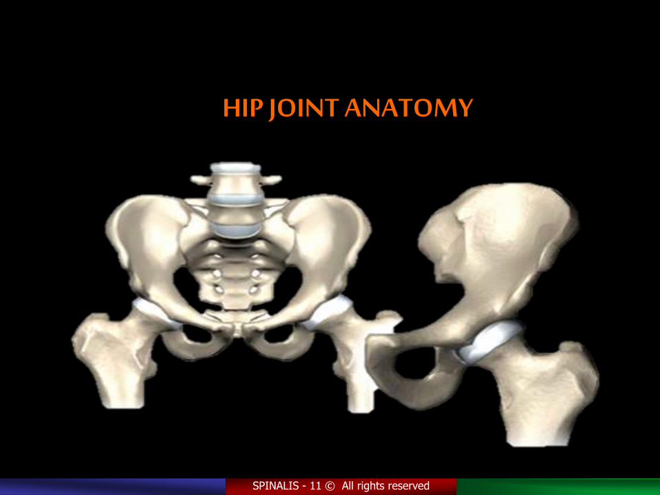
HIP JOINT ANATOMY
SPINALIS - 11 © All rights reserved
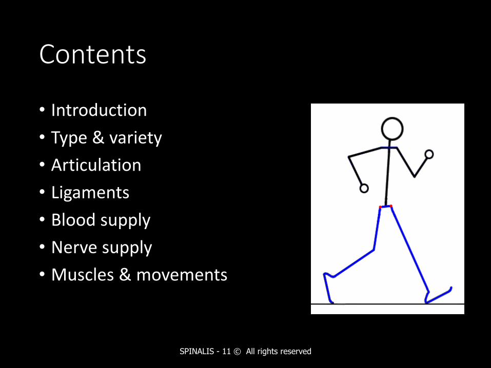
Contents
• Introduction
• Type & variety
• Articulation
• Ligaments
• Blood supply
• Nerve supply
• Muscles & movements
SPINALIS - 11 © All rights reserved
Introduction
• The hip joint allows the same movement as the mobile shoulder joint, but the range of movement is restricted
SPINALIS - 11 © All rights reserved
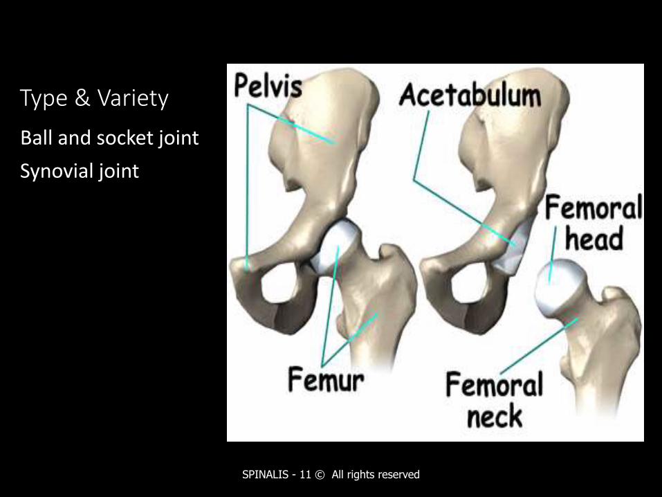
Type & Variety
Ball and socket joint
Synovial joint
SPINALIS - 11 © All rights reserved
SPINALIS - 11 © All rights reserved
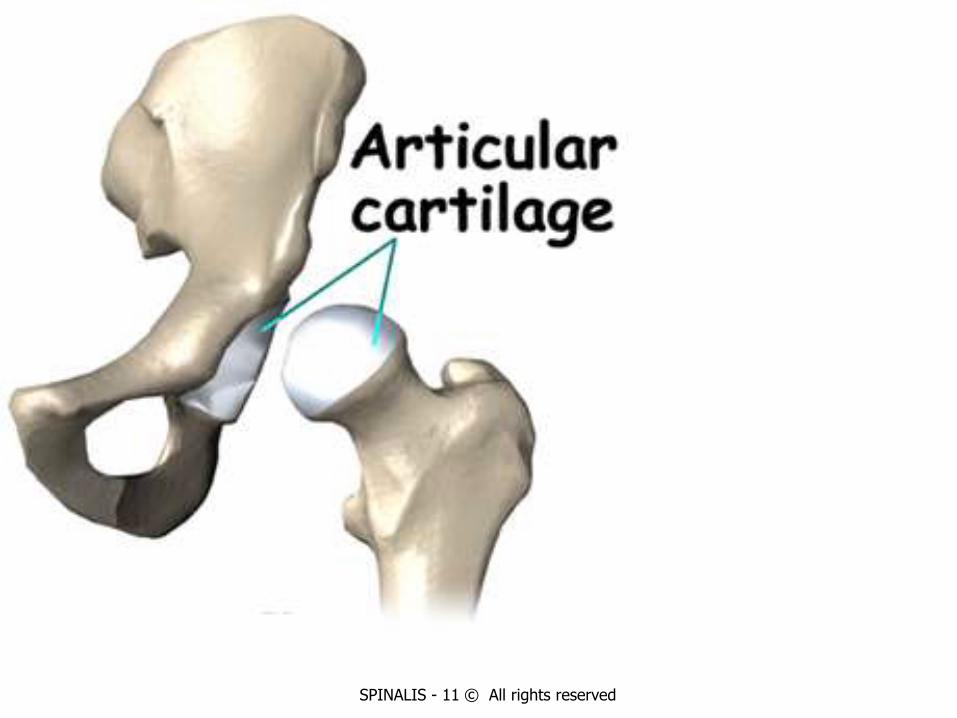
Articulation
• The head of the femur articulate with the acetabulum of the hip bone to form the hip joint
• The head of the femur forms more than half a sphere, and is covered with hyaline cartilage except at the fovea capitis
• The acetabulum presence a horseshoe shaped, lunate articular surface, an acetabular notch and an acetabular fossa
SPINALIS - 11 © All rights reserved
SPINALIS - 11 © All rights reserved
Stability
• The hip joint is unique in having a high degree of stability as well as mobility
• The stability or strength depends upon:• Depth of the acetabulum and the narrowing of its
mouth by the acetabular labrum
• Tension and strength of ligaments
• Strength of the surrounding muscles
• Length and obliquity of the neck of the femur
• Atmosphere pressure
SPINALIS - 11 © All rights reserved
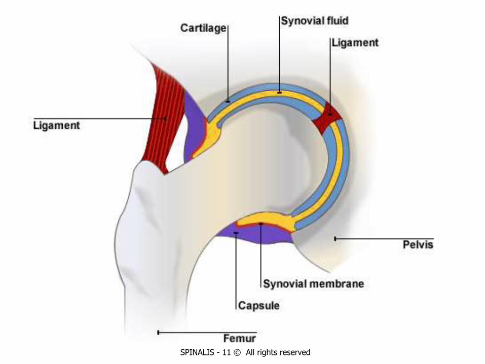
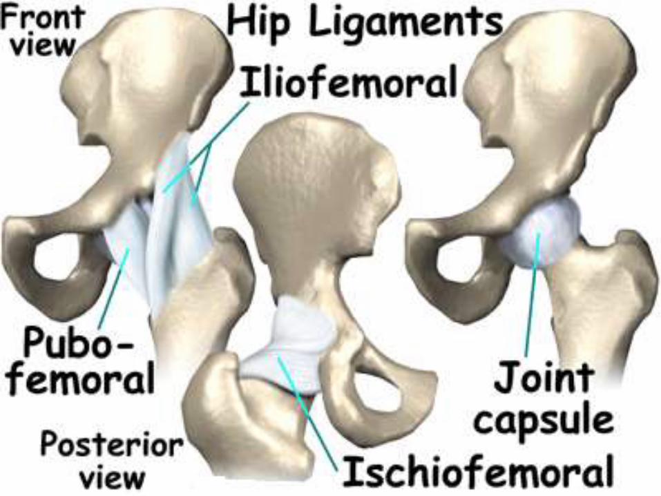
Ligaments
• Fibrous capsule
• Iliofemoral ligament
• Pubofemoral ligament
• Ischiofemoral ligament
• Ligament of the head of the femur
• Acetabular labrum
• Transverse acetabular ligament
SPINALIS - 11 © All rights reserved
Fibrous capsule
• It is attached on the hip bone to the acetabular labrum including the transverse acetabular ligament, and to bone above and behind the acetabulum; and an the femur to the intertrochanderic line in front, and 1cm medial to the intertrochanderic crest behind
• Anterosuperiorly, the capsule is thick and firmly attached
• This part is subjected to maximum tension in standing posture
SPINALIS - 11 © All rights reserved
• Posteroinferiorly, the capsule is thin and loosely attached to bone
• The capsule is made up of two types of fibres :• Outer fibres – longitudinal
• Inner fibres – zona orbicularis
• The joint cavity communicates with a bursae lying deep to the tendon of psoas major
SPINALIS - 11 © All rights reserved
Iliofemoral ligament
• It is inverted y shaped ligament of bigelow , lies anteriorly
• It is one of the strongest ligament of the body
• It prevents the trunk from falling backwards in the standing posture
• It is triangular in shape
SPINALIS - 11 © All rights reserved
Pubofemoral ligament
• It is support the joint inferomedially
• It is also triangular in shape
• Superiorly, it is attached to the iliopubic eminence, the obturator crest and the obturator membrane
• inferiorly, it merges with the anteroinferior part of the capsule and with the band of the Iliofemoral ligament
SPINALIS - 11 © All rights reserved
Ischiofemoral ligament
• It covers the joint posteriorly
• It is fibres or twisted and extend from the ischium to the acetabulum
• The fibres of the ligament form the zona orbicularis
• some of them are attached to the greater trochander
SPINALIS - 11 © All rights reserved
SPINALIS - 11 © All rights reserved
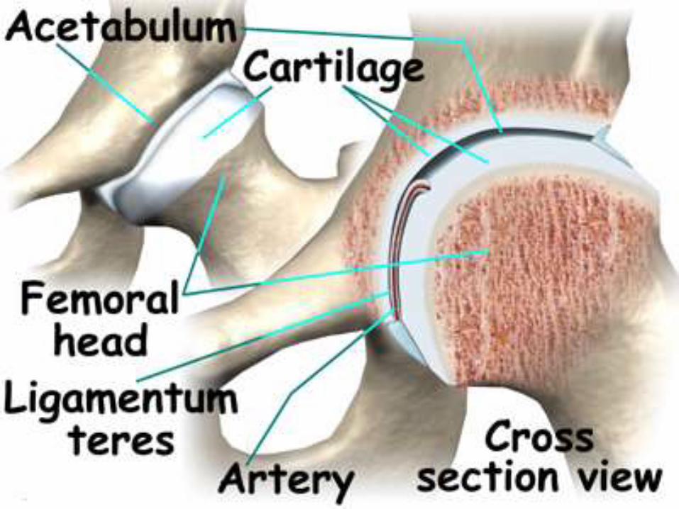
Ligament of the head of the femur• It is round ligament or ligamentum teres is a flat
and triangular ligament
• The apex is attached to the fovea capitis, and the base to the transversr ligament and the margins of the acetabular notch
SPINALIS - 11 © All rights reserved
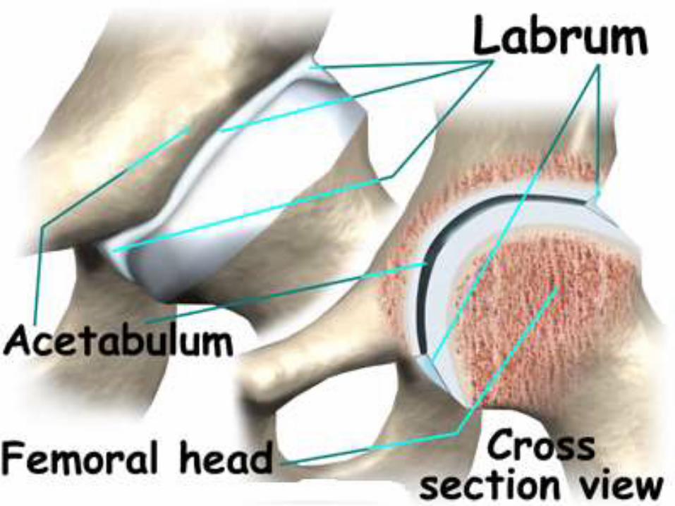
Acetabular labrum
• It is a fibro cartilaginous rim attached to the margins of the acetabulum
• It narrows the mouth of the acetabulum
• This helps in holding the head of the femur in position
SPINALIS - 11 © All rights reserved
SPINALIS - 11 © All rights reserved
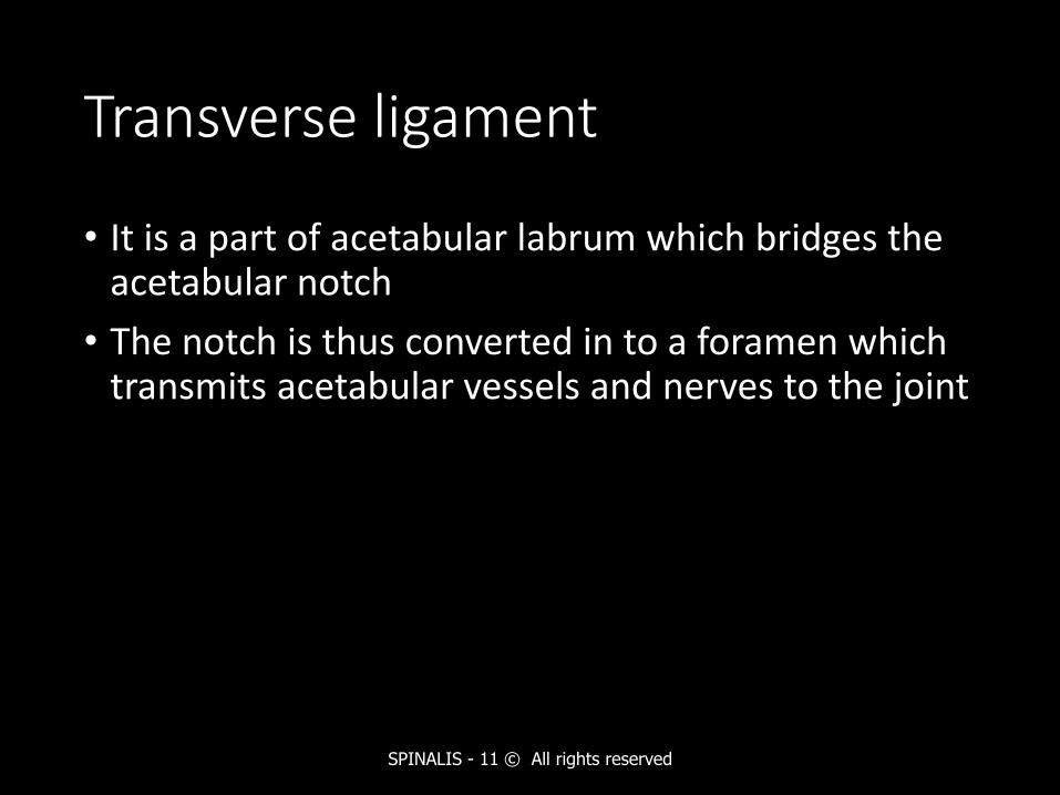
Transverse ligament
• It is a part of acetabular labrum which bridges the acetabular notch
• The notch is thus converted in to a foramen which transmits acetabular vessels and nerves to the joint
SPINALIS - 11 © All rights reserved
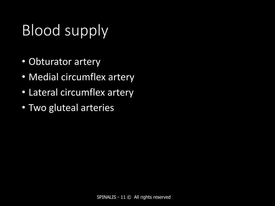
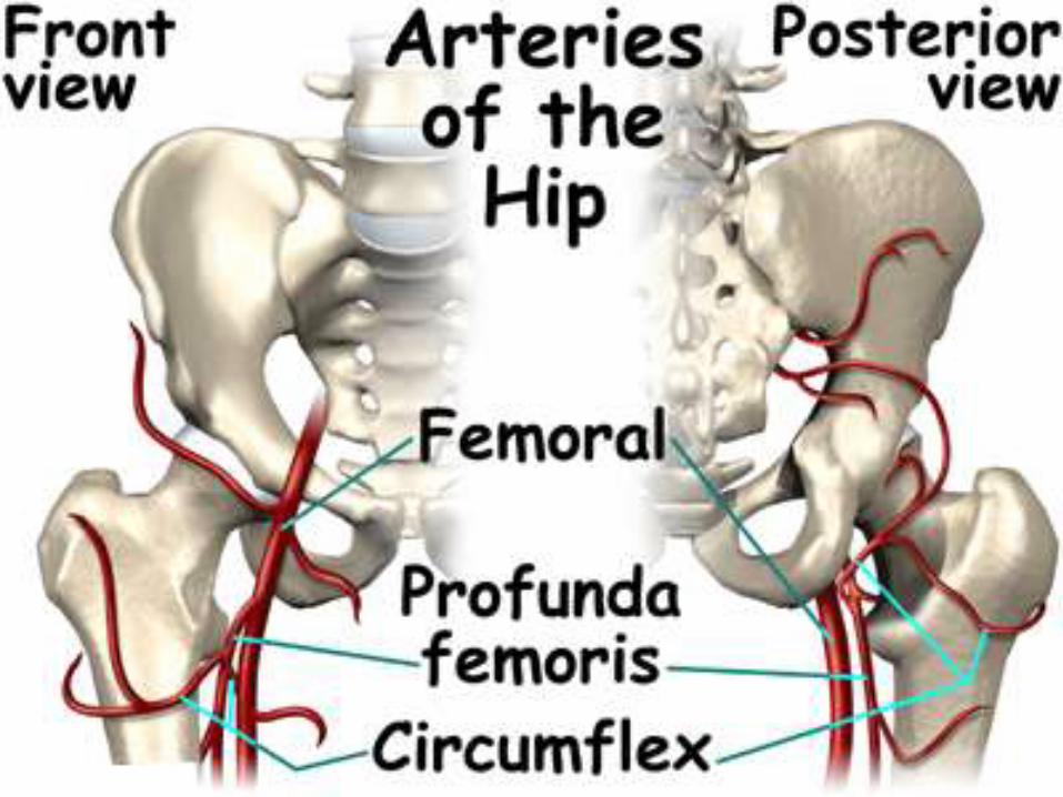
Blood supply
• Obturator artery
• Medial circumflex artery
• Lateral circumflex artery
• Two gluteal arteries
SPINALIS - 11 © All rights reserved
SPINALIS - 11 © All rights reserved
SPINALIS - 11 © All rights reserved
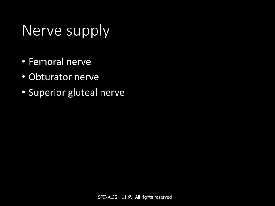
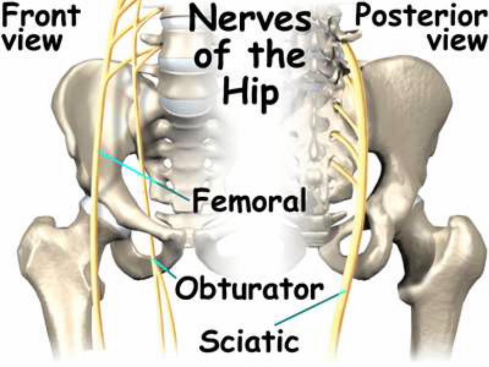
Nerve supply
• Femoral nerve
• Obturator nerve
• Superior gluteal nerve
SPINALIS - 11 © All rights reserved
SPINALIS - 11 © All rights reserved
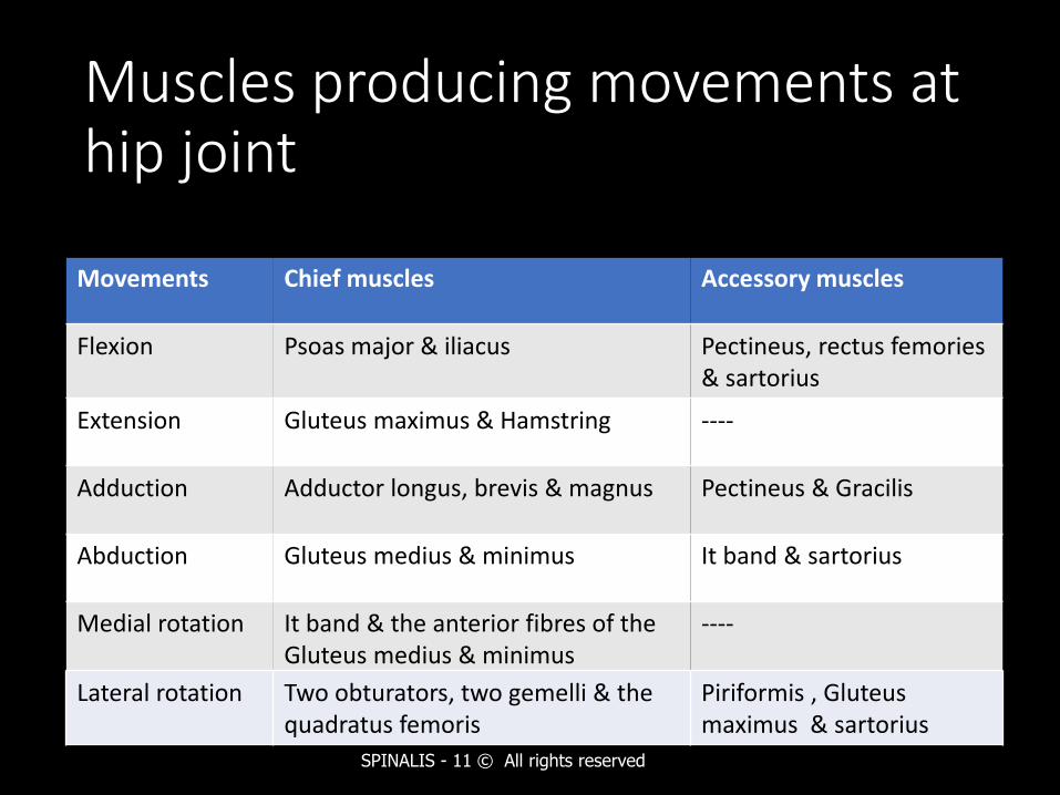
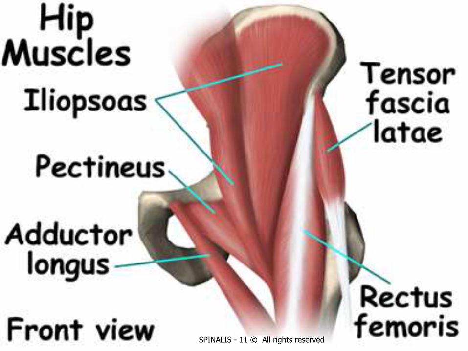
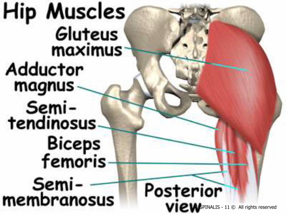
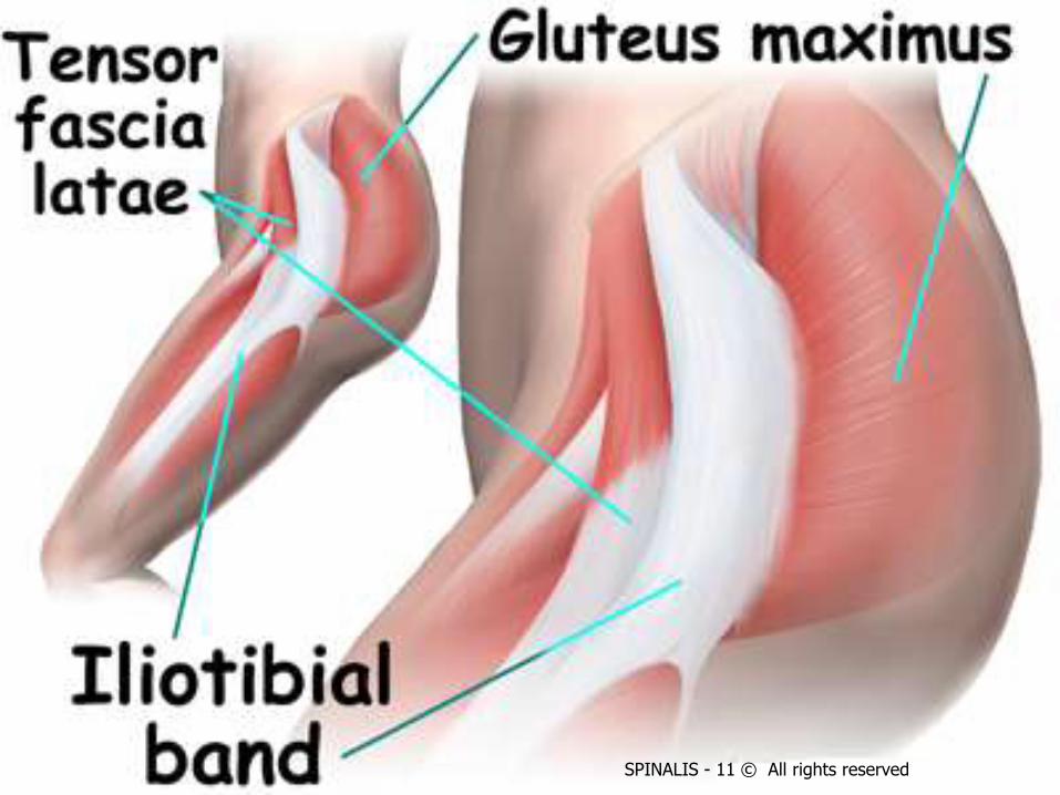
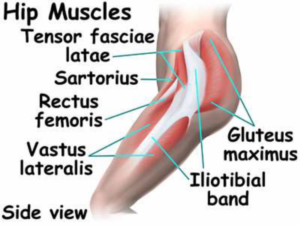
Muscles producing movements at hip joint
Movements Chief muscles Accessory muscles
Flexion Psoas major & iliacus Pectineus, rectus femories& sartorius
Extension Gluteus maximus & Hamstring ----
Adduction Adductor longus, brevis & magnus Pectineus & Gracilis
Abduction Gluteus medius & minimus It band & sartorius
Medial rotation It band & the anterior fibres of the Gluteus medius & minimus
----
SPINALIS - 11 © All rights reserved
Lateral rotation Two obturators, two gemelli & the quadratus femoris
Piriformis , Gluteus maximus & sartorius
SPINALIS - 11 © All rights reserved
SPINALIS - 11 © All rights reserved
SPINALIS - 11 © All rights reserved
SPINALIS - 11 © All rights reserved
SPINALIS - 11 © All rights reservedJoin www.fb.com/physicaltherapyorg