Embed Size (px)
Citation preview
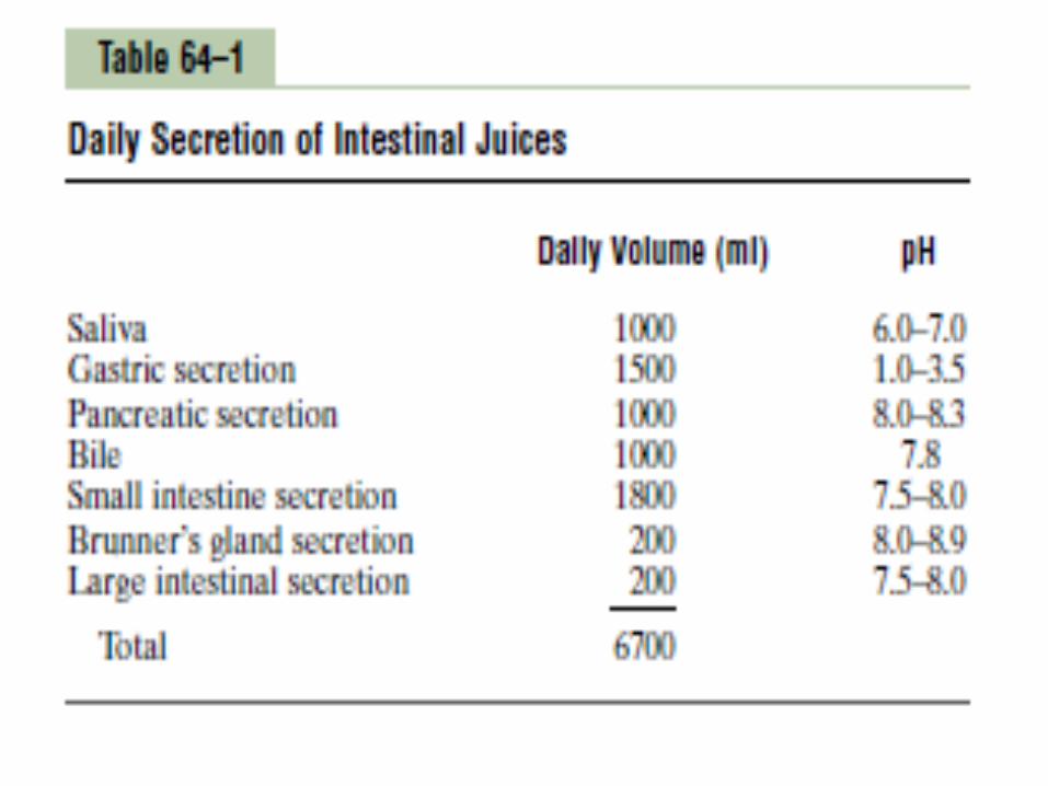
Gastrointestinal secretions
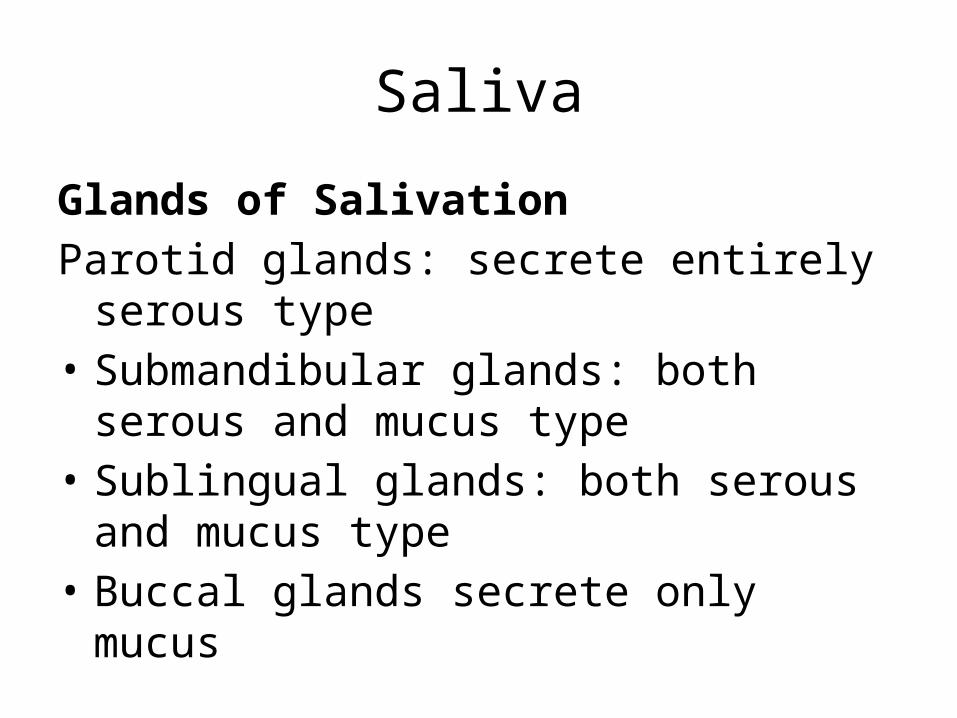
Saliva
Glands of SalivationParotid glands: secrete entirely serous type• Submandibular glands: both serous and
mucus type• Sublingual glands: both serous and mucus
type• Buccal glands secrete only mucus

Types of secretion• Serous type of secretions contains ptyalin, an
enzyme for the digesting starches• Mucus secretions contain mucin for
lubrication and for surface protective purposes

Composition and function of saliva• Sodium ions are actively reabsorbed from all the salivary ducts
and potassium ions are actively secreted in exchange for sodium.• Bicarbonate ions are secreted by ductal epithelium into lumen of
the duct• Ptyalin (α amylase) is responsible for the digestion of
carbohydrates• Mucus, glycoproteins• Thiocyanate ions• Lysozymes• Immunoglobulins (IgA)

Functions of Saliva• Preparation of food for swallowing• Appreciation of taste• Digestive function• Cleansing and protective functions• Role in speech• Excretory function• Regulation of body temperature• Regulation of water balance

Regulation of salivation• Salivation is mainly controlled by autonomic
nervous system. Both branches of autonomic nervous system stimulate salivation, but parasympathetic nervous system stimulate much more strongly than the sympathetic nervous system

Reflex regulation of salivary secretion• Unconditioned reflex• Conditioned reflex

Nervous Regulation of Salivary Secretion• Salivary glands are controlled mainly by parasympathetic
nervous signals all the way from the superior and inferior salivatory nuclei in the brain stem.
• The salivatory nuclei are located approximately at the juncture of the medulla and pons and are excited by both taste and tactile stimuli from the tongue and other areas of the mouth and pharynx. Many taste stimuli, especially the sour taste (caused by acids), elicit copious secretion of saliva—often 8 to 20 times the basal rate of secretion.
• Salivation also occurs in response to reflexes originating in the stomach and upper small intestines, particularly when irritating foods are swallowed or when a person is nauseated because of some gastrointestinal abnormality.
• Esophageal Secretion• The esophageal secretions are entirely mucous
in character and principally provide lubrication for swallowing. The main body of the esophagus is lined with many simple mucous glands. At the gastric end and to a lesser extent in the initial portion of the esophagus, there are also many compound mucous glands.
• The mucus secreted by the compound glands in the upper esophagus prevents mucosal excoriation by newly entering food, whereas the compound glands located near the esophagogastric junction protect the esophageal wall from digestion by acidic gastric juices that often reflux from the stomach back into the lower esophagus.

Gastric Secretion
• Oxyntic glands: these secrete hydrochloric acid, pepsinogen, intrinsic factor and mucus. These are located on the inside surfaces of body and fundus of the stomach
• Pyloric glands: mainly secrete mucus and hormone gastrin. These are located in the antral portion of the stomach
Secretion from oxyntic glandsA typical oxyntic gland is composed of three types
of cells• Mucus cells which secrete mainly mucus but also
some pepsinogen• Peptic or chief cells which secrete mainly large
quantities of pepsinogen• Parietal or oxyntic cells which secrete
hydrochloric acid and intrinsic factor
• Mucous Cells• The thick mucous present in the gastric juice is
responsible for the protection of the gastric wall. The mucous protects the stomach wall from irritation or mechanical injury. It prevents the back diffusion of hydrogen ions into the gastric mucosa. Beneath the mucous layer, a layer rich in bicarbonate ion neutralizes hydrogen ions.
• Chief Cells• Protein digestion begins in the stomach
because of the activity of chief cells. These cells secrete the inactive precursor protein, pepsinogen, which is activated to the proteolytic enzyme, pepsin, in the presence of acid and small amounts of active pepsin. Pepsin functions optimally at a pH of approximately 2.

Parietal CellsParietal cells• Secrete hydrogen ions• Activate protein digestive enzymes such as pepsinogen.• Create a harsh environment for bacterial growth.• Secrete intrinsic factor which binds vitamin B12 in
protein rich foods, such as meat, to prevent its degradation in small intestine and to allow absorption in the terminal ileum


Basic mechanism of hydrochloric acid secretion• Parietal cells secrete HCl into the lumen of the stomach
and, concurrently, absorb bicarbonate ion into the blood stream as follows
• In the parietal cells, CO2 and water are converted to hydrogen ions and bicarbonate ions, catalyzed by carbonic anhydrase
• Hydrogen ion is secreted into the lumen by H+-K+ pump (H+-K+ ATPase). Chloride is secreted along with the hydrogen ions, thus the secretion product of parietal cells is HCl

• The bicarbonate ions produced in the cells is absorbed into the bloodstream in exchange for chloride ions (Cl- - HCO3
- exchange). As bicarbonate ion is added to the venous blood, the pH of the blood increases (“alkanline tide”). (Eventually this bicarbonate ion will be secreted in pancreatic secretions to neutralize hydrogen ions in the small intestine.)


Pyloric Glands• They are responsible for the secretion of large
amount of thin mucous that helps to lubricate food movement as well as to protect the stomach wall from digestion by gastric enzymes. They also secrete the hormone gastrin which plays a key role in controlling gastric secretion.

Stimulation of gastric hydrogen ion secretion• Vagal stimulation– Increases hydrogen ion secretion by a direct
pathway and an indirect pathway.– In the direct path, the vagus nerve innervates G
cells and stimulates gastrin secretion directly. The neurotransmitter at these synapses is Acetylcholine.

• In the indirect path, the vagus nerve innervates G cells and stimulates gastrin secretion, which then stimulates hydrogen ion secretion by an endocrine action. The neurotransmitter at these synapses is Gastrin releasing peptide.

Gastrin– Is released in response to eating a meal (small
peptides, distention of the stomach, vagal stimulation).
– Stimulates hydrogen ion secretion by interacting with cholecystokininB (CCKB) receptor on the parietal cells.
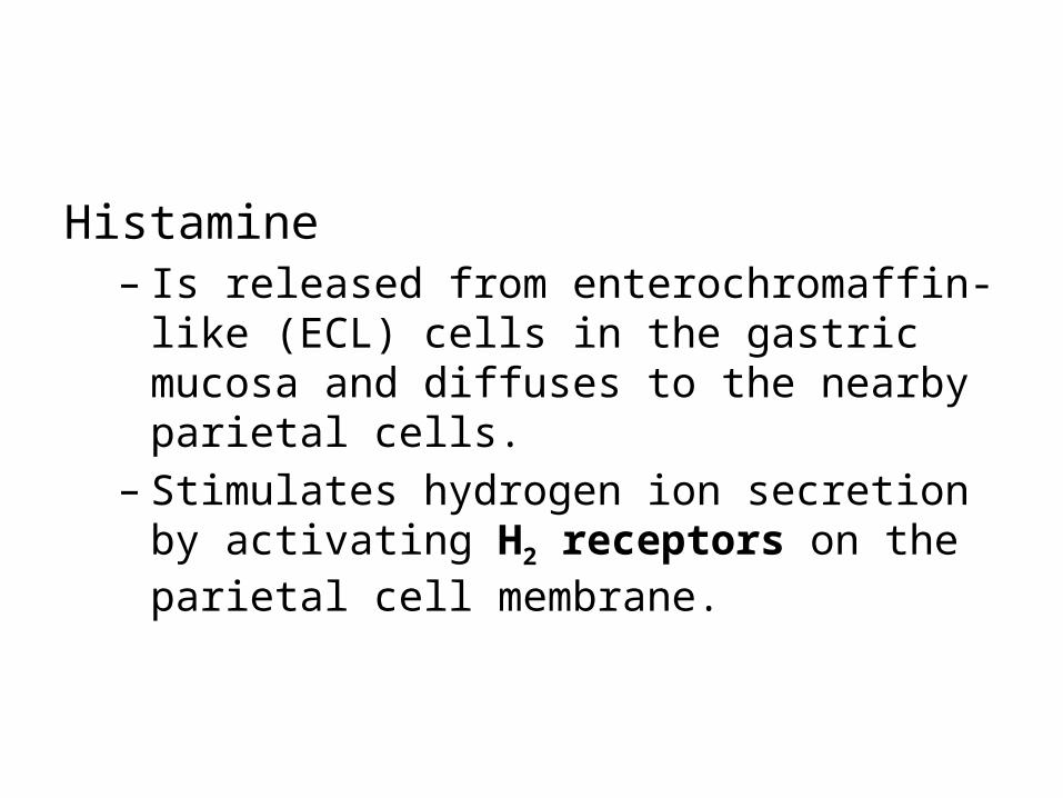
Histamine– Is released from enterochromaffin-like (ECL) cells
in the gastric mucosa and diffuses to the nearby parietal cells.
– Stimulates hydrogen ion secretion by activating H2 receptors on the parietal cell membrane.

Inhibition of gastric hydrogen ion secretion• Negative feedback mechanisms inhibit the secretion of
hydrogen ions by parietal cells.• The presence of food in the small intestine initiates a
reverse enterogastric reflex, transmitted through the myenteric nervous system as well as through extrinsic sympathetic and vagus nerves, that inhibits stomach secretion. This reflex can be initiated by distending the small bowel, by the presence of acid in the upper intestine, by the presence of protein breakdown products, or by irritation of the mucosa.

• Low pH (<3.0) in the stomach– Inhibits gastrin secretion and thereby inhibits
hydrogen ion secretion. After a meal in ingested, hydrogen ion secretion is stimulated by certain mechanisms. After the meal is digested and the stomach emptied, further hydrogen ion secretion decreases the pH of the stomach contents. When the pH of the stomach contents is <3.0, gastrin secretion is inhibited and , by negative feedback, inhibits further hydrogen ion secretion.

Somatostatin– Inhibits gastric hydrogen ion secretion by a direct
pathway and an indirect pathway.– In the direct pathway, somatostatin antagonizes
the stimulatory action of histamine on hydrogen ion secretion.
– In the indirect pathway, somatostatin inhibits release of histamine and gastrin, thus decreasing hydrogen ion secretion indirectly.
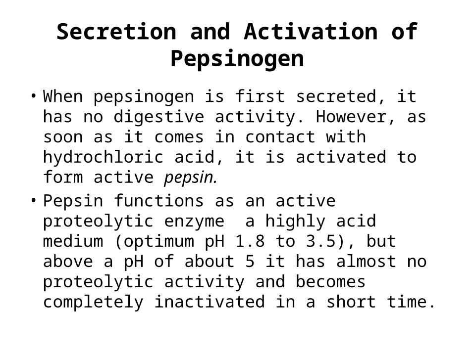
Secretion and Activation of Pepsinogen
• When pepsinogen is first secreted, it has no digestive activity. However, as soon as it comes in contact with hydrochloric acid, it is activated to form active pepsin.
• Pepsin functions as an active proteolytic enzyme a highly acid medium (optimum pH 1.8 to 3.5), but above a pH of about 5 it has almost no proteolytic activity and becomes completely inactivated in a short time.

• Secretion of Intrinsic Factor. The substance intrinsic factor, essential for absorption of vitamin B12 in the ileum, is secreted by the parietal cells along with the secretion of hydrochloric acid.
• When the acid-producing parietal cells of the stomach are destroyed the person develops not only achlorhydria (lack of stomach acid secretion) but often also pernicious anemia because of failure of maturation of the red blood cells in the absence of vitamin B12 stimulation of the bone marrow.

Pancreas
• Pancreas is a dual organ having two functions• Endocrine function: involves production of
hormones like insulin• Exocrine function: involves secretion of
digestive juice- pancreatic juice

Anatomy of the exocrine part of the pancreas• It is made up of acinar cells• Acinar cells contain zymogen granules, which possess
digestive juices• The ducts arising from acini join together to form
intralobular duct• Intralobular ducts unite to form main duct of pancreas
called Wirsung’s duct• Wirsung’s duct joins common bile duct to form ampulla
of vater which opens into the duodenum

Pancreatic secretion• Contains high concentration of HCO3 whose
purpose is to neutralize the digestive enzymes reaching the duodenum
• Contains enzymes for digesting all 3 major types of food: proteins, carbohydrates and fats

Enzymatic components• The more important of enzymes are• Trypsin which is activated from trypsinogen in
the presence of enzyme called enterokinase. Enterokinase is secreted by intestinal mucosa when chyme comes on contact with mucosa
• Chymotrypsinogen is activated by trypsin to form chymotrypsin
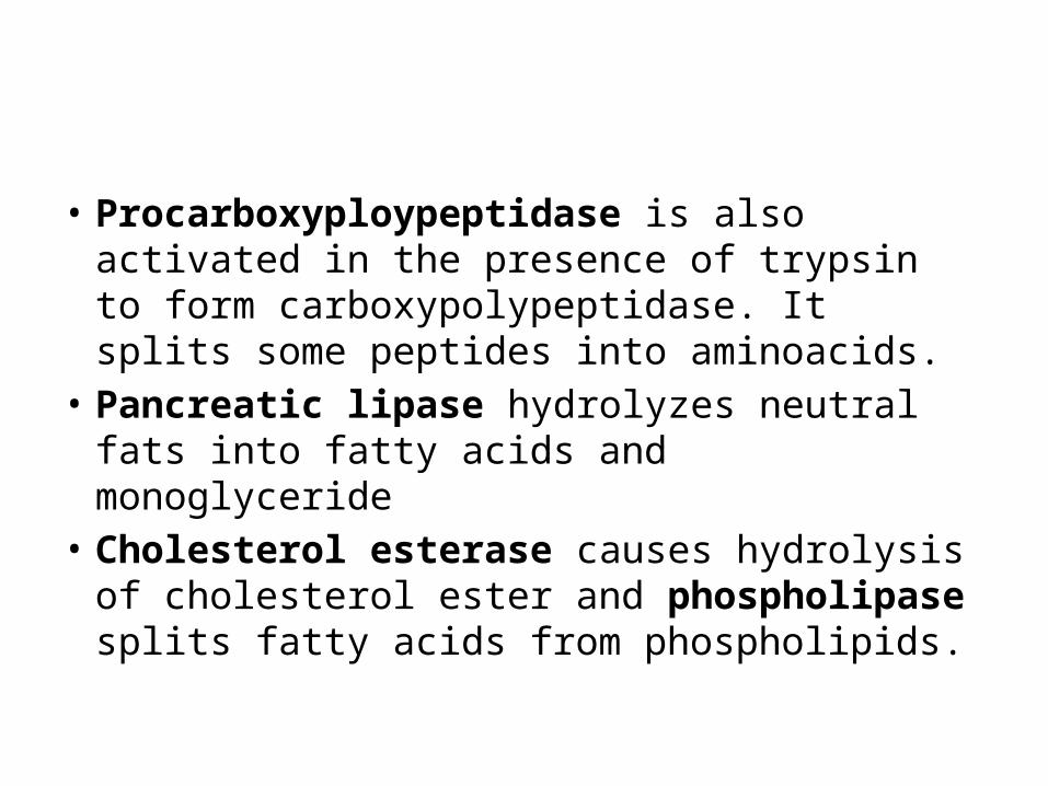
• Procarboxyploypeptidase is also activated in the presence of trypsin to form carboxypolypeptidase. It splits some peptides into aminoacids.
• Pancreatic lipase hydrolyzes neutral fats into fatty acids and monoglyceride
• Cholesterol esterase causes hydrolysis of cholesterol ester and phospholipase splits fatty acids from phospholipids.

• Pancreatic amylase hydrolyzes starches, glycogen and most other carbohydrates except cellulose to form disaccharides and a few trisaccharides.
• Trypsin inhibitor is secreted into the acini of pancreas and it prevents the activation of trypsin both inside the secretory cells and in the acini and ducts of the pancreas

Secretion of Bicarbonate ions• Carbon dioxide diffuses to the interior of the
cell from the blood and combines with the water in the presence of carbonic anhydrase to form carbonic acid. This carbonic acid in turn dissociates into bicarbonate ions and hydrogen ions. The bicarbonate ions are actively transported in exchange for the chloride ions and enters into the lumen of the duct


Regulation of pancreatic secretion• Stimuli of pancreatic secretion are• Acetylcholine• Choleccystokinin secreted when food enters small
intestine• Secretin is secreted in response to acidic food• Acetylcholine and cholecystokinin cause production of
large quantities of digestive enzymes whereas secretin stimulates secretion of large quantities of water and bicarbonate

Phases of pancreatic secretion• Cephalic phase• Gastric phase• Intestinal phase

Secretions of bile by liver
• Normally 600-1200 ml /day bile is secreted by the liver. Bile contains bile salts, phospholipids, cholesterol, and bile pigments.
• Secretion of bile Bile is secreted in two stages• Initial portion is secreted by liver hepatocytes.
This secretion contains large amounts of bile acids, cholesterol and other organic constituents. It is secreted into minute bile canaliculi.

• The bile flows toward the interlobular septa, where canaliculi empty into terminal bile ducts and then progressively into larger ducts, finally reaching the hepatic duct and common bile duct, from which the bile either empties directly into the duodenum or is diverted through the cystic duct into the gall bladder


Storage of bile• Most of the bile from liver enters the gallbladder
where it is stored. It is released from gallbladder into the duodenum whenever required. The maximum volume of the gallbladder is 30 -60 ml. A large amount of water and electrolytes (except calcium and potassium) are absorbed resulting in high concentration of bile salts, bile pigments, cholesterol, fatty acids and lecithin

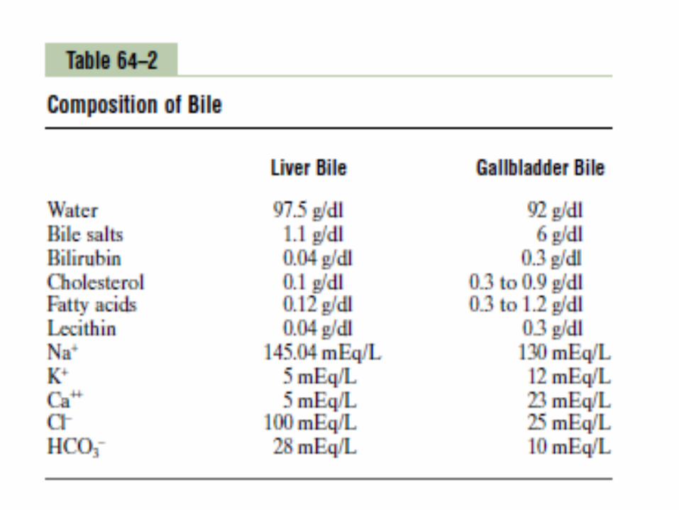
Composition and function of bile• The most abundant substances secreted in the
bile are bile salts, which account for about one half of the total solutes also in the bile. Also secreted or excreted in large concentrations are bilirubin, cholesterol, lecithin, and the usual electrolytes of plasma.

• Bile salts • The liver cells synthesize about 6 grams of bile salts
daily. The precursor of the bile salts is cholesterol, which is either present in the diet or synthesized in the liver cells during the course of fat metabolism.
• Bile salts are amphipathic molecules and are emulsifier. They are potassium or sodium salts of bile acids, which are conjugated with glycine and to lesser amount with taurine

• The precursor of the bile salts is cholesterol, which is either present in the diet or synthesized in the liver cells during the course of fat metabolism.
• The cholesterol is first converted to cholic acid or chenodeoxycholic acid in about equal quantities.

These acids in turn combine principally with glycine and to a lesser extent with taurine to form glyco- and tauro conjugated bile acids. The salts of these acids, mainly sodium salts, are then secreted in the bile.
Due to bacterial action in the intestine the primary bile acids are converted into secondary bile acids which are transported back to the liver through enterohepatic circulation.

• Role of Secretin in Helping to Control Bile Secretion.
• In addition to the strong stimulating effect of bile acids to cause bile secretion, the hormone secretin that also stimulates pancreatic secretion increases bile secretion, sometimes more than doubling its secretion for several hours after a meal.

• Role of secretin in controlling bile • Bile acids have strong stimulating effect bile
secretion. In addition hormone secretin also increases bile secretion. The hormone secretin increases bile secretion, sometimes more than doubling the secretion rate for several hours. This increase in secretion represents almost entirely secretion of a bicarbonate-rich watery solution by the epithelial cells of bile ductules and ducts and not increased secretion by the liver parenchymal cells themselves.

• Bile pigments• Bile pigments are the excretory products in the bile.
Bilirubin and biliverdin are the two bile pigments and bilirubin is the major bile pigment in human being.
• Bilirubin: A major bile pigment, bilirubin is a lipid soluble metabolite of haemoglobin. Transported to the liver attached to the protein, it is then conjugated and excreted as water soluble glucuronides. These give a golden color to bile.

• Biliverdin: Heme splits into iron and pigment biliverdin which then reduces to bilirubin.
• Stercobilin: It is produced from metabolism of bilirubin by intestinal bacteria. It gives brown color to the stool.

• Phospholipids (mainly lecithin)• It is insoluble in water but are solubilized by
bile salt micelles• Cholesterol• It is present in small amount. It is insoluble in
water and must be solubilized by bile salt micelles before it can be secreted in the bile.
Control of bile secretion and gall bladder contraction
• Secretin causes secretion of bicarbonate ions and fluid into bile canalicular ducts
• Secretion of bile salts by hepatocytes is directly proportional to hepatic portal vein concentration of bile salts
• Choleccystokinin causes gallbladder contraction and sphincter of Oddi relaxation

Functions of bile salts• Emulsification of fats• Absorption of fats• Choleretic action• Cholagogue action• Laxative action• Prevention of gallstone formation
Functions of liverThe Liver Functions as a Blood Reservoir• Liver is an expandable organ so large quantities of blood can be
stored in its blood vessels. Its normal blood volume, including both in hepatic veins and in the hepatic sinuses, is about 450 milliliters, or almost 10 per cent of the body’s total blood volume. When high pressure in the right atrium causes backpressure in the liver, the liver expands, and 0.5 to 1 liter of extra blood is occasionally stored in the hepatic veins and sinuses. This occurs especially in cardiac failure with peripheral congestion. Thus, in effect, the liver is a large, expandable, venous organ capable of acting as a valuable blood reservoir in times of excess blood volume and capable of supplying extra blood in times of diminished blood volume.

• The Liver Has Very High Lymph Flow• Because the pores in the hepatic sinusoids are very
permeable and allow ready passage of both fluid and proteins into the spaces of Disse, the lymph draining from the liver usually has a protein concentration of about 6 g/dl, which is only slightly less than the protein concentration of plasma. Also, the extreme permeability of the liver sinusoid epithelium allows large quantities of lymph to form. Therefore, about half of all the lymph formed in the body under resting conditions arises in the liver.


• Regulation of Liver Mass—Regeneration• The liver possesses a remarkable ability to restore
itself after significant hepatic tissue loss from either partial hepatectomy or acute liver injury, as long as the injury is uncomplicated by viral infection or inflammation. During liver regeneration, hepatocytes are estimated to replicate once or twice, and after the original size and volume of the liver are achieved, the hepatocytes revert to their usual quiet state.

Hepatic Macrophage System Serves a Blood-Cleansing Function• Blood flowing through the intestinal capillaries
picks up many bacteria from the intestines. • The Kupffer cells, the large phagocytic
macrophages that line the hepatic venous sinuses, efficiently cleanse blood as it passes through the sinuses; when a bacterium comes into momentary contact with a Kupffer cell
Carbohydrate Metabolism• In carbohydrate metabolism, the liver performs the
following functions:1. Storage of large amounts of glycogen2. Conversion of galactose and fructose to glucose3. Gluconeogenesis4. Formation of many chemical compounds from
intermediate products of carbohydrate metabolism

• Gluconeogenesis in the liver is also important in maintaining a normal blood glucose concentration, because gluconeogenesis occurs to a significant extent only when the glucose concentration falls below normal. In such a case, large amounts of amino acids and glycerol from triglycerides are converted into glucose, thereby helping to maintain a normal blood glucose concentration.

• The liver is especially important for maintaining a normal blood glucose concentration. Storage of glycogen allows the liver to remove excess glucose from the blood, store it, and then return it to the blood when the blood glucose concentration begins to fall too low. This is called the glucose buffer function of the liver.

Fat Metabolism• Oxidation of fatty acids to supply energy for
other body functions• Synthesis of large quantities of cholesterol,
phospholipids, and most lipoproteins• Synthesis of fat from proteins and
carbohydrates
• To derive energy from neutral fats, the fat is first split into glycerol and fatty acids; then the fatty acids are split by beta-oxidation into two-carbon acetyl radicals that form acetyl coenzyme A (acetyl-CoA). This can enter the citric acid cycle and be oxidized to liberate tremendous amounts of energy. Beta-oxidation can take place in all cells of the body, but it occurs especially rapidly in the hepatic cells.

Protein Metabolism1. Deamination of amino acids2. Formation of urea for removal of ammonia
from the body fluids3. Formation of plasma proteins4. Interconversions of the various amino acids
and synthesis of other compounds from amino acids

• Deamination of amino acids is required before they can be used for energy or converted into carbohydrates or fats
• Formation of urea by the liver removes ammonia from the body fluids. Large amounts of ammonia are formed by the deamination process, and additional amounts are continually formed in the gut by bacteria and then absorbed into the blood. Therefore, if the liver does not form urea, the plasma ammonia concentration rises rapidly and results in hepatic coma and death.

The Liver Stores Iron as Ferritin. • Except for the iron in the hemoglobin of the blood, by
far the greatest proportion of iron in the body is stored in the liver in the form of ferritin. The hepatic cells contain large amounts of a protein called apoferritin, which is capable of combining reversibly with iron. When the iron in the circulating body fluids reaches a low level, the ferritin releases the iron. Thus, the apoferritin- ferritin system of the liver acts as a blood iron buffer, as well as an iron storage medium.
The Liver Forms a Large Proportion of the Blood Substances Used in Coagulation. • Substances formed in the liver that are used in
the coagulation process include fibrinogen, prothrombin, accelerator globulin, Factor VII, and several other important factors. Vitamin K is required by the metabolic processes of the liver for the formation of several of these substances, especially prothrombin and Factors VII, IX, and X.
The Liver Removes or Excretes Drugs, Hormones, and Other Substances.• The active chemical medium of the liver is well known
for its ability to detoxify or excrete into the bile many drugs, including sulfonamides, penicillin, ampicillin, and erythromycin. In a similar manner, several of the hormones secreted by the endocrine glands are either chemically altered or excreted by the liver, including thyroxine and essentially all the steroid hormones, such as estrogen, cortisol, and aldosterone.

Small intestine secretions
Secretion of mucus by Brunner’s glands• A large number of compound mucous glands,
called Brunner’s glands, is located in the wall of the first few centimeters of the duodenum, mainly between the pylorus of the stomach and the papilla of Vater where pancreatic secretion and bile empty into the duodenum.

• These glands secrete large amounts of alkaline mucus in response to
• (1) tactile or irritating stimuli on the duodenal mucosa;
• (2) vagal stimulation, which causes increased Brunner’s glands secretion concurrently with increase in stomach secretion; and
• (3) gastrointestinal hormones, especially secretin.

• The function of the mucus secreted by Brunner’s glands is to protect the duodenal wall from digestion by the highly acid gastric juice emptying from the stomach.
• Mucus also contains large amount of bicarbonate ions which neutralize the acid entering from the stomach.

Secretion of Intestinal DigestiveJuices by the Crypts of Lieberkühn

• Over the entire surface of the small intestine are located small pits called crypts of Lieberkühn
• These crypts lie between the intestinal villi. The surfaces of both the crypts and the villi are covered by an epithelium composed of two types of cells:
• (1) a moderate number of goblet cells, which secrete mucus that lubricates and protects the
• intestinal surfaces
• (2) a large number of enterocytes, which, in the crypts, secrete large quantities of water and electrolytes and, over the surfaces of adjacent villi, reabsorb the water and electrolytes along with end products of digestion.
• Digestive Enzymes in the Small Intestinal Secretion
• The enterocytes of the mucosa, especially those that cover the villi, do contain digestive enzymes that digest specific food substances while they are being absorbed through the epithelium.

• These enzymes are the following:• (1) several peptidases for splitting small peptides
into amino acids• (2) four enzymes—sucrase, maltase isomaltase,
and lactase—for splitting disaccharides into monosaccharides
• (3) small amounts of intestinal lipase for splitting neutral fats into glycerol and fatty acids.

• Regulating small intestine secretion are controlled by local enteric nervous reflexes, especially reflexes initiated by tactile or irritative stimuli from the chyme in the intestines.

Secretions of the Large Intestine
• Mucus Secretion. The mucosa of the large intestine, like that of the small intestine, has many crypts of Lieberkühn; however, unlike the small intestine, there are no villi. The epithelial cells contain almost no enzymes. Instead, they consist mainly of mucous cells that secrete only mucus.
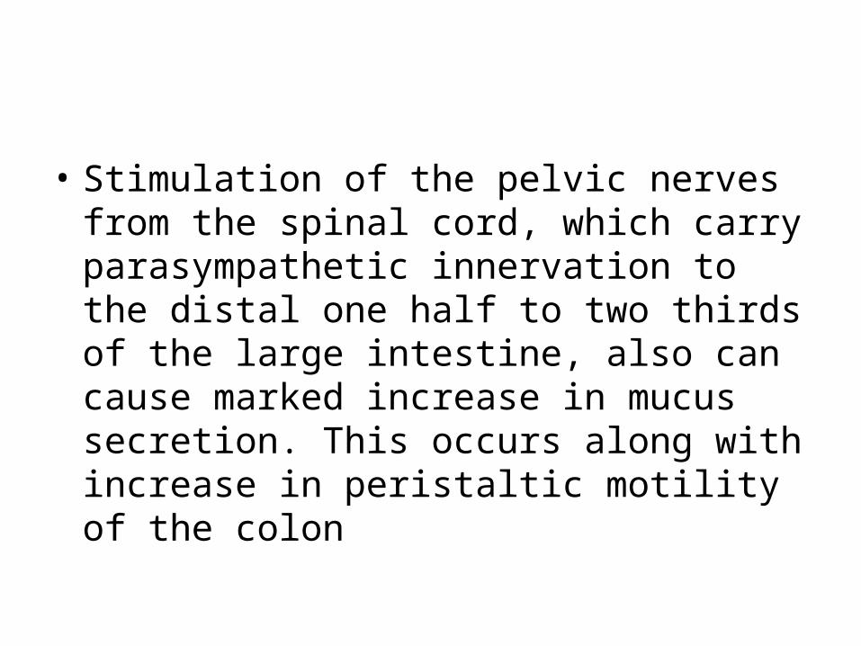
• Stimulation of the pelvic nerves from the spinal cord, which carry parasympathetic innervation to the distal one half to two thirds of the large intestine, also can cause marked increase in mucus secretion. This occurs along with increase in peristaltic motility of the colon

Functions of mucus
• Mucus in the large intestine protects the intestinal wall against excoriation,
• It provides an adherent medium for holding fecal matter together.
• It protects the intestinal wall from the great amount of bacterial activity that takes place inside the feces

• The mucus plus the alkalinity of the secretion (pH of 8.0 caused by large amounts of sodium bicarbonate) provides a barrier to keep acids formed in the feces from attacking the intestinal wall.


![Git LFS - acailly.github.io · $ git config --list [...] filter.lfs.clean=git-lfs clean -- %f filter.lfs.smudge=git-lfs smudge -- %f filter.lfs.process=git-lfs filter-process filter.lfs.required=true](https://img.pdfslide.us/doc/110x75/60bd0c0fa3a22721690a1c10/git-lfs-git-config-list-filterlfscleangit-lfs-clean-f-filterlfssmudgegit-lfs.jpg)