Embed Size (px)
Citation preview

Dr. Nithin Mathew

Dr. Nithin Mathew – Gingiva
Introduction Definition Macroscopic anatomy
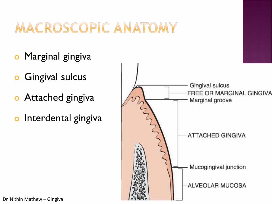
1. Marginal gingiva2. Gingival sulcus3. Attached gingiva4. Interdental gingiva

Dr. Nithin Mathew – Gingiva
Periodontiumperi = aroundodontos = tooth
i.e structures surrounding the tooth
Comprises Gingiva Periodontal ligament Cementum Alveolar bone

Dr. Nithin Mathew – Gingiva
Oral mucosa - three zones:• the gingiva and the covering of the hard
palate : masticatory mucosa
• the dorsum of the tongue, covered by specialized mucosa
• the oral mucous membrane lining the remainder of the oral cavity.

Dr. Nithin Mathew – Gingiva
Gingiva is that part of oral mucosa that covers thealveolar processes of the jaws and surrounds thenecks of the teeth.
Dr. Nithin Mathew – Gingiva
Marginal gingiva
Gingival sulcus
Attached gingiva
Interdental gingiva
Dr. Nithin Mathew – Gingiva
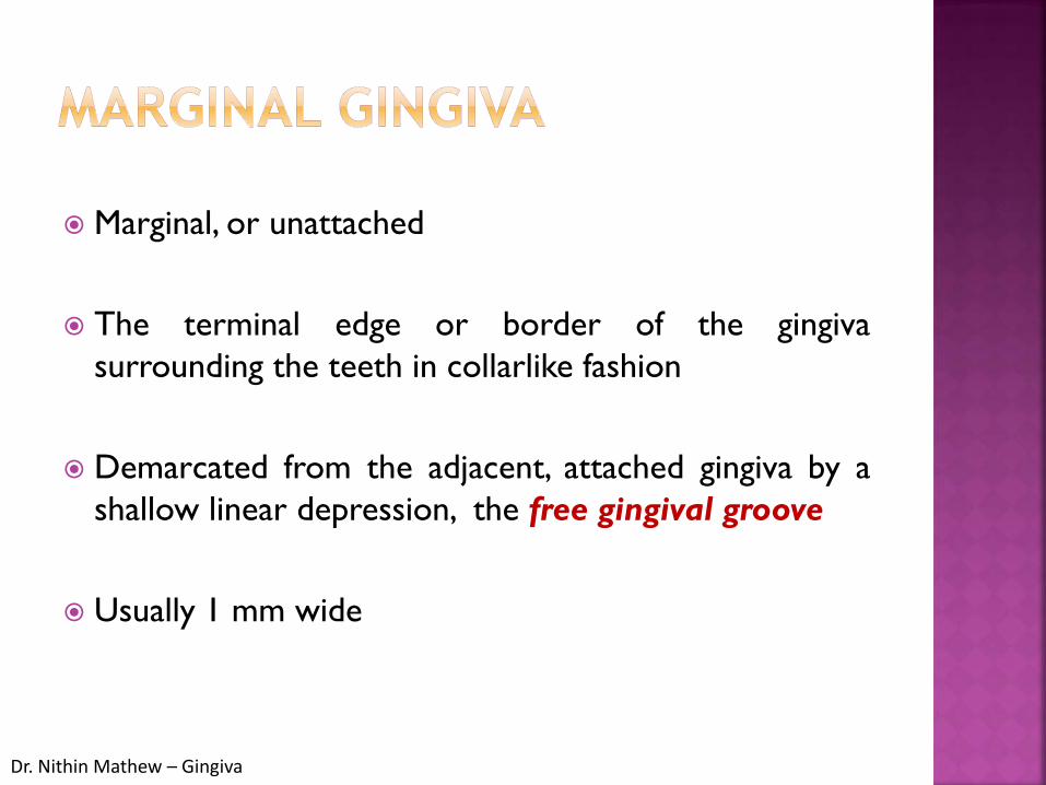
Marginal, or unattached
The terminal edge or border of the gingivasurrounding the teeth in collarlike fashion
Demarcated from the adjacent, attached gingiva by ashallow linear depression, the free gingival groove
Usually 1 mm wide
Dr. Nithin Mathew – Gingiva
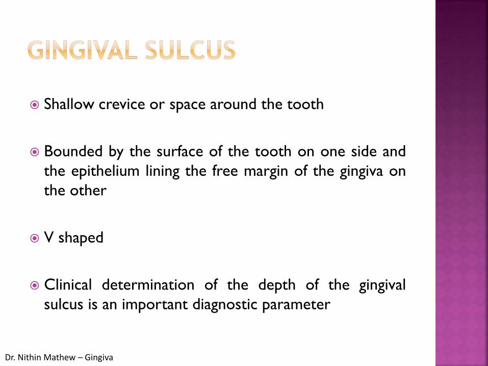
Shallow crevice or space around the tooth
Bounded by the surface of the tooth on one side andthe epithelium lining the free margin of the gingiva onthe other
V shaped
Clinical determination of the depth of the gingivalsulcus is an important diagnostic parameter

Dr. Nithin Mathew – Gingiva
Depth of this sulcus, as determined in histologicsections, has been reported as 1.8 mm,
The clinical maneuver used to determine the depth ofthe sulcus is the introduction of the periodontalprobe-and the estimation of the distance it penetrates.
The histologic depth of a sulcus need not be exactlyequal to the depth of penetration of the probe.
The so-called probing depth of a clinically normalgingival sulcus in humans is 2 to 3 mm

Dr. Nithin Mathew – Gingiva
It is the distance between the mucogingival junctionand the projection on the external surface at thebottom of the gingival sulcus or the periodontalpocket
The attached gingiva is continuous with the marginalgingiva.
It is firm, resilient and tightly bound to the underlyingperiosteum of alveolar bone.

Dr. Nithin Mathew – Gingiva
The facial aspect of the attached gingiva extends to therelatively loose and movable alveolar mucosa fromwhich it is demarcated by the mucogingival junction.
The width of the attached gingiva is another importantclinical parameter.
It is the distance between the mucogingival junctionand the projection on the external surface of thebottom of the gingival sulcus or the periodontalpocket

Dr. Nithin Mathew – Gingiva
greatest in the incisor region : 3.5 to 4.5 mm in the maxilla 3.3 to 3.9 mm in the mandible
less in the posterior segments with the least width inthe first premolar area : 1.9 mm in the maxilla 1.8 mm in the mandible
Width of the attached gingiva increases with age' andin supraerupted teeth

Dr. Nithin Mathew – Gingiva
Lingual aspect of the mandible, the attached gingivaterminates at the junction with the lingual alveolarmucosa, which is continuous with the mucousmembrane lining the floor of the mouth.
The palatal surface of the attached gingiva in themaxilla blends imperceptibly with the equally firm,resilient palatal mucosa.

Dr. Nithin Mathew – Gingiva
Occupies the gingival embrasure Can be pyramidal or have a "col" shape
Shape depends on the contact point and the presenceor absence of some degree of recession

Dr. Nithin Mathew – Gingiva
‘col’ it presents a valley like depression that connectsthe facial and lingual papilla and conforms to the shapeof interproximal contact.
Dr. Nithin Mathew – Gingiva
Measurement approach By using Schiller’s potassium iodide solution Tension test Roll test

Dr. Nithin Mathew – Gingiva
Broadly speaking gingiva is made up of epithelium andconnective tissue.
The gingival epithelium can be studied under threeheadings:
Outer or oral epithelium Sulcular epithelium Junctional epithelium

Dr. Nithin Mathew – Gingiva
Function Mechanical, chemical, water and microbial barrier Signalling function
Major cell type : keratinocyte
Other cells : langerhan cells, merkel cells, melanocytes.
Cell to cell attachments : desmosomes, tight jn, gap jn.

Dr. Nithin Mathew – Gingiva
Covers the crest and the outer surface of marginal gingivaand surface of attached gingiva.
4 layers Stratum basale:- cuboidal cells Stratum spinosum:- large polyhedral cells→Desmosomes
Stratum granulosum Stratum corneum:- superficial most layer Large, wide,
flat and lacking nucleus.
0.2 – 0.3mm thickness
keratinization varies


Dr. Nithin Mathew – Gingiva
Lines the gingival sulcus
Non keratinized stratified squamous epithelium
It is not keratinized due to constant irritation ofplaque
Extends from the coronal area of the junctionalepithelium to the free margin of the gingival.
Epithelium lacks heavy ridges and papillae.


Dr. Nithin Mathew – Gingiva
Collar-like band
0.25 – 1.35mm
Formed by the confluence of oral epithelium and reducedenamel epithelium.
Epithelial attachment – internal basal laminae
3-4 layers thick in early life, but the number of layers increaseswith age to 10 or even 20 layers
Junctional epithelium + gingival fibres = dento-gingival unit

Dr. Nithin Mathew – Gingiva
Attached to the tooth surface (epithelial attachment) bymeans of an internal basal lamina and to the gingivalconnective tissue by an external basal lamina
The attachment of the junctional epithelium to the toothis reinforced by the gingival fibers, which brace themarginal gingiva against the tooth surface
Junctional epithelium + gingival fibres = dento-gingivalunit


Dr. Nithin Mathew – Gingiva
The gingival sulcus is the shallow, V-shaped space orgroove between the tooth and gingiva that encircles thenewly erupted tip of the crown.
In the fully erupted tooth, only the junctional epitheliumpersists.
Sulcus consists of the shallow space that is coronal tothe attachment of the junctional epithelium and boundedby the tooth on one side and the Sulcular epithelium onthe other.
The coronal extent of the gingival sulcus is the gingivalmargin

Dr. Nithin Mathew – Gingiva
Undergoes continuous renewal.
Thickness is maintained by a balance between new cellformation in the basal and spinous layers and theshedding of old cells at the surface.
The mitotic activity exhibits a 24-hour periodicity, withhighest and lowest rates occurring in the morning andevening, respectively.
The mitotic rate is higher in nonkeratinized areas andis increased in gingivitis

Dr. Nithin Mathew – Gingiva
The gingival sulcus contains a fluid that seeps into itfrom the gingival connective tissue through the thinSulcular epithelium.
Functions Cleanse debris from the sulcus Contain plasma proteins that may improve
adhesion of the epithelium to the tooth Possess antimicrobial properties, and exert
antibody activity to defend the gingiva

Dr. Nithin Mathew – Gingiva
Connective tissue of the gingiva is known as theLamina Propria
Consists of two layers: a papillary layer subjacent to the epithelium, which
consists of papillary projections between the epithelialrete pegs
a reticular layer contiguous with the periosteum of thealveolar bone.
Connective tissue has a cellular and an extracellularcompartment composed of fibers and groundsubstance

Dr. Nithin Mathew – Gingiva
The ground substance fills the space between fibersand cells, is amorphous, and has a high content ofwater.
Composed of proteoglycans, mainly hyaluronic acidand chondroitin sulfate, and glycoproteins, mainlyfibronectin.
Fibronectin binds fibroblasts to the fibers and manyother components of the intercellular matrix, helpingmediate cell adhesion and migration.
Laminin is another glycoprotein found in the basallaminae, which serves to attach it to epithelial cells
Dr. Nithin Mathew – Gingiva
The three types of connective tissue fibers arecollagen, reticular, and elastic.
Collagen type I forms the bulk of the lamina propriaand provides the tensile strength to the gingival tissue.
Type IV collagen branches between the collagen type Ibundles and is continuous with fibers of the basementmembrane and blood vessel walls

Dr. Nithin Mathew – Gingiva
Connective tissue of the marginal gingiva is denselycollagenous, containing a prominent system of collagenfiber bundles called the gingival fibers
Functions: To brace the marginal gingiva firmly against the tooth To provide the rigidity necessary to withstand the
forces of mastication without being deflected awayfrom the tooth surface
To unite the free marginal gingiva with the cementumof the root and the adjacent attached gingiva

Dr. Nithin Mathew – Gingiva
The gingival fibers are arranged in three groups:dentogingival, circular and transseptal
Dentogingival
Facial, lingual, and interproximal surfaces.
Embedded in the cementum just beneath theepithelium at the base of the gingival sulcus.
On the facial and lingual surfaces, they project fromthe cementum in fanlike conformation toward thecrest and outer surface of the marginal gingiva,terminating short of the epithelium
Dr. Nithin Mathew – Gingiva
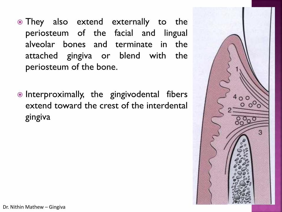
They also extend externally to theperiosteum of the facial and lingualalveolar bones and terminate in theattached gingiva or blend with theperiosteum of the bone.
Interproximally, the gingivodental fibersextend toward the crest of the interdentalgingiva

Circular Group
The circular fibers course through the connective tissue of the marginal and interdental gingivae and encircle the tooth in ringlikefashion

Dr. Nithin Mathew – Gingiva
Transseptal Group
Located interproximally,
Transseptal fibers form horizontal bundles that extendbetween the cementum of approximating teeth intowhich they are embedded.
Lie in the area between the epithelium at the base ofthe gingival sulcus and the crest of the interdentalbone

Dr. Nithin Mathew – Gingiva
Alveologingival group
The fibers run from the crest of the alveolar boneand interdental septum, radiating coronally into theoverlying lamina propria of the gingiva
Dentoperiosteal group
The fibers only occur in vestibular and lingual gingiva.They arise from cementum and pass over the alveolarcrest to insert into the periosteum

Dr. Nithin Mathew – Gingiva
Semicircular group
The fibers emanate from cementum nearthe cementenamel junction, cross the freemarginal gingiva, and insert into a similar position onthe opposite side of the tooth.
Transgingival group
The fibers reinforce the circular and semicircularfibers. The fibers arise from the cervical cementum andextend into the marginal gingiva of the adjacent tooth,merging with the circular fibers

Dr. Nithin Mathew – Gingiva
Longitudinal group
The fibers extend for long distances within thefree gingiva, some possibly for the whole length of thearch.
Interdental group
The fibers pass through the coronal portion ofthe interdental gingiva in the buccolingual direction,connecting buccal and lingual papillae.

Dr. Nithin Mathew – Gingiva
Vertical group
The fibers arise in alveolar mucosa orattached gingiva and pass coronally towards themarginal gingiva and interdental papilla.

Dr. Nithin Mathew – Gingiva
Three sources of blood supply to the gingiva areasfollows
Supraperiosteal arterioles along the facial and lingualsurfaces of the alveolar bone, from which capillariesextend along the sulcular epithelium and between therete pegs of the external gingival surface .
Occasional branches of the arterioles pass through thealveolar bone to the periodontal ligament or run overthe crest of the alveolar bone

Dr. Nithin Mathew – Gingiva
Vessels of the periodontal ligament, which extendinto the gingiva and anastomose with capillaries in thesulcus area.
Arterioles, which emerge from the crest of theinterdental septa and extend parallel to the crest ofthe bone to anastomose with vessels of theperiodontal ligament, with capillaries in the gingivalcrevicular areas and vessels that run over the alveolarcrest.

Dr. Nithin Mathew – Gingiva
Maxillary
Nasopalatine nerveSupplies facial aspect of anterior teeth
Posterior superior alveolar nerveSupplies facial aspect of posterior teeth
Greater palatine nerveSupplies lingual aspect of posterior teeth
Anterior palatine nerveSupplies lingual aspect of anterior teeth

Dr. Nithin Mathew – Gingiva
Mandibular
Inferior Alveolar nerve

Dr. Nithin Mathew – Gingiva
Color
Healthy gingiva : "coral pink." Other colours like red, white, and blue can signify
inflammation (gingivitis) or pathology.
Normal racial pigmentation makes the gingiva appeardarker.
Because the color of gingiva varies due to racialpigmentation, uniformity of color is more importantthan the underlying color itself.

Dr. Nithin Mathew – Gingiva
Produced by vascular supply, the thickness and degreeof keratinization of the epithelium and the presence ofpigment containing cells : melanin

Dr. Nithin Mathew – Gingiva
Size
Corresponds to the sum total of the bulk of cellularand intercellular elements and their vascular supply.
Contour
Depends on the shape of the teeth and their alignmentin the arch, the location and size of the area of theproximal contact and the dimensions of the facial andlingual embrasures.
Marginal gingiva-scalloped outline on facial and lingualsurfaces, straight line along the teeth with flat surfaces.

Dr. Nithin Mathew – Gingiva
On teeth with pronounced mesiodistal convexity orteeth in labial version, the normal arcuate contour isaccentuated and the gingival is located farther apically.
On teeth in lingual version the gingiva is horizontal andthickened.
Shape
Governed by the contour of the proximal toothsurfaces and the location and shape of the gingivalembrasures.
The height of the interdental gingival varies with thelocation of the proximal contact.

Dr. Nithin Mathew – Gingiva
Consistency
Firm and resilient because it is bound to theunderlying bone except movable free margin.
Collagenous nature of lamina propria and its contiguitywith the mucoperiosteum of the alveolar bonedetermine the firmness of the attached gingiva.
Gingival fibers contribute to the firmness of gingivalmargin.

Dr. Nithin Mathew – Gingiva
SurfaceTexture
Orange peel-referred to as being stippled.
It is best viewed by drying the gingiva.
Attached gingiva is stippled, marginal is not.
Central portion of interdental papilla is usually stippledbut the marginal borders are smooth.
Stippling is less prominent in lingual surfaces.

Dr. Nithin Mathew – Gingiva
Position
The position of the gingiva refers to the level at whichthe gingival margin is attached to the tooth
![Relationship of Facial Skin Complexion with Gingiva …...Ibuski, reported that the color of the gingiva varied with the position of the papillary, marginal and attached gingiva [9]](https://img.pdfslide.us/doc/110x75/5e61b02ebfe26e503169c604/relationship-of-facial-skin-complexion-with-gingiva-ibuski-reported-that-the.jpg)