Embed Size (px)
Citation preview
Project: Ghana Emergency Medicine Collaborative Document Title: Pulmonary Embolism Part 2 (2012) Author(s): Rockefeller A. Oteng, M.D., University of Michigan License: Unless otherwise noted, this material is made available under the terms of the Creative Commons Attribution Share Alike-3.0 License: http://creativecommons.org/licenses/by-sa/3.0/
We have reviewed this material in accordance with U.S. Copyright Law and have tried to maximize your ability to use, share, and adapt it. These lectures have been modified in the process of making a publicly shareable version. The citation key on the following slide provides information about how you may share and adapt this material. Copyright holders of content included in this material should contact [email protected] with any questions, corrections, or clarification regarding the use of content. For more information about how to cite these materials visit http://open.umich.edu/privacy-and-terms-use. Any medical information in this material is intended to inform and educate and is not a tool for self-diagnosis or a replacement for medical evaluation, advice, diagnosis or treatment by a healthcare professional. Please speak to your physician if you have questions about your medical condition. Viewer discretion is advised: Some medical content is graphic and may not be suitable for all viewers.
1
Attribution Key
for more information see: http://open.umich.edu/wiki/AttributionPolicy
Use + Share + Adapt
Make Your Own Assessment
Creative Commons – Attribution License
Creative Commons – Attribution Share Alike License
Creative Commons – Attribution Noncommercial License
Creative Commons – Attribution Noncommercial Share Alike License
GNU – Free Documentation License
Creative Commons – Zero Waiver
Public Domain – Ineligible: Works that are ineligible for copyright protection in the U.S. (17 USC § 102(b)) *laws in your jurisdiction may differ
Public Domain – Expired: Works that are no longer protected due to an expired copyright term.
Public Domain – Government: Works that are produced by the U.S. Government. (17 USC § 105)
Public Domain – Self Dedicated: Works that a copyright holder has dedicated to the public domain.
Fair Use: Use of works that is determined to be Fair consistent with the U.S. Copyright Act. (17 USC § 107) *laws in your jurisdiction may differ Our determination DOES NOT mean that all uses of this 3rd-party content are Fair Uses and we DO NOT guarantee that your use of the content is Fair. To use this content you should do your own independent analysis to determine whether or not your use will be Fair.
{ Content the copyright holder, author, or law permits you to use, share and adapt. }
{ Content Open.Michigan believes can be used, shared, and adapted because it is ineligible for copyright. }
{ Content Open.Michigan has used under a Fair Use determination. }
2
Pulmonary Embolism Part 2
Rockefeller A. Oteng Ghana Emergency Medicine
Collabora@ve
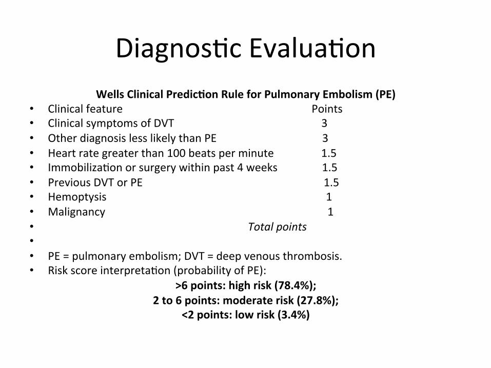
Diagnos@c Evalua@on Wells Clinical Predic.on Rule for Pulmonary Embolism (PE)
• Clinical feature Points • Clinical symptoms of DVT 3 • Other diagnosis less likely than PE 3 • Heart rate greater than 100 beats per minute 1.5 • Immobiliza@on or surgery within past 4 weeks 1.5 • Previous DVT or PE 1.5 • Hemoptysis 1 • Malignancy 1 • Total points • • PE = pulmonary embolism; DVT = deep venous thrombosis. • Risk score interpreta@on (probability of PE):
>6 points: high risk (78.4%); 2 to 6 points: moderate risk (27.8%);
<2 points: low risk (3.4%)
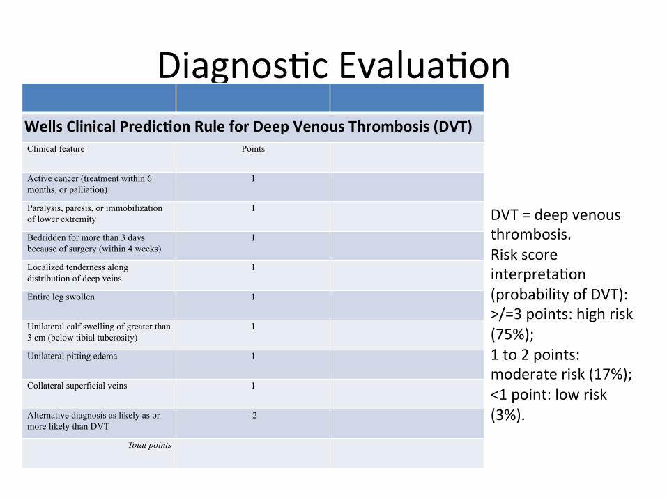
Diagnos@c Evalua@on Wells Clinical Predic.on Rule for Deep Venous Thrombosis (DVT) Clinical feature Points
Active cancer (treatment within 6 months, or palliation)
1
Paralysis, paresis, or immobilization of lower extremity
1
Bedridden for more than 3 days because of surgery (within 4 weeks)
1
Localized tenderness along distribution of deep veins
1
Entire leg swollen 1
Unilateral calf swelling of greater than 3 cm (below tibial tuberosity)
1
Unilateral pitting edema 1
Collateral superficial veins 1
Alternative diagnosis as likely as or more likely than DVT
-2
Total points
DVT = deep venous thrombosis. Risk score interpreta@on (probability of DVT): >/=3 points: high risk (75%); 1 to 2 points: moderate risk (17%); <1 point: low risk (3%).
Diagnos@c Evalua@on
• Laboratory: – Rou@ne laboratory findings are nonspecific. – Include leukocytosis – Increased erythrocyte sedimenta@on rate (ESR), and an elevated serum LDH or AST (SGOT)
– normal serum bilirubin.
Diagnos@c Evalua@on
• Arterial blood gas – Arterial blood gas (ABG) measurements and pulse oximetry have a limited role in diagnosing PE.
– ABGs usually reveal hypoxemia • Hypocapnia, • Respiratory alkalosis.
Diagnos@c Evalua@on
• Troponin : – Serum troponin I and troponin T are elevated in 30 to 50 percent of pa@ents who have a moderate to large pulmonary embolism.
– Presumed mechanism is acute right heart overload.
• Brain Nature.c Pep.de: – Very non specific pep@de – Large eleva@on can suggest poor prognosis
Diagnos@c Evalua@on
• Electrocardiogram – ECG abnormali@es common in pa@ents with and without PE
– limi@ng the diagnos@c usefulness of the ECG – Most common Ekg finding is a sinus tachycardia
• Or non specific ST and T wave changes – abnormali@es historically considered to be sugges@ve of PE
• S1Q3T3 pa_ern, right ventricular strain, new incomplete right bundle branch block
Diagnos@c Evalua@on
• V/Q scan : – The most extensive evalua@on of the accuracy of the ven@la@on-‐perfusion (V/Q) scan was the Prospec@ve Inves@ga@on of Pulmonary Embolism Diagnosis (PIOPED)
– Accuracy was based on comparison with the gold standard test of Pulmonary angiogram
– The found clincally accuracy was best when combined with pretest probabili@es
Diagnos@c Evalua@on
• V/Q scan : • Pa@ents with high clinical probability of PE and a high-‐probability V/Q scan had a 95 percent likelihood of having PE
• Pa@ents with low clinical probability of PE and a low-‐probability V/Q scan had only a 4 percent likelihood of having PE
• A normal V/Q scan virtually excluded PE
Diagnos@c Evalua@on
• Ultrasound: – In some pa@ents clinicians have a_empted to use lower extremity Doppler's to evaluate
– Studies show that many pa@ents with PE are missed
– Bilateral lower extremity doppler’s will decrease the rate of missed DVT
– Operator dependent
Diagnos@c Evalua@on • D-‐dimer:
– D-‐dimer is a degrada@on product of cross-‐linked fibrin. It can be detected in serum using a variety of different assays:
– Enzyme-‐linked immunosorbent assay (ELISA) (results in >8 hrs)
– Quan@ta@ve rapid ELISA (results in 30 min) – Semi-‐quan@ta@ve rapid ELISA (results in 10 min) – Qualita@ve rapid ELISA (results in 10 min) – Quan@ta@ve latex agglu@na@on assay (results in 10 to 15 min)
– Semi-‐quan@ta@ve latex agglu@na@on assay (results in 5 min)
Diagnos@c Evalua@on
• D-‐Dimer: • For the quan@ta@ve assays, a level >500 ng/mL is usually considered abnormal
• They are best characterized as having good sensi@vity and nega@ve predic@ve value
• Poor specificity and posi@ve predic@ve value.
Diagnos@c Evalua@on
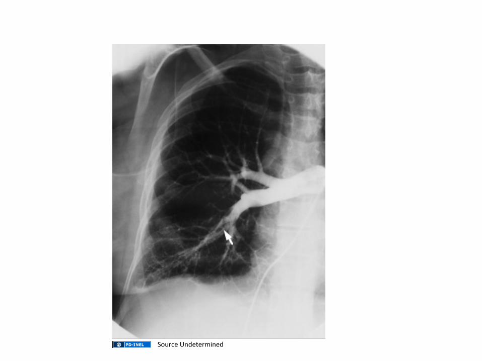
• Angiography : – Pulmonary angiography is the defini@ve diagnos@c technique or "gold standard" in the diagnosis of acute PE.
– It is performed by injec@ng contrast into a pulmonary artery branch ajer percutaneous catheteriza@on, usually via the femoral vein. A filling defect or abrupt cutoff of a small vessel is indica@ve of PE.
Diagnos@c Evalua@on
• Angiography: – A nega@ve pulmonary angiogram excludes clinically relevant PE.
– Pulmonary angiography is generally safe and well tolerated in the absence of hemodynamic instability caused by acute, severe pulmonary hypertension
– Radia@on exposure depends on the length and complexity of the procedure, and greater than CT.
Source Undetermined
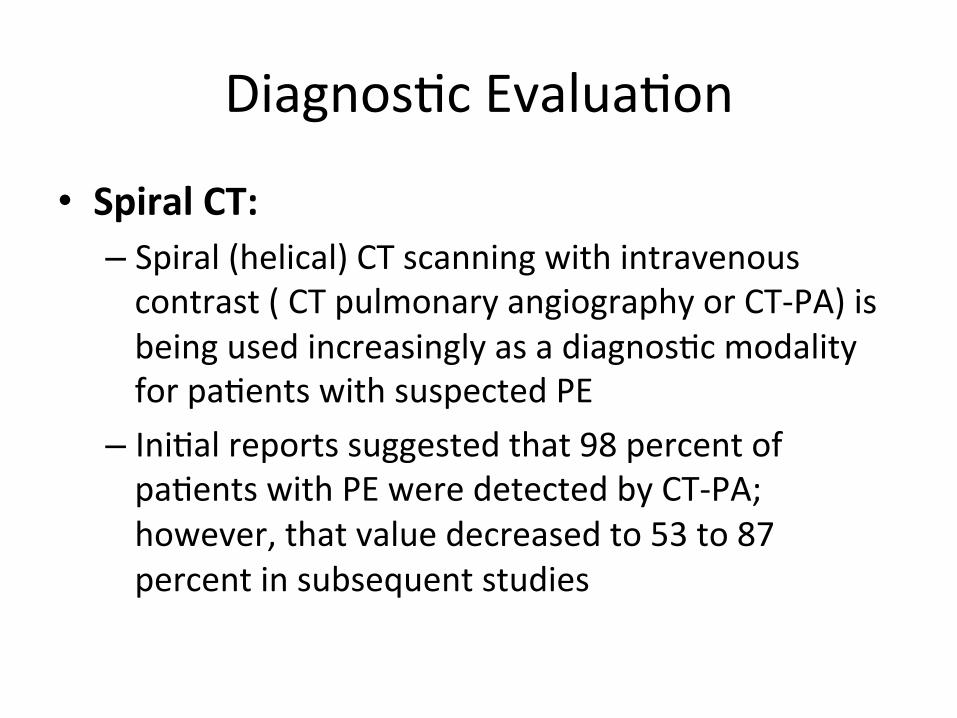
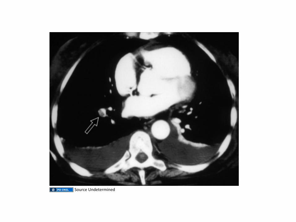
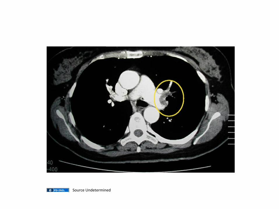
Diagnos@c Evalua@on
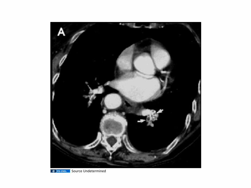
• Spiral CT: – Spiral (helical) CT scanning with intravenous contrast ( CT pulmonary angiography or CT-‐PA) is being used increasingly as a diagnos@c modality for pa@ents with suspected PE
– Ini@al reports suggested that 98 percent of pa@ents with PE were detected by CT-‐PA; however, that value decreased to 53 to 87 percent in subsequent studies
Source Undetermined
Source Undetermined
Source Undetermined
Source Undetermined
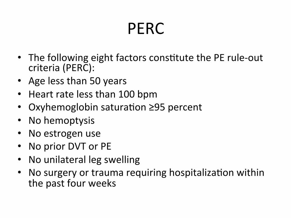
PERC • The following eight factors cons@tute the PE rule-‐out criteria (PERC):
• Age less than 50 years • Heart rate less than 100 bpm • Oxyhemoglobin satura@on ≥95 percent • No hemoptysis • No estrogen use • No prior DVT or PE • No unilateral leg swelling • No surgery or trauma requiring hospitaliza@on within the past four weeks