Embed Size (px)
Citation preview
Project: Ghana Emergency Medicine Collaborative Document Title: Cardiovascular Emergencies Author(s): Susan Anne Bell (University of Michigan), RN 2012 License: Unless otherwise noted, this material is made available under the terms of the Creative Commons Attribution Share Alike-3.0 License: http://creativecommons.org/licenses/by-sa/3.0/
We have reviewed this material in accordance with U.S. Copyright Law and have tried to maximize your ability to use, share, and adapt it. These lectures have been modified in the process of making a publicly shareable version. The citation key on the following slide provides information about how you may share and adapt this material. Copyright holders of content included in this material should contact [email protected] with any questions, corrections, or clarification regarding the use of content. For more information about how to cite these materials visit http://open.umich.edu/privacy-and-terms-use. Any medical information in this material is intended to inform and educate and is not a tool for self-diagnosis or a replacement for medical evaluation, advice, diagnosis or treatment by a healthcare professional. Please speak to your physician if you have questions about your medical condition. Viewer discretion is advised: Some medical content is graphic and may not be suitable for all viewers.
1
Attribution Key
for more information see: http://open.umich.edu/wiki/AttributionPolicy
Use + Share + Adapt
Make Your Own Assessment
Creative Commons – Attribution License
Creative Commons – Attribution Share Alike License
Creative Commons – Attribution Noncommercial License
Creative Commons – Attribution Noncommercial Share Alike License
GNU – Free Documentation License
Creative Commons – Zero Waiver
Public Domain – Ineligible: Works that are ineligible for copyright protection in the U.S. (17 USC § 102(b)) *laws in your jurisdiction may differ
Public Domain – Expired: Works that are no longer protected due to an expired copyright term.
Public Domain – Government: Works that are produced by the U.S. Government. (17 USC § 105)
Public Domain – Self Dedicated: Works that a copyright holder has dedicated to the public domain.
Fair Use: Use of works that is determined to be Fair consistent with the U.S. Copyright Act. (17 USC § 107) *laws in your jurisdiction may differ Our determination DOES NOT mean that all uses of this 3rd-party content are Fair Uses and we DO NOT guarantee that your use of the content is Fair. To use this content you should do your own independent analysis to determine whether or not your use will be Fair.
{ Content the copyright holder, author, or law permits you to use, share and adapt. }
{ Content Open.Michigan believes can be used, shared, and adapted because it is ineligible for copyright. }
{ Content Open.Michigan has used under a Fair Use determination. }
2
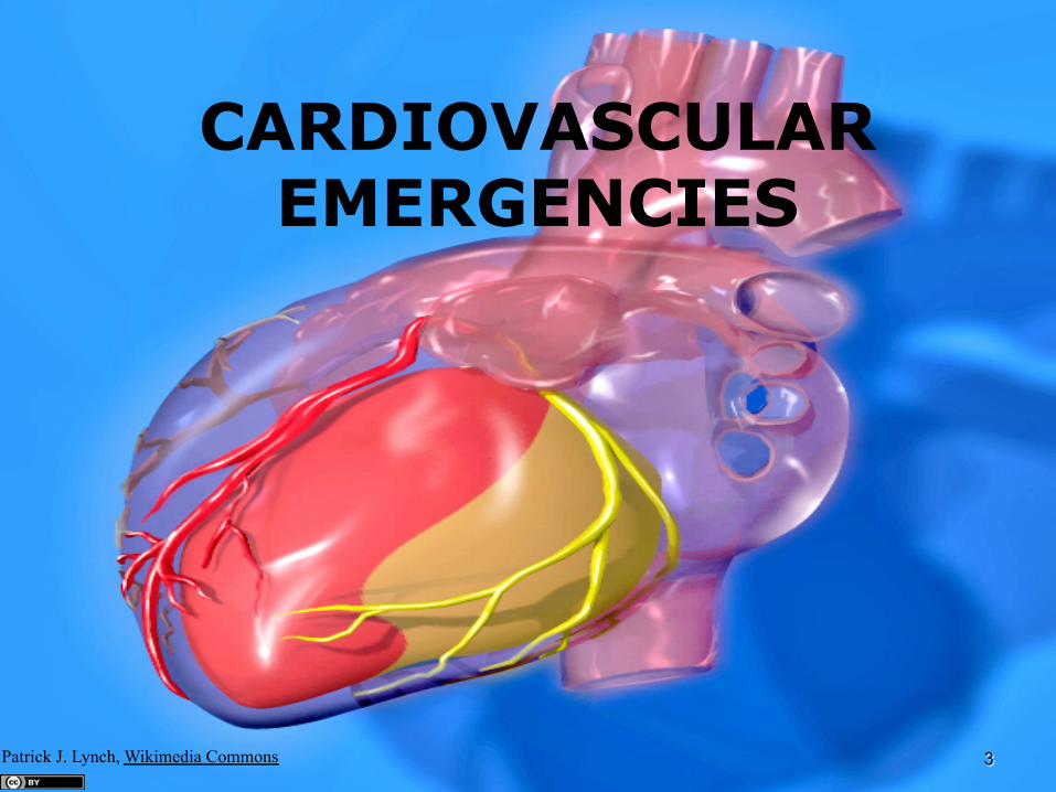
CARDIOVASCULAR EMERGENCIES
Patrick J. Lynch, Wikimedia Commons 3
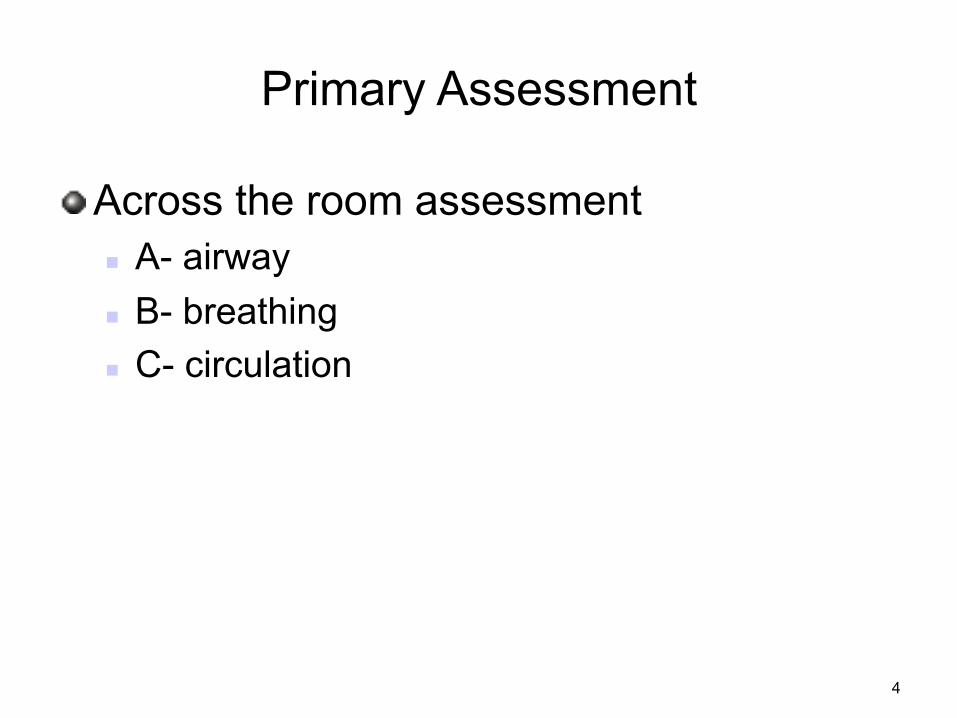
Primary Assessment
! Across the room assessment n A- airway n B- breathing n C- circulation
4
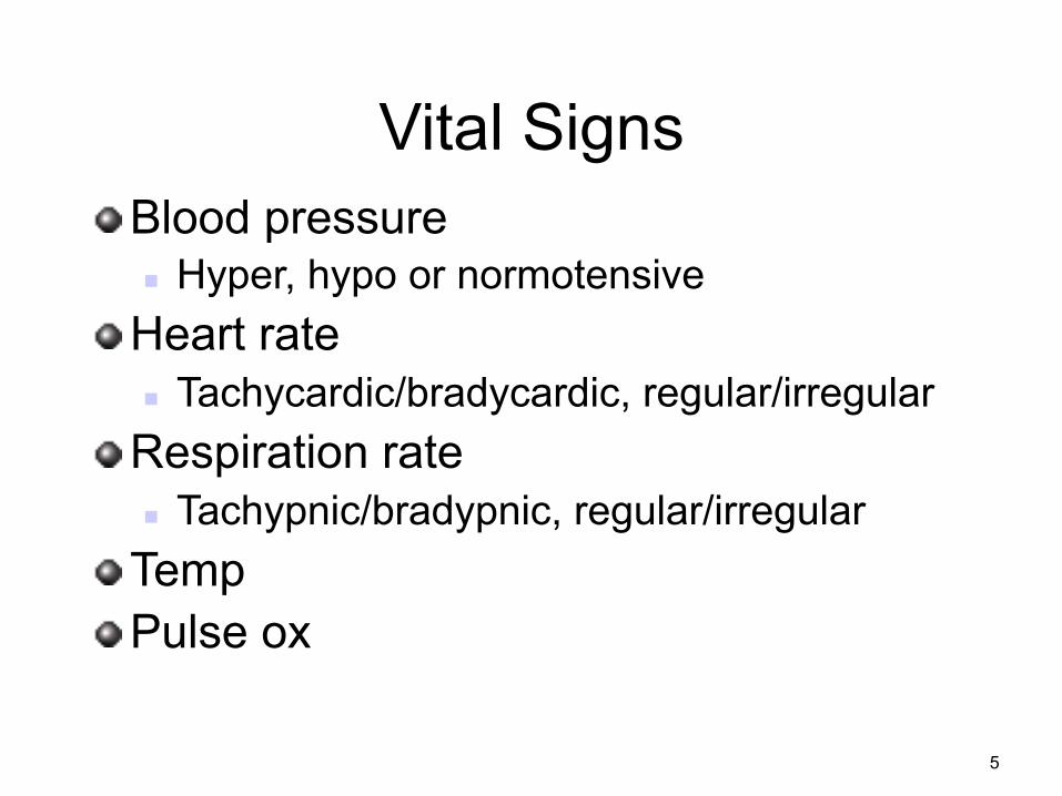
Vital Signs ! Blood pressure
n Hyper, hypo or normotensive ! Heart rate
n Tachycardic/bradycardic, regular/irregular ! Respiration rate
n Tachypnic/bradypnic, regular/irregular ! Temp ! Pulse ox
5
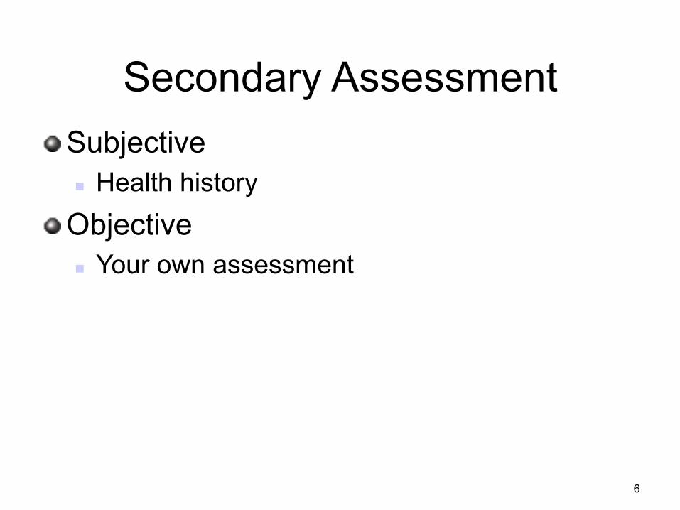
Secondary Assessment ! Subjective
n Health history ! Objective
n Your own assessment
6
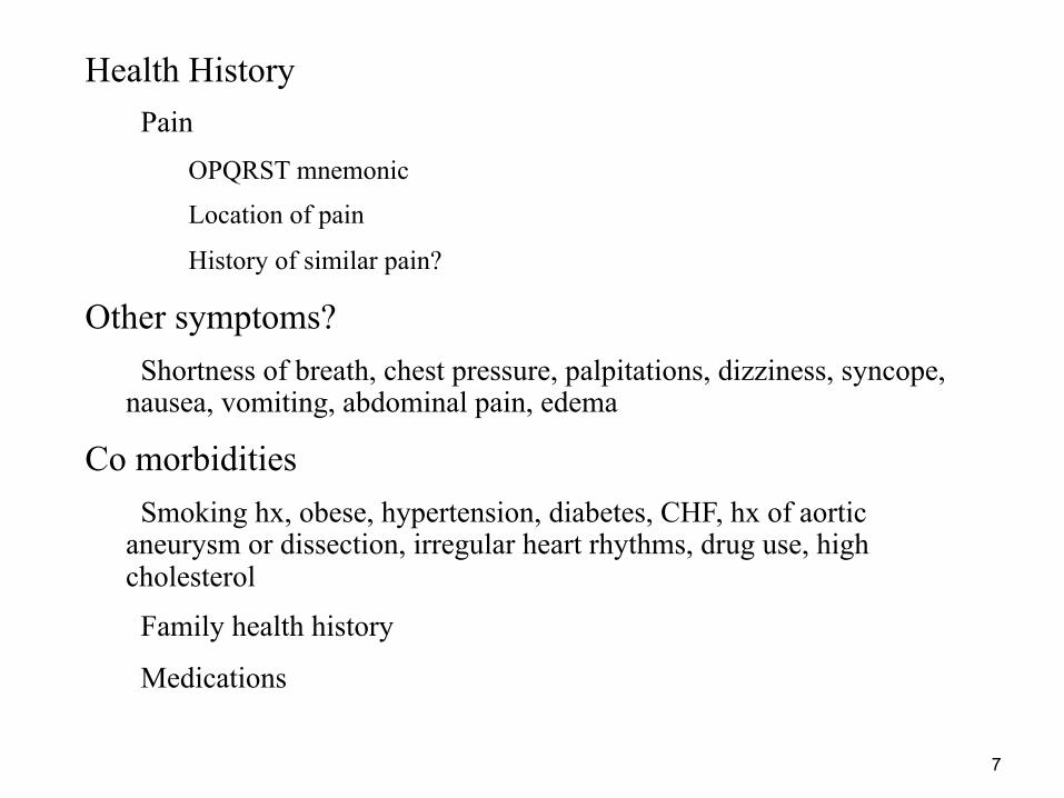
• Health History – Pain
• OPQRST mnemonic
• Location of pain
• History of similar pain?
• Other symptoms? – Shortness of breath, chest pressure, palpitations, dizziness, syncope, nausea, vomiting, abdominal pain, edema
• Co morbidities – Smoking hx, obese, hypertension, diabetes, CHF, hx of aortic aneurysm or dissection, irregular heart rhythms, drug use, high cholesterol
– Family health history
– Medications
7
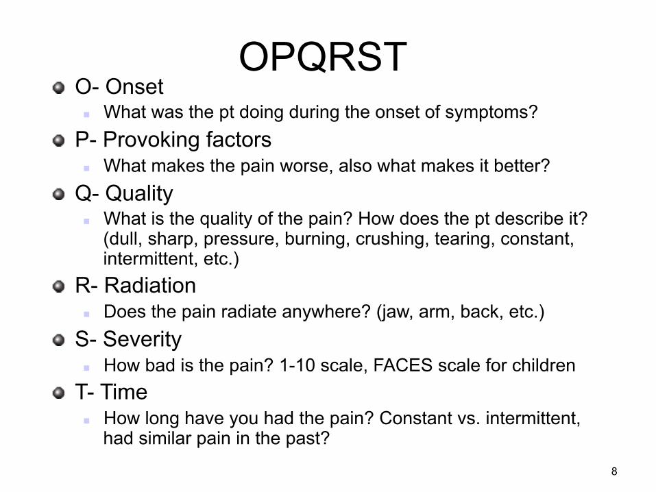
OPQRST ! O- Onset
n What was the pt doing during the onset of symptoms? ! P- Provoking factors
n What makes the pain worse, also what makes it better? ! Q- Quality
n What is the quality of the pain? How does the pt describe it? (dull, sharp, pressure, burning, crushing, tearing, constant, intermittent, etc.)
! R- Radiation n Does the pain radiate anywhere? (jaw, arm, back, etc.)
! S- Severity n How bad is the pain? 1-10 scale, FACES scale for children
! T- Time n How long have you had the pain? Constant vs. intermittent,
had similar pain in the past?
8
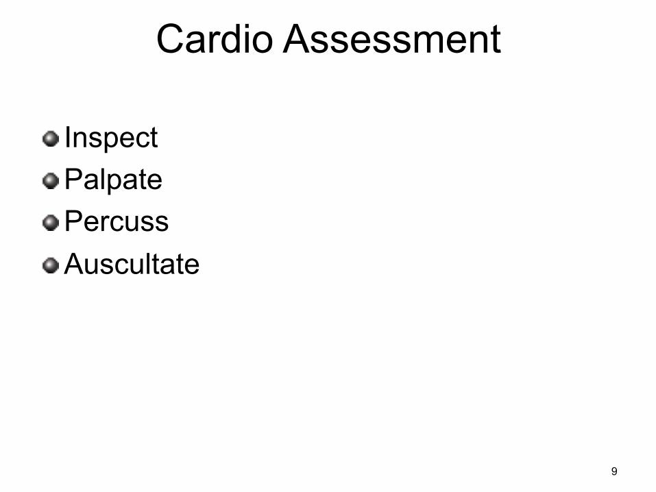
Cardio Assessment
! Inspect ! Palpate ! Percuss ! Auscultate
9
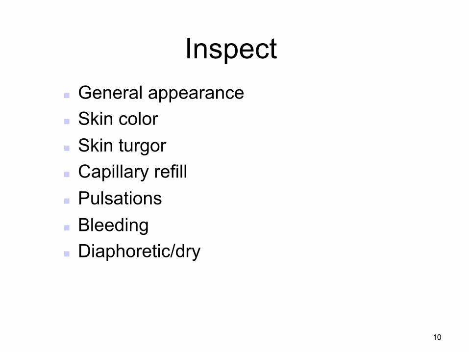
Inspect n General appearance n Skin color n Skin turgor n Capillary refill n Pulsations n Bleeding n Diaphoretic/dry
10
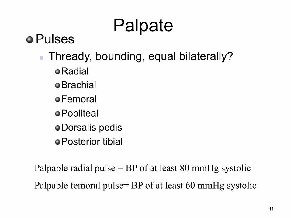
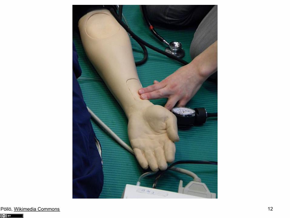
Palpate ! Pulses
n Thready, bounding, equal bilaterally? ! Radial ! Brachial ! Femoral ! Popliteal ! Dorsalis pedis ! Posterior tibial
Palpable radial pulse = BP of at least 80 mmHg systolic
Palpable femoral pulse= BP of at least 60 mmHg systolic
11
12 Pöllö, Wikimedia Commons
Percussion
! Can percuss for cardiac borders if needed-
n Begin at axillary line and percuss along 5th intercostal space toward sternum.
n Resonance to dullness at L border of heart, cannot usually hear R border d/t sternum.
13
Auscultation ! Rate
n Tachycardic, bradycardic ! Rhythm
n Regular, irregular ! Heart sounds
14 OCAL, clker.com
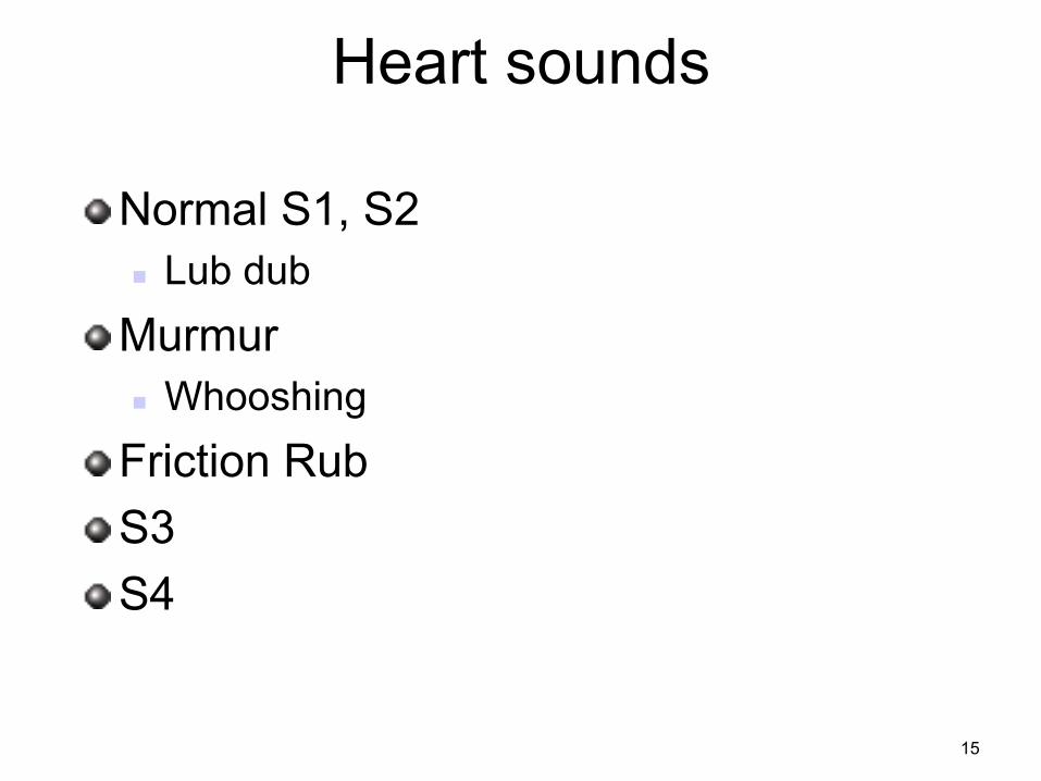
Heart sounds
! Normal S1, S2 n Lub dub
! Murmur n Whooshing
! Friction Rub ! S3 ! S4
15
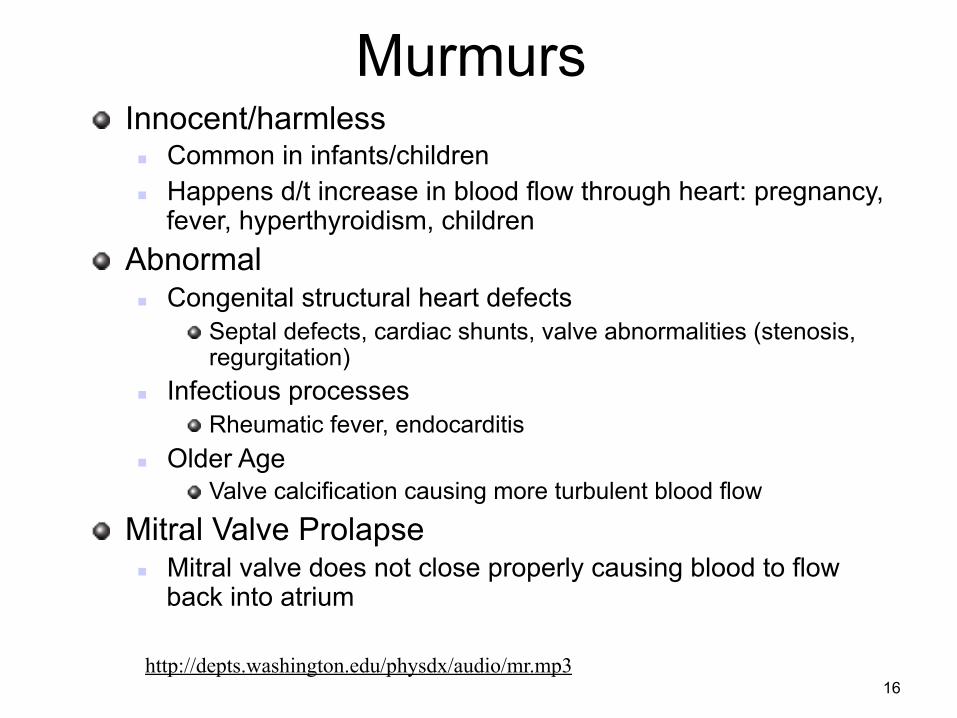
Murmurs ! Innocent/harmless
n Common in infants/children n Happens d/t increase in blood flow through heart: pregnancy,
fever, hyperthyroidism, children ! Abnormal
n Congenital structural heart defects ! Septal defects, cardiac shunts, valve abnormalities (stenosis,
regurgitation) n Infectious processes
! Rheumatic fever, endocarditis n Older Age
! Valve calcification causing more turbulent blood flow
! Mitral Valve Prolapse n Mitral valve does not close properly causing blood to flow
back into atrium
http://depts.washington.edu/physdx/audio/mr.mp3 16
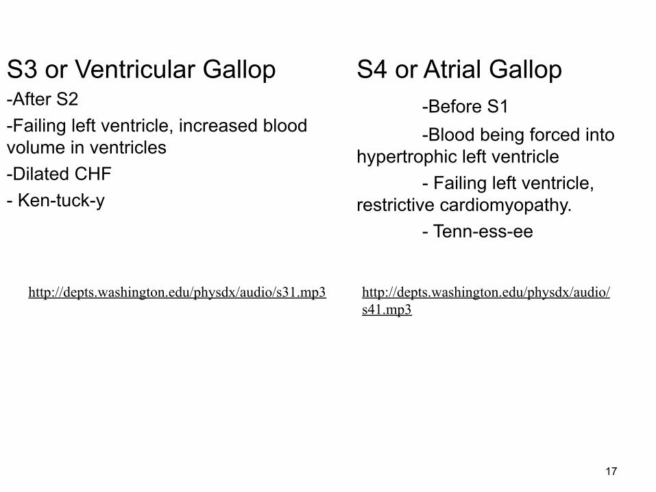
S3 or Ventricular Gallop -After S2 -Failing left ventricle, increased blood volume in ventricles -Dilated CHF - Ken-tuck-y
S4 or Atrial Gallop -Before S1 -Blood being forced into
hypertrophic left ventricle - Failing left ventricle,
restrictive cardiomyopathy. - Tenn-ess-ee
http://depts.washington.edu/physdx/audio/s31.mp3 http://depts.washington.edu/physdx/audio/s41.mp3
17
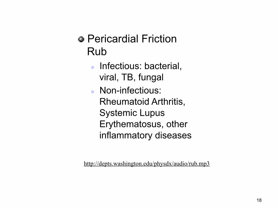
! Pericardial Friction Rub n Infectious: bacterial,
viral, TB, fungal n Non-infectious:
Rheumatoid Arthritis, Systemic Lupus Erythematosus, other inflammatory diseases
http://depts.washington.edu/physdx/audio/rub.mp3
18
Diagnostic Procedures
19
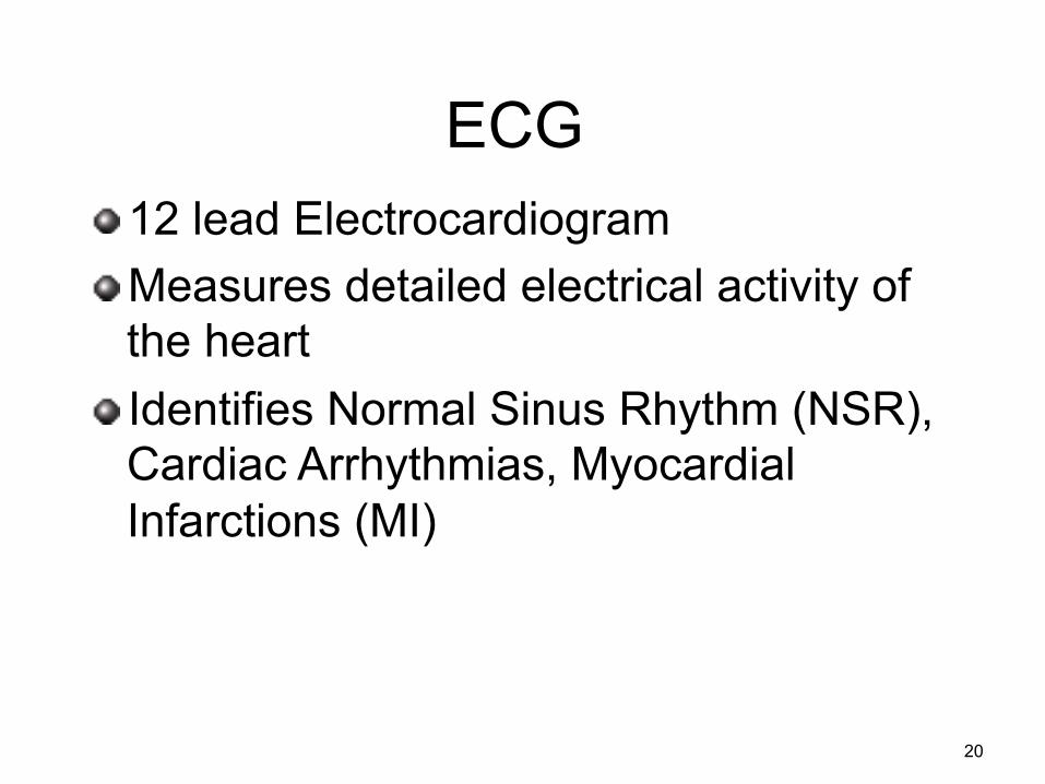
ECG ! 12 lead Electrocardiogram ! Measures detailed electrical activity of
the heart ! Identifies Normal Sinus Rhythm (NSR),
Cardiac Arrhythmias, Myocardial Infarctions (MI)
20
Reasons to obtain ECG
! Chest pain/pressure ! Shortness of breath/difficulty breathing ! Palpitations or pounding of heart ! Tachycardia/bradycardia ! Syncope
21
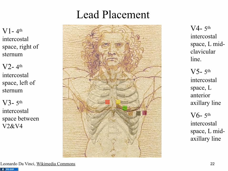
Lead Placement
V1- 4th intercostal space, right of sternum
V2- 4th intercostal space, left of sternum
V3- 5th intercostal space between V2&V4
V4- 5th intercostal space, L mid-clavicular line.
V5- 5th intercostal space, L anterior axillary line
V6- 5th intercostal space, L mid-axillary line
Leonardo Da Vinci, Wikimedia Commons 22
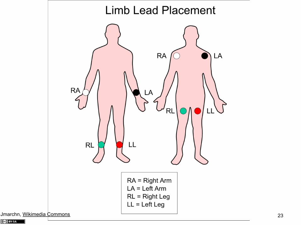
23 Jmarchn, Wikimedia Commons
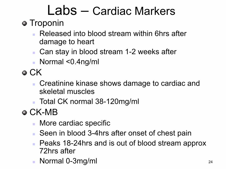
Labs – Cardiac Markers ! Troponin
n Released into blood stream within 6hrs after damage to heart
n Can stay in blood stream 1-2 weeks after n Normal <0.4ng/ml
! CK n Creatinine kinase shows damage to cardiac and
skeletal muscles n Total CK normal 38-120mg/ml
! CK-MB n More cardiac specific n Seen in blood 3-4hrs after onset of chest pain n Peaks 18-24hrs and is out of blood stream approx
72hrs after n Normal 0-3mg/ml 24
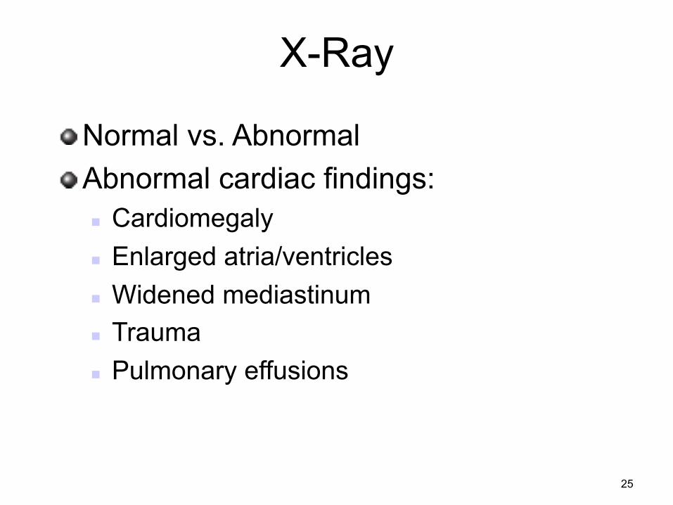
X-Ray
! Normal vs. Abnormal ! Abnormal cardiac findings:
n Cardiomegaly n Enlarged atria/ventricles n Widened mediastinum n Trauma n Pulmonary effusions
25
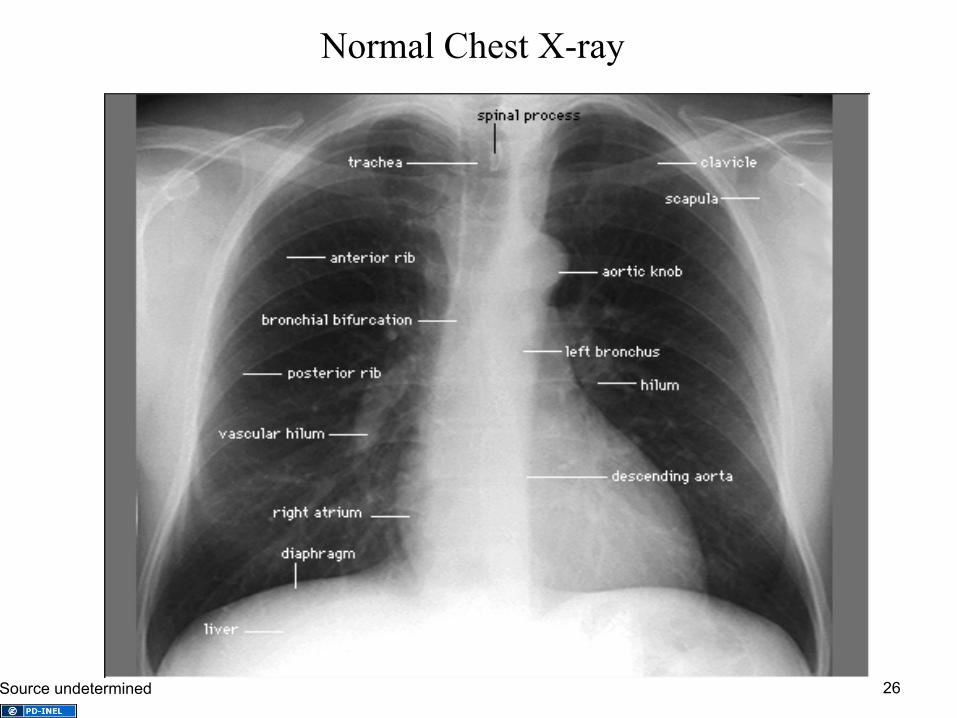
Normal Chest X-ray
26 Source undetermined
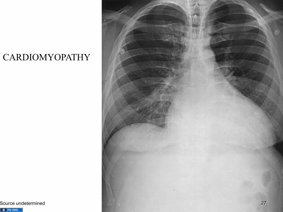
CARDIOMYOPATHY
27 Source undetermined
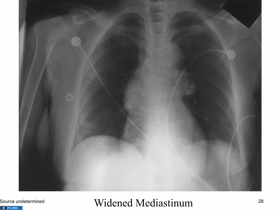
Widened Mediastinum 28 Source undetermined
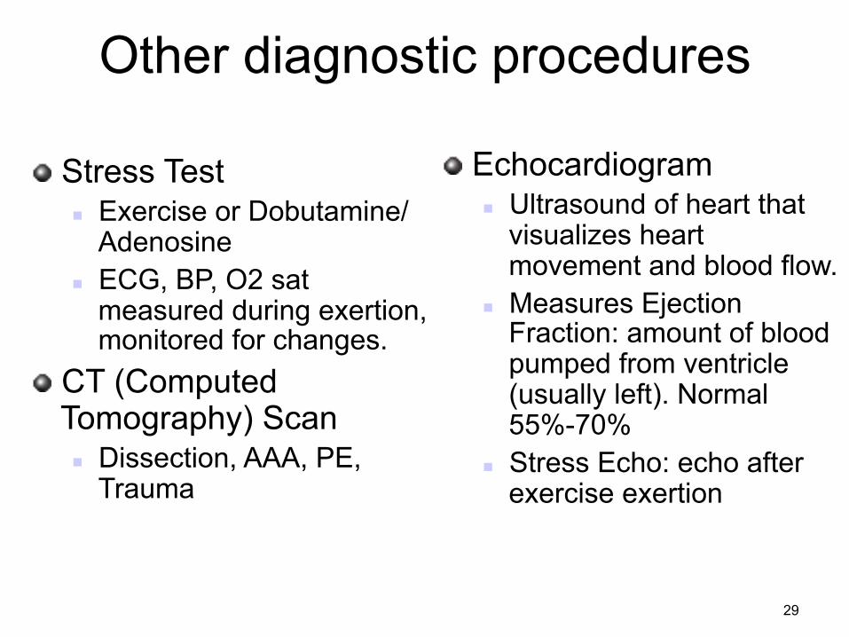
Other diagnostic procedures
! Stress Test n Exercise or Dobutamine/
Adenosine n ECG, BP, O2 sat
measured during exertion, monitored for changes.
! CT (Computed Tomography) Scan n Dissection, AAA, PE,
Trauma
! Echocardiogram n Ultrasound of heart that
visualizes heart movement and blood flow.
n Measures Ejection Fraction: amount of blood pumped from ventricle (usually left). Normal 55%-70%
n Stress Echo: echo after exercise exertion
29
Cardiovascular Nursing Diagnoses
& Collaborative Problems
30
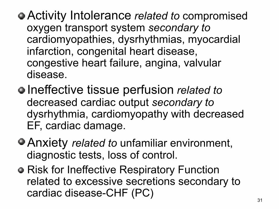
! Activity Intolerance related to compromised oxygen transport system secondary to cardiomyopathies, dysrhythmias, myocardial infarction, congenital heart disease, congestive heart failure, angina, valvular disease.
! Ineffective tissue perfusion related to decreased cardiac output secondary to dysrhythmia, cardiomyopathy with decreased EF, cardiac damage.
! Anxiety related to unfamiliar environment, diagnostic tests, loss of control.
! Risk for Ineffective Respiratory Function related to excessive secretions secondary to cardiac disease-CHF (PC)
31
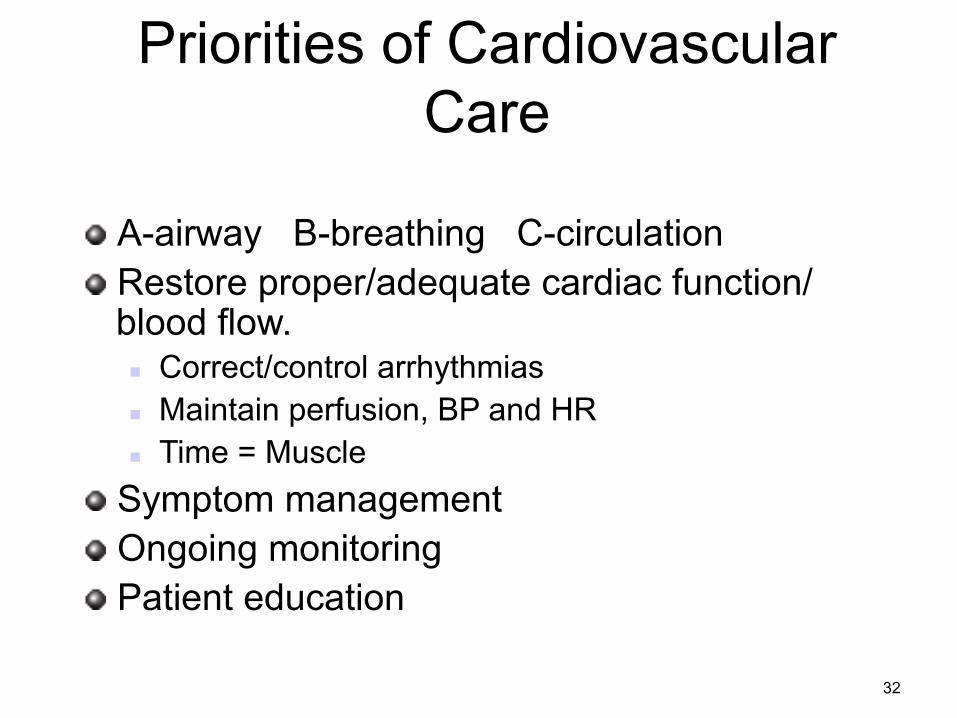
Priorities of Cardiovascular Care
! A-airway B-breathing C-circulation ! Restore proper/adequate cardiac function/
blood flow. n Correct/control arrhythmias n Maintain perfusion, BP and HR n Time = Muscle
! Symptom management ! Ongoing monitoring ! Patient education
32
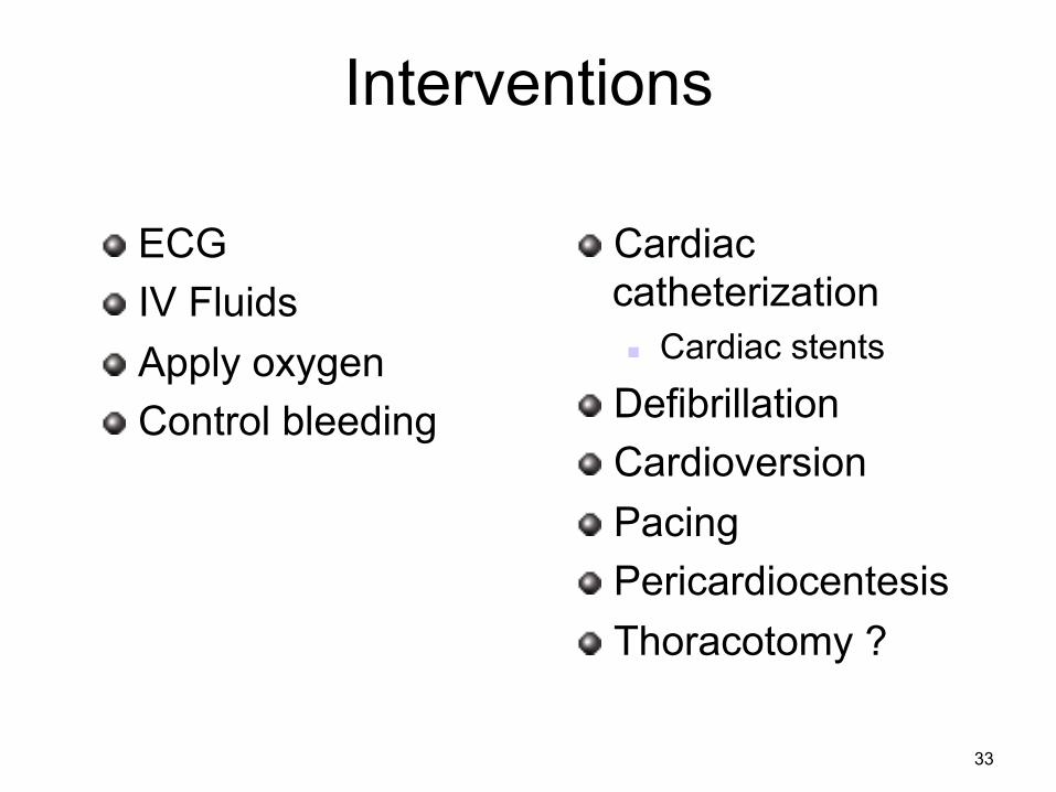
Interventions
! Cardiac catheterization n Cardiac stents
! Defibrillation ! Cardioversion ! Pacing ! Pericardiocentesis ! Thoracotomy ?
! ECG ! IV Fluids ! Apply oxygen ! Control bleeding
33
Medications
34 John Baker, Wikimedia Commons
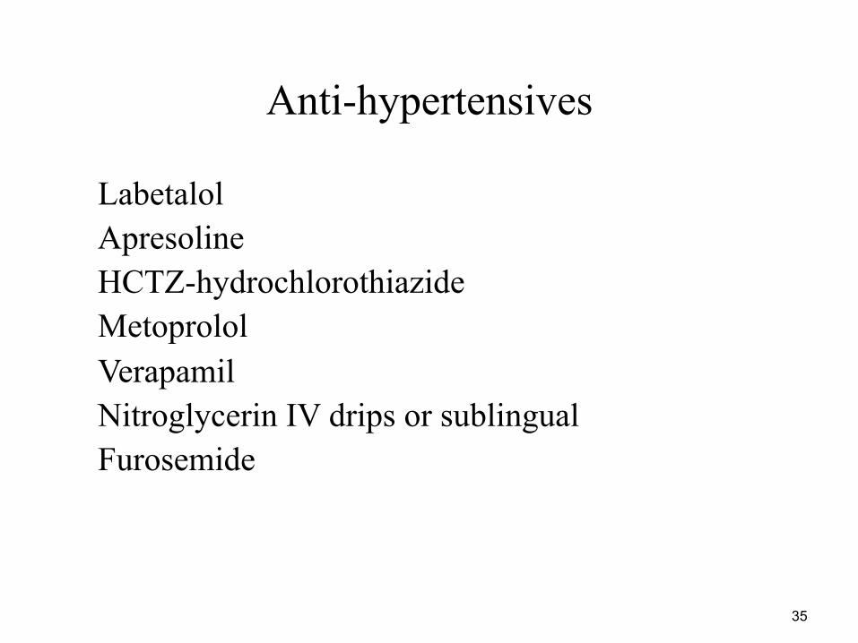
Anti-hypertensives • Labetalol • Apresoline • HCTZ-hydrochlorothiazide • Metoprolol • Verapamil • Nitroglycerin IV drips or sublingual • Furosemide
35
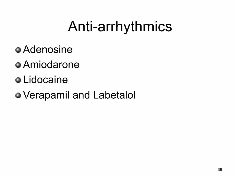
Anti-arrhythmics ! Adenosine ! Amiodarone ! Lidocaine ! Verapamil and Labetalol
36
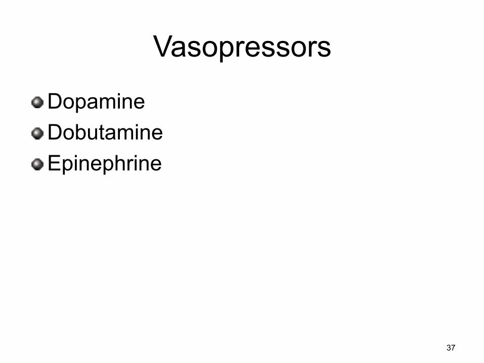
Vasopressors
! Dopamine ! Dobutamine ! Epinephrine
37
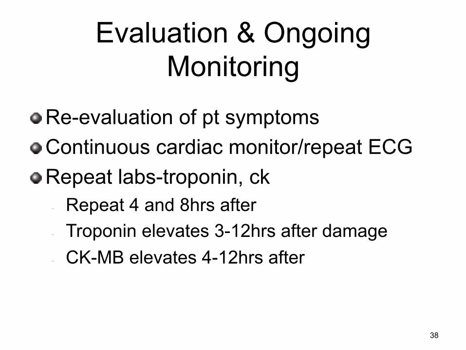
Evaluation & Ongoing Monitoring
! Re-evaluation of pt symptoms ! Continuous cardiac monitor/repeat ECG ! Repeat labs-troponin, ck
- Repeat 4 and 8hrs after - Troponin elevates 3-12hrs after damage - CK-MB elevates 4-12hrs after
38
Documentation ! Vital signs ! Cardiac Rhythm ! Airway/Airway adjuncts ! Pain score! ! Interventions ! Pt tolerance of interventions ! Pt condition
39
Documentation Example:
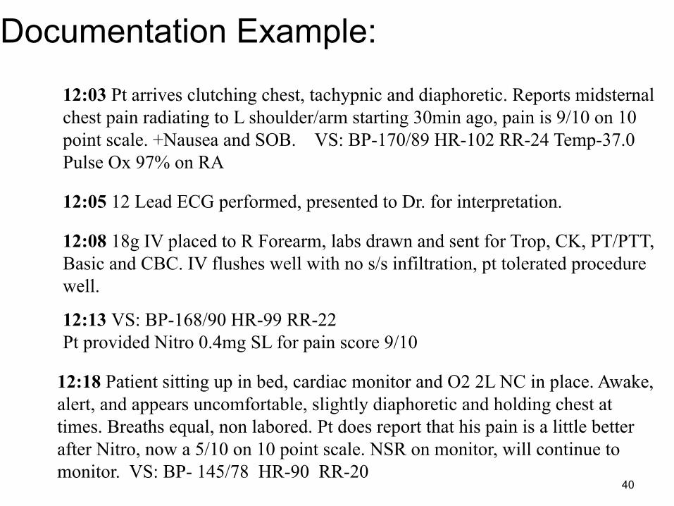
12:18 Patient sitting up in bed, cardiac monitor and O2 2L NC in place. Awake, alert, and appears uncomfortable, slightly diaphoretic and holding chest at times. Breaths equal, non labored. Pt does report that his pain is a little better after Nitro, now a 5/10 on 10 point scale. NSR on monitor, will continue to monitor. VS: BP- 145/78 HR-90 RR-20
12:03 Pt arrives clutching chest, tachypnic and diaphoretic. Reports midsternal chest pain radiating to L shoulder/arm starting 30min ago, pain is 9/10 on 10 point scale. +Nausea and SOB. VS: BP-170/89 HR-102 RR-24 Temp-37.0 Pulse Ox 97% on RA
12:05 12 Lead ECG performed, presented to Dr. for interpretation.
12:08 18g IV placed to R Forearm, labs drawn and sent for Trop, CK, PT/PTT, Basic and CBC. IV flushes well with no s/s infiltration, pt tolerated procedure well.
12:13 VS: BP-168/90 HR-99 RR-22 Pt provided Nitro 0.4mg SL for pain score 9/10
40
Patient Education ! Healthy diet and exercise ! Know your risk factors ! Know your body
n Chest pain, difficulty breathing, pain/numbness/tingling down L arm, jaw pain, palpitations or racing heart, dizziness, nausea/vomiting, fatigue, sweating.
41
Age Related Considerations
Jessicafm, Flickr 42
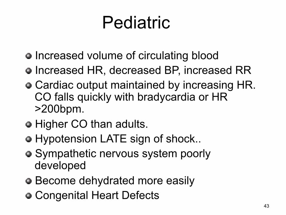
Pediatric
! Increased volume of circulating blood ! Increased HR, decreased BP, increased RR ! Cardiac output maintained by increasing HR.
CO falls quickly with bradycardia or HR >200bpm.
! Higher CO than adults. ! Hypotension LATE sign of shock.. ! Sympathetic nervous system poorly
developed ! Become dehydrated more easily ! Congenital Heart Defects
43
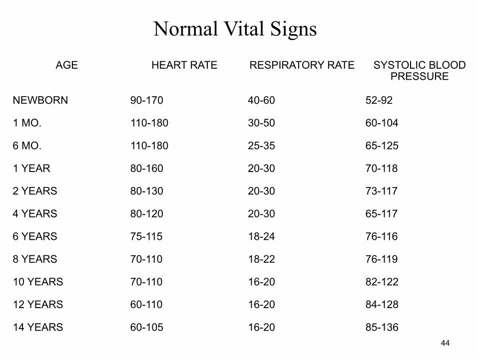
AGE HEART RATE RESPIRATORY RATE SYSTOLIC BLOOD PRESSURE
NEWBORN 90-170 40-60 52-92
1 MO. 110-180 30-50 60-104
6 MO. 110-180 25-35 65-125
1 YEAR 80-160 20-30 70-118
2 YEARS 80-130 20-30 73-117
4 YEARS 80-120 20-30 65-117
6 YEARS 75-115 18-24 76-116
8 YEARS 70-110 18-22 76-119
10 YEARS 70-110 16-20 82-122
12 YEARS 60-110 16-20 84-128
14 YEARS 60-105 16-20 85-136
Normal Vital Signs
44
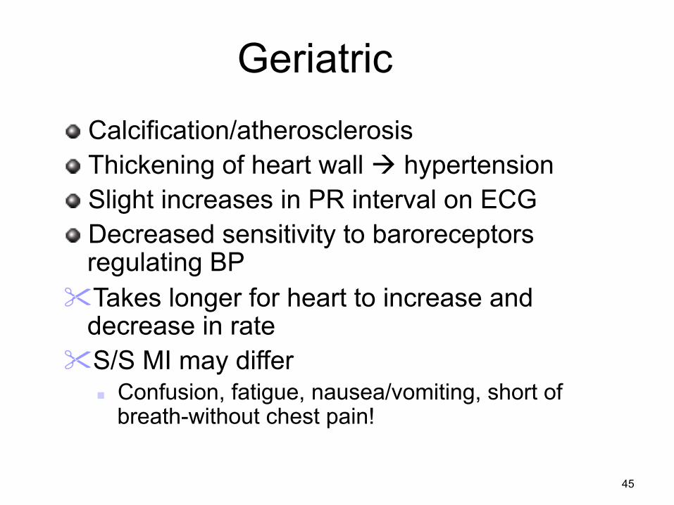
Geriatric ! Calcification/atherosclerosis ! Thickening of heart wall à hypertension ! Slight increases in PR interval on ECG ! Decreased sensitivity to baroreceptors
regulating BP " Takes longer for heart to increase and
decrease in rate " S/S MI may differ
n Confusion, fatigue, nausea/vomiting, short of breath-without chest pain!
45