Embed Size (px)
Citation preview
DNA Recombination Mechanisms
Recombination
Present in prokaryotic and eukaryotic cells
Only poorly understood
Why do chromosomes undergo recombination?
1. include roles in specialized DNA repair systems,
2. specialized activities in DNA replication,
3. regulation of expression of certain genes,
4. facilitation of proper chromosome segregation during
eukaryotic cell division,
5. maintenance of genetic diversity,
6. and implementation of programmed genetic
rearrangements during embryonic development.
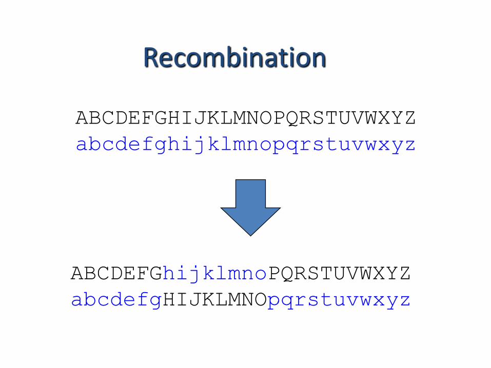
Recombination
ABCDEFGhijklmnoPQRSTUVWXYZ
abcdefgHIJKLMNOpqrstuvwxyz
ABCDEFGHIJKLMNOPQRSTUVWXYZ
abcdefghijklmnopqrstuvwxyz
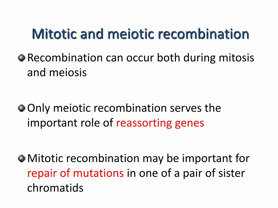
Mitotic and meiotic recombination
Recombination can occur both during mitosis and meiosis
Only meiotic recombination serves the important role of reassorting genes
Mitotic recombination may be important for repair of mutations in one of a pair of sister chromatids
Recombination mechanisms
Best studied in yeast, bacteria and phage
Recombination is mediated by the breakage and joining of DNA strands
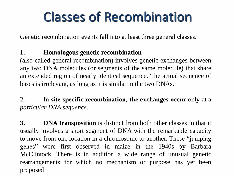
Genetic recombination events fall into at least three general classes.
1. Homologous genetic recombination
(also called general recombination) involves genetic exchanges between
any two DNA molecules (or segments of the same molecule) that share
an extended region of nearly identical sequence. The actual sequence of
bases is irrelevant, as long as it is similar in the two DNAs.
2. In site-specific recombination, the exchanges occur only at a
particular DNA sequence.
3. DNA transposition is distinct from both other classes in that it
usually involves a short segment of DNA with the remarkable capacity
to move from one location in a chromosome to another. These “jumping
genes” were first observed in maize in the 1940s by Barbara
McClintock. There is in addition a wide range of unusual genetic
rearrangements for which no mechanism or purpose has yet been
proposed
Classes of Recombination
Homologous recombination thus serves at least three
identifiable functions:
(1) it contributes to the repair of several types of DNA
damage;
(2) it provides, in eukaryotic cells, a transient physical
link between chromatids that promotes the orderly
segregation of chromosomes at the first meiotic cell
division; and
(3) it enhances genetic diversity in a population.
1. Homologous Recombination
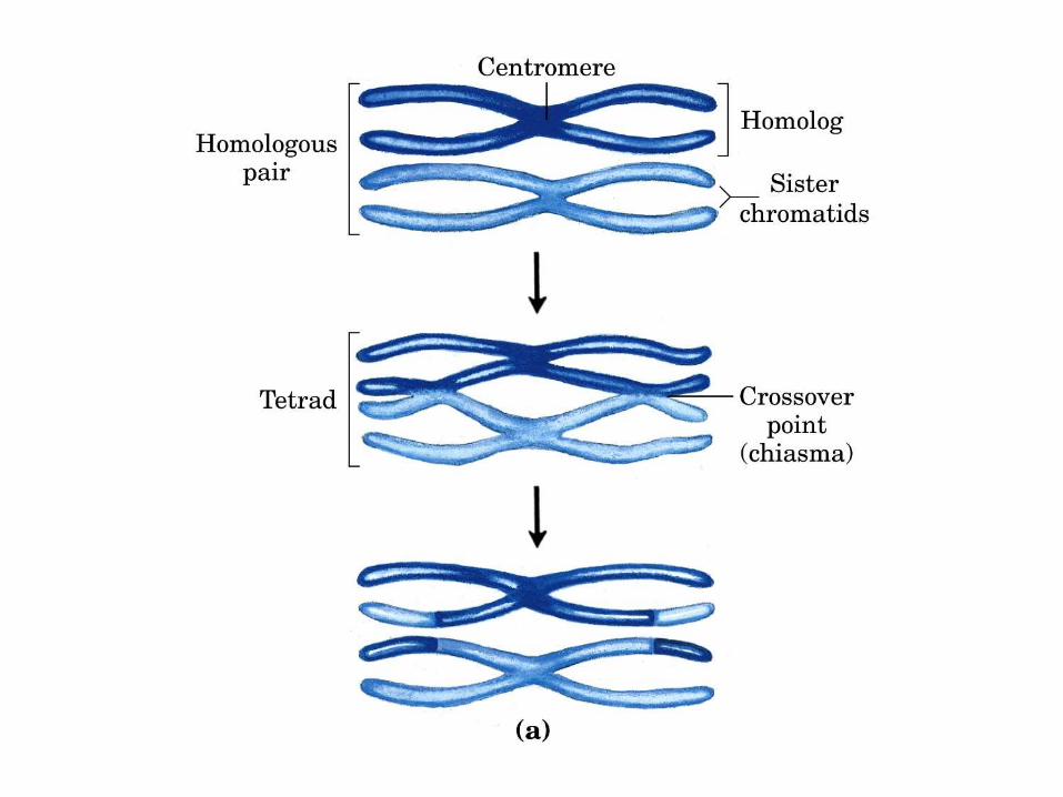
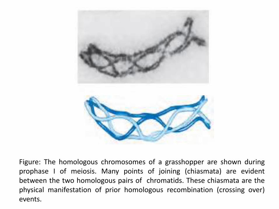
Figure: The homologous chromosomes of a grasshopper are shown duringprophase I of meiosis. Many points of joining (chiasmata) are evidentbetween the two homologous pairs of chromatids. These chiasmata are thephysical manifestation of prior homologous recombination (crossing over)events.
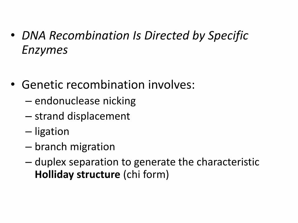
• DNA Recombination Is Directed by Specific Enzymes
• Genetic recombination involves:– endonuclease nicking
– strand displacement
– ligation
– branch migration
– duplex separation to generate the characteristic Holliday structure (chi form)
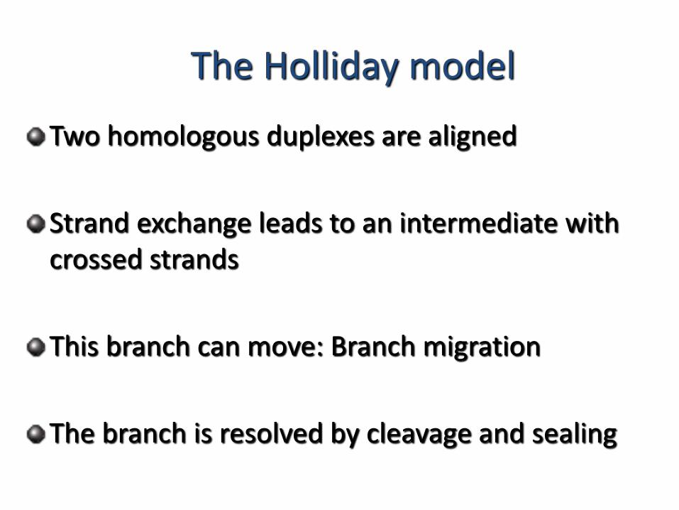
The Holliday model
Two homologous duplexes are aligned
Strand exchange leads to an intermediate with crossed strands
This branch can move: Branch migration
The branch is resolved by cleavage and sealing
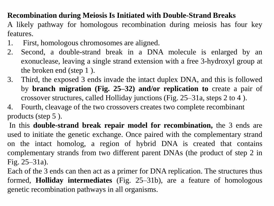
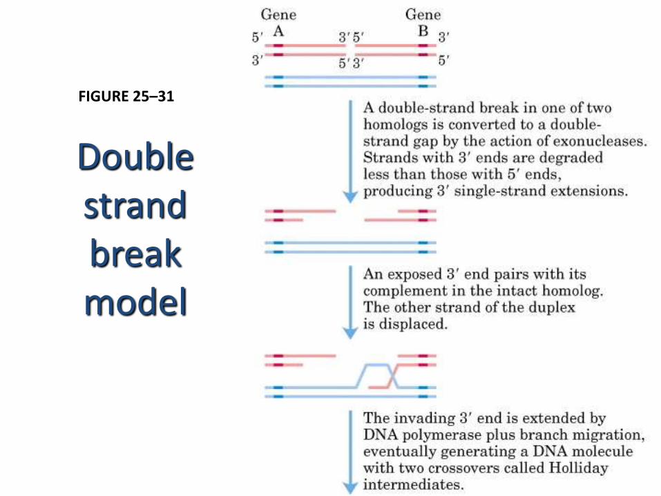
Recombination during Meiosis Is Initiated with Double-Strand Breaks
A likely pathway for homologous recombination during meiosis has four key
features.
1. First, homologous chromosomes are aligned.
2. Second, a double-strand break in a DNA molecule is enlarged by an
exonuclease, leaving a single strand extension with a free 3-hydroxyl group at
the broken end (step 1 ).
3. Third, the exposed 3 ends invade the intact duplex DNA, and this is followed
by branch migration (Fig. 25–32) and/or replication to create a pair of
crossover structures, called Holliday junctions (Fig. 25–31a, steps 2 to 4 ).
4. Fourth, cleavage of the two crossovers creates two complete recombinant
products (step 5 ).
In this double-strand break repair model for recombination, the 3 ends are
used to initiate the genetic exchange. Once paired with the complementary strand
on the intact homolog, a region of hybrid DNA is created that contains
complementary strands from two different parent DNAs (the product of step 2 in
Fig. 25–31a).
Each of the 3 ends can then act as a primer for DNA replication. The structures thus
formed, Holliday intermediates (Fig. 25–31b), are a feature of homologous
genetic recombination pathways in all organisms.
Double strand break model
FIGURE 25–31
Double strand break model
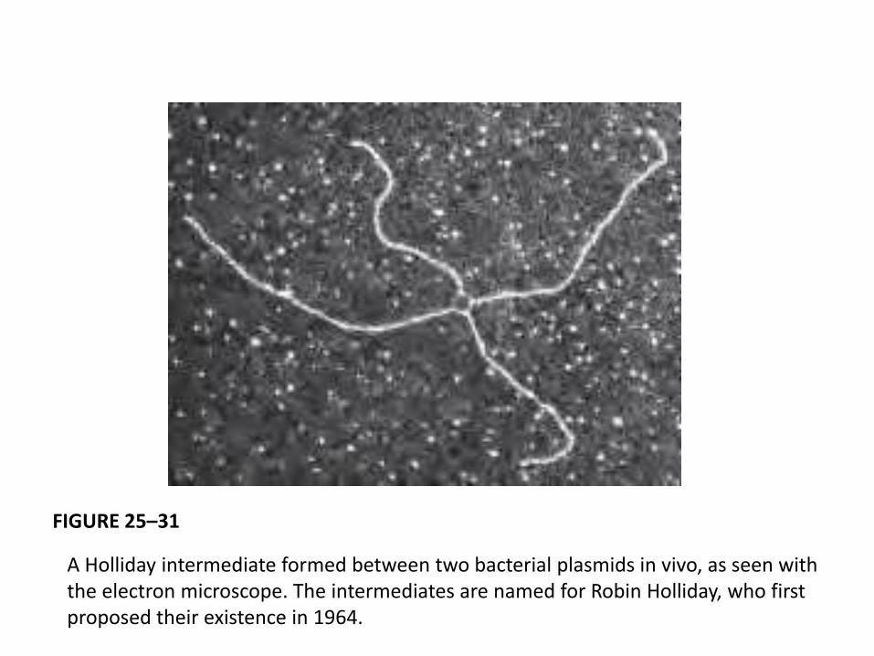
A Holliday intermediate formed between two bacterial plasmids in vivo, as seen with the electron microscope. The intermediates are named for Robin Holliday, who first proposed their existence in 1964.
FIGURE 25–31
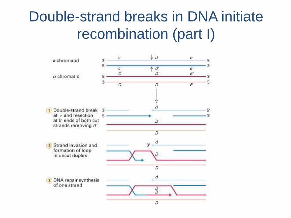
Double-strand breaks in DNA initiate
recombination (part I)
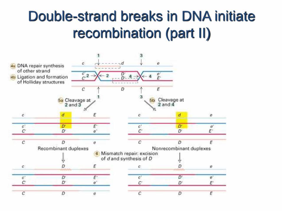
Double-strand breaks in DNA initiate
recombination (part II)
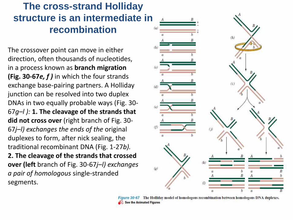
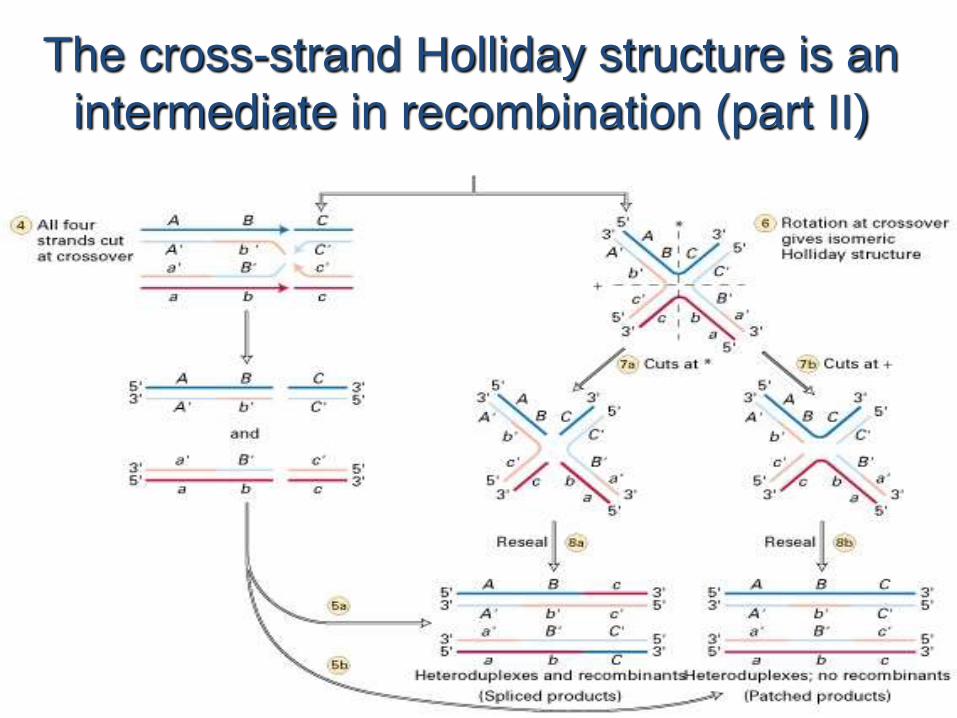
The cross-strand Holliday
structure is an intermediate in
recombination
The crossover point can move in either direction, often thousands of nucleotides, in a process known as branch migration (Fig. 30-67e, f ) in which the four strandsexchange base-pairing partners. A Holliday junction can be resolved into two duplexDNAs in two equally probable ways (Fig. 30-67g–l ): 1. The cleavage of the strands that did not cross over (right branch of Fig. 30-67j–l) exchanges the ends of the original duplexes to form, after nick sealing, the traditional recombinant DNA (Fig. 1-27b).2. The cleavage of the strands that crossed over (left branch of Fig. 30-67j–l) exchanges a pair of homologous single-stranded segments.
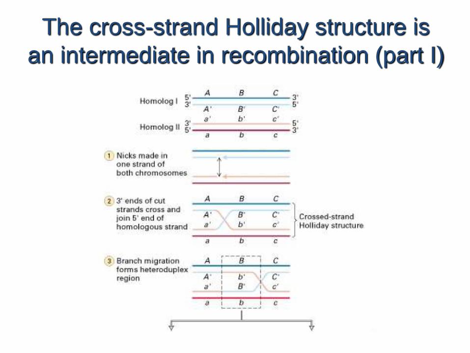
The cross-strand Holliday structure is
an intermediate in recombination (part I)
The cross-strand Holliday structure is an
intermediate in recombination (part II)
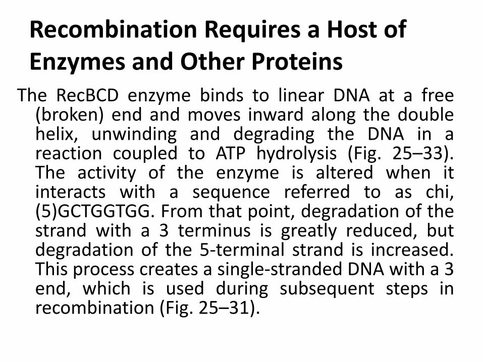
Recombination Requires a Host of Enzymes and Other Proteins
The RecBCD enzyme binds to linear DNA at a free(broken) end and moves inward along the doublehelix, unwinding and degrading the DNA in areaction coupled to ATP hydrolysis (Fig. 25–33).The activity of the enzyme is altered when itinteracts with a sequence referred to as chi,(5)GCTGGTGG. From that point, degradation of thestrand with a 3 terminus is greatly reduced, butdegradation of the 5-terminal strand is increased.This process creates a single-stranded DNA with a 3end, which is used during subsequent steps inrecombination (Fig. 25–31).
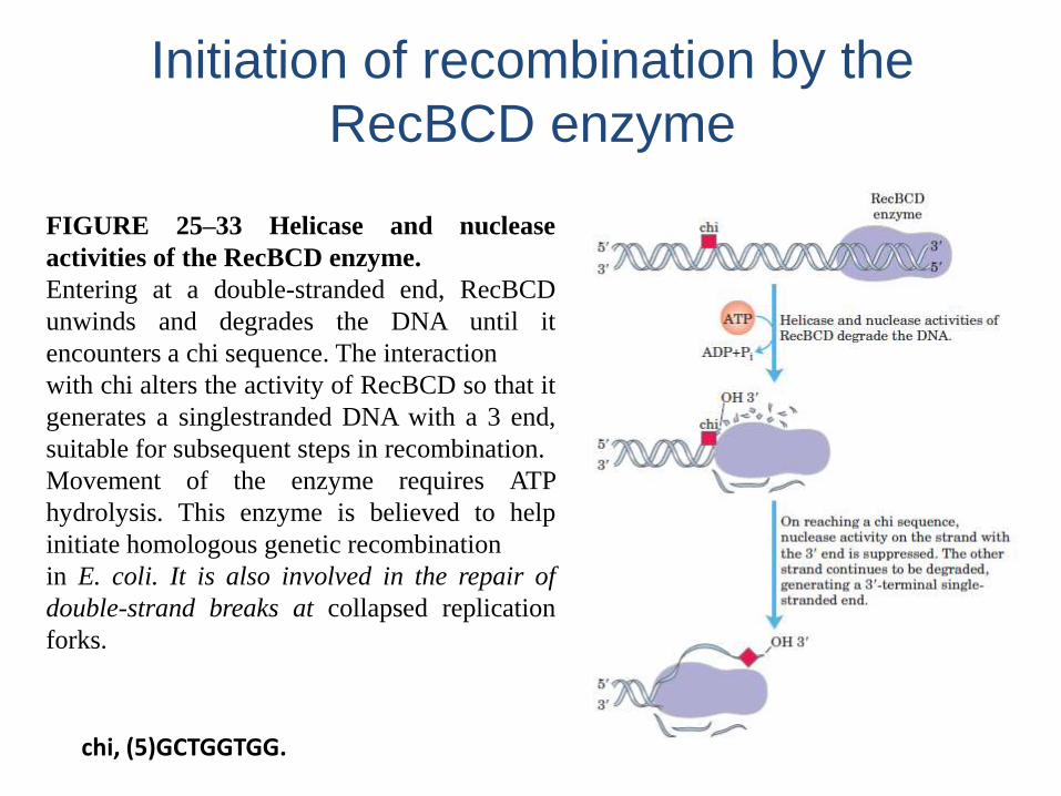
Initiation of recombination by the
RecBCD enzyme
chi, (5)GCTGGTGG.
FIGURE 25–33 Helicase and nuclease
activities of the RecBCD enzyme.
Entering at a double-stranded end, RecBCD
unwinds and degrades the DNA until it
encounters a chi sequence. The interaction
with chi alters the activity of RecBCD so that it
generates a singlestranded DNA with a 3 end,
suitable for subsequent steps in recombination.
Movement of the enzyme requires ATP
hydrolysis. This enzyme is believed to help
initiate homologous genetic recombination
in E. coli. It is also involved in the repair of
double-strand breaks at collapsed replication
forks.
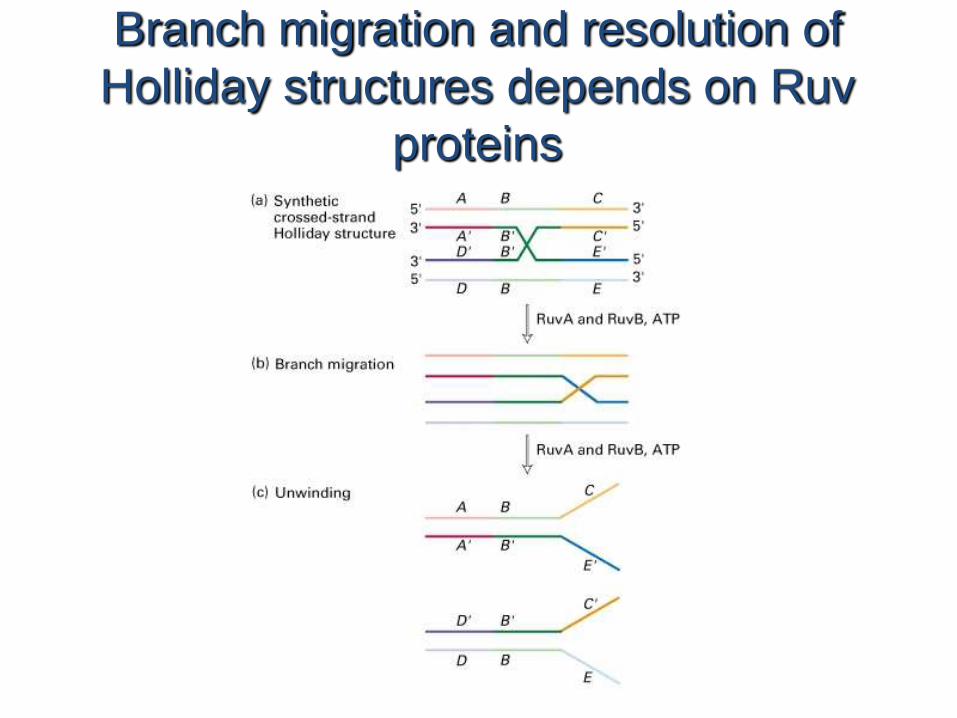
Branch migration and resolution of
Holliday structures depends on Ruv
proteins
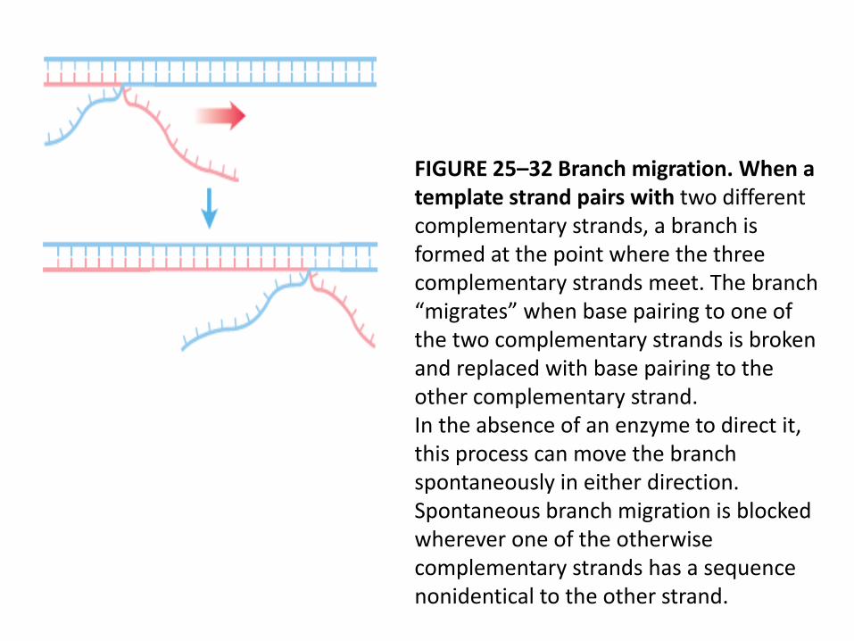
FIGURE 25–32 Branch migration. When a template strand pairs with two different complementary strands, a branch is formed at the point where the three complementary strands meet. The branch “migrates” when base pairing to one of the two complementary strands is broken and replaced with base pairing to the other complementary strand.In the absence of an enzyme to direct it, this process can move the branch spontaneously in either direction. Spontaneous branch migration is blocked wherever one of the otherwise complementary strands has a sequence nonidentical to the other strand.
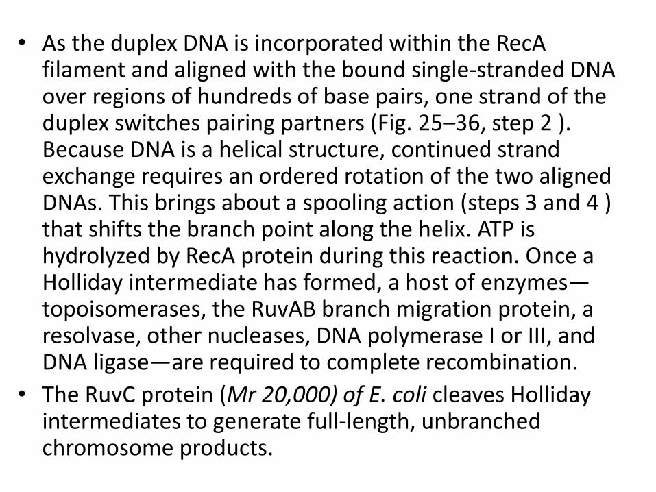
• As the duplex DNA is incorporated within the RecAfilament and aligned with the bound single-stranded DNA over regions of hundreds of base pairs, one strand of the duplex switches pairing partners (Fig. 25–36, step 2 ). Because DNA is a helical structure, continued strand exchange requires an ordered rotation of the two aligned DNAs. This brings about a spooling action (steps 3 and 4 ) that shifts the branch point along the helix. ATP is hydrolyzed by RecA protein during this reaction. Once a Holliday intermediate has formed, a host of enzymes—topoisomerases, the RuvAB branch migration protein, a resolvase, other nucleases, DNA polymerase I or III, and DNA ligase—are required to complete recombination.
• The RuvC protein (Mr 20,000) of E. coli cleaves Holliday intermediates to generate full-length, unbranchedchromosome products.
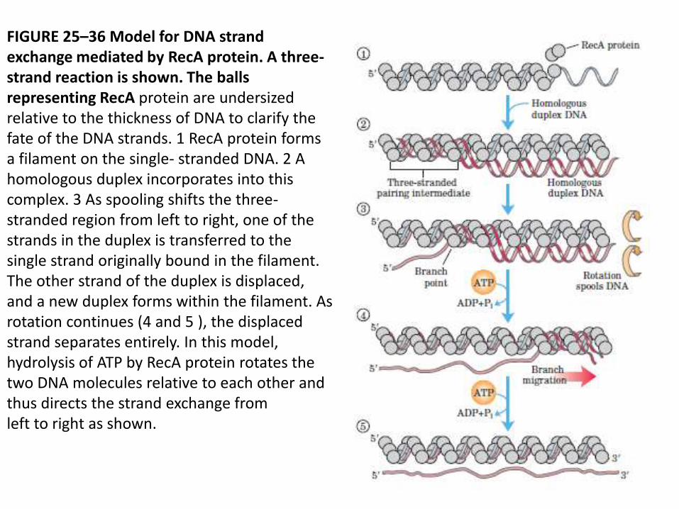
FIGURE 25–36 Model for DNA strand exchange mediated by RecA protein. A three-strand reaction is shown. The balls representing RecA protein are undersized relative to the thickness of DNA to clarify thefate of the DNA strands. 1 RecA protein forms a filament on the single- stranded DNA. 2 A homologous duplex incorporates into thiscomplex. 3 As spooling shifts the three-stranded region from left to right, one of the strands in the duplex is transferred to the single strand originally bound in the filament. The other strand of the duplex is displaced,and a new duplex forms within the filament. As rotation continues (4 and 5 ), the displaced strand separates entirely. In this model, hydrolysis of ATP by RecA protein rotates the two DNA molecules relative to each other and thus directs the strand exchange fromleft to right as shown.
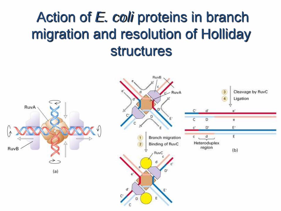
Action of E. coli proteins in branch
migration and resolution of Holliday
structures
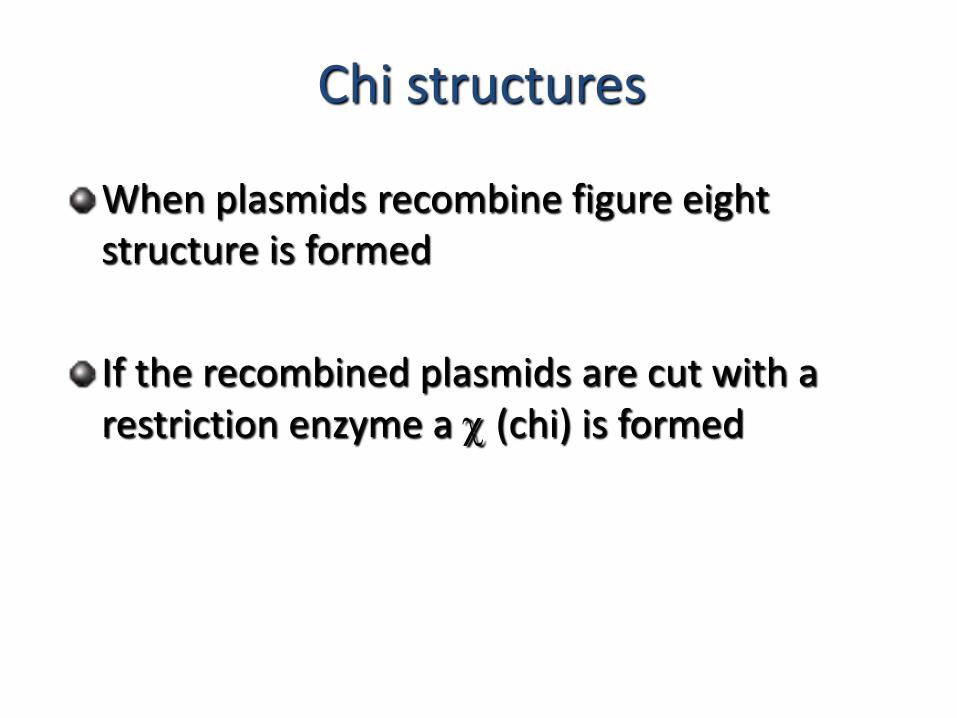
Chi structures
When plasmids recombine figure eight structure is formed
If the recombined plasmids are cut with a restriction enzyme a c (chi) is formed
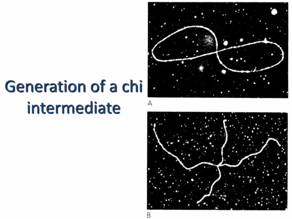
Generation of a chi intermediate
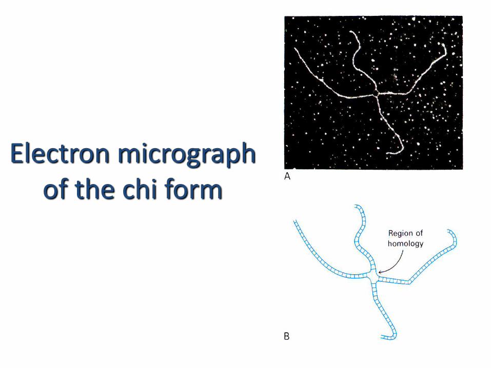
Electron micrograph of the chi form
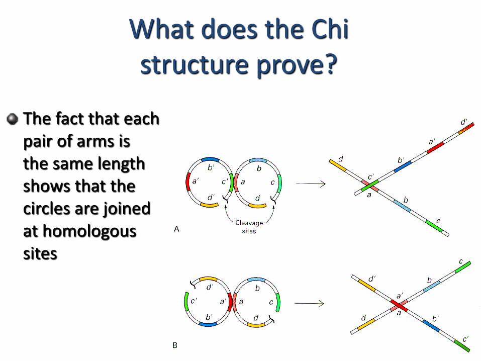
What does the Chi structure prove?
The fact that each pair of arms is the same length shows that the circles are joined at homologous sites
Recombination between homologous
DNA sites
Recombination provides a means by which a genome can
change to generate new combinations of genes
Homologous recombination allows for the exchange of blocks
of genes between homologous chromosomes and thereby is
a mechanism for generating genetic diversity
Recombination occurs randomly between two homologous
sequences and the frequency of recombination between two
sites is proportional to the distance between the sites
2. Site specific recombination
Viruses and transposable elements often integrate their genomes into the host chromosome
Site specific recombination is used by both eukaryotes and prokaryotes to regulate gene expression and to increase the organisms genetic range
Site specific recombination
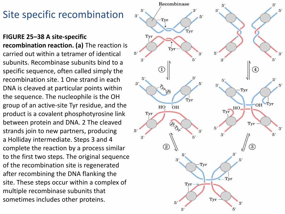
FIGURE 25–38 A site-specific recombination reaction. (a) The reaction is carried out within a tetramer of identical subunits. Recombinase subunits bind to a specific sequence, often called simply the recombination site. 1 One strand in each DNA is cleaved at particular points within the sequence. The nucleophile is the OH group of an active-site Tyr residue, and the product is a covalent phosphotyrosine link between protein and DNA. 2 The cleaved strands join to new partners, producinga Holliday intermediate. Steps 3 and 4 complete the reaction by a process similar to the first two steps. The original sequenceof the recombination site is regenerated after recombining the DNA flanking the site. These steps occur within a complex of multiple recombinase subunits that sometimes includes other proteins.
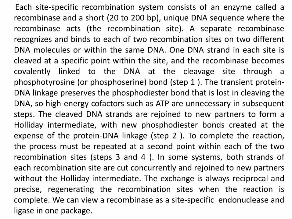
Each site-specific recombination system consists of an enzyme called arecombinase and a short (20 to 200 bp), unique DNA sequence where therecombinase acts (the recombination site). A separate recombinaserecognizes and binds to each of two recombination sites on two differentDNA molecules or within the same DNA. One DNA strand in each site iscleaved at a specific point within the site, and the recombinase becomescovalently linked to the DNA at the cleavage site through aphosphotyrosine (or phosphoserine) bond (step 1 ). The transient protein-DNA linkage preserves the phosphodiester bond that is lost in cleaving theDNA, so high-energy cofactors such as ATP are unnecessary in subsequentsteps. The cleaved DNA strands are rejoined to new partners to form aHolliday intermediate, with new phosphodiester bonds created at theexpense of the protein-DNA linkage (step 2 ). To complete the reaction,the process must be repeated at a second point within each of the tworecombination sites (steps 3 and 4 ). In some systems, both strands ofeach recombination site are cut concurrently and rejoined to new partnerswithout the Holliday intermediate. The exchange is always reciprocal andprecise, regenerating the recombination sites when the reaction iscomplete. We can view a recombinase as a site-specific endonuclease andligase in one package.
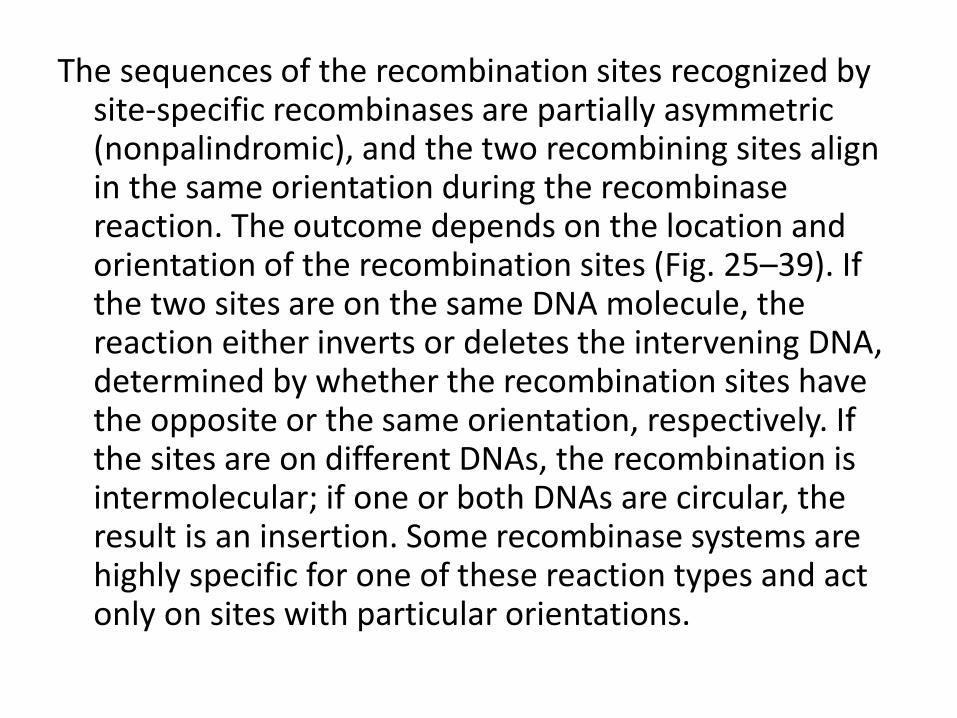
The sequences of the recombination sites recognized by site-specific recombinases are partially asymmetric (nonpalindromic), and the two recombining sites align in the same orientation during the recombinasereaction. The outcome depends on the location and orientation of the recombination sites (Fig. 25–39). If the two sites are on the same DNA molecule, the reaction either inverts or deletes the intervening DNA, determined by whether the recombination sites have the opposite or the same orientation, respectively. If the sites are on different DNAs, the recombination is intermolecular; if one or both DNAs are circular, the result is an insertion. Some recombinase systems are highly specific for one of these reaction types and act only on sites with particular orientations.
FIGURE 25–39 Effects of site-specific recombination. The outcome of site-specific
recombination depends on the location and orientation of the recombination sites (red
and green) in a double-stranded DNA molecule. Orientation here (shown by arrowheads)
refers to the order of nucleotides in the recombination site, not the 5n3 direction.
(a) Recombination sites with opposite orientation in the same DNA molecule. The
result is an inversion. (b) Recombination sites with the same orientation, either on one
DNA molecule, producing a deletion, or on two DNA molecules, producing an insertion.
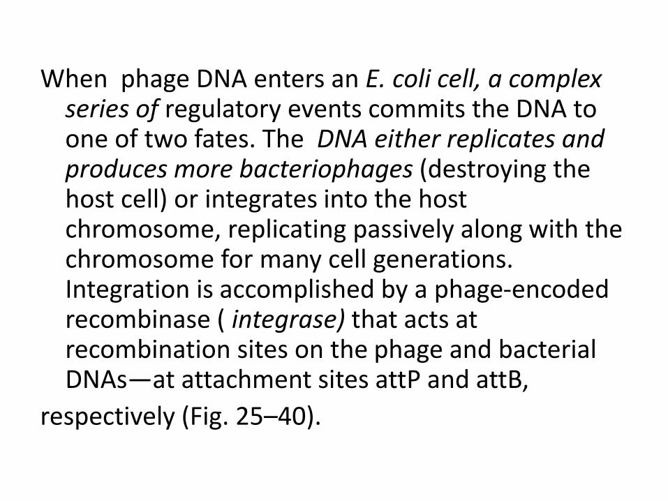
When phage DNA enters an E. coli cell, a complex series of regulatory events commits the DNA to one of two fates. The DNA either replicates and produces more bacteriophages (destroying the host cell) or integrates into the host chromosome, replicating passively along with the chromosome for many cell generations. Integration is accomplished by a phage-encoded recombinase ( integrase) that acts at recombination sites on the phage and bacterial DNAs—at attachment sites attP and attB,
respectively (Fig. 25–40).
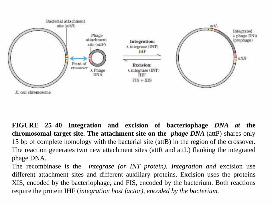
FIGURE 25–40 Integration and excision of bacteriophage DNA at the
chromosomal target site. The attachment site on the phage DNA (attP) shares only
15 bp of complete homology with the bacterial site (attB) in the region of the crossover.
The reaction generates two new attachment sites (attR and attL) flanking the integrated
phage DNA.
The recombinase is the integrase (or INT protein). Integration and excision use
different attachment sites and different auxiliary proteins. Excision uses the proteins
XIS, encoded by the bacteriophage, and FIS, encoded by the bacterium. Both reactions
require the protein IHF (integration host factor), encoded by the bacterium.
DNA Transposition
recombination that allows the movement of transposableelements, or transposons. These segments of DNA, found invirtually all cells, move, or “jump,” from one place on achromosome (the donor site) to another on the same or adifferent chromosome (the target site). DNA sequencehomology is not usually required for this movement, calledtransposition; the new location is determined more or lessrandomly. Insertion of a transposon in an essential gene couldkill the cell, so transposition is tightly regulated and usuallyvery infrequent. Transposons are perhaps the simplest ofmolecular parasites, adapted to replicate passively within thechromosomes of host cells. In some cases they carry genesthat are useful to the host cell, and thus exist in a kind ofsymbiosis with the host
Bacteria have two classes of transposons.
1. simple transposons
Insertion sequences contain only the sequences required for transposition and the
genes for proteins (transposases) that promote the process.
2. Complex transposons contain one or more genes in addition to those
needed for transposition. These extra genes might, for example, confer resistance
to antibiotics and thus enhance the survival chances of the host cell. The spread of
antibiotic-resistance elements among disease-causing bacterial populations that is
rendering some antibiotics ineffectual is mediated in part by transposition.
Bacterial transposons vary in structure, but most have short repeated sequences at
each end that serve as binding sites for the transposase. When transposition
occurs, a short sequence at the target site (5 to 10 bp) is duplicated to form an
additional short repeated sequence that flanks each end of the inserted transposon
(Fig. 25–42). These duplicated segments result from the cutting mechanism used
to insert a transposon into the DNA at a new location.
Classes of Transposons
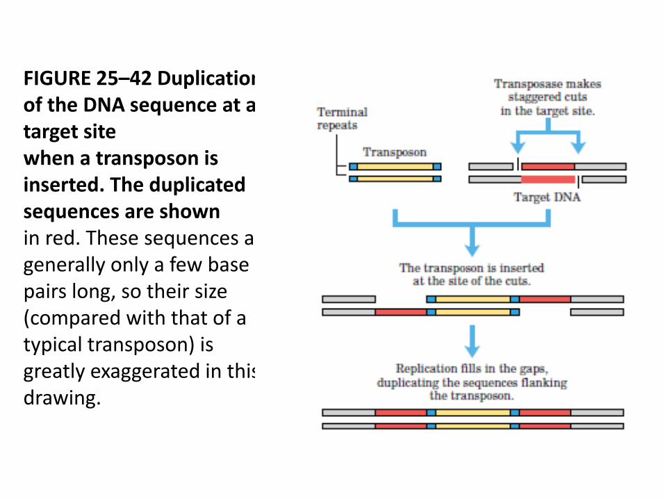
FIGURE 25–42 Duplication of the DNA sequence at a target sitewhen a transposon is inserted. The duplicated sequences are shownin red. These sequences are generally only a few base pairs long, so their size (compared with that of a typical transposon) is greatly exaggerated in this drawing.
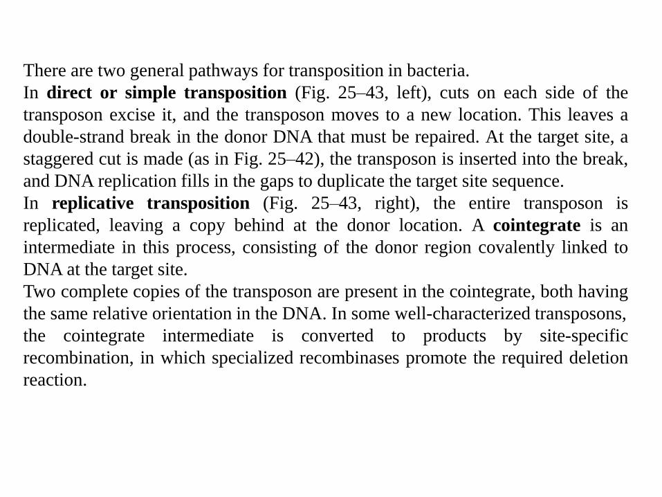
There are two general pathways for transposition in bacteria.
In direct or simple transposition (Fig. 25–43, left), cuts on each side of the
transposon excise it, and the transposon moves to a new location. This leaves a
double-strand break in the donor DNA that must be repaired. At the target site, a
staggered cut is made (as in Fig. 25–42), the transposon is inserted into the break,
and DNA replication fills in the gaps to duplicate the target site sequence.
In replicative transposition (Fig. 25–43, right), the entire transposon is
replicated, leaving a copy behind at the donor location. A cointegrate is an
intermediate in this process, consisting of the donor region covalently linked to
DNA at the target site.
Two complete copies of the transposon are present in the cointegrate, both having
the same relative orientation in the DNA. In some well-characterized transposons,
the cointegrate intermediate is converted to products by site-specific
recombination, in which specialized recombinases promote the required deletion
reaction.
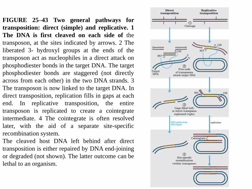
FIGURE 25–43 Two general pathways for
transposition: direct (simple) and replicative. 1
The DNA is first cleaved on each side of the
transposon, at the sites indicated by arrows. 2 The
liberated 3- hydroxyl groups at the ends of the
transposon act as nucleophiles in a direct attack on
phosphodiester bonds in the target DNA. The target
phosphodiester bonds are staggered (not directly
across from each other) in the two DNA strands. 3
The transposon is now linked to the target DNA. In
direct transposition, replication fills in gaps at each
end. In replicative transposition, the entire
transposon is replicated to create a cointegrate
intermediate. 4 The cointegrate is often resolved
later, with the aid of a separate site-specific
recombination system.
The cleaved host DNA left behind after direct
transposition is either repaired by DNA end-joining
or degraded (not shown). The latter outcome can be
lethal to an organism.