Embed Size (px)
Citation preview

Presented by:Dr. Jamal GiriResident (1st Year)
Development of Branchial
arches
Contents Introduction.
Basic embryology.
Development of Branchial arches.
Derivatives of Branchial arches.
2
Embryology
Embryo Greek word “Embryon”= unborn
The branch of biology that deals with the formation, early growth, and development of living organisms.
3

Ovum Embryo Foetus
4
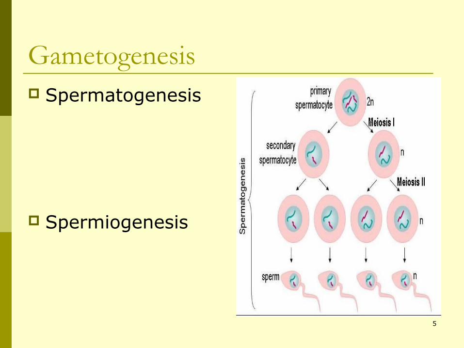
Gametogenesis Spermatogenesis
Spermiogenesis
5
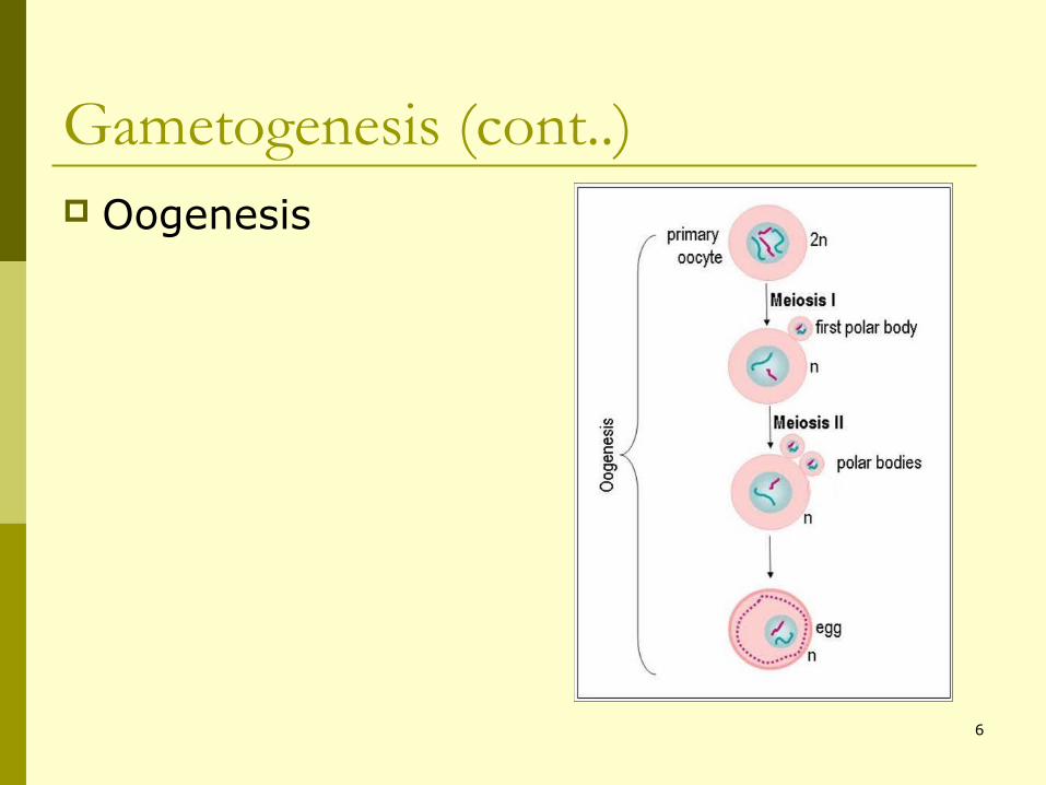
Gametogenesis (cont..) Oogenesis
6
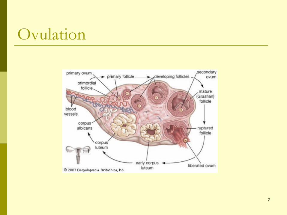
Ovulation
7
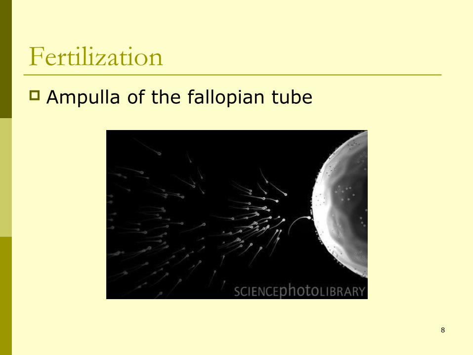
Fertilization Ampulla of the fallopian tube
8
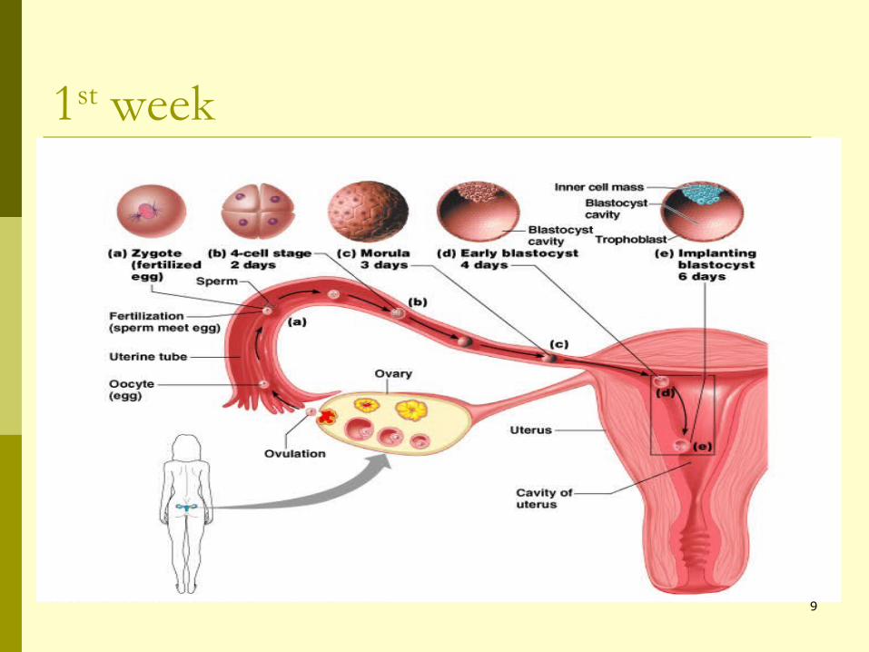
1st week
9
10
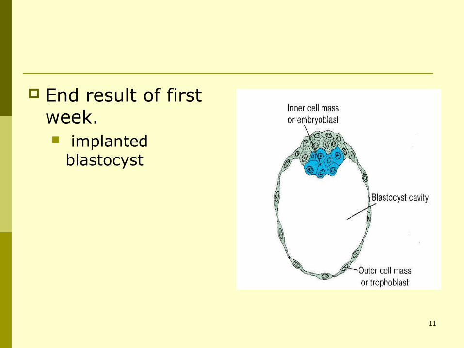
End result of first week. implanted
blastocyst
11
2nd week (week of two’s) Trophoblast differentiates into two layers:
cytotrophoblast and syncytiotrophoblast.
Embryoblast forms two layers: epiblast and hypoblast. (Bilaminar)
Formation of 2 cavities: amniotic and yolk sac cavities.
12
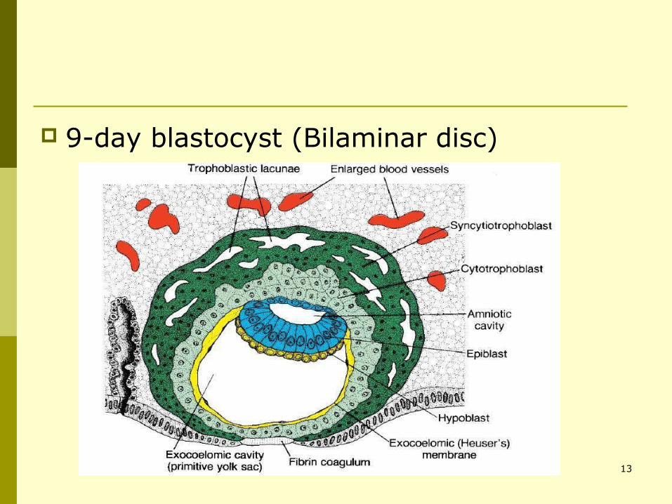
9-day blastocyst (Bilaminar disc)
13
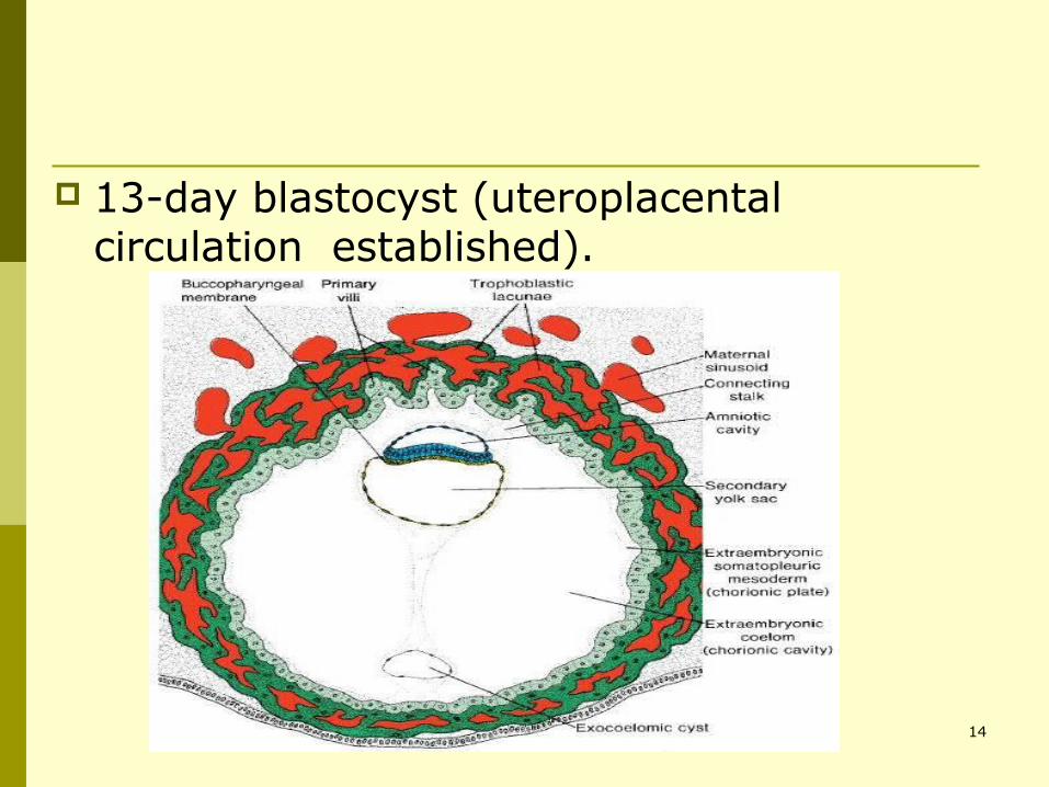
13-day blastocyst (uteroplacental circulation established).
14
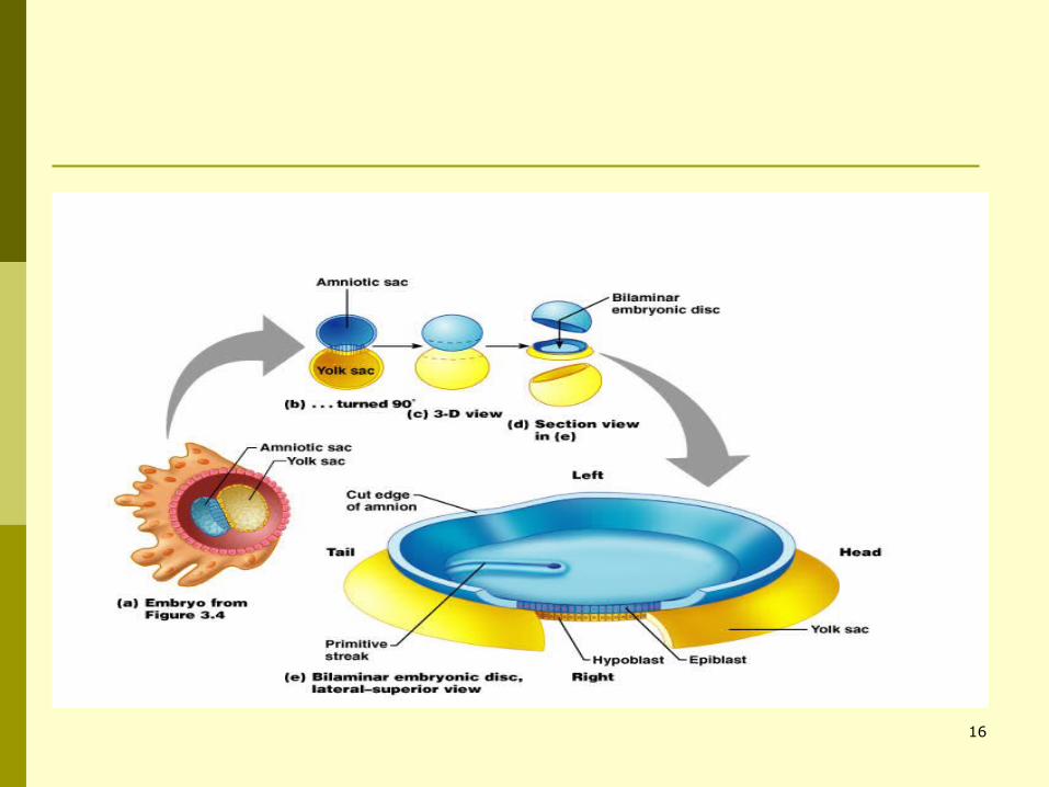
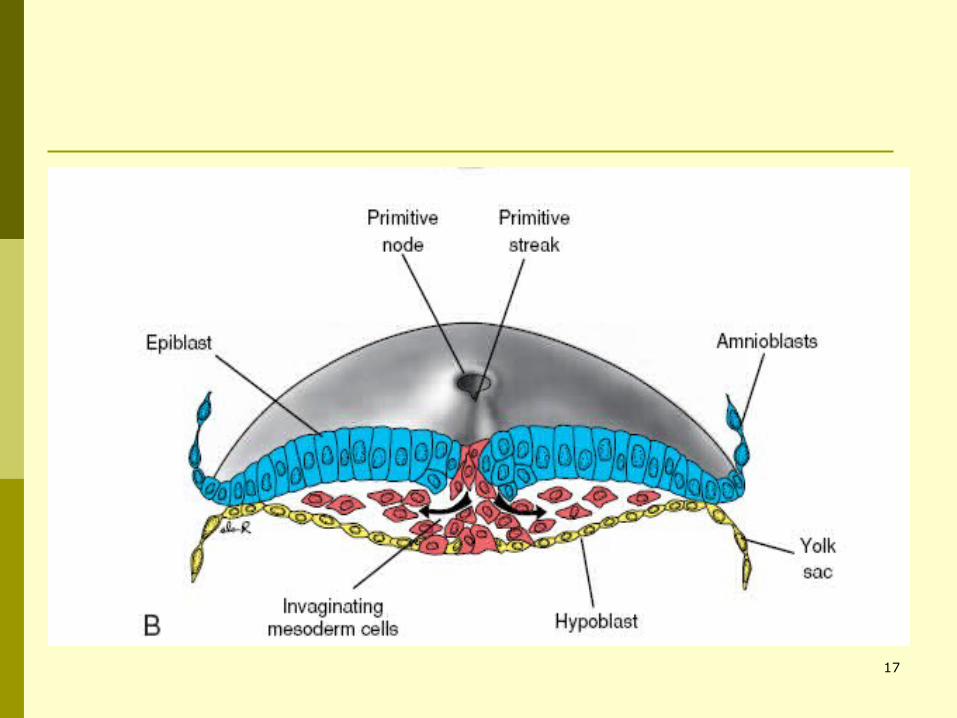
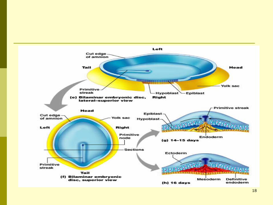
3rd week Gastrulation:
Primitive streak formation. Invasion of epiblast cells. Formation of 3 germ layers
15
16
17
18
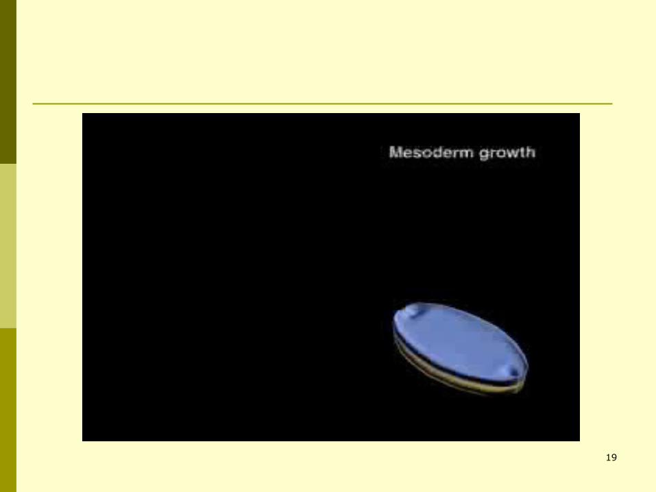
19

Which is the most important time of human life?
"It is not birth, marriage, or death, but gastrulation, which is truly the most
important time in your life."
Lewis Wolpert (1986) 20
21
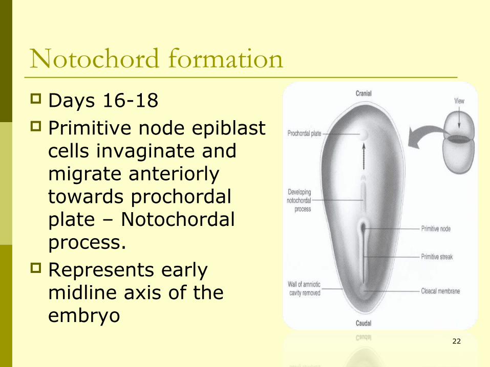
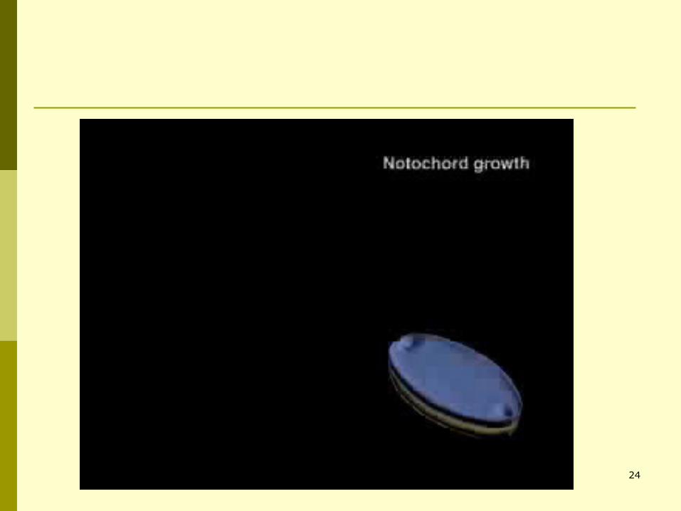
Notochord formation Days 16-18 Primitive node epiblast
cells invaginate and migrate anteriorly towards prochordal plate – Notochordal process.
Represents early midline axis of the embryo
22
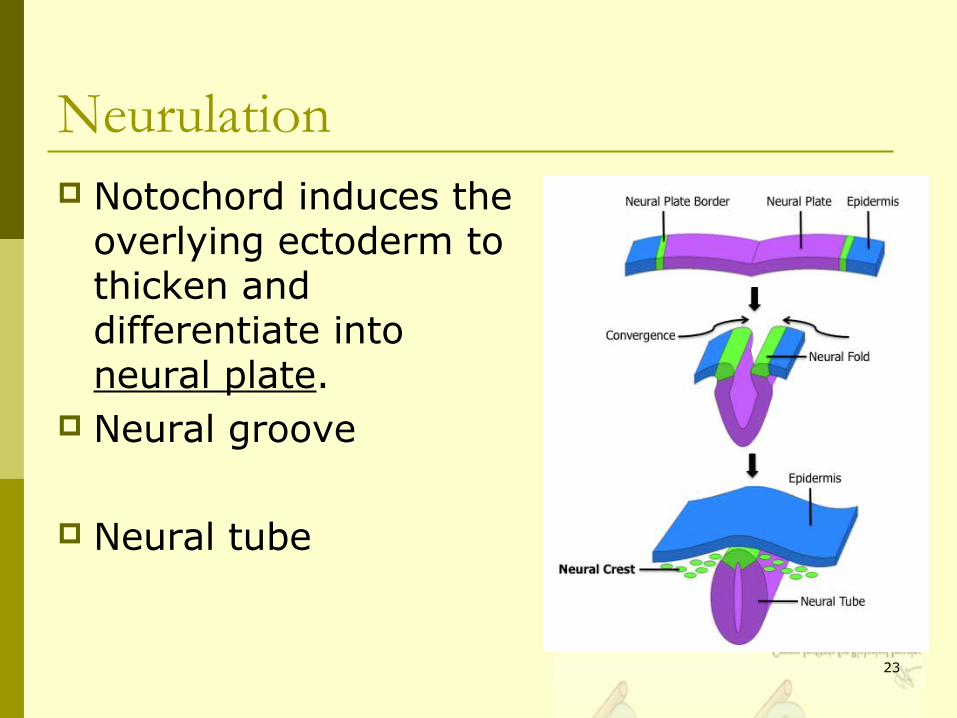
Neurulation Notochord induces the
overlying ectoderm to thicken and differentiate into neural plate.
Neural groove
Neural tube
23
24
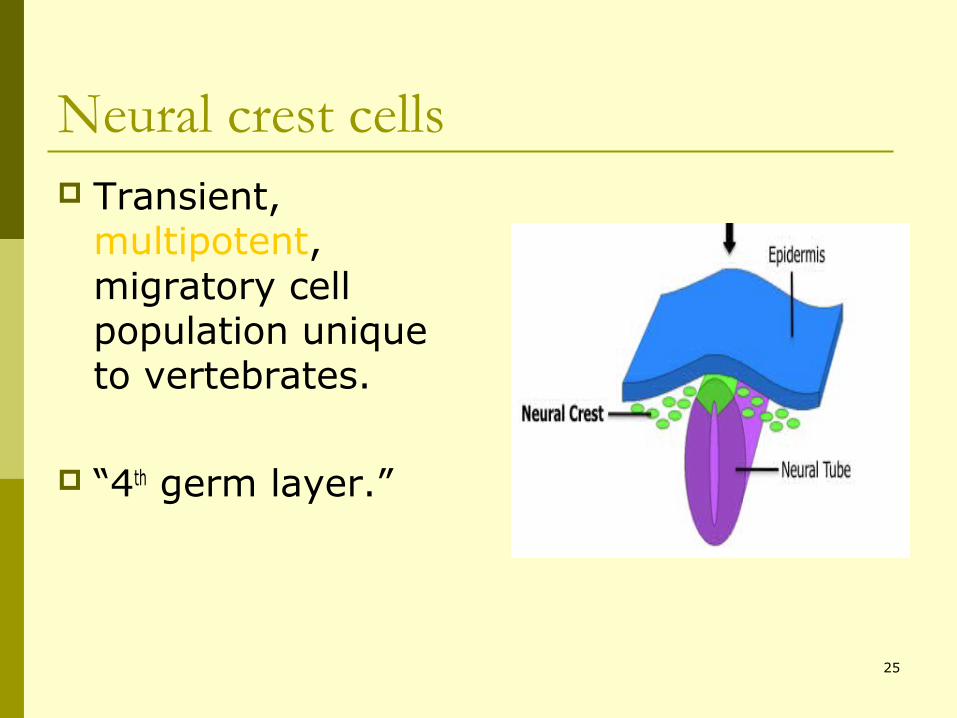
Neural crest cells Transient,
multipotent, migratory cell population unique to vertebrates.
“4th germ layer.”
25
26
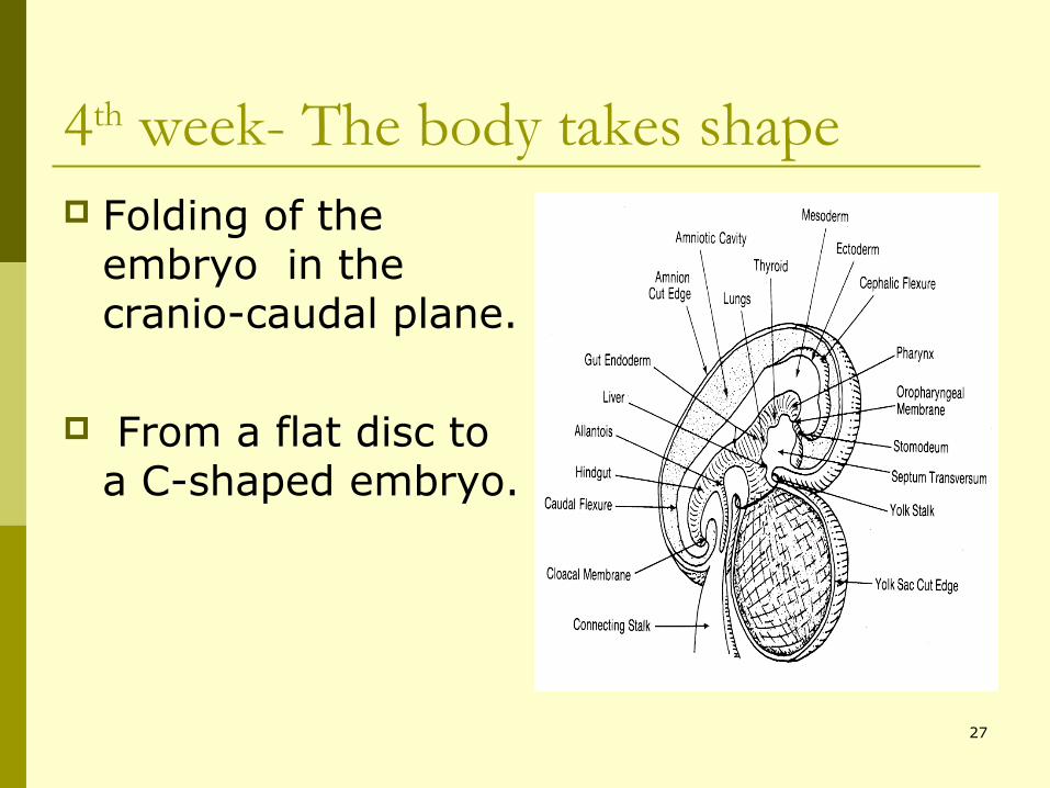
4th week- The body takes shape Folding of the
embryo in the cranio-caudal plane.
From a flat disc to a C-shaped embryo.
27
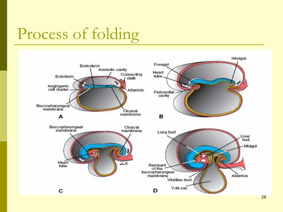
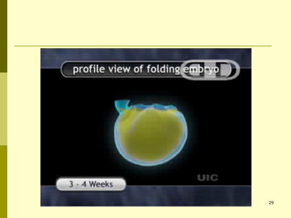
Process of folding
28
29
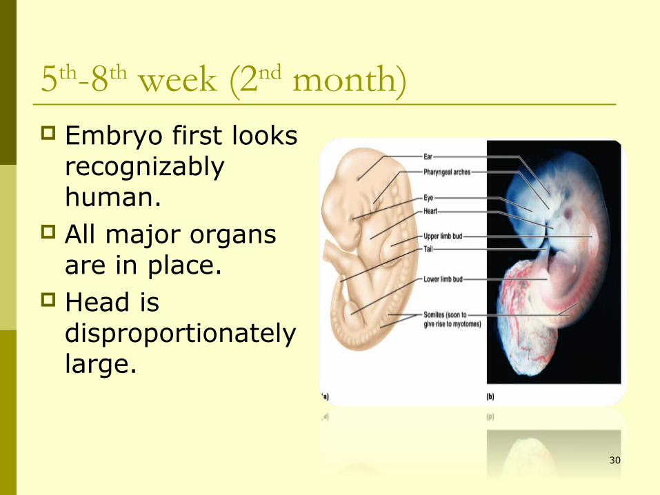
5th-8th week (2nd month) Embryo first looks
recognizably human.
All major organs are in place.
Head is disproportionately large.
30
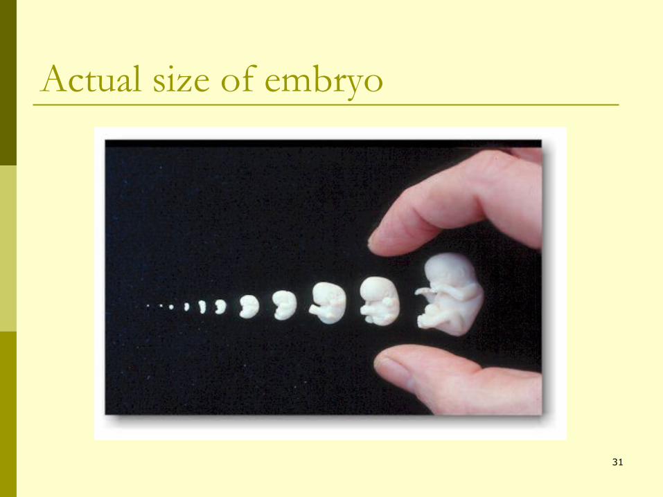
Actual size of embryo
31
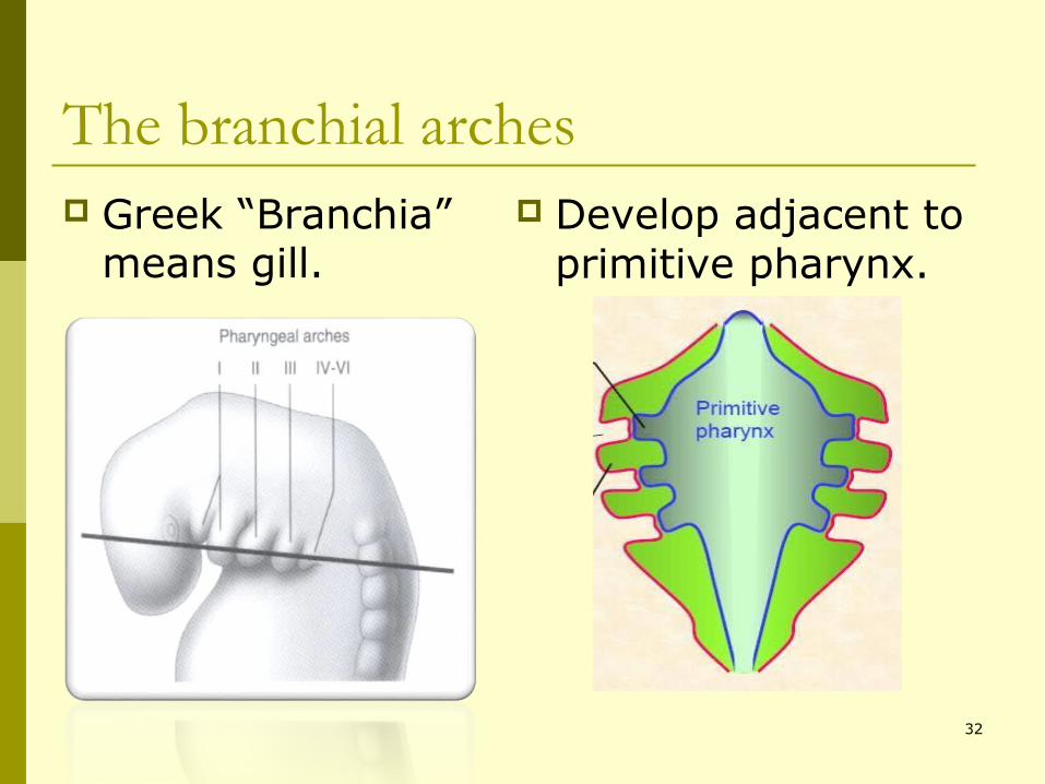
The branchial arches Greek “Branchia”
means gill. Develop adjacent to
primitive pharynx.
32
33
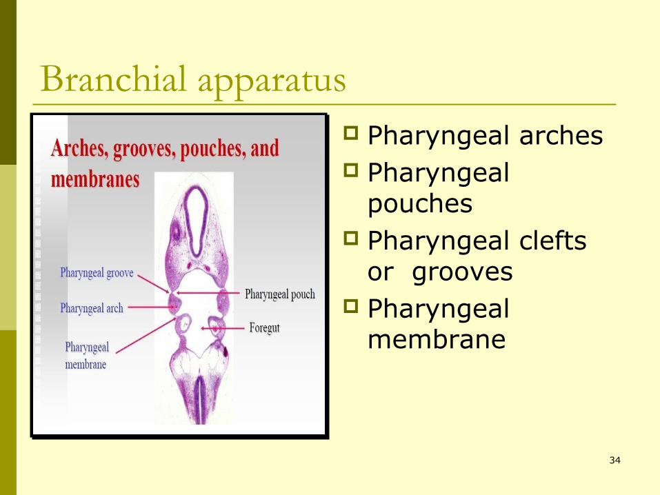
Branchial apparatus Pharyngeal arches Pharyngeal
pouches Pharyngeal clefts
or grooves Pharyngeal
membrane
34
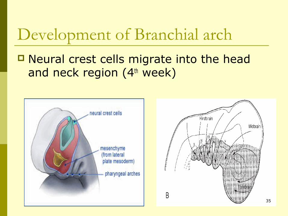
Development of Branchial arch Neural crest cells migrate into the head
and neck region (4th week)
35
The endodermal wall of the foregut is separated from the surface ectoderm by a layer of mesoderm.
Soon after, the mesoderm is arranged in the form of six bars.
5th arch soon degenerates.
36
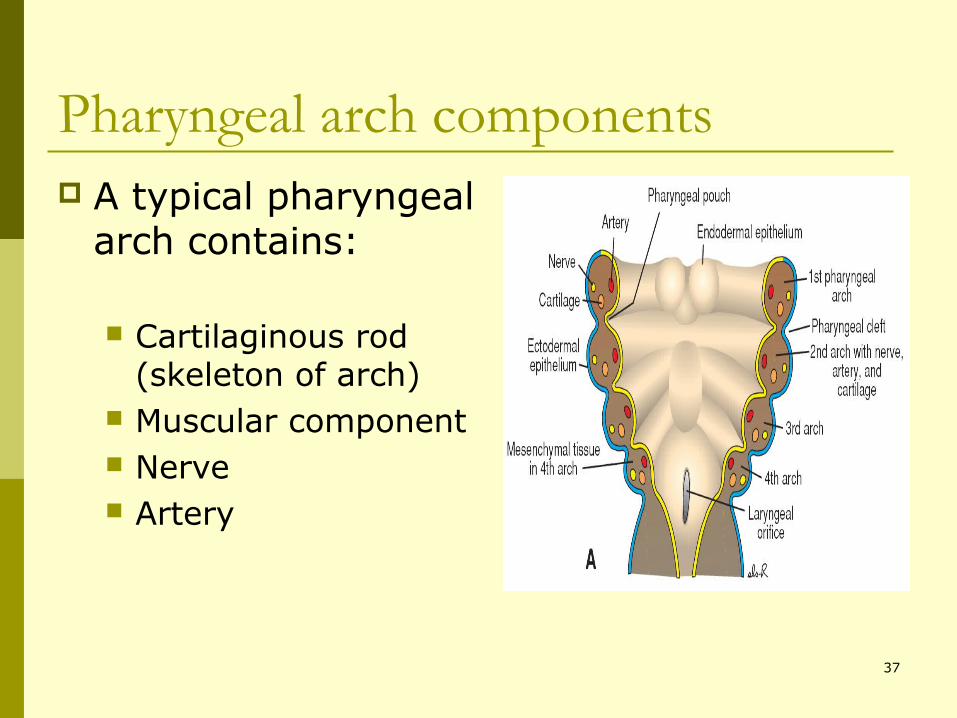
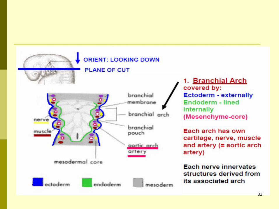
Pharyngeal arch components A typical pharyngeal
arch contains:
Cartilaginous rod (skeleton of arch)
Muscular component Nerve Artery
37
Derivatives of branchial arches Head and neck region of human beings
develop from these branchial arches.
Skeletal derivatives Muscular derivatives Nerves supply Arterial supply
38
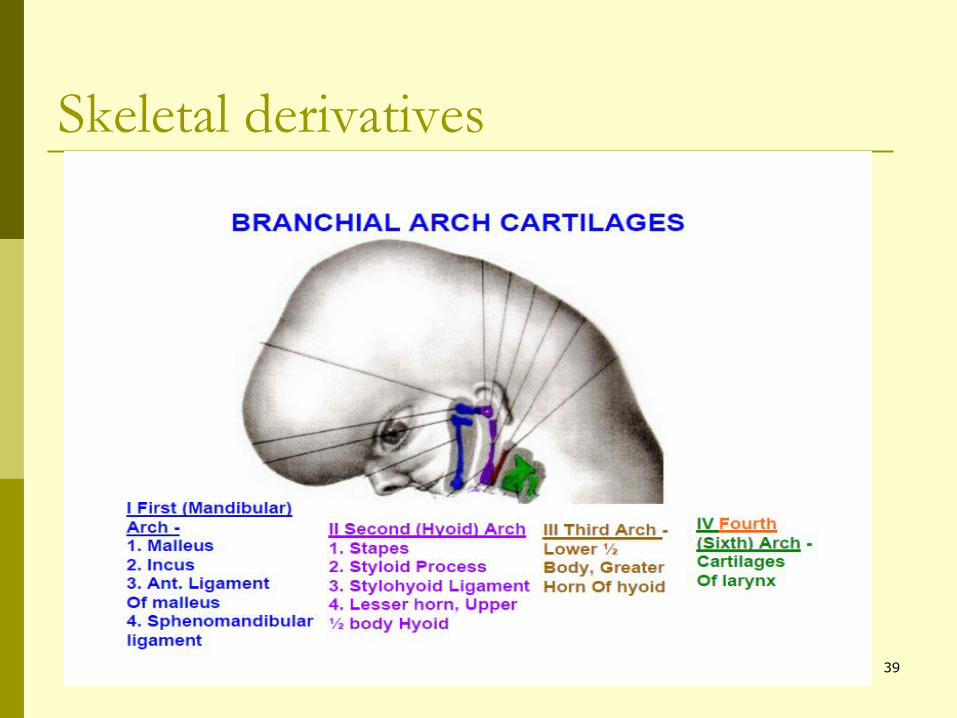
Skeletal derivatives
39
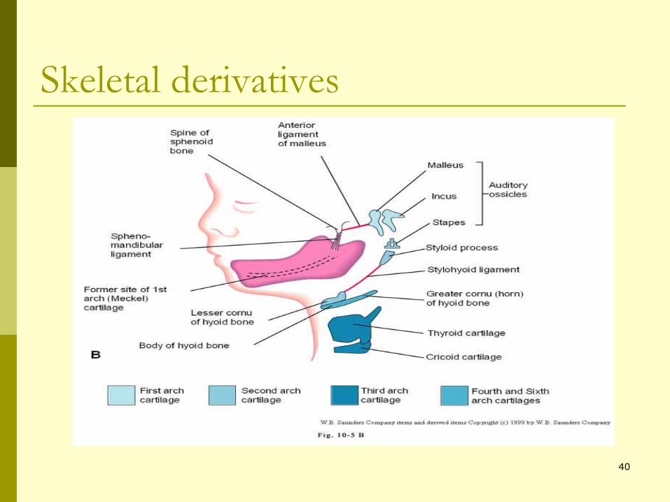
Skeletal derivatives
40
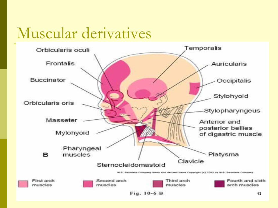
Muscular derivatives
41
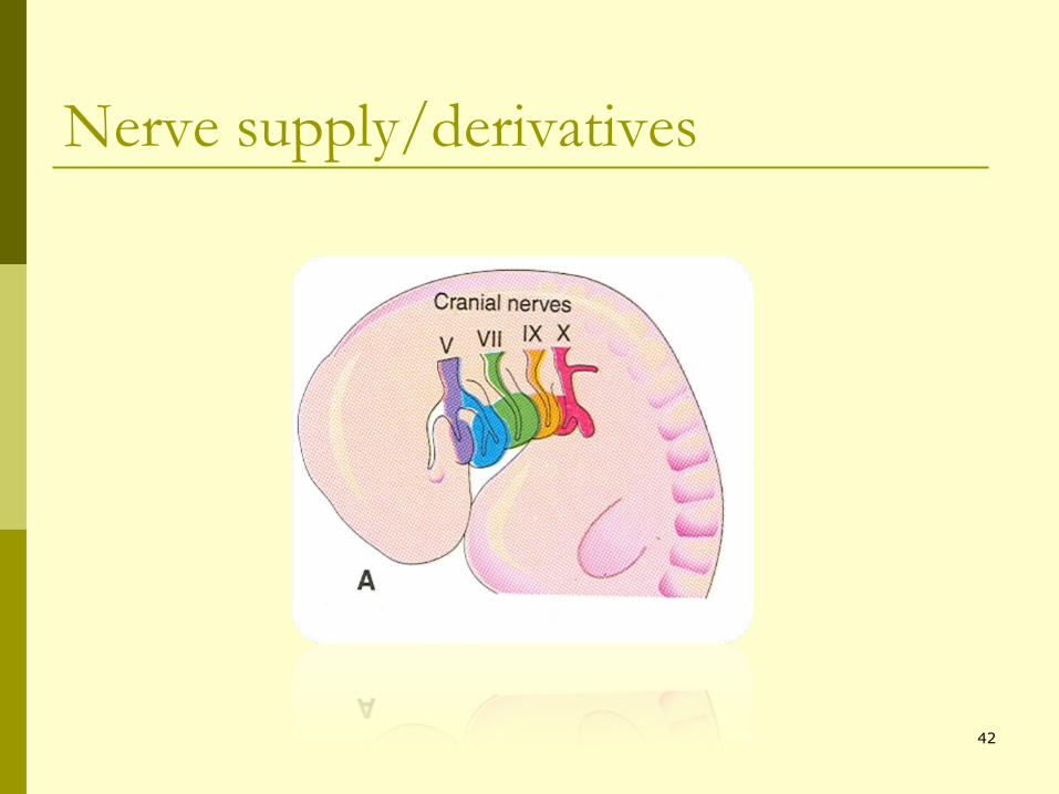
Nerve supply/derivatives
42
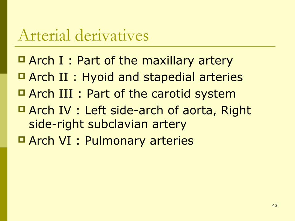
Arterial derivatives Arch I : Part of the maxillary artery Arch II : Hyoid and stapedial arteries Arch III : Part of the carotid system Arch IV : Left side-arch of aorta, Right
side-right subclavian artery Arch VI : Pulmonary arteries
43

44
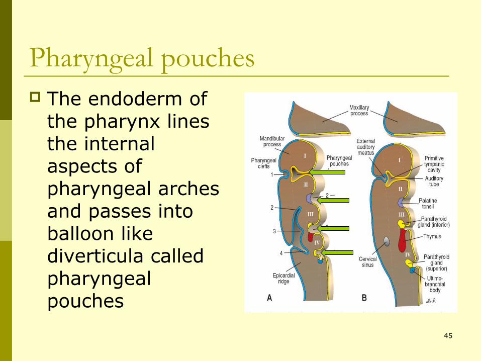
Pharyngeal pouches The endoderm of
the pharynx lines the internal aspects of pharyngeal arches and passes into balloon like diverticula called pharyngeal pouches
45
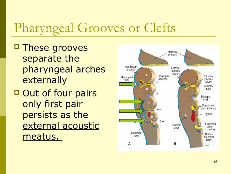
Pharyngeal Grooves or Clefts These grooves
separate the pharyngeal arches externally
Out of four pairs only first pair persists as the external acoustic meatus.
46
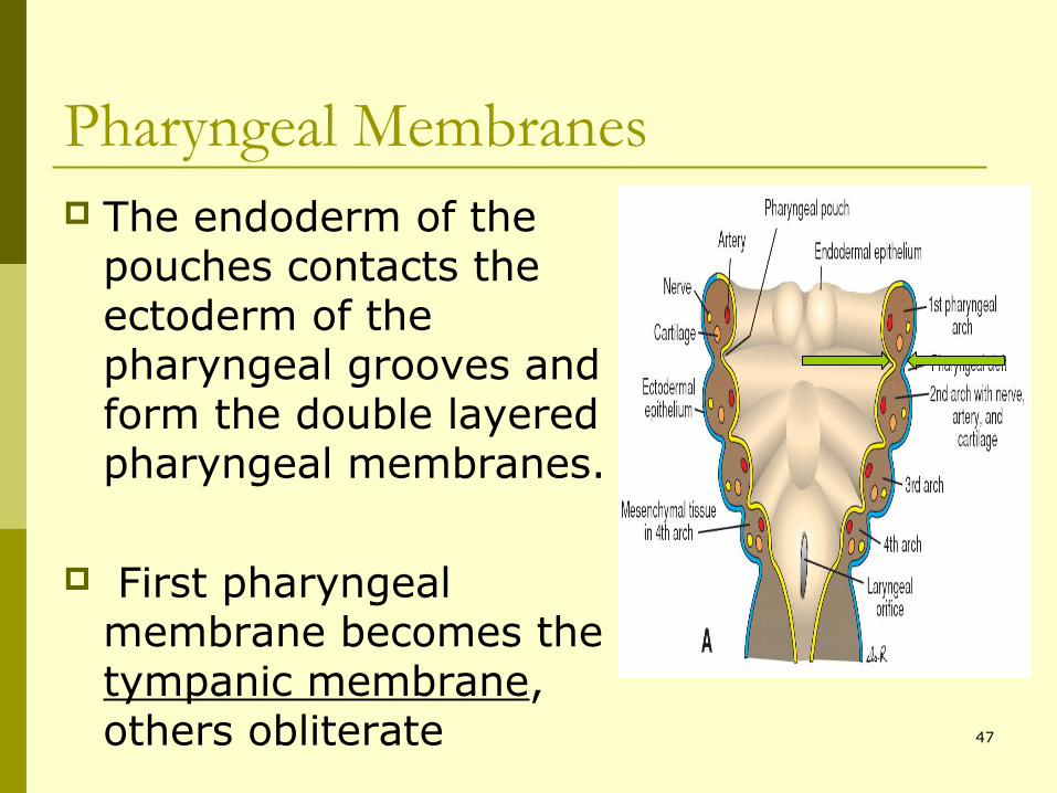
Pharyngeal Membranes The endoderm of the
pouches contacts the ectoderm of the pharyngeal grooves and form the double layered pharyngeal membranes.
First pharyngeal membrane becomes the tympanic membrane, others obliterate 47

48