Embed Size (px)
Citation preview
Presenter:Dr. Jamal GiriResident (1st Year)
Development of dentition
Contents
1. Development of dentition and supporting structures.
2. Mechanism of tooth eruption.3. Proffit’s contributions regarding
eruption.4. Clinical implications.
2Dept. of dentistry-PG section, IOM (2011)
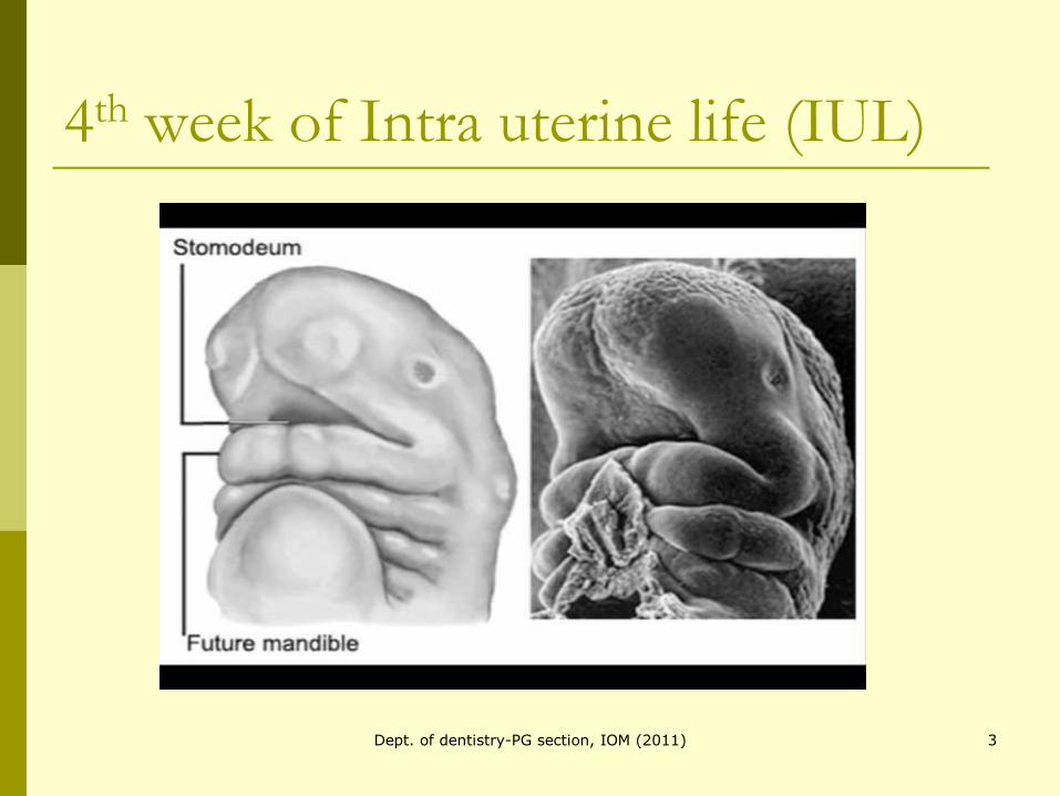
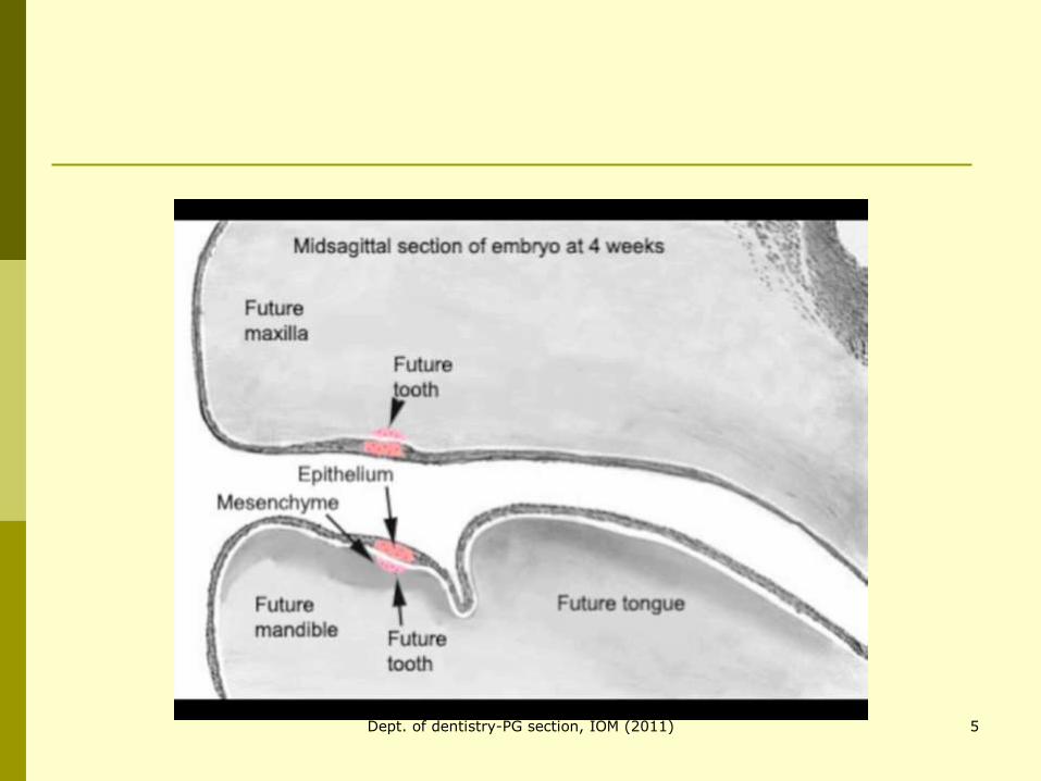
4th week of Intra uterine life (IUL)
3Dept. of dentistry-PG section, IOM (2011)
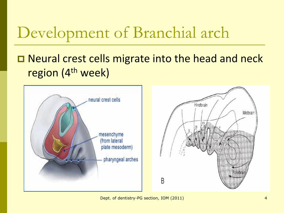
Development of Branchial arch
Neural crest cells migrate into the head and neck region (4th week)
4Dept. of dentistry-PG section, IOM (2011)
5Dept. of dentistry-PG section, IOM (2011)
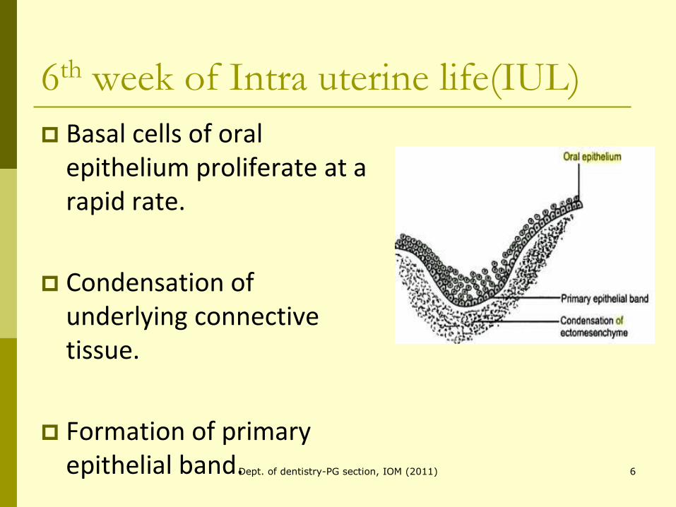
6th week of Intra uterine life(IUL)
Basal cells of oral epithelium proliferate at a rapid rate.
Condensation of underlying connective tissue.
Formation of primary epithelial band. 6Dept. of dentistry-PG section, IOM (2011)
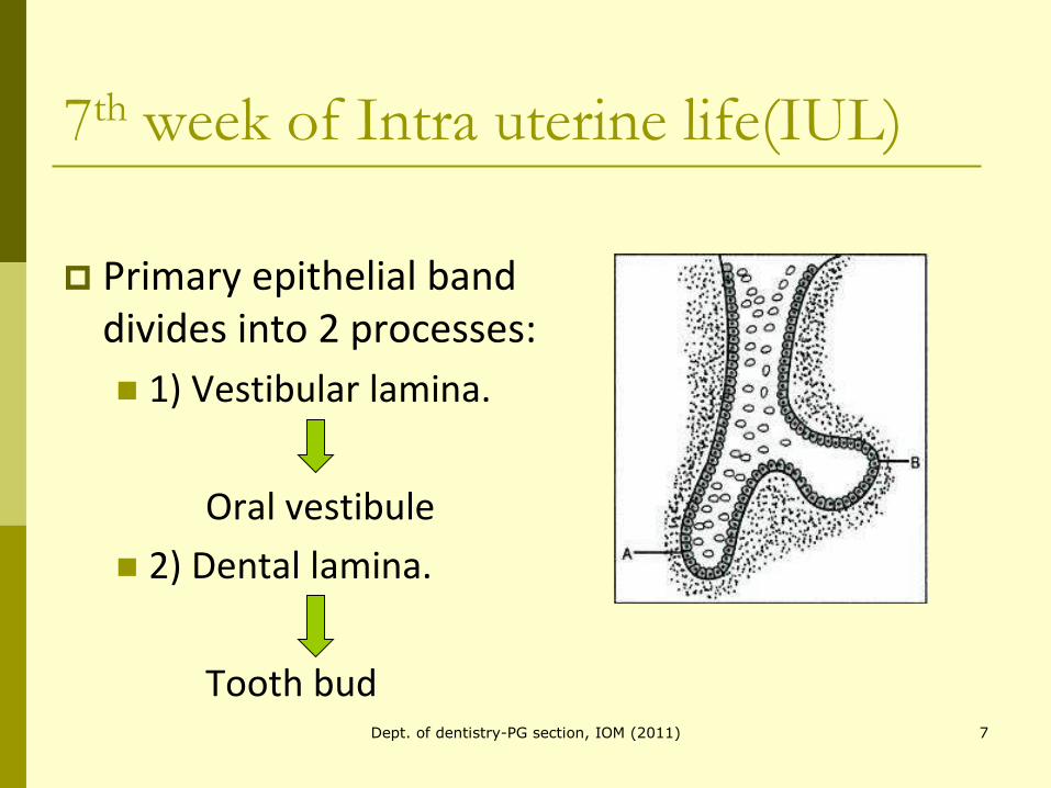
7th week of Intra uterine life(IUL)
Primary epithelial band divides into 2 processes:
1) Vestibular lamina.
Oral vestibule
2) Dental lamina.
Tooth bud7Dept. of dentistry-PG section, IOM (2011)
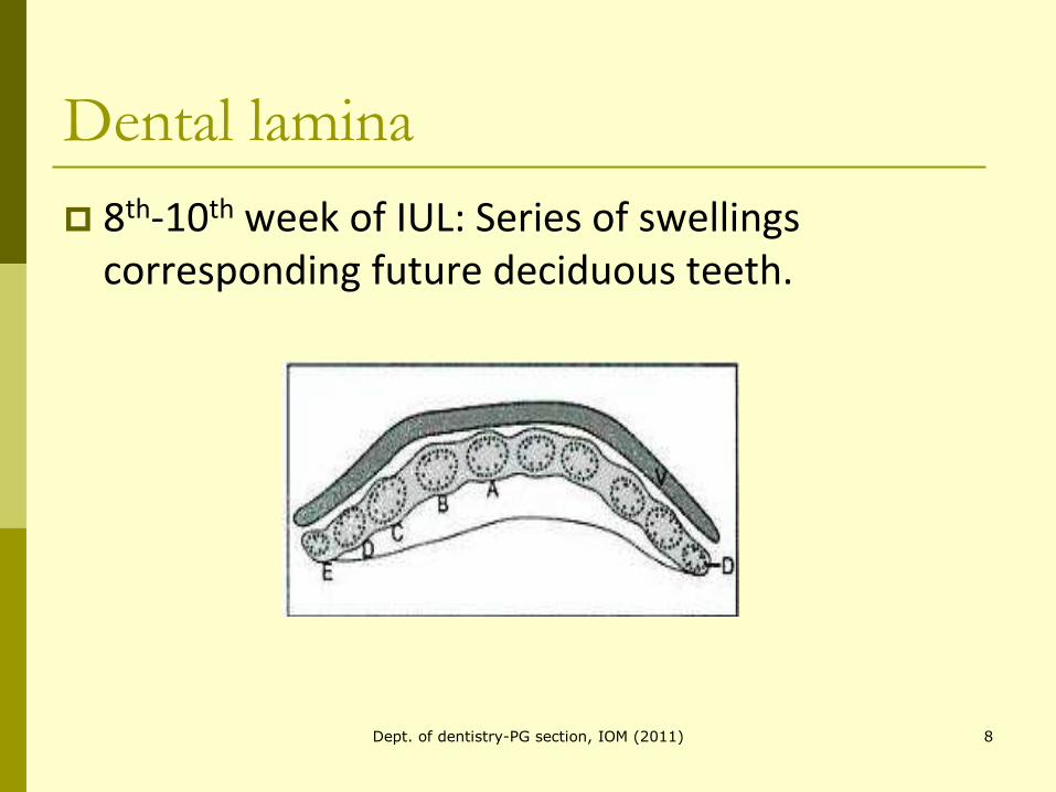
Dental lamina
8th-10th week of IUL: Series of swellings corresponding future deciduous teeth.
8Dept. of dentistry-PG section, IOM (2011)
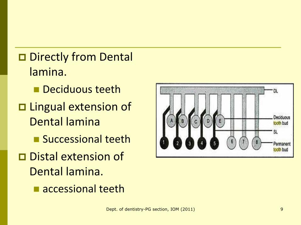
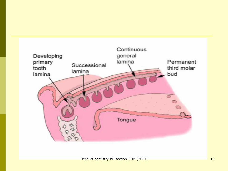
Directly from Dental lamina.
Deciduous teeth
Lingual extension of Dental lamina
Successional teeth
Distal extension of Dental lamina.
accessional teeth
9Dept. of dentistry-PG section, IOM (2011)
10Dept. of dentistry-PG section, IOM (2011)
Stages of tooth development1)Bud stage.
2)Cap stage.
3)Bell stage.
4)Stage of apposition.
5)Stage of root formation and eruption.
11Dept. of dentistry-PG section, IOM (2011)
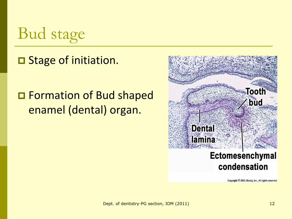
Bud stage
Stage of initiation.
Formation of Bud shaped enamel (dental) organ.
12Dept. of dentistry-PG section, IOM (2011)
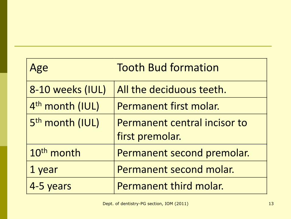
Age Tooth Bud formation
8-10 weeks (IUL) All the deciduous teeth.
4th month (IUL) Permanent first molar.
5th month (IUL) Permanent central incisor to first premolar.
10th month Permanent second premolar.
1 year Permanent second molar.
4-5 years Permanent third molar.
13Dept. of dentistry-PG section, IOM (2011)
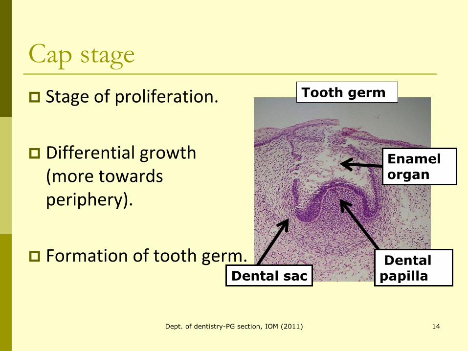
Cap stage
Stage of proliferation.
Differential growth (more towards periphery).
Formation of tooth germ. Dental papillaDental sac
Enamel organ
Tooth germ
14Dept. of dentistry-PG section, IOM (2011)
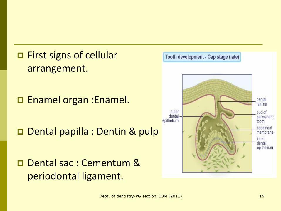
First signs of cellular arrangement.
Enamel organ :Enamel.
Dental papilla : Dentin & pulp
Dental sac : Cementum & periodontal ligament.
15Dept. of dentistry-PG section, IOM (2011)
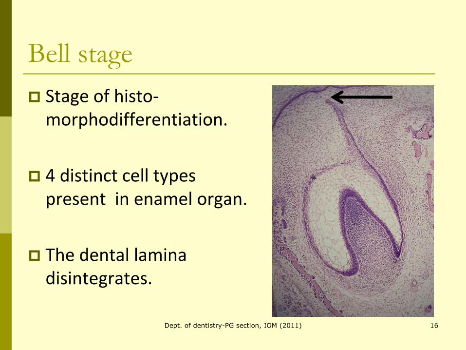
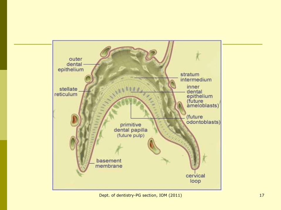
Bell stage
Stage of histo-morphodifferentiation.
4 distinct cell types present in enamel organ.
The dental lamina disintegrates.
16Dept. of dentistry-PG section, IOM (2011)
17Dept. of dentistry-PG section, IOM (2011)
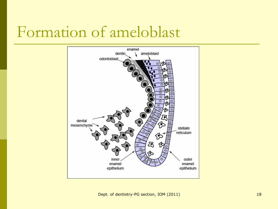
Formation of ameloblast
18Dept. of dentistry-PG section, IOM (2011)
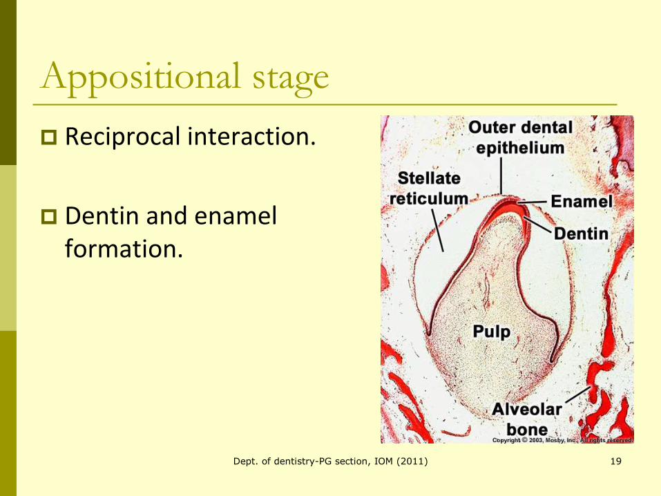
Appositional stage
Reciprocal interaction.
Dentin and enamel formation.
19Dept. of dentistry-PG section, IOM (2011)
20Dept. of dentistry-PG section, IOM (2011)
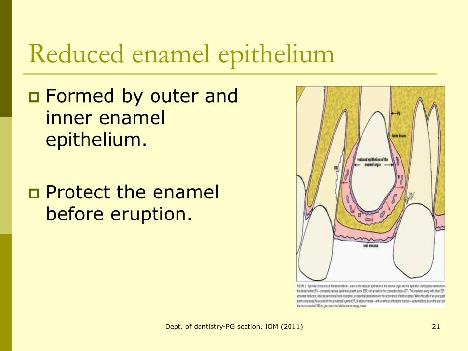
Reduced enamel epithelium
Formed by outer and inner enamel epithelium.
Protect the enamel before eruption.
21Dept. of dentistry-PG section, IOM (2011)
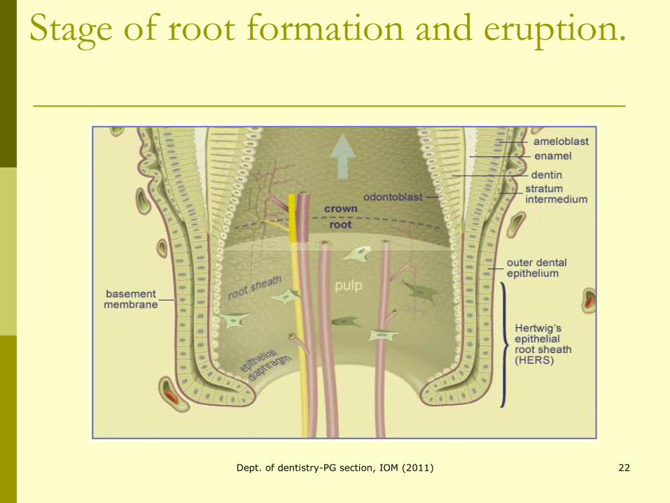
Stage of root formation and eruption.
22Dept. of dentistry-PG section, IOM (2011)
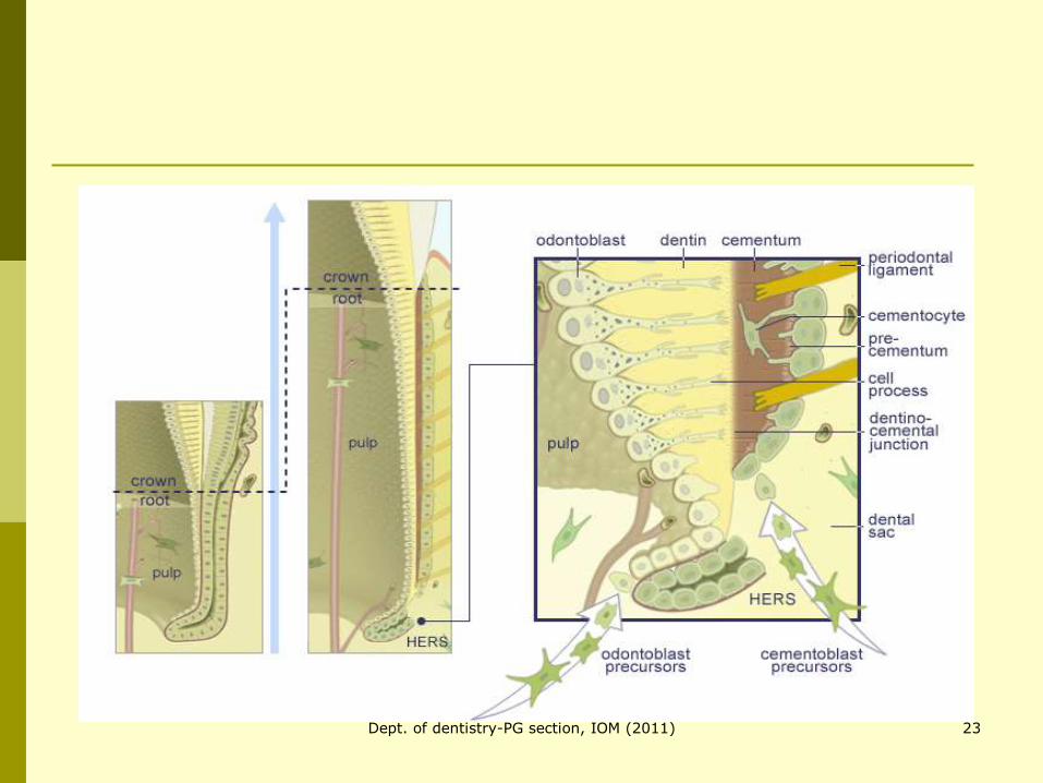
23Dept. of dentistry-PG section, IOM (2011)
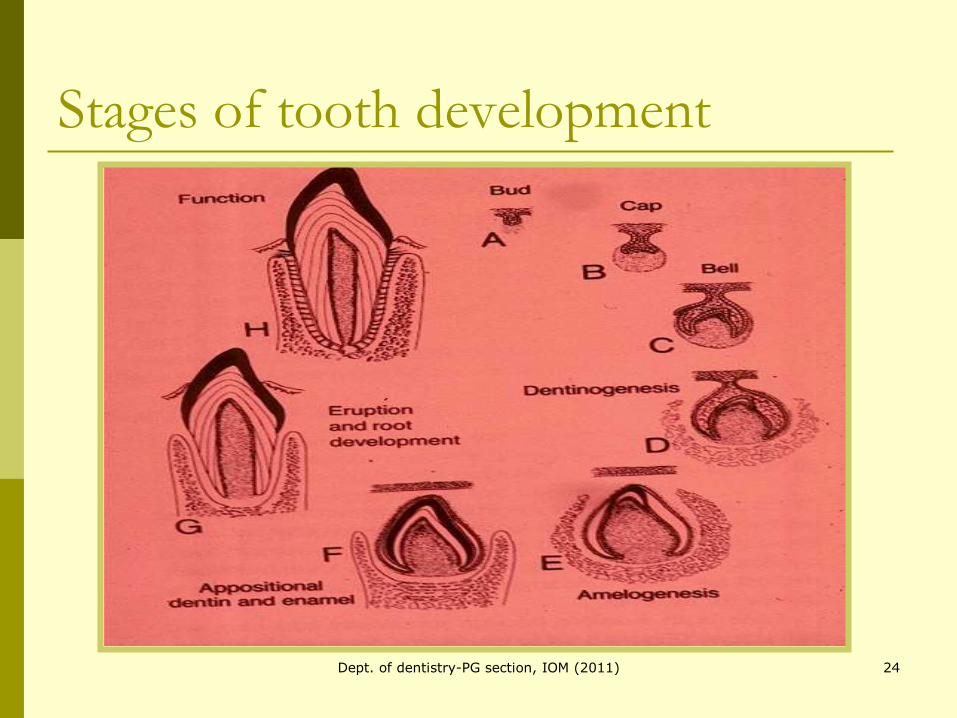
Stages of tooth development
24Dept. of dentistry-PG section, IOM (2011)

Genetic basis of tooth development.
More than 200 genes involved.
Homeobox genes.
(http://bite-it.helsinki.fi)
Oral Biosci Med 2/2004, S. 77-91
25Dept. of dentistry-PG section, IOM (2011)

Patterning of Dentition
26Dept. of dentistry-PG section, IOM (2011)
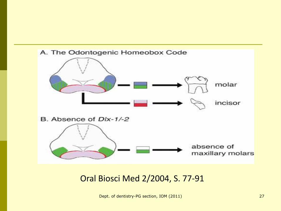
Oral Biosci Med 2/2004, S. 77-91
27Dept. of dentistry-PG section, IOM (2011)
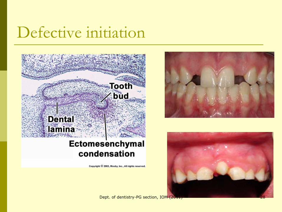
Defective initiation
28Dept. of dentistry-PG section, IOM (2011)
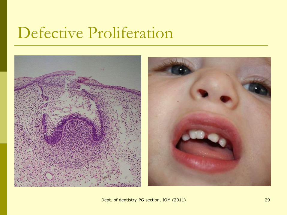
Defective Proliferation
29Dept. of dentistry-PG section, IOM (2011)
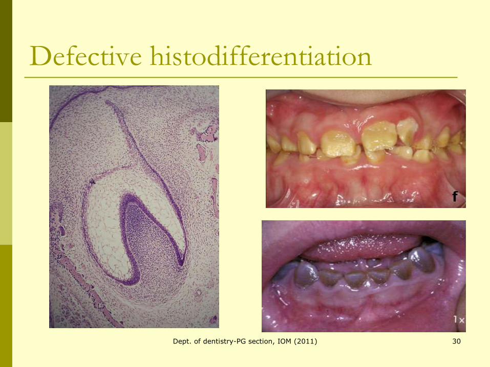
Defective histodifferentiation
30Dept. of dentistry-PG section, IOM (2011)
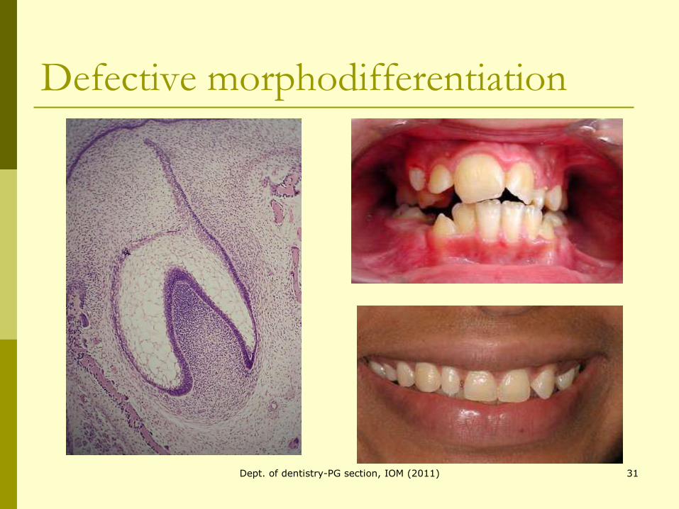
Defective morphodifferentiation
31Dept. of dentistry-PG section, IOM (2011)
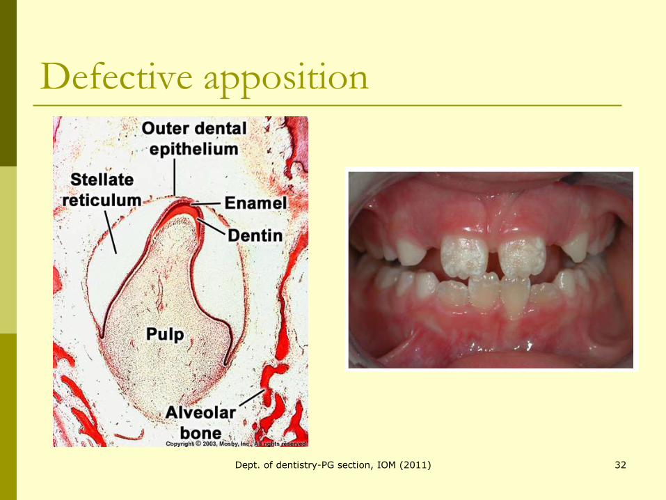
Defective apposition
32Dept. of dentistry-PG section, IOM (2011)
Tooth eruption
Greek word:
Erumpere- to break out
The process whereby a tooth moves from its developmental position within the jaws to emerge in the oral cavity.
33Dept. of dentistry-PG section, IOM (2011)
Theories of tooth eruption.
Role of root formation.
Role of bone remodeling.
Role of periodontal ligament.
Role of hydrostatic pressure.
Role of dental follicle.34Dept. of dentistry-PG section, IOM (2011)
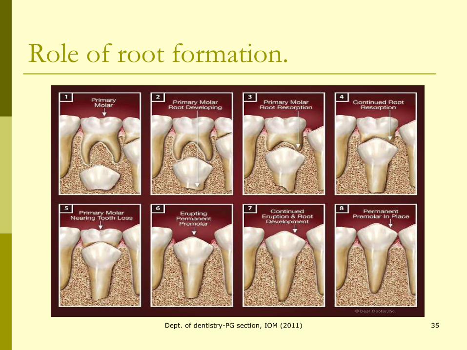
Role of root formation.
35Dept. of dentistry-PG section, IOM (2011)
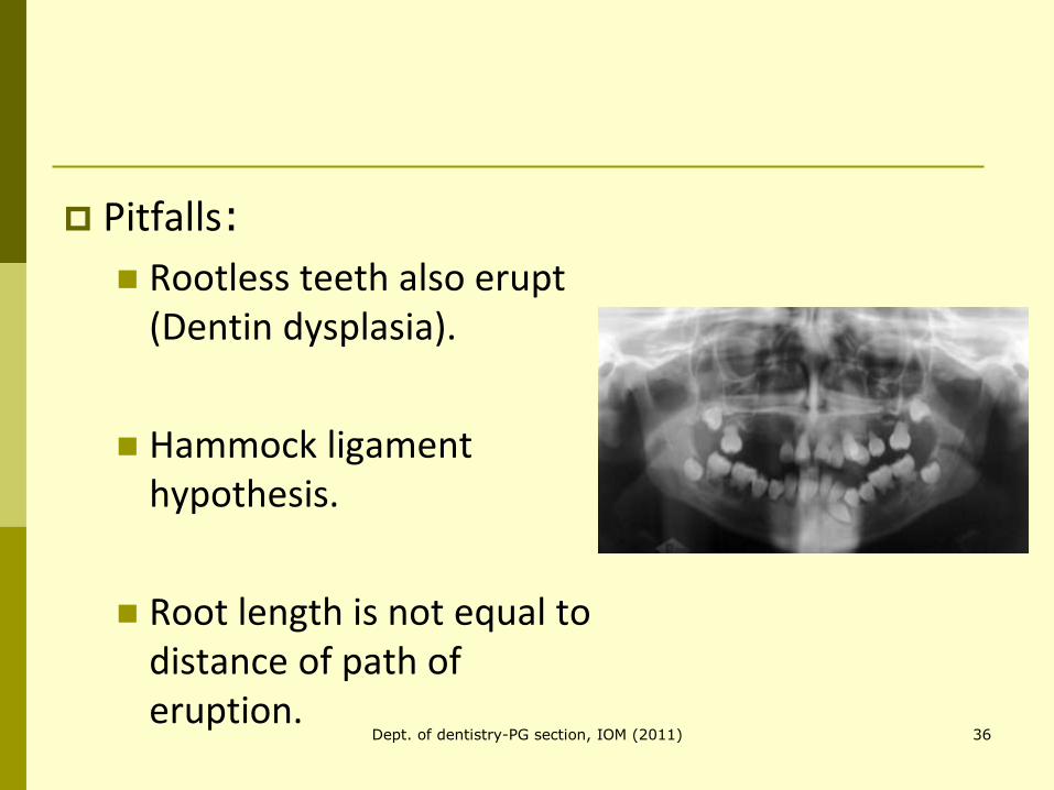
Pitfalls:
Rootless teeth also erupt (Dentin dysplasia).
Hammock ligament hypothesis.
Root length is not equal to distance of path of eruption.
36Dept. of dentistry-PG section, IOM (2011)
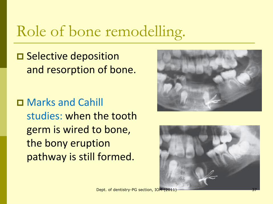
Role of bone remodelling.
Selective deposition and resorption of bone.
Marks and Cahill studies: when the tooth germ is wired to bone, the bony eruption pathway is still formed.
37Dept. of dentistry-PG section, IOM (2011)
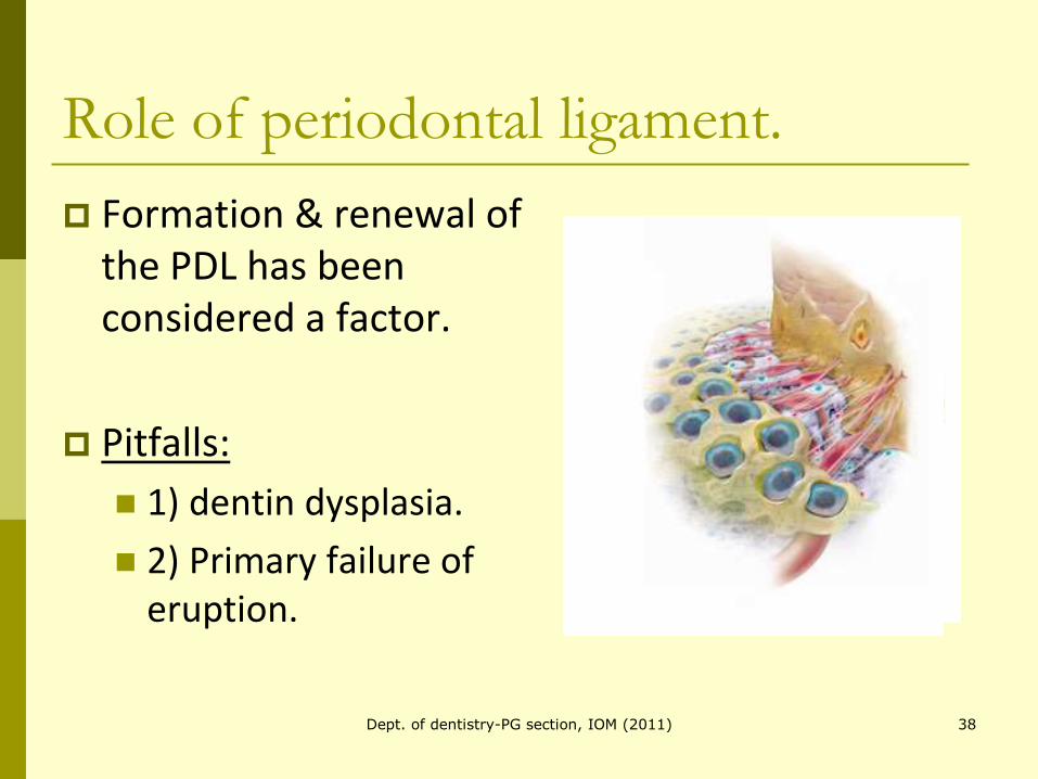
Role of periodontal ligament.
Formation & renewal of the PDL has been considered a factor.
Pitfalls:
1) dentin dysplasia.
2) Primary failure of eruption.
38Dept. of dentistry-PG section, IOM (2011)
Role of hydrostatic pressure.
Van Hassel and McMinn (1972)
Hydrostatic pressure gradient apico-occlusally.
Pitfall:
Relatively small study (6 dogs).
39Dept. of dentistry-PG section, IOM (2011)
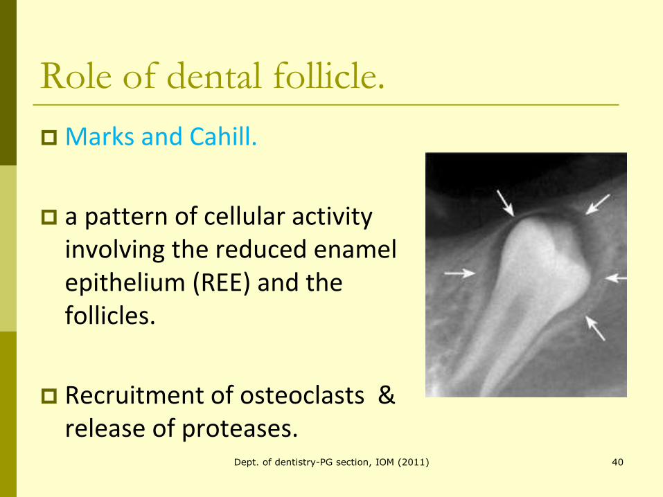
Role of dental follicle.
Marks and Cahill.
a pattern of cellular activity involving the reduced enamel epithelium (REE) and the follicles.
Recruitment of osteoclasts & release of proteases.
40Dept. of dentistry-PG section, IOM (2011)
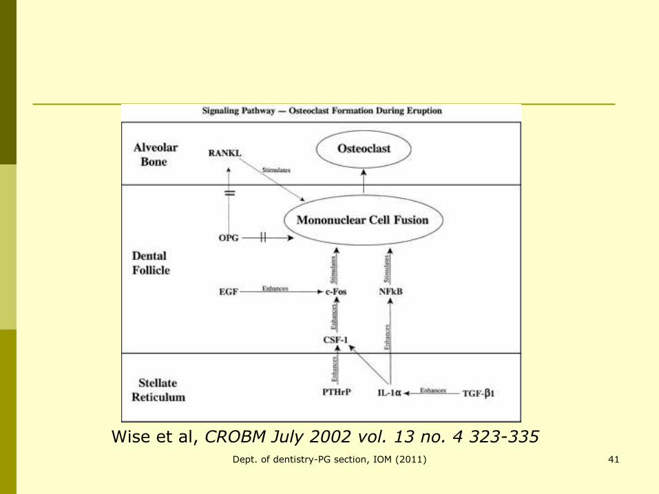
Wise et al, CROBM July 2002 vol. 13 no. 4 323-335
41Dept. of dentistry-PG section, IOM (2011)
Study on dog’s premolar.
Removal of the follicle from the unerupted tooth prevented the tooth from erupting (Cahill and Marks, 1980).
Leaving the follicle intact and substituting an inert object for the tooth resulted in eruption of the inert object (Marks and Cahill, 1984).
42Dept. of dentistry-PG section, IOM (2011)
Proffit’s contribution
Mechanism and control of tooth eruption: overview and clinical implications, OrthodCraniofac Res 2009;12:59–66.
Tooth eruption is difficult to study:
1) erupt slowly.
2) Inaccessible for clinical examination.
43Dept. of dentistry-PG section, IOM (2011)
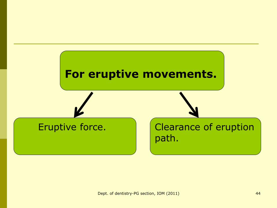
For eruptive movements.
Eruptive force. Clearance of eruption path.
44Dept. of dentistry-PG section, IOM (2011)
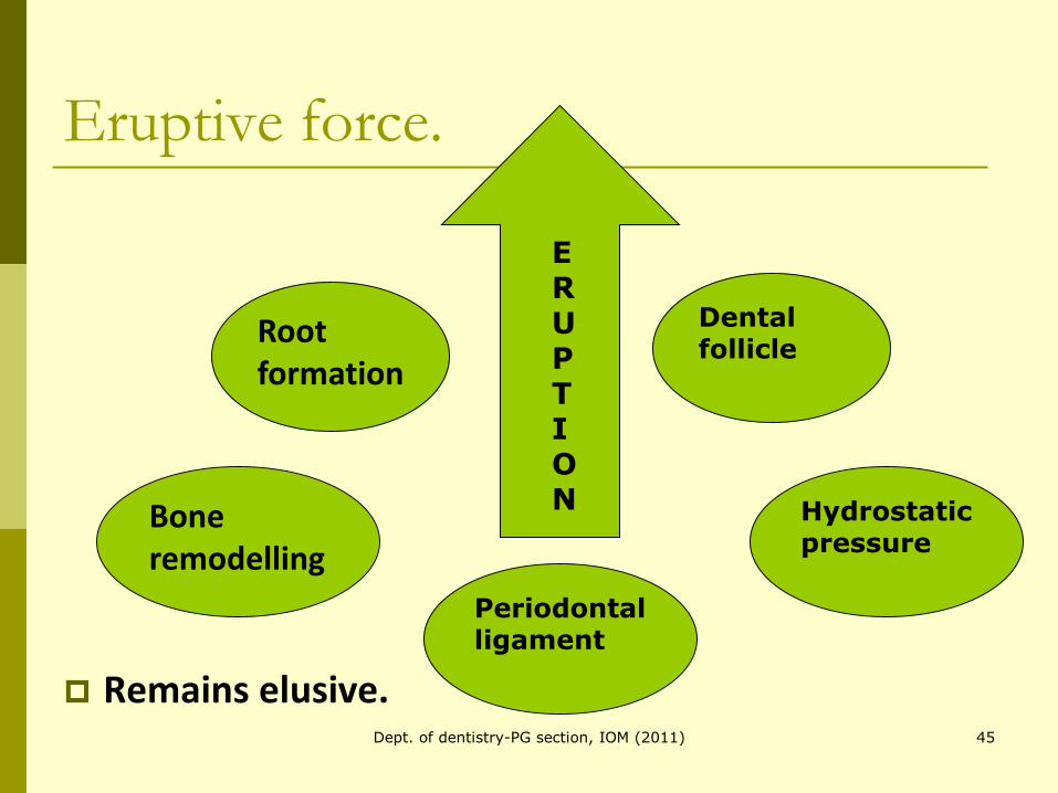
Eruptive force.
Remains elusive.
ERUPTION
Root formation
Bone remodelling
Dental follicle
Hydrostatic pressure
Periodontal ligament
45Dept. of dentistry-PG section, IOM (2011)
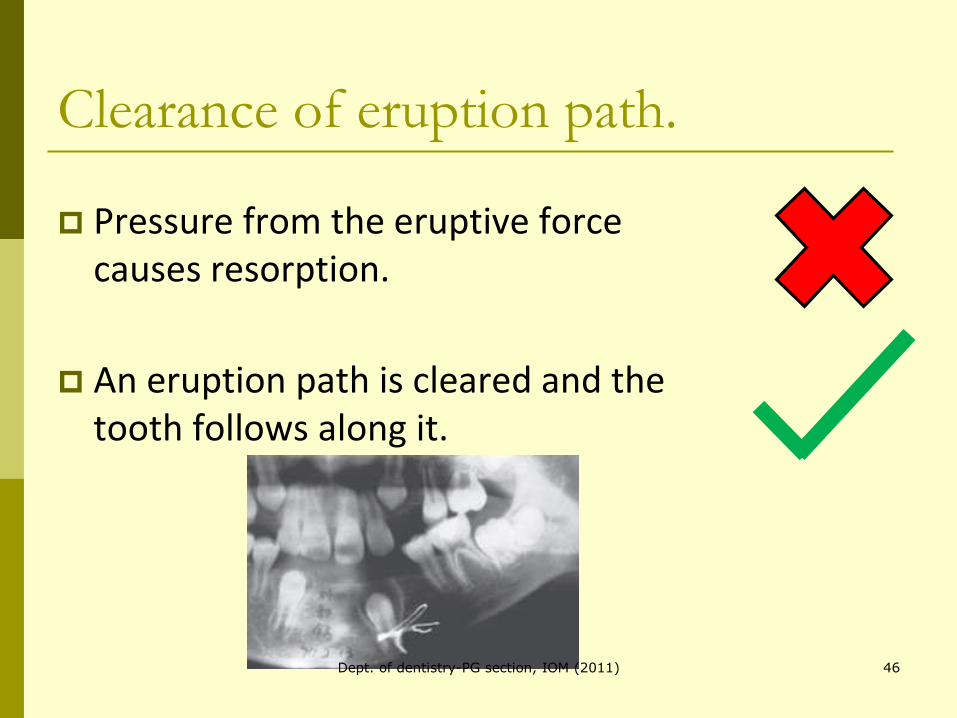
Clearance of eruption path.
Pressure from the eruptive force causes resorption.
An eruption path is cleared and the tooth follows along it.
46Dept. of dentistry-PG section, IOM (2011)
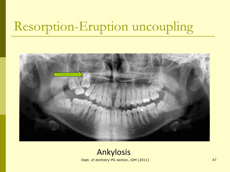
Resorption-Eruption uncoupling
Ankylosis47Dept. of dentistry-PG section, IOM (2011)
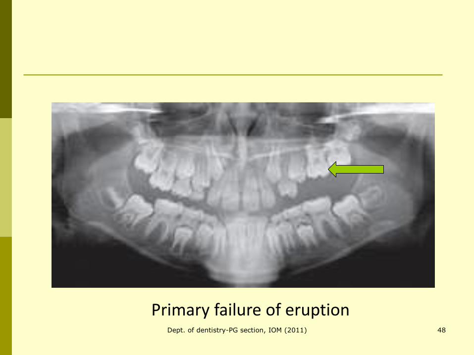
Primary failure of eruption48Dept. of dentistry-PG section, IOM (2011)
The daily rhythm of tooth eruption
AJODO, Volume 107, Issue 1, January 1995, Pages 38-47.
17 subjects ( 10 boys, 7 girls)
41 hour video microscopic observation.
49Dept. of dentistry-PG section, IOM (2011)
Results
Mean daily eruption: 71um/day
Small amount of intrusion during Daytime.
Net eruption at night.
Transient intrusion associated with breakfast, lunch, dinner.
50Dept. of dentistry-PG section, IOM (2011)

51