Embed Size (px)
Citation preview
CHAPTER 20: LYMPHATIC
SYSTEM, LYMPHOID
ORGANS & TISSUES
Human Anatomy and Physiology II –
BIOL153
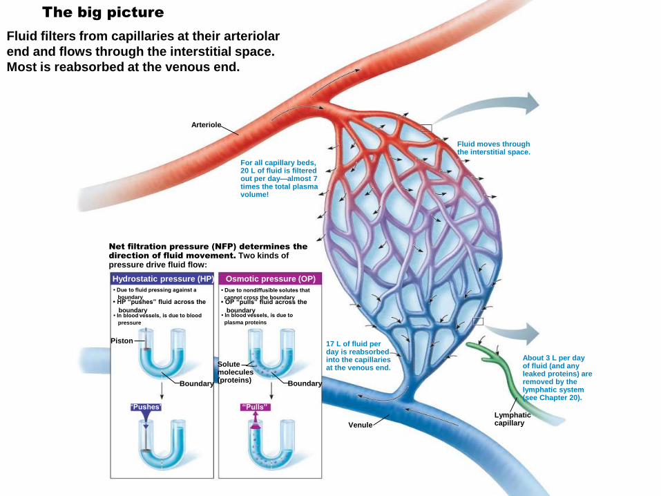
The big picture
Fluid filters from capillaries at their arteriolar
end and flows through the interstitial space.
Most is reabsorbed at the venous end.
For all capillary beds, 20 L of fluid is filteredout per day—almost 7times the total plasmavolume!
• Due to fluid pressing against a
boundary• HP “pushes” fluid across the
boundary• In blood vessels, is due to blood
pressure
• Due to nondiffusible solutes that
cannot cross the boundary• OP “pulls” fluid across the
boundary• In blood vessels, is due to
plasma proteins
Piston
Boundary
Solutemolecules(proteins)
Boundary
“Pushes” “Pulls”
Hydrostatic pressure (HP) Osmotic pressure (OP)
17 L of fluid per day is reabsorbedinto the capillariesat the venous end.
LymphaticcapillaryVenule
About 3 L per day of fluid (and anyleaked proteins) areremoved by thelymphatic system(see Chapter 20).
Arteriole
Fluid moves through the interstitial space.
Net filtration pressure (NFP) determines the
direction of fluid movement. Two kinds of pressure drive fluid flow:
Goals/Objectives
Describe the structure and distribution of lymphatic vessels
Describe the source of lymph and mechanism(s) and pathway of lymph transport
Describe the general location, histological structure, and functions of lymph nodes
Describe the basic structure and cellular population of lymphoid tissue
Name and describe the other lymphoid organs of the body. Compare and contrast them with lymph nodes, structurally and functionally.
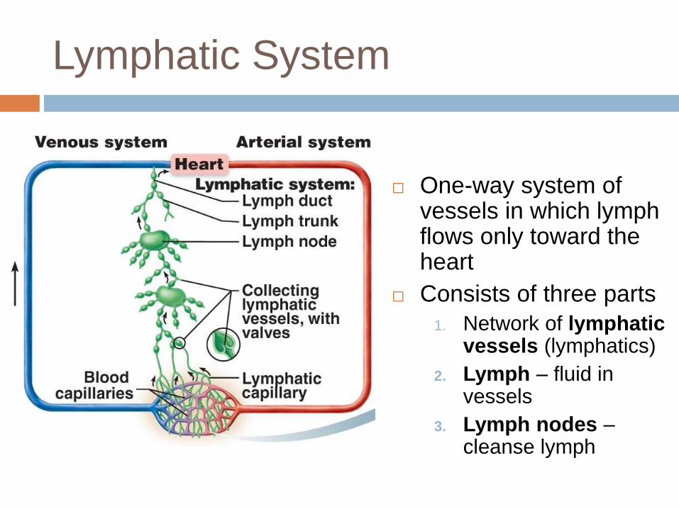
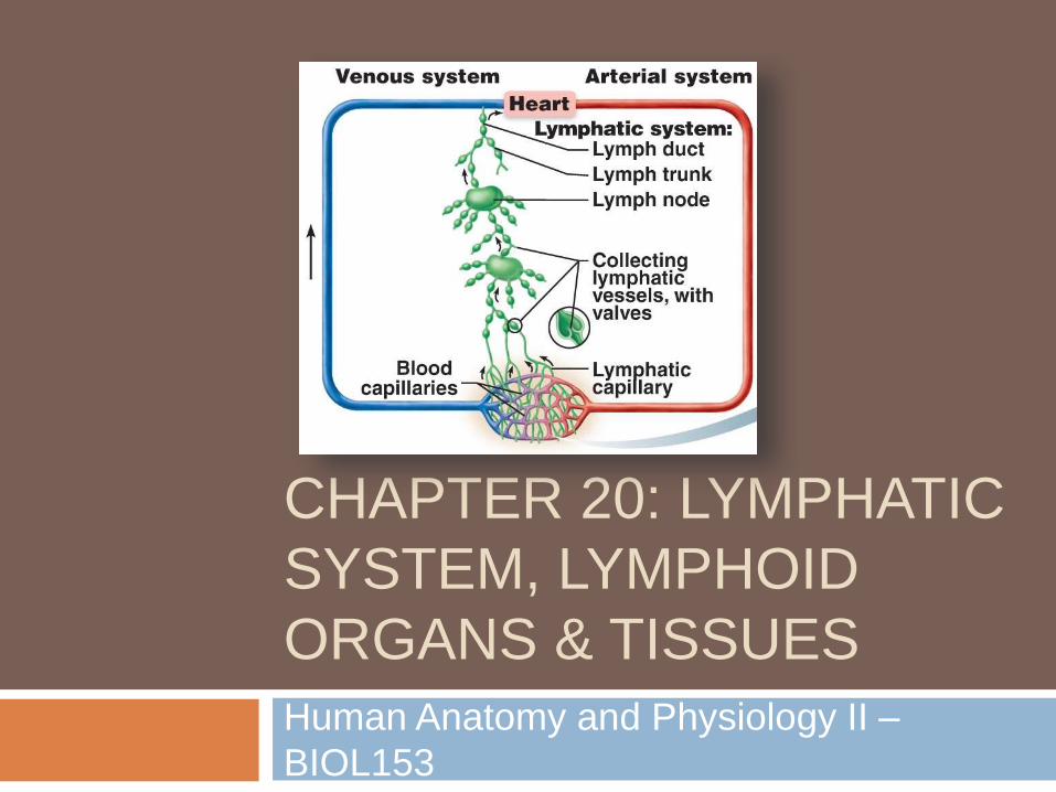
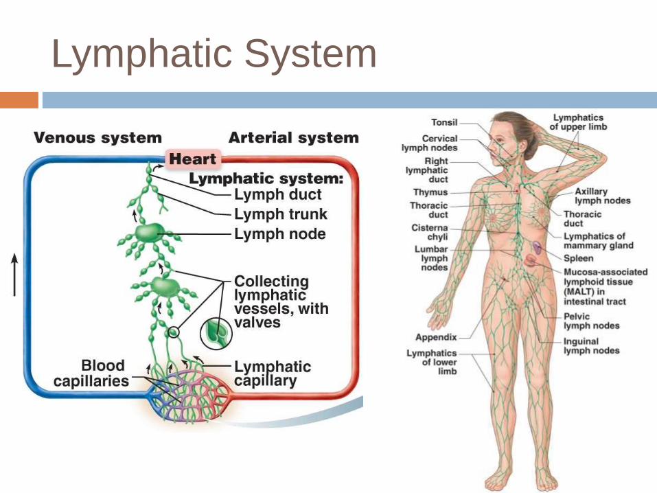
Lymphatic System
One-way system of vessels in which lymph flows only toward the heart
Consists of three parts
1. Network of lymphatic vessels (lymphatics)
2. Lymph – fluid in vessels
3. Lymph nodes –cleanse lymph
Lymphatic System
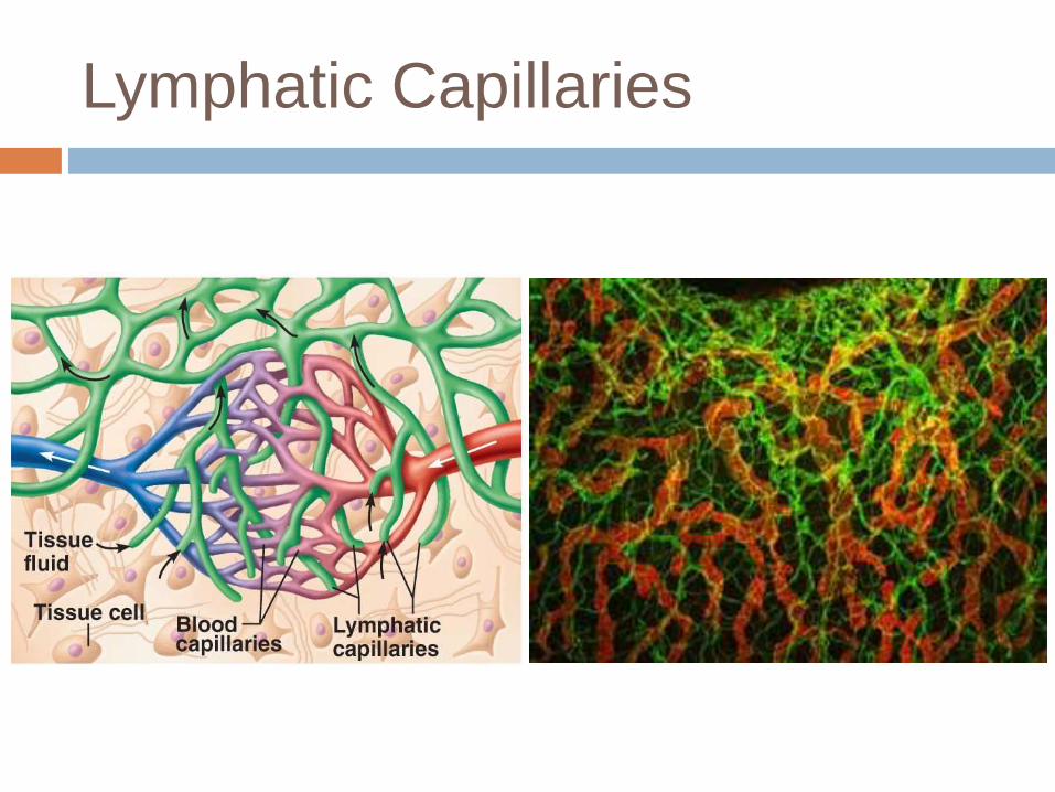
Lymphatic Capillaries
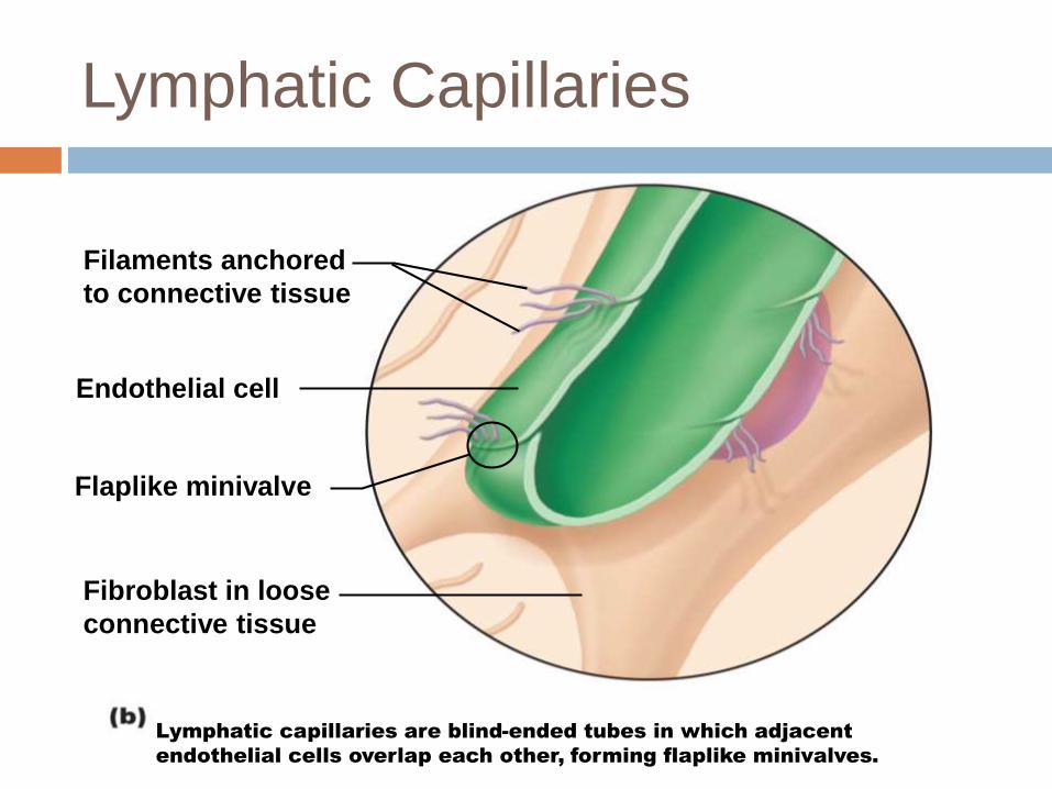
Lymphatic Capillaries
Filaments anchored
to connective tissue
Endothelial cell
Flaplike minivalve
Fibroblast in loose
connective tissue
Lymphatic capillaries are blind-ended tubes in which adjacent
endothelial cells overlap each other, forming flaplike minivalves.
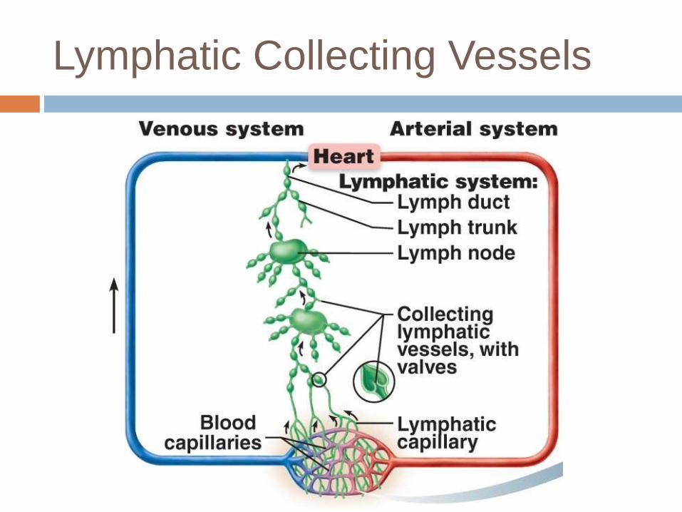
Lymphatic Collecting Vessels
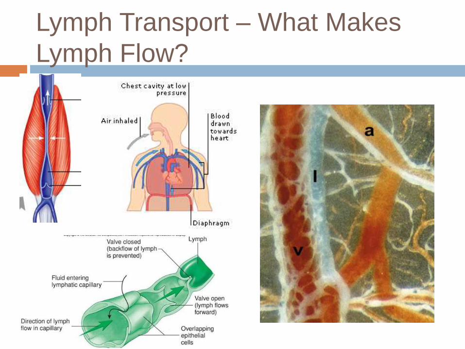
Lymph Transport – What Makes
Lymph Flow?
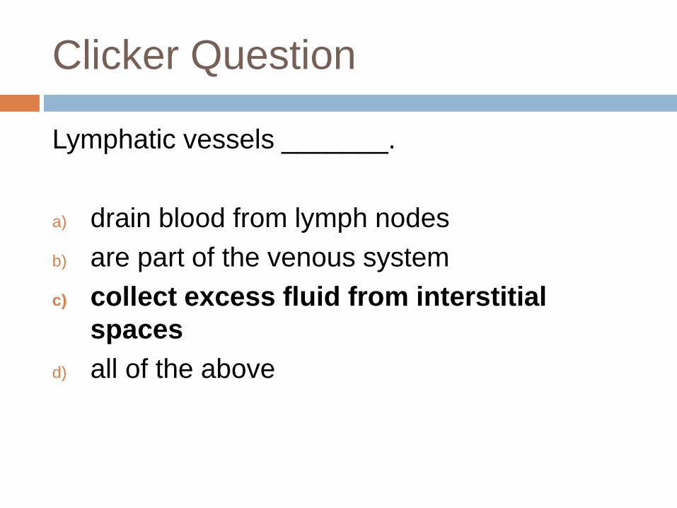
Clicker Question
Lymphatic vessels _______.
a) drain blood from lymph nodes
b) are part of the venous system
c) collect excess fluid from interstitial
spaces
d) all of the above
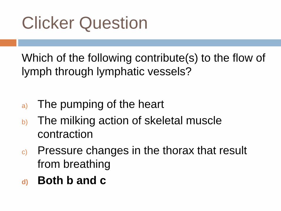
Clicker Question
Which of the following contribute(s) to the flow of
lymph through lymphatic vessels?
a) The pumping of the heart
b) The milking action of skeletal muscle
contraction
c) Pressure changes in the thorax that result
from breathing
d) Both b and c
Goals/Objectives
Describe the structure and distribution of lymphatic vessels
Describe the source of lymph and mechanism(s) and pathway of lymph transport
Describe the general location, histological structure, and functions of lymph nodes
Describe the basic structure and cellular population of lymphoid tissue
Name and describe the other lymphoid organs of the body. Compare and contrast them with lymph nodes, structurally and functionally.
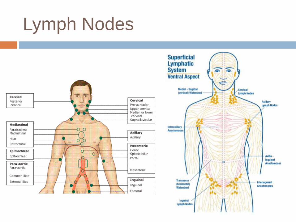
Lymph Nodes
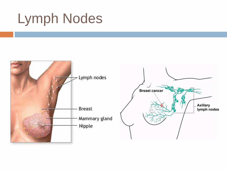
Lymph Nodes
Lymph Nodes
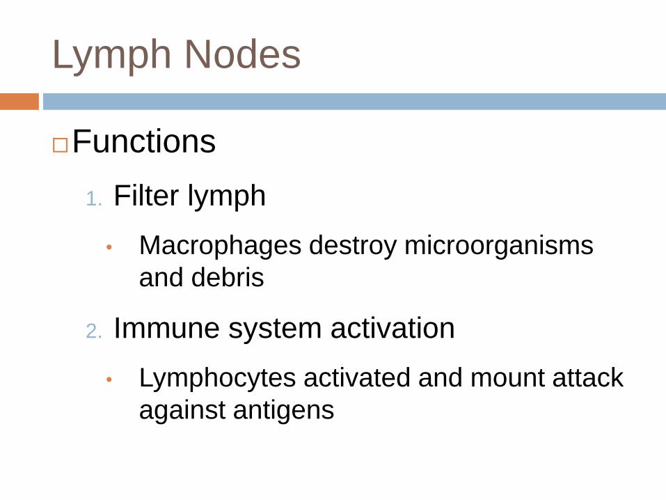
Functions
1. Filter lymph
• Macrophages destroy microorganisms
and debris
2. Immune system activation
• Lymphocytes activated and mount attack
against antigens
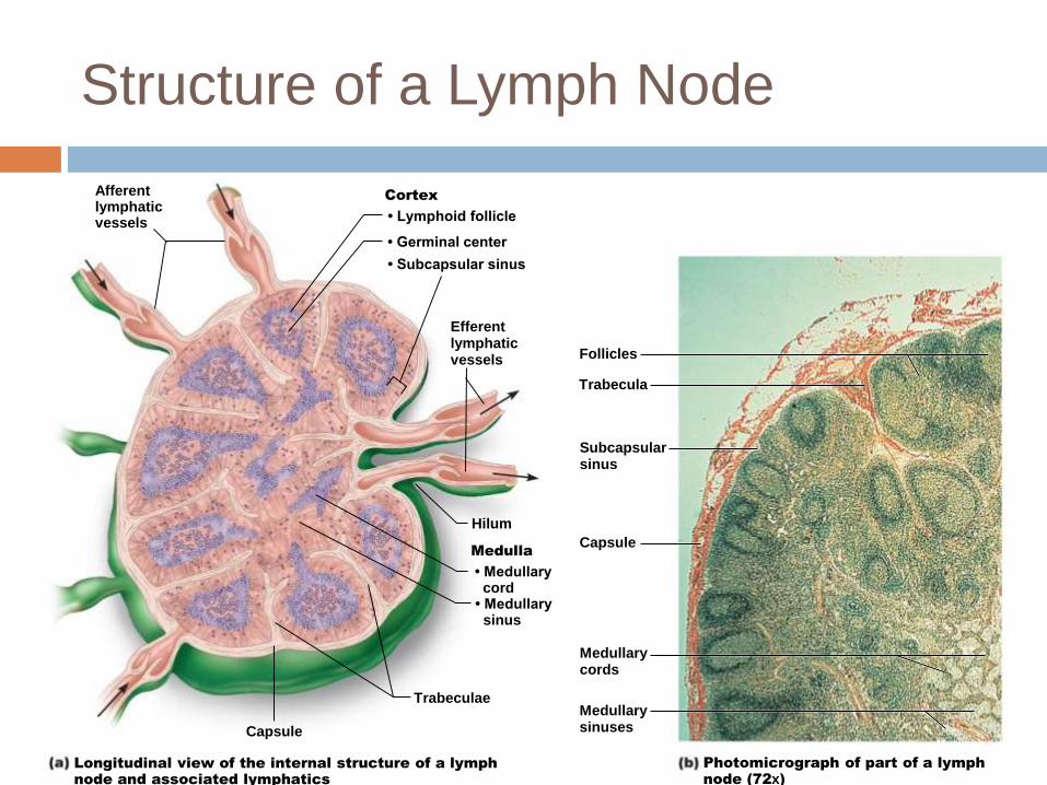
Structure of a Lymph Node
Afferentlymphaticvessels
Cortex
• Lymphoid follicle
• Germinal center
• Subcapsular sinus
Efferentlymphaticvessels Follicles
Trabecula
Subcapsularsinus
Capsule
Medullarycords
Medullarysinuses
Trabeculae
• Medullarysinus
• Medullarycord
Medulla
Hilum
Longitudinal view of the internal structure of a lymph
node and associated lymphatics
Photomicrograph of part of a lymph
node (72x)
Capsule
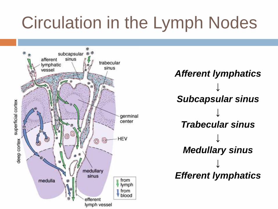
Circulation in the Lymph Nodes
Afferent lymphatics
↓
Subcapsular sinus
↓
Trabecular sinus
↓
Medullary sinus
↓
Efferent lymphatics
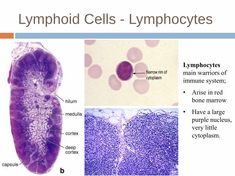
Lymphoid Cells - Lymphocytes
Lymphocytes
main warriors of
immune system;
• Arise in red
bone marrow
• Have a large
purple nucleus,
very little
cytoplasm.
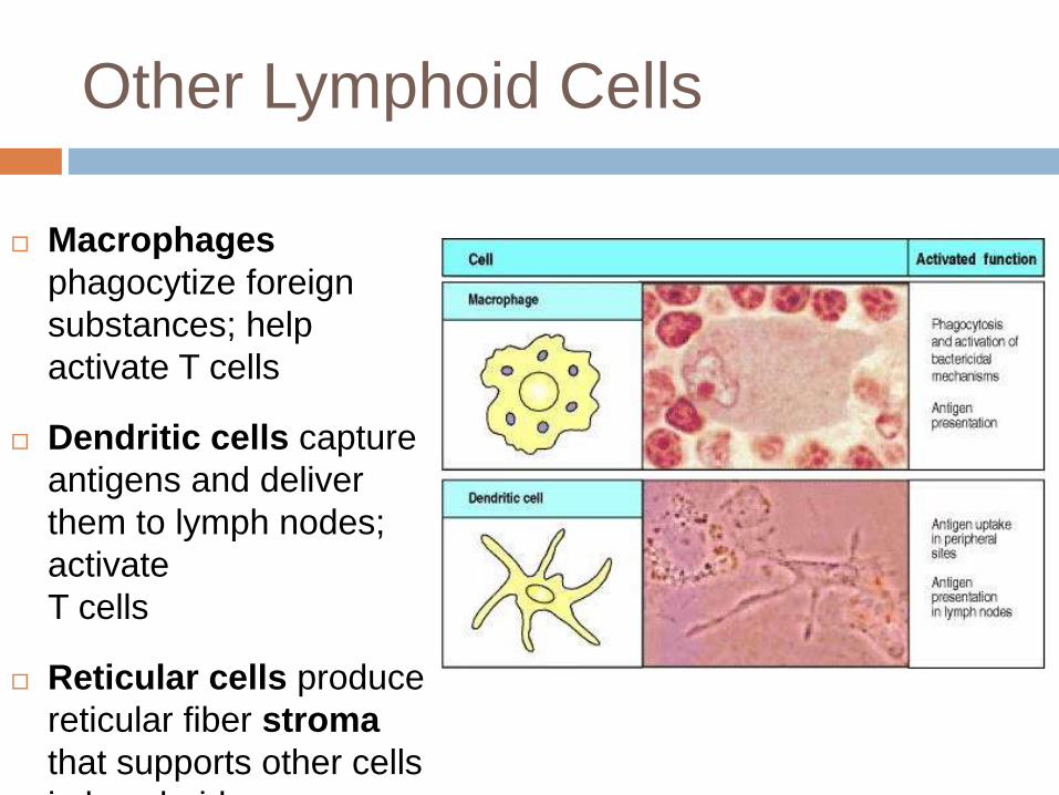
Other Lymphoid Cells
Macrophages
phagocytize foreign
substances; help
activate T cells
Dendritic cells capture
antigens and deliver
them to lymph nodes;
activate
T cells
Reticular cells produce
reticular fiber stroma
that supports other cells
in lymphoid organs

Structure of a Lymph Node
Afferentlymphaticvessels
Cortex
• Lymphoid follicle
• Germinal center
• Subcapsular sinus
Efferentlymphaticvessels Follicles
Trabecula
Subcapsularsinus
Capsule
Medullarycords
Medullarysinuses
Trabeculae
• Medullarysinus
• Medullarycord
Medulla
Hilum
Longitudinal view of the internal structure of a lymph
node and associated lymphatics
Photomicrograph of part of a lymph
node (72x)
Capsule
Goals/Objectives
Describe the structure and distribution of lymphatic vessels
Describe the source of lymph and mechanism(s) and pathway of lymph transport
Describe the general location, histological structure, and functions of lymph nodes
Describe the basic structure and cellular population of lymphoid tissue
Name and describe the other lymphoid organs of the body. Compare and contrast them with lymph nodes, structurally and functionally.
Other Lymphoid Tissue and
Organs
Houses, and provides proliferation site for,
lymphocytes
Surveillance vantage point for
lymphocytes and macrophages
Largely reticular connective tissue –
type of loose connective tissue
Two main types of lymphoid tissue
Diffuse lymphoid tissue (more scattered);
Lymphoid follicles (tight collections of B/T
cells)
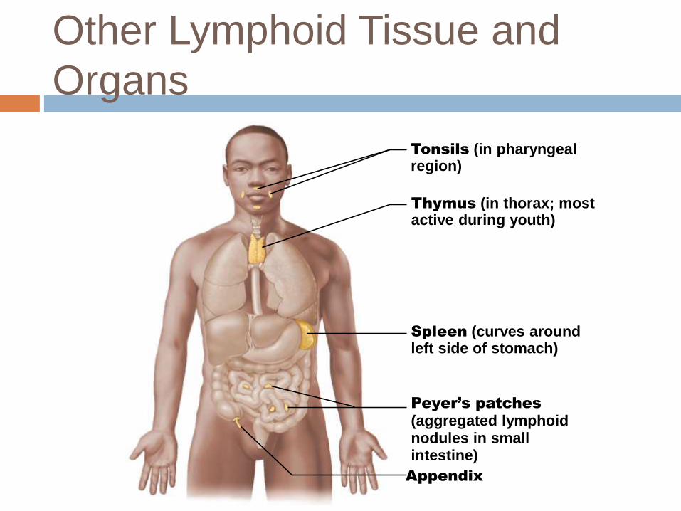
Other Lymphoid Tissue and
Organs
© 2013 Pearson Education, Inc.
Tonsils (in pharyngealregion)
Thymus (in thorax; most active during youth)
Spleen (curves around left side of stomach)
Peyer’s patches
(aggregated lymphoidnodules in smallintestine)
Appendix
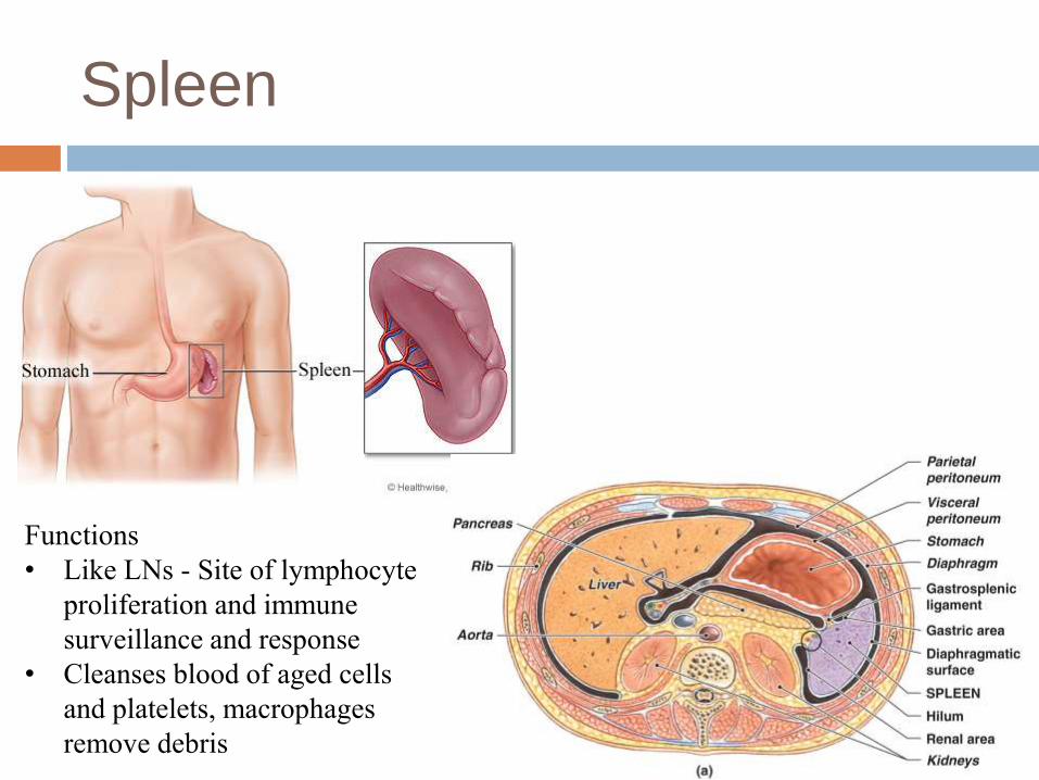
Spleen
Functions
• Like LNs - Site of lymphocyte
proliferation and immune
surveillance and response
• Cleanses blood of aged cells
and platelets, macrophages
remove debris
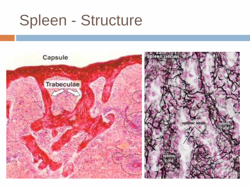
Spleen - Structure
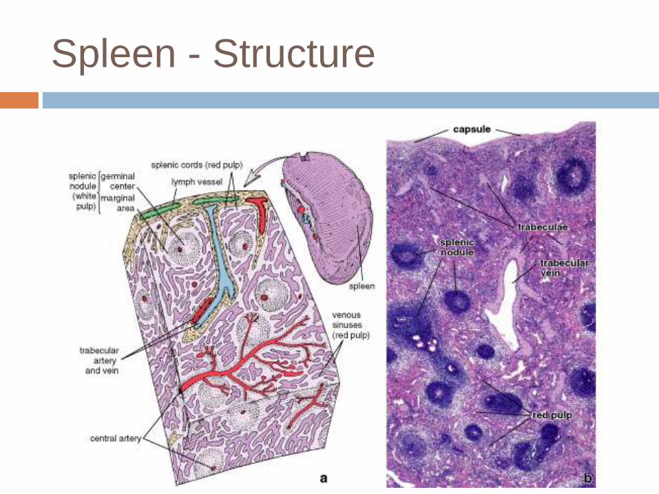
Spleen - Structure
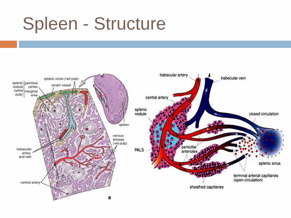
Spleen - Structure
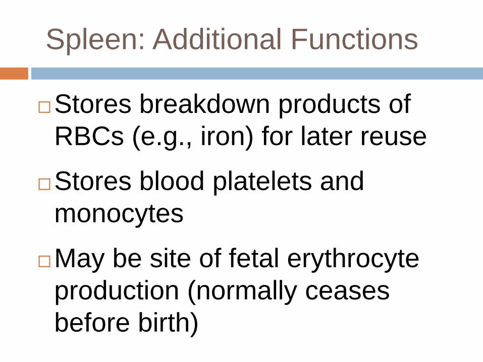
Spleen: Additional Functions
Stores breakdown products of
RBCs (e.g., iron) for later reuse
Stores blood platelets and
monocytes
May be site of fetal erythrocyte
production (normally ceases
before birth)
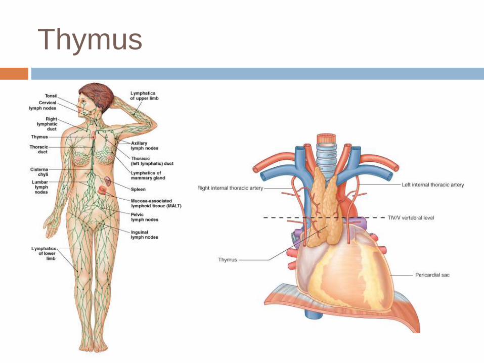
Thymus
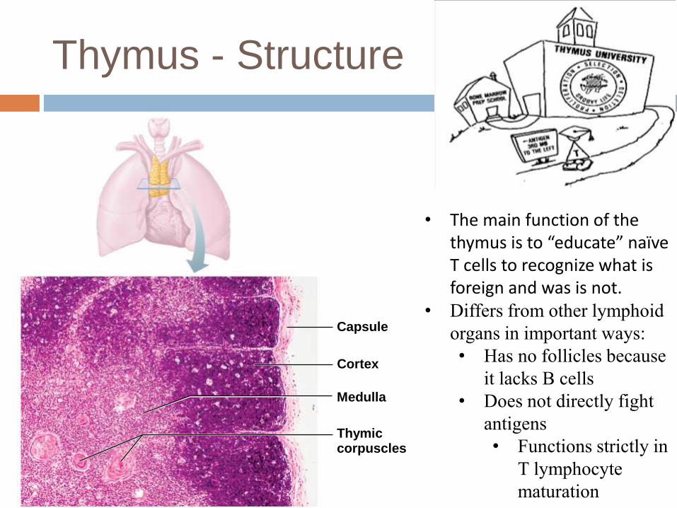
Thymus - Structure
Capsule
Cortex
Medulla
Thymiccorpuscles
• The main function of the thymus is to “educate” naïve T cells to recognize what is foreign and was is not.
• Differs from other lymphoid
organs in important ways:
• Has no follicles because
it lacks B cells
• Does not directly fight
antigens
• Functions strictly in
T lymphocyte
maturation
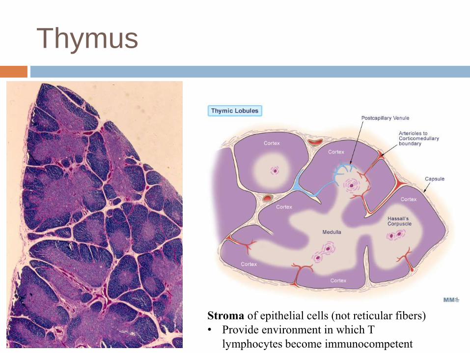
Thymus
Stroma of epithelial cells (not reticular fibers)
• Provide environment in which T
lymphocytes become immunocompetent
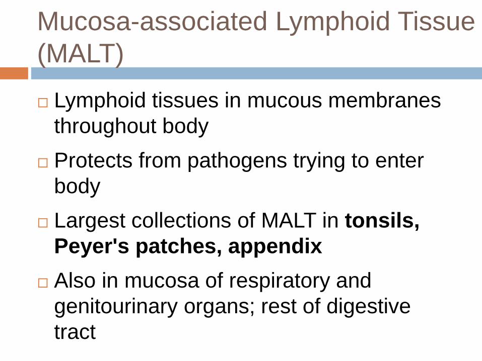
Mucosa-associated Lymphoid Tissue
(MALT)
Lymphoid tissues in mucous membranes
throughout body
Protects from pathogens trying to enter
body
Largest collections of MALT in tonsils,
Peyer's patches, appendix
Also in mucosa of respiratory and
genitourinary organs; rest of digestive
tract
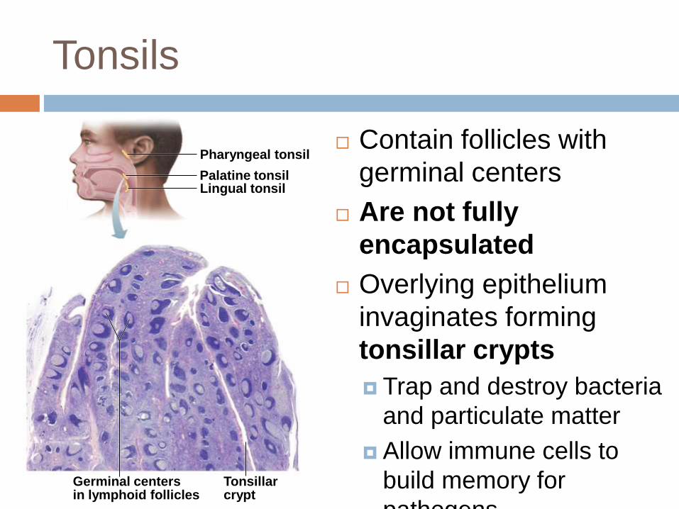
Tonsils
Pharyngeal tonsil
Palatine tonsilLingual tonsil
Germinal centersin lymphoid follicles
Tonsillarcrypt
Contain follicles with
germinal centers
Are not fully
encapsulated
Overlying epithelium
invaginates forming
tonsillar crypts
Trap and destroy bacteria
and particulate matter
Allow immune cells to
build memory for
pathogens
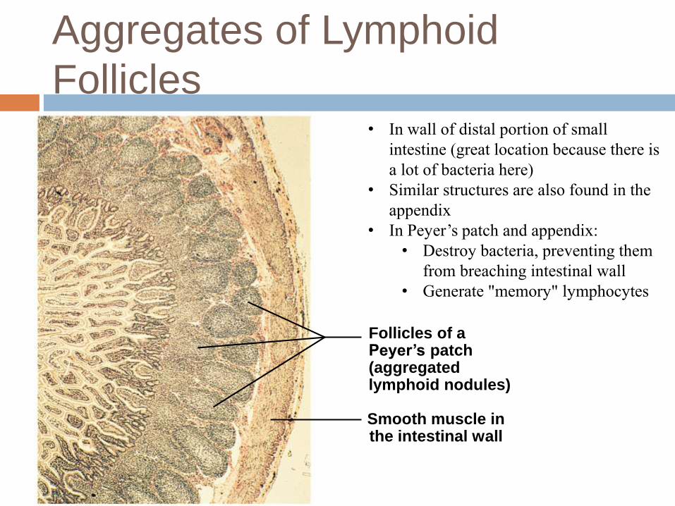
Aggregates of Lymphoid
Follicles
Follicles of aPeyer’s patch(aggregatedlymphoid nodules)
Smooth muscle inthe intestinal wall
• In wall of distal portion of small
intestine (great location because there is
a lot of bacteria here)
• Similar structures are also found in the
appendix
• In Peyer’s patch and appendix:
• Destroy bacteria, preventing them
from breaching intestinal wall
• Generate "memory" lymphocytes
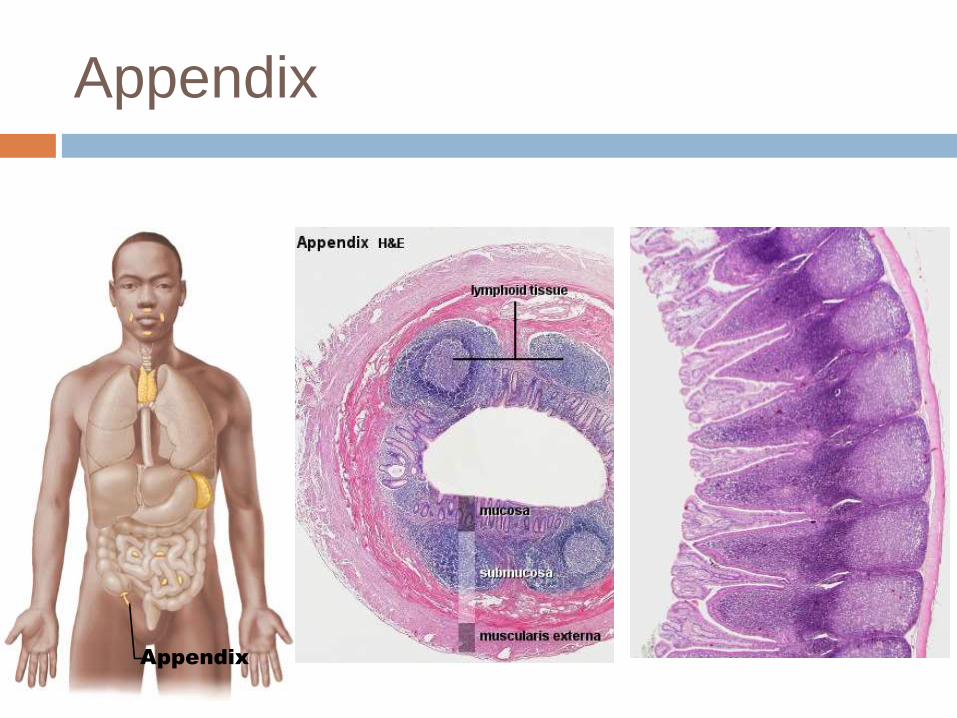
Appendix
Appendix
Clicker Question
The thymus is important for _______.
a) removal of foreign antigens
b) B lymphocyte maturation
c) T lymphocyte maturation
d) secretion of hormones that promote B
lymphocyte immunocompetence