Embed Size (px)
Citation preview
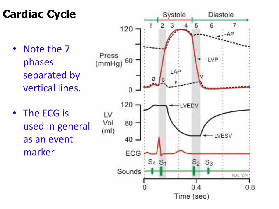
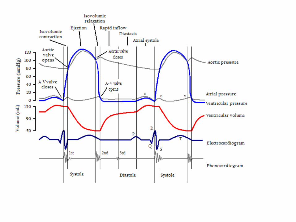
Cardiac Cycle
• Note the 7 phases separated by vertical lines.
• The ECG is used in general as an event marker

Cardiac Cycle
1. Atrial systole• Is preceded by the P wave (electrical activation of atria)• Contributes to ventricle filling• Produce the a wave of the JVP
2. Isovolumetric contraction• Begins after the onset of the QRS of the ECG• When ventricle pressure exceeds that of the atria, AV
valves close producing the 1st heart sound• Ventricular pressure increases isovolumetrically while
all four valves are closed • c wave of the JVP occurs due to high ventricular
pressure
Cardiac Cycle
3. Rapid ventricular ejection• When ventricle pressure exceeds aortic pressure, aortic
valve open• Rapid ejection of blood to aorta occurs• Most of the stroke volume is ejected during this phase• Same time, atrial filling begins.• T wave of the ECG occurs and the ventricles start
repolarising
4. Reduced ventricular ejection• Blood continues to be ejected slowly• Both ventricular and aortic pressure starts dropping• Atrial filling continues
Cardiac Cycle
5. Isovolumetric ventricular relaxation• Repolarisation of ventricle is now complete• Semilunar valves close and the 2nd heart sound occur• All 4 valves are closed while the ventricle relaxes –
causing a rapid drop in pressure• Dicrotic notch in the aortic pressure occur• When ventricle pressure becomes lower than the atrial
pressure mitral valve opens• v wave of the JVP occur at the end due to atrial filling
6. Rapid ventricular filling• Mitral valve open and ventricle fill from the atria
rapidly
Cardiac Cycle
7. Reduced ventricular filling (diastasis)• Longest phase of the cardiac cycle• Ventricle fill at a slower rate• The time for this varies with the heart rate
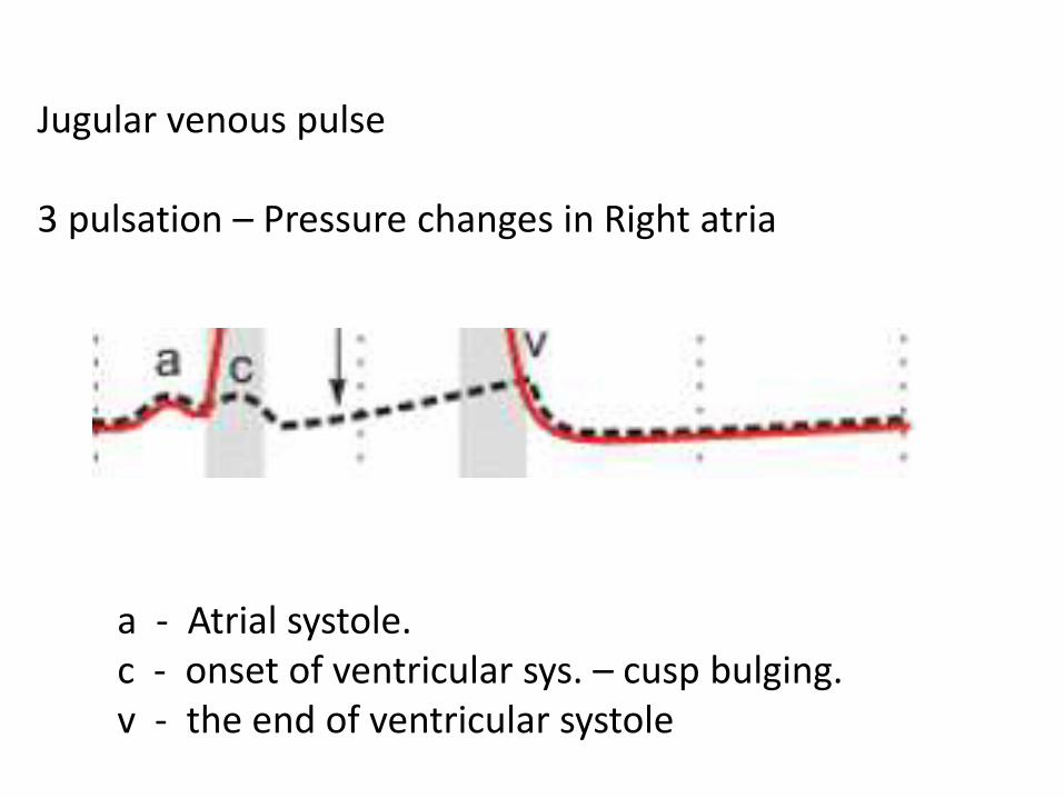
Jugular venous pulse
3 pulsation – Pressure changes in Right atria
a - Atrial systole.c - onset of ventricular sys. – cusp bulging.v - the end of ventricular systole
Vascular system
•Humans have Parallel vascular arrangement which is important.
•Flow in different capillary beds can be selectively altered
•Flow to vital organs can maintained at expense of other organs
•Constriction of a significant fraction of capillary bed can Increase total peripheral resistance
•This is not possible if the pumps are in series
Arteries• Thick walled, extensive elastic tissue & smooth muscle
• Under high pressure
• The blood volume contained in them are called the stressed volume
Arterioles
• Smallest branches of the arteries
• Site of highest resistance in the cardiovascular system
• Have smooth muscle walls which have extensive autonomic innervation
• 1 adrenergic - in the skin and splanchnic arterioles
• Β2 adrenergic – in the skeletal muscle arterioles
Components of the vasculature
Capillaries•Have the largest cross sectional surface area
• Consist of a single layer of cells – thin walled
•Are the site of exchange of nutrients, water & gases
Venules
•Are formed from merged capillaries
Components of the vasculature
Veins• Progressively forms larger and larger veins
•Are thin walled
•Are under low pressure
• Contains the highest proportion of the blood in the cardiovascular system
• Blood volume in the veins is called the unstressed volume
•Are innervated by autonomic fibres
Components of the vasculature
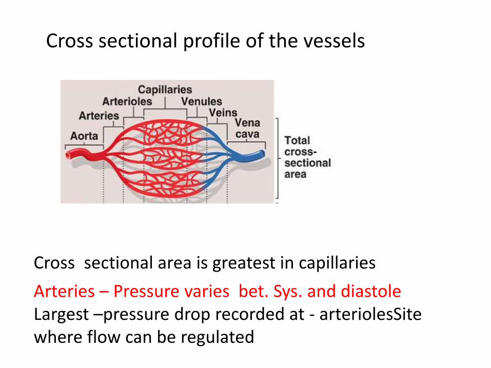
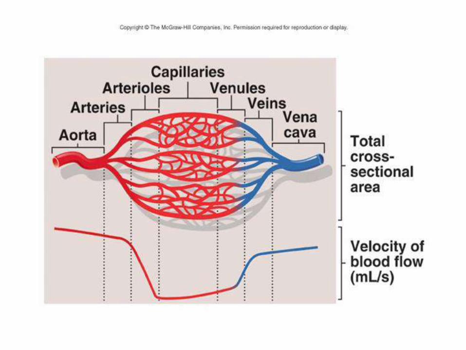
Cross sectional profile of the vessels
Cross sectional area is greatest in capillaries
Arteries – Pressure varies bet. Sys. and diastoleLargest –pressure drop recorded at - arteriolesSitewhere flow can be regulated

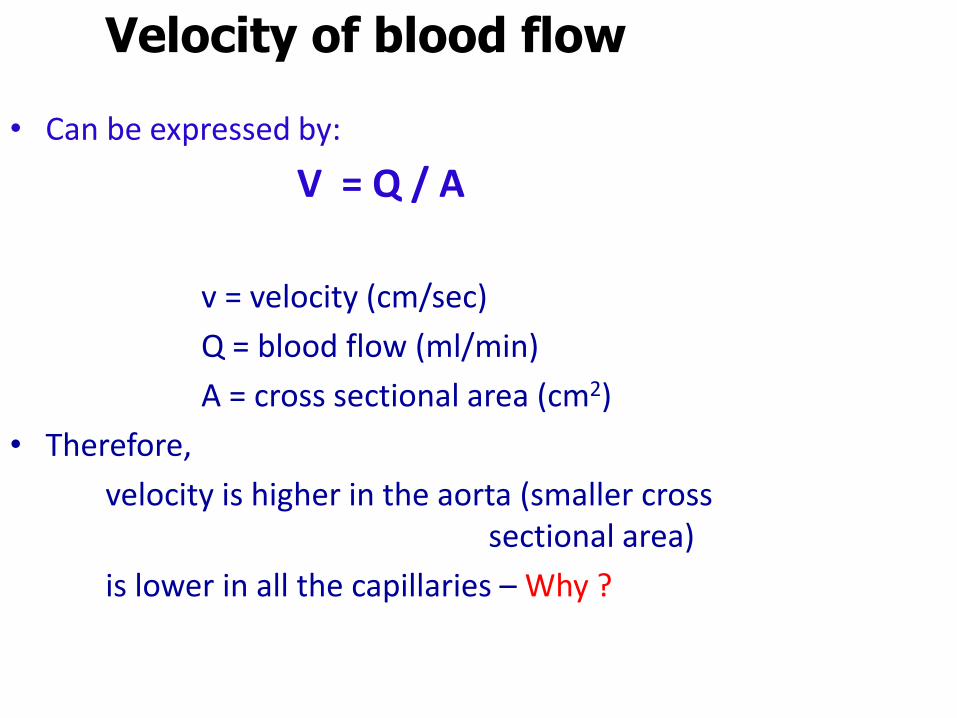
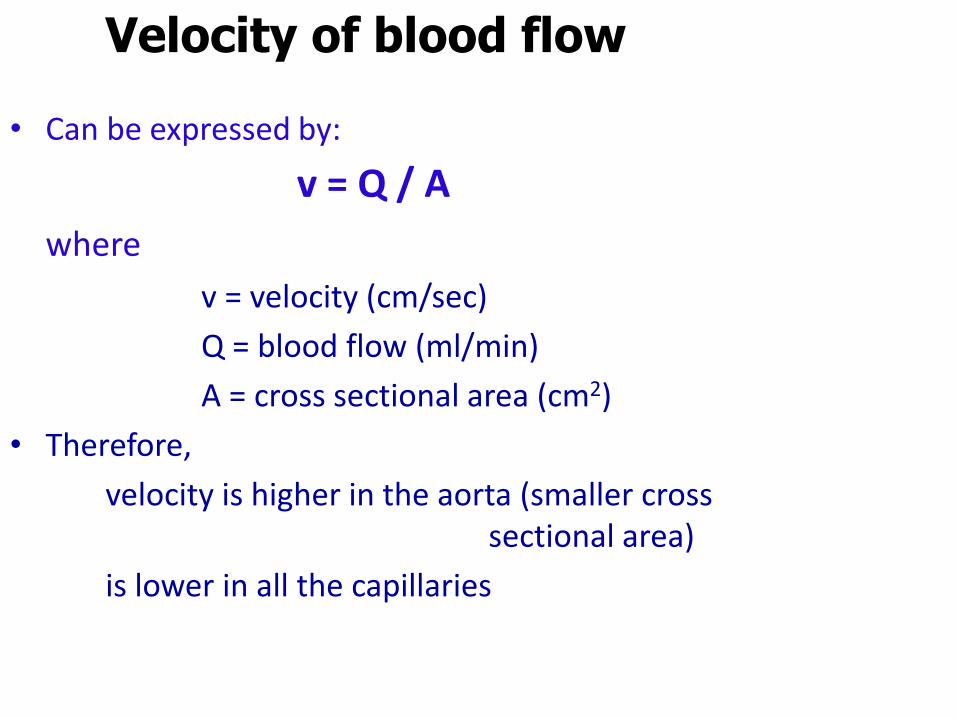
Velocity of blood flow
• Can be expressed by:
V = Q / A
v = velocity (cm/sec)
Q = blood flow (ml/min)
A = cross sectional area (cm2)
• Therefore,
velocity is higher in the aorta (smaller cross sectional area)
is lower in all the capillaries – Why ?
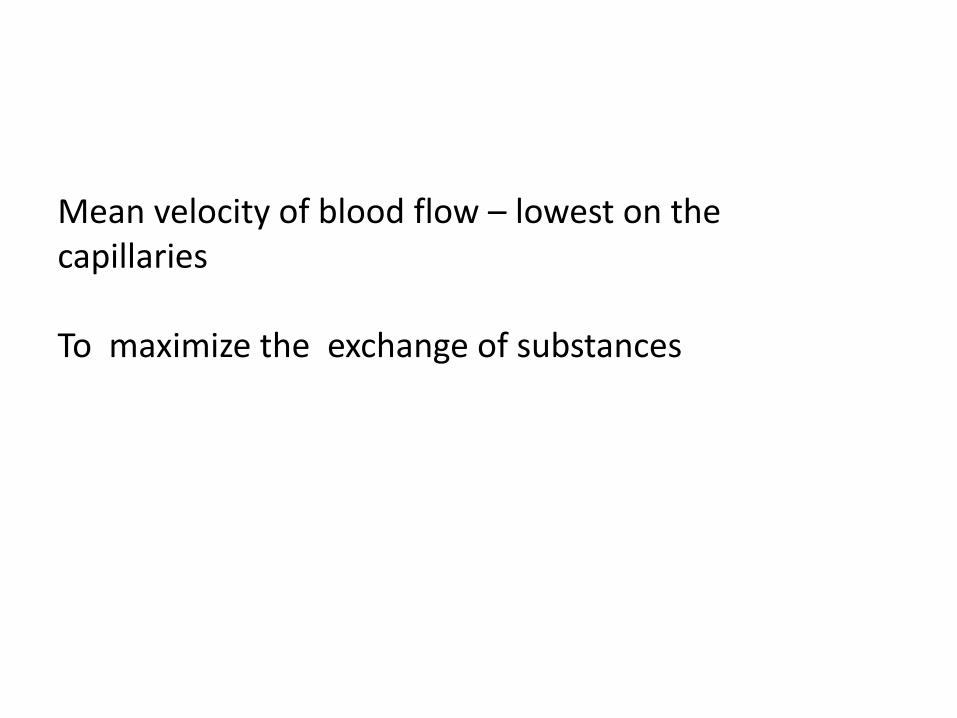
Mean velocity of blood flow – lowest on the capillaries
To maximize the exchange of substances
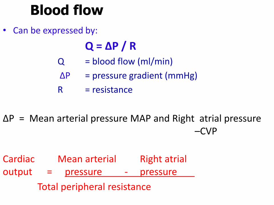
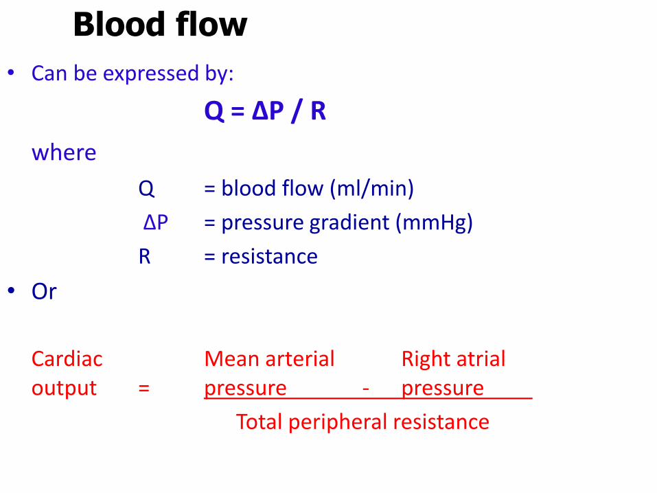
Blood flow
• Can be expressed by:
Q = ∆P / RQ = blood flow (ml/min)
∆P = pressure gradient (mmHg)
R = resistance
ΔP = Mean arterial pressure MAP and Right atrial pressure –CVP
Cardiac Mean arterial Right atrialoutput = pressure - pressure
Total peripheral resistance
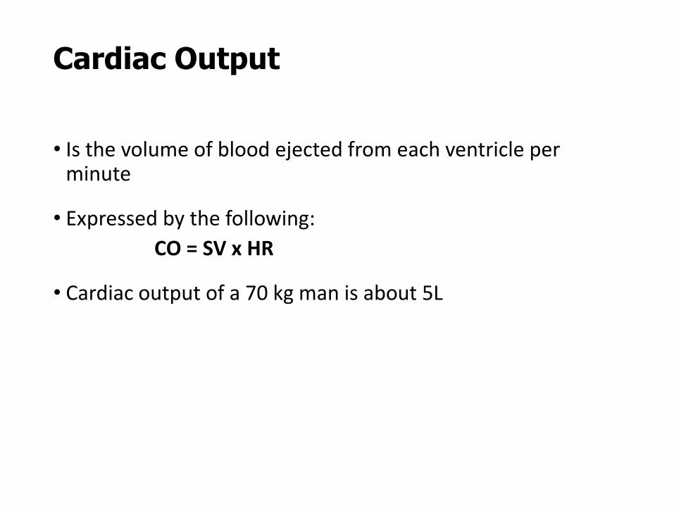
Cardiac Output
• Is the volume of blood ejected from each ventricle per minute
• Expressed by the following:
CO = SV x HR
• Cardiac output of a 70 kg man is about 5L
Stroke volume
• Is the volume of blood ejected from each ventricle on each beat
• Expressed by the following:
Stroke volume = EDV – ESV
• Normally is about 70 ml
(as EDV = 140 ml & ESV = 70 ml)
• SV = (~2 x pulse pressure)
Cardiac Index
•Expressed by the following:
cardiac index = CO / body surface area
•Gives a correct estimation of the cardiac output depending on the size of the person
Ejection fraction
• Is the fraction of end-diastolic volume ejected in each stroke volume
• Is normally 0.55 or 55%
• Is expressed by the following equation:
Ejection fraction = SV
End-diastolic volume
Stroke work
• Is the work the heart performs on each beat
• Is expressed by;
Stroke work = Aortic pressure x Stroke volume
• Fatty acids are the primary energy source for stroke work
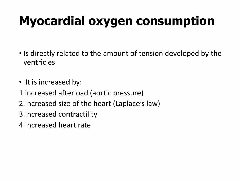
Myocardial oxygen consumption
• Is directly related to the amount of tension developed by the ventricles
• It is increased by:
1.increased afterload (aortic pressure)
2.Increased size of the heart (Laplace’s law)
3.Increased contractility
4.Increased heart rate
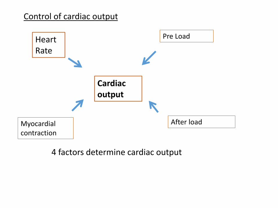
Control of cardiac output
Cardiac output
Heart Rate
Pre Load
Myocardial contraction
After load
4 factors determine cardiac output
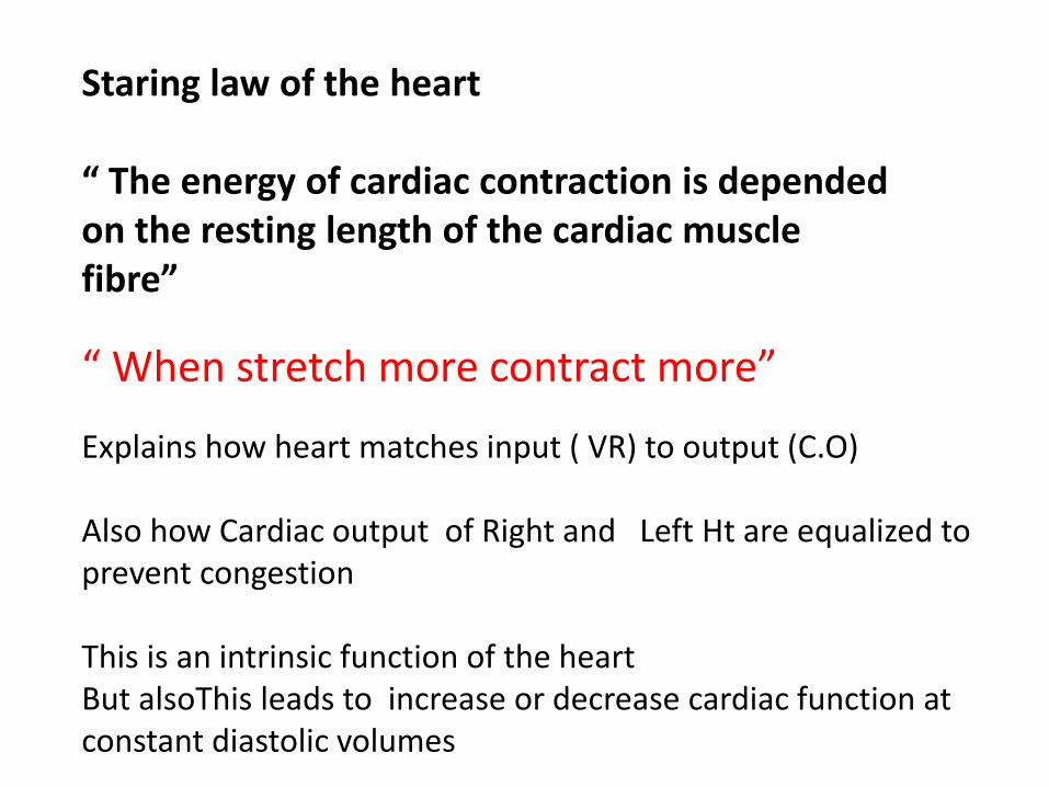
Staring law of the heart
“ The energy of cardiac contraction is depended on the resting length of the cardiac muscle fibre”
“ When stretch more contract more”
Explains how heart matches input ( VR) to output (C.O)
Also how Cardiac output of Right and Left Ht are equalized to prevent congestion
This is an intrinsic function of the heart But alsoThis leads to increase or decrease cardiac function at constant diastolic volumes
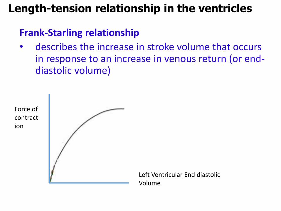
Frank-Starling relationship
• describes the increase in stroke volume that occurs in response to an increase in venous return (or end-diastolic volume)
Length-tension relationship in the ventricles
Force of contraction
Left Ventricular End diastolic Volume
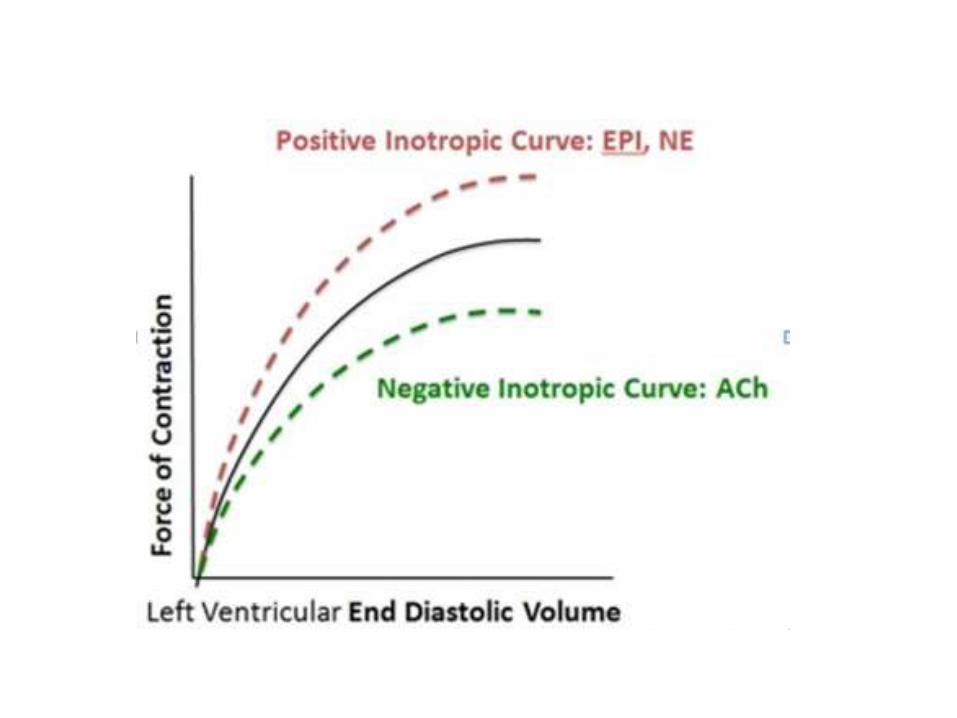
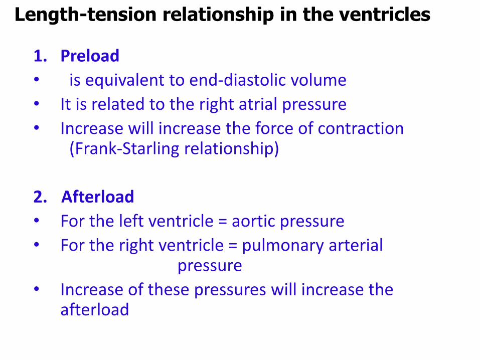
1. Preload
• is equivalent to end-diastolic volume
• It is related to the right atrial pressure
• Increase will increase the force of contraction (Frank-Starling relationship)
2. Afterload
• For the left ventricle = aortic pressure
• For the right ventricle = pulmonary arterial pressure
• Increase of these pressures will increase the afterload
Length-tension relationship in the ventricles
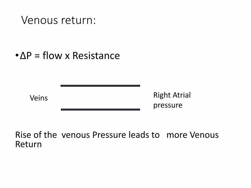
Venous return:
•ΔP = flow x Resistance
Rise of the venous Pressure leads to more Venous Return
Right Atrial pressure
Veins
Venous Return
RAP
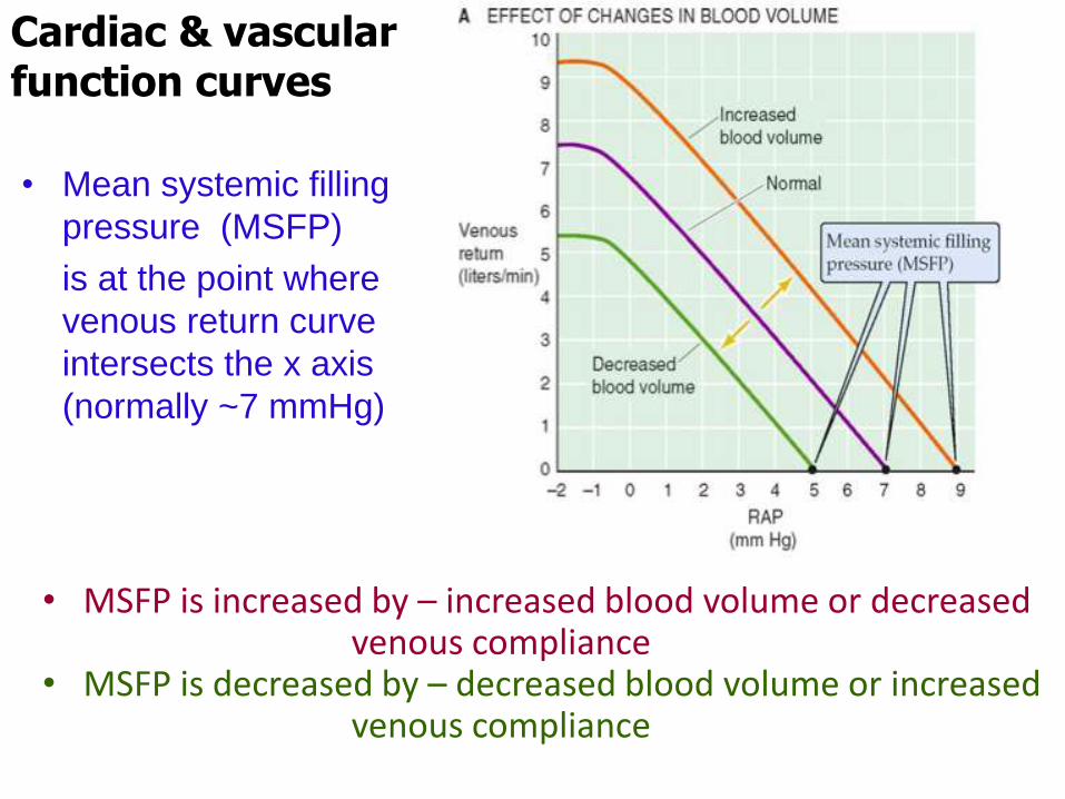
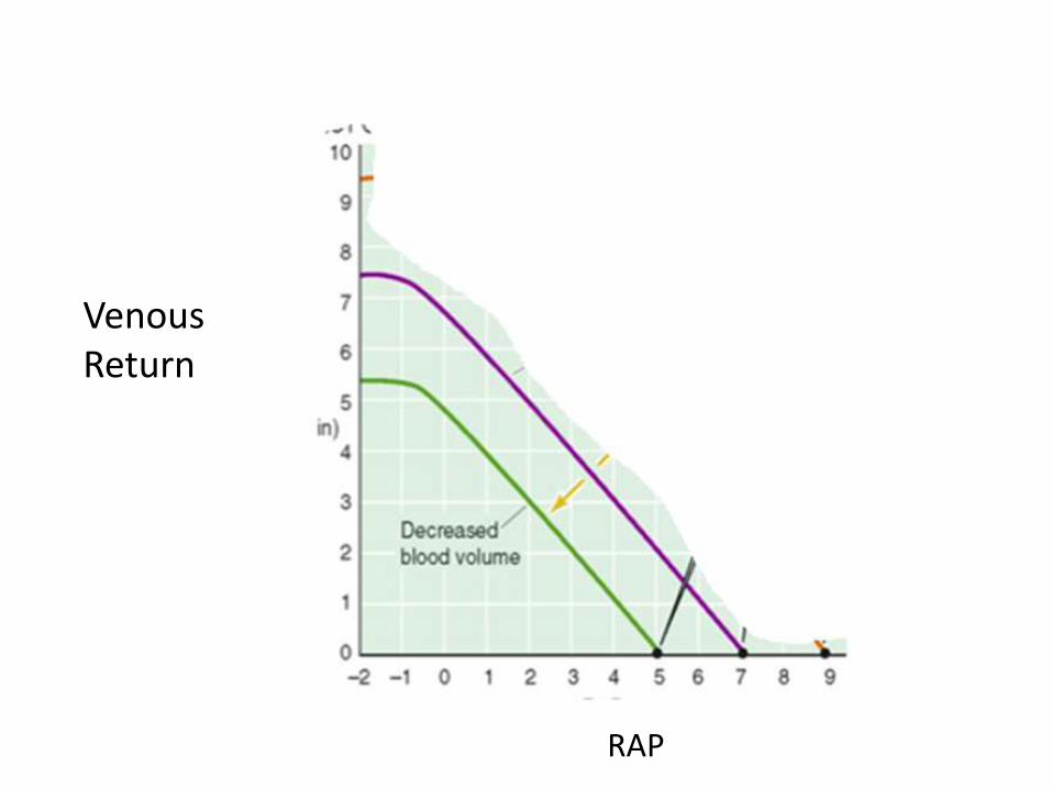
• MSFP is increased by – increased blood volume or decreased venous compliance
• MSFP is decreased by – decreased blood volume or increased venous compliance
Cardiac & vascular function curves
• Mean systemic filling
pressure (MSFP)
is at the point where
venous return curve
intersects the x axis
(normally ~7 mmHg)
Control of cardiac output
2 relationships
1. Between Venous Return and Rt atrial pressure
2. Between cardiac output and preload (Starling's law)

Cardiac & vascular function curves
• Its a simultaneous plot of cardiac output and venous return as a function of right atrial pressure or end diastolic volume
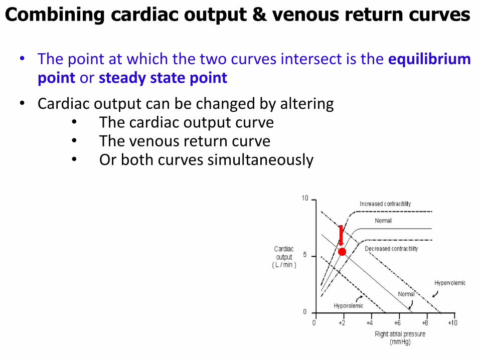
• The point at which the two curves intersect is the equilibrium point or steady state point
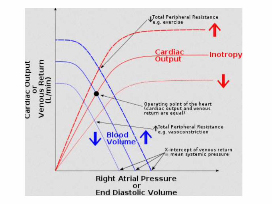
• Cardiac output can be changed by altering • The cardiac output curve• The venous return curve• Or both curves simultaneously
Combining cardiac output & venous return curves
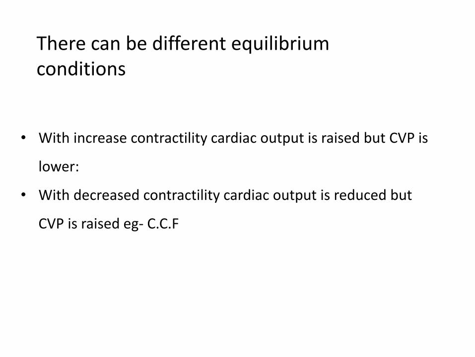
There can be different equilibrium conditions
• With increase contractility cardiac output is raised but CVP is
lower:
• With decreased contractility cardiac output is reduced but
CVP is raised eg- C.C.F
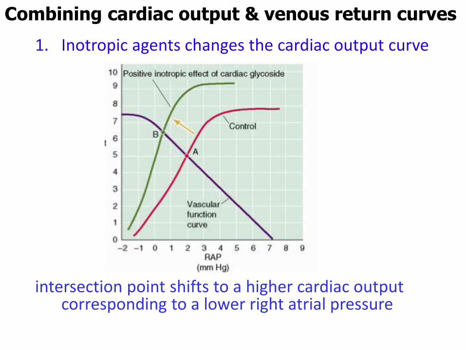
1. Inotropic agents changes the cardiac output curve
intersection point shifts to a higher cardiac output corresponding to a lower right atrial pressure
Combining cardiac output & venous return curves
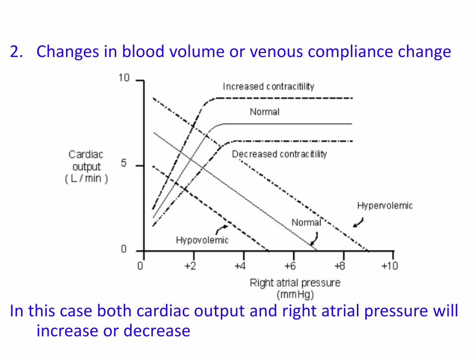
2. Changes in blood volume or venous compliance change
In this case both cardiac output and right atrial pressure will increase or decrease
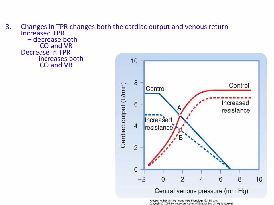
3. Changes in TPR changes both the cardiac output and venous returnIncreased TPR
– decrease both CO and VR
Decrease in TPR – increases both
CO and VR
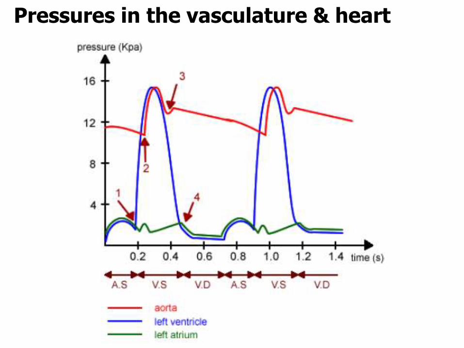
Pressures in the vasculature & heart
Velocity of blood flow
• Can be expressed by:
v = Q / A
where
v = velocity (cm/sec)
Q = blood flow (ml/min)
A = cross sectional area (cm2)
• Therefore,
velocity is higher in the aorta (smaller cross sectional area)
is lower in all the capillaries
Blood flow
• Can be expressed by:
Q = ∆P / R
where
Q = blood flow (ml/min)
∆P = pressure gradient (mmHg)
R = resistance
• Or
Cardiac Mean arterial Right atrialoutput = pressure - pressure
Total peripheral resistance
Resistance
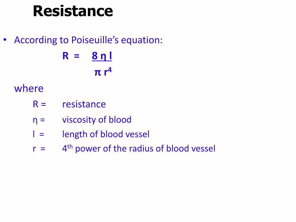
• According to Poiseuille’s equation:
R = 8 η l
π r4
where
R = resistance
η = viscosity of blood
l = length of blood vessel
r = 4th power of the radius of blood vessel
Resistance
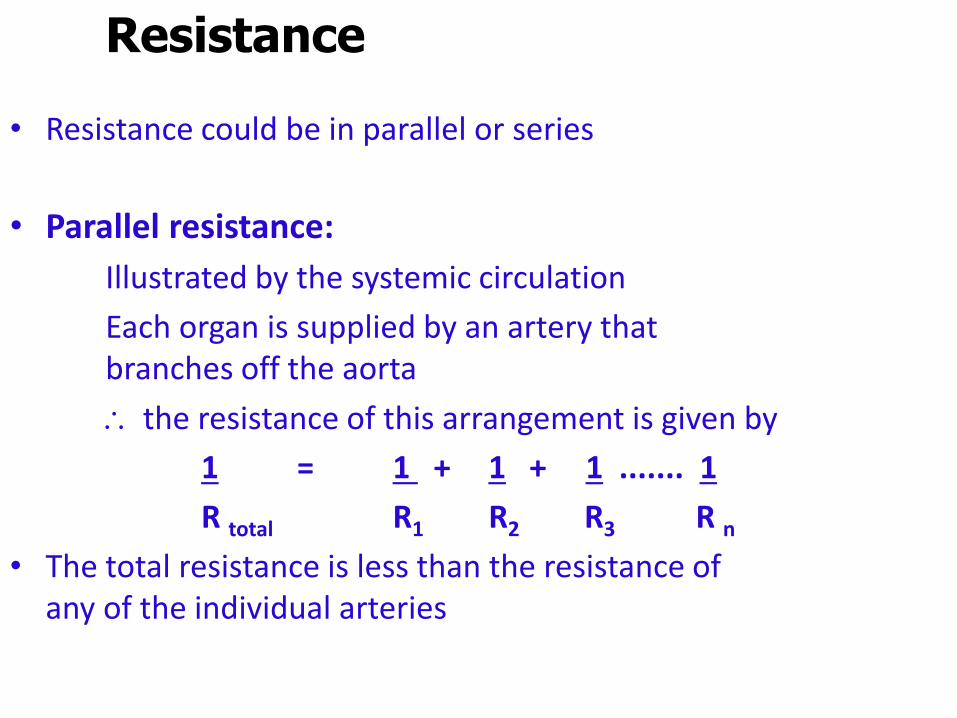
• Resistance could be in parallel or series
• Parallel resistance:
Illustrated by the systemic circulation
Each organ is supplied by an artery that branches off the aorta
the resistance of this arrangement is given by
1 = 1 + 1 + 1 ....... 1
R total R1 R2 R3 R n• The total resistance is less than the resistance of
any of the individual arteries
Resistance
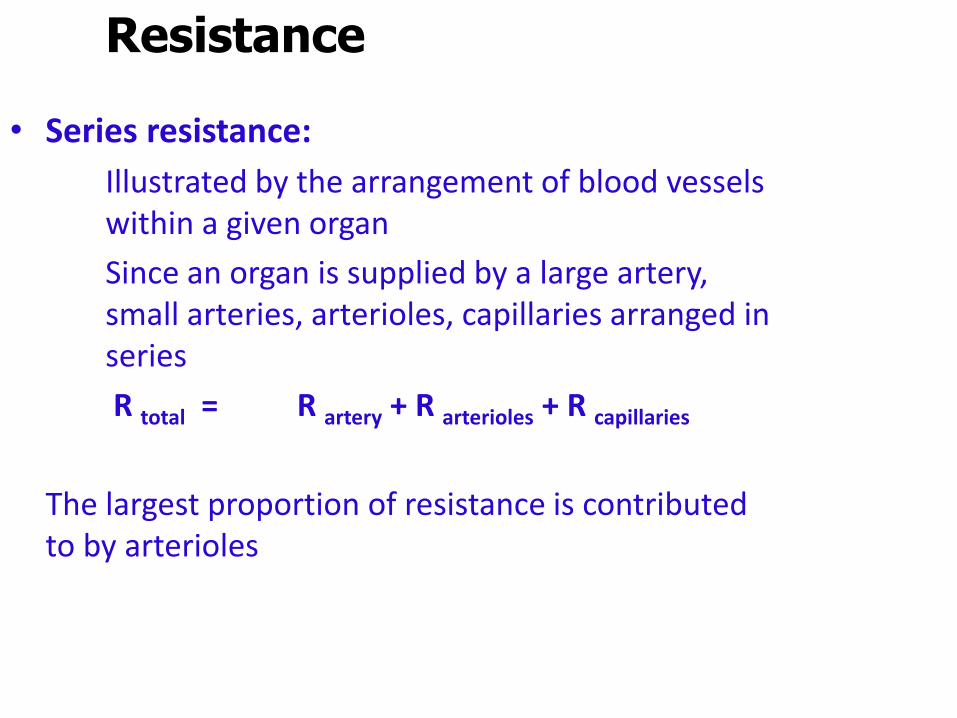
• Series resistance:
Illustrated by the arrangement of blood vessels within a given organ
Since an organ is supplied by a large artery, small arteries, arterioles, capillaries arranged in series
R total = R artery + R arterioles + R capillaries
The largest proportion of resistance is contributed to by arterioles
Blood flow – laminar vs turbulent
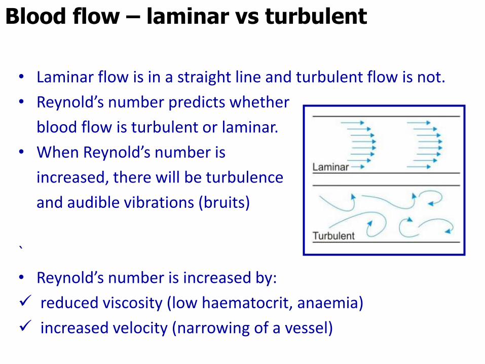
• Laminar flow is in a straight line and turbulent flow is not.
• Reynold’s number predicts whether
blood flow is turbulent or laminar.
• When Reynold’s number is
increased, there will be turbulence
and audible vibrations (bruits)
`
• Reynold’s number is increased by:
reduced viscosity (low haematocrit, anaemia)
increased velocity (narrowing of a vessel)
C =
Capacitance (compliance)
• Describes the distensibility of blood vessels
• Is inversely related to elastance
• Capacitance is given by:
C = V / P
where
C = capacitance (ml/mmHg)
V = volume (ml)
P = pressure (mmHg)
• Describes how volume changes in response to changes in pressure
C =• Capacitance is much greater for veins than for
arteries
• Changes in venous capacitance changes the venous blood volume
• Eg: decrease in venous capacitance decreases the unstressed volume (venous volume) and increases the stressed volume (arterial volume)
• Capacitance of arteries decreases with age. Arteries become stiffer and less distensible
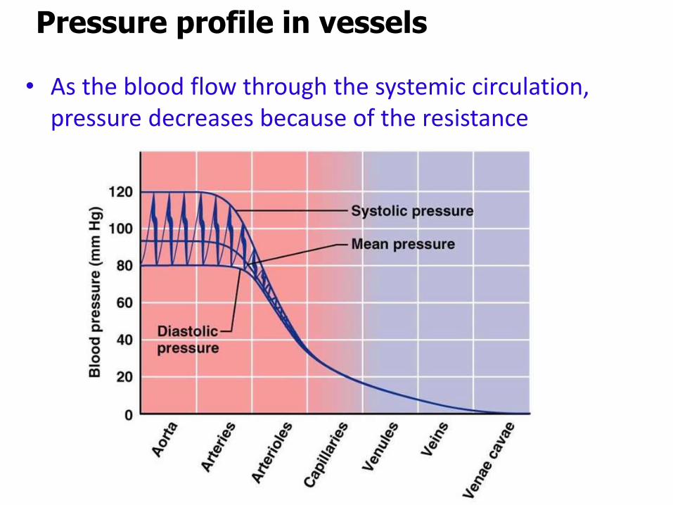
Pressure profile in vessels
• As the blood flow through the systemic circulation, pressure decreases because of the resistance
C =• Pressure is highest in the aorta and lowest in
the venae cavae
• The largest decrease in pressure occurs across the arterioles (site of highest resistance)
• Mean pressures:
aorta - 100 mmHg
end of arterioles - 30 mmHg
vena cava - 4 mmHg
Pressure profile in vessels
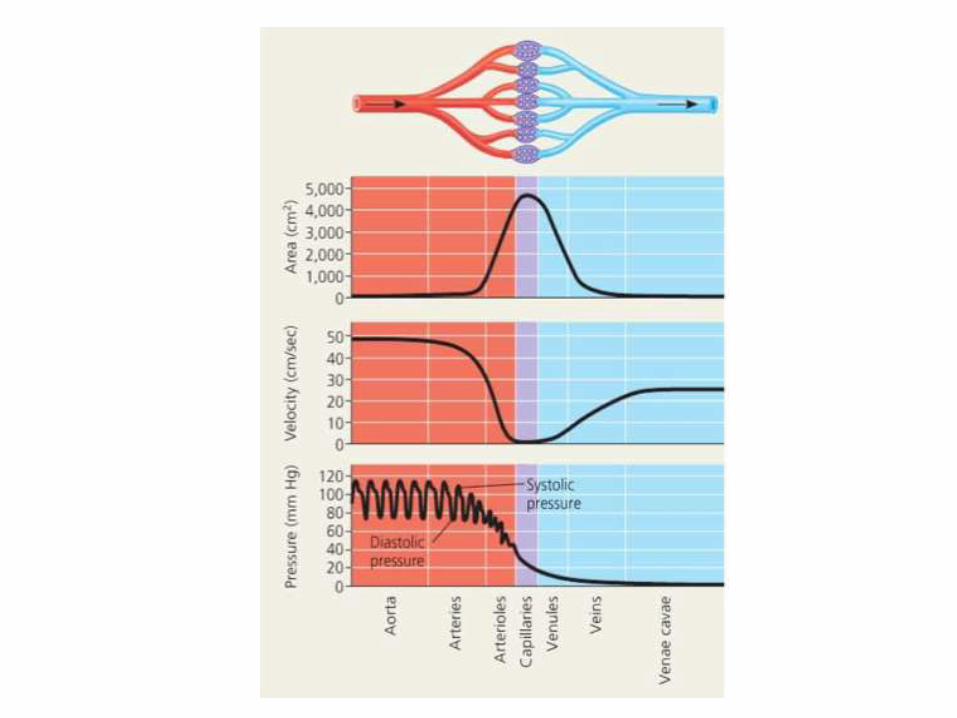
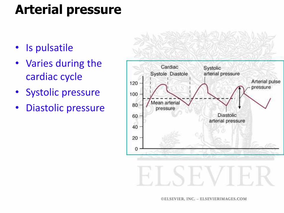
• Is pulsatile
• Varies during the cardiac cycle
• Systolic pressure
• Diastolic pressure
Arterial pressure
C =
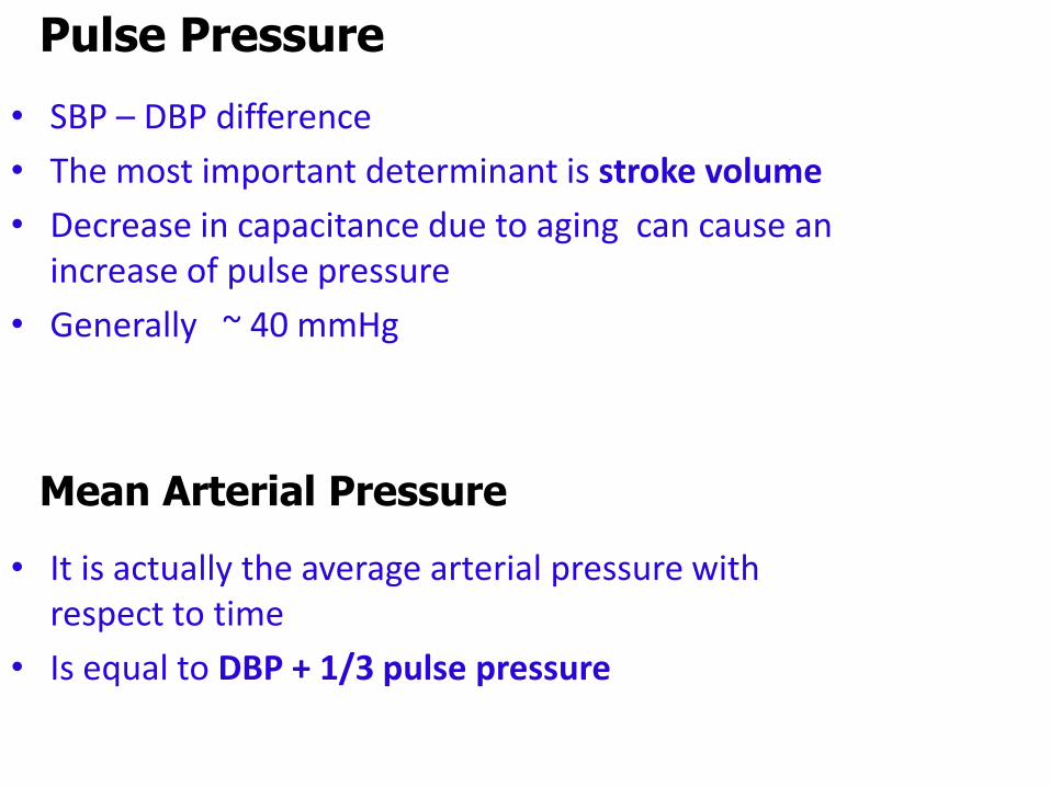
Pulse Pressure
• SBP – DBP difference
• The most important determinant is stroke volume
• Decrease in capacitance due to aging can cause an increase of pulse pressure
• Generally ~ 40 mmHg
Mean Arterial Pressure
• It is actually the average arterial pressure with respect to time
• Is equal to DBP + 1/3 pulse pressure
C =
Venous pressure
• Is very low
• Has a high capacitance
• Able to hold a large volume without an increase in pressure

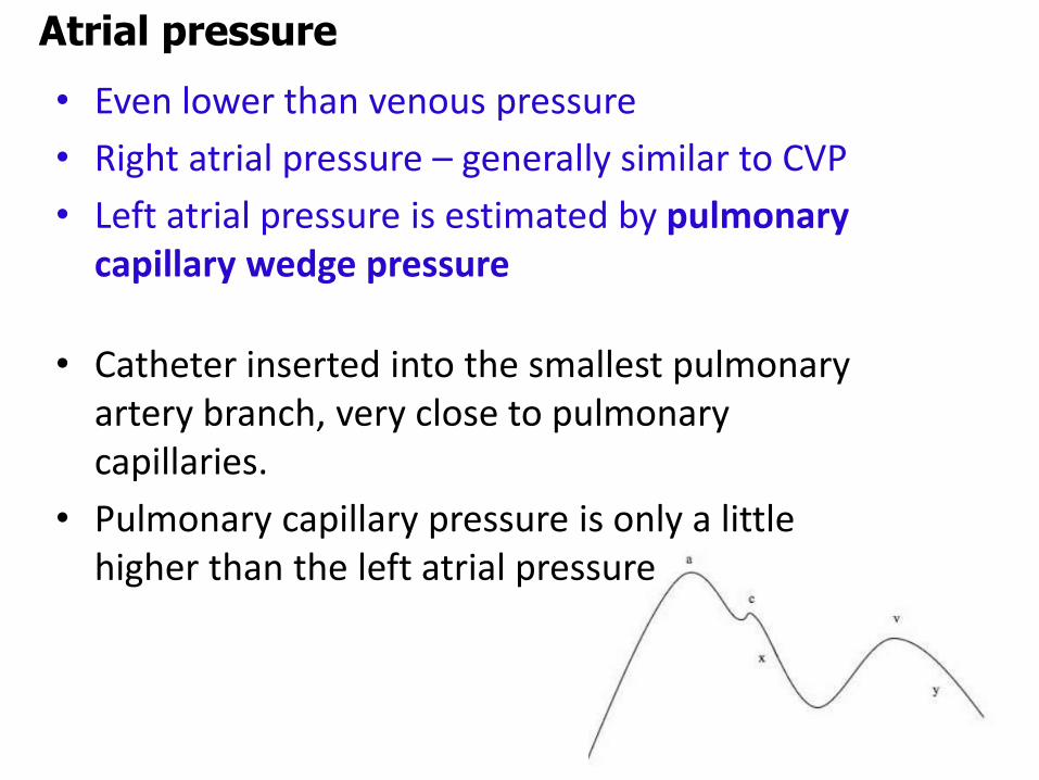
Atrial pressure
• Even lower than venous pressure
• Right atrial pressure – generally similar to CVP
• Left atrial pressure is estimated by pulmonary capillary wedge pressure
• Catheter inserted into the smallest pulmonary artery branch, very close to pulmonary capillaries.
• Pulmonary capillary pressure is only a little higher than the left atrial pressure

C =
Venous pressure
• Is very low
• Has a high capacitance
• Able to hold a large volume without an increase in pressure
Atrial pressure
• Even lower than venous pressure
• Right atrial pressure – generally similar to CVP
• Left atrial pressure is estimated by pulmonary capillary wedge pressure
• Catheter inserted into the smallest pulmonary artery branch, very close to pulmonary capillaries.
• Pulmonary capillary pressure is only a little higher than the left atrial pressure
•Most important mechanisms are:
• the fast neurally mediated baroreceptor mechanism
• the slower hormonally mediated renal mechanisms
•Other mechanisms include;
Atrial stretch receptors
local vasoconstrictors and dilators
Regulation of arterial blood pressure
•Most of the vasculature is innervated by sympathetics
•Sympathetic noradrenergic fibres terminate on resistant vessels – mediates vasoconstriction
•Exceptions:- Skeletal muscle vessels undergo vasodilatation via β2
due to circulating adrenaline- parasympathetic innervation is seen in
some erectile tissue of reproductive organs, uterine vesselssome facial vesselsblood vessels in the salivary glands
•Sympathetic innervation of veins cause a reduction in capacitance and an increase in venous return
Innervation of blood vessels
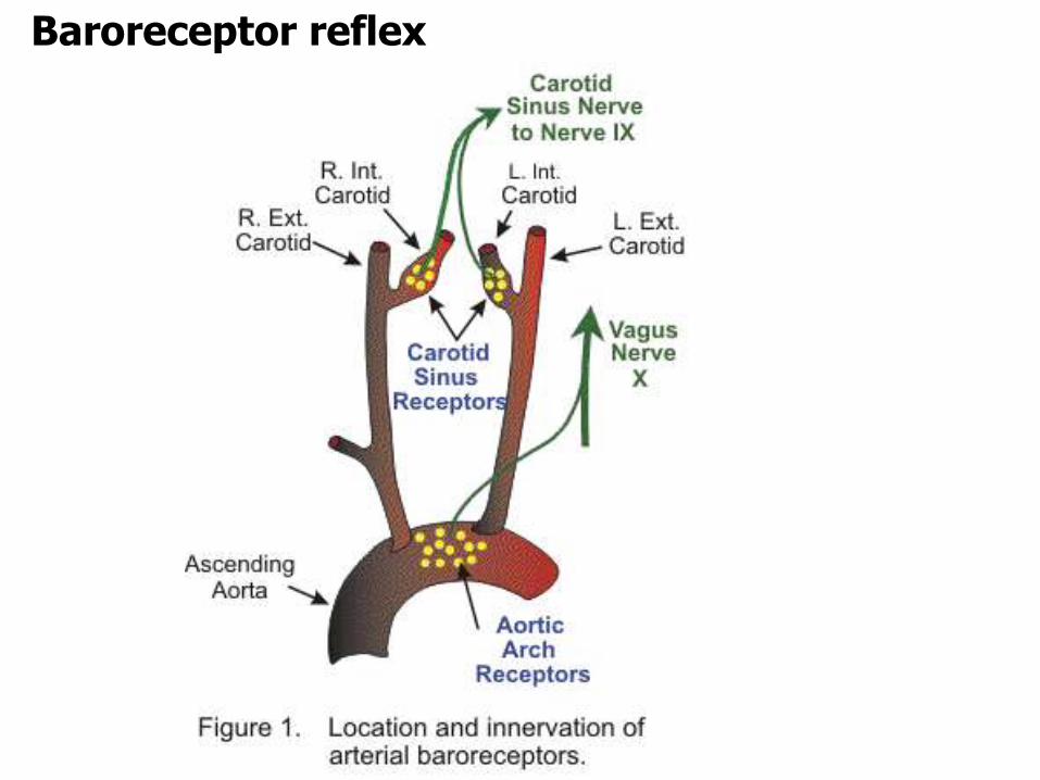
•There are pressure receptors located in the cardiovascular system
•Those that monitor arterial pressure:In the carotid sinus & aortic arch
• Low-pressure receptors (cardiopulmonary receptors)In walls of right atria at vena caval entrancewall of left atriapulmonary circulation
•BaroreceptorsLocated within the walls of the carotid sinus near the bifurcation of the common carotid artery and aortic arch
Baroreceptor reflex
Baroreceptor reflex

Baroreceptor reflex
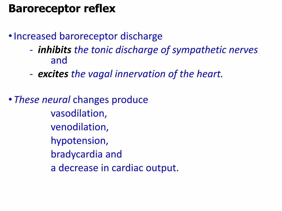
• Increased baroreceptor discharge - inhibits the tonic discharge of sympathetic nerves
and - excites the vagal innervation of the heart.
•These neural changes produce vasodilation, venodilation, hypotension, bradycardia and a decrease in cardiac output.
Baroreceptor reflex
•There are two types of stretch receptors in the atria
Those discharging in atrial systole &
In late diastole during atrial filling
•Effect of increase discharge from the include;
vasodilatation & a fall in BP
But, an increase in heart rate
Cardiopulmonary receptors
• Peripheral chemoreceptors found in the;
Aortic & carotid bodies
•Have a very high blood flow
•Activated by: low PaO2, PCO2 and pH
• Stimulated by hypoxic hypoxia
•Main effects are on respiration, but also leads to vasoconstriction
•Direct effect of chemoreceptor activation is
hypoxia, increased catecholamines from medulla which increases HR and BP
Peripheral chemoreceptor reflex
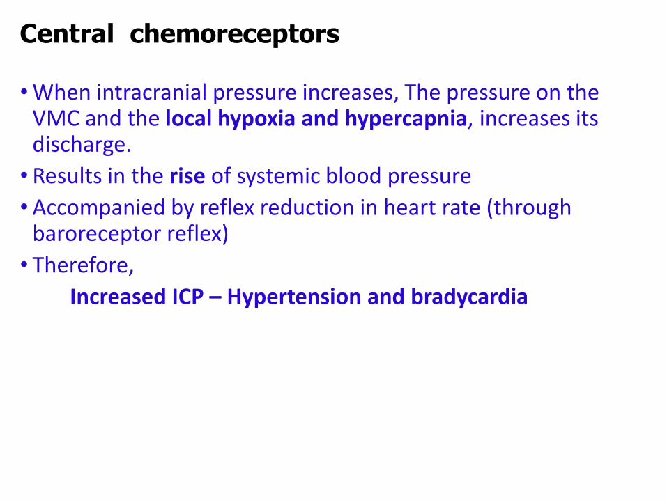
•When intracranial pressure increases, The pressure on the VMC and the local hypoxia and hypercapnia, increases its discharge.
• Results in the rise of systemic blood pressure
•Accompanied by reflex reduction in heart rate (through baroreceptor reflex)
• Therefore,
Increased ICP – Hypertension and bradycardia
Central chemoreceptors
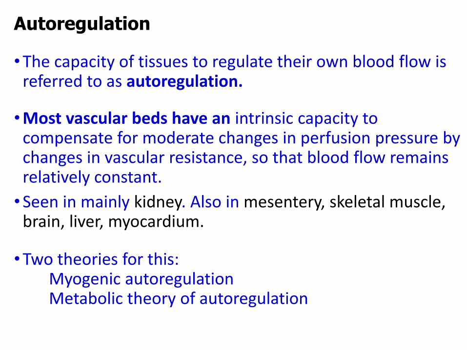
•The capacity of tissues to regulate their own blood flow is referred to as autoregulation.
•Most vascular beds have an intrinsic capacity to compensate for moderate changes in perfusion pressure by changes in vascular resistance, so that blood flow remains relatively constant.
•Seen in mainly kidney. Also in mesentery, skeletal muscle, brain, liver, myocardium.
•Two theories for this:Myogenic autoregulationMetabolic theory of autoregulation
Autoregulation
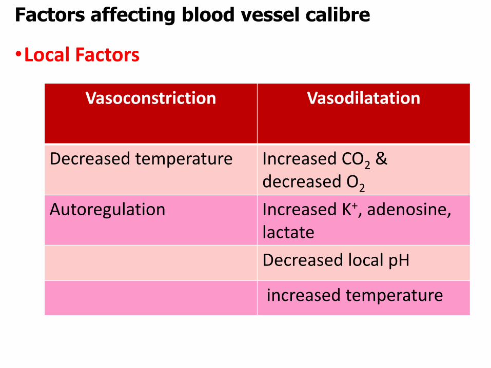
•Local Factors
Factors affecting blood vessel calibre
Vasoconstriction Vasodilatation
Decreased temperature Increased CO2 & decreased O2
Autoregulation Increased K+, adenosine, lactate
Decreased local pH
increased temperature
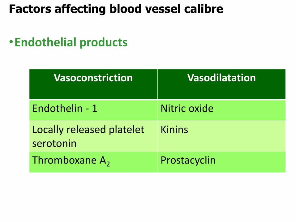
•Endothelial products
Factors affecting blood vessel calibre
Vasoconstriction Vasodilatation
Endothelin - 1 Nitric oxide
Locally released platelet serotonin
Kinins
Thromboxane A2 Prostacyclin
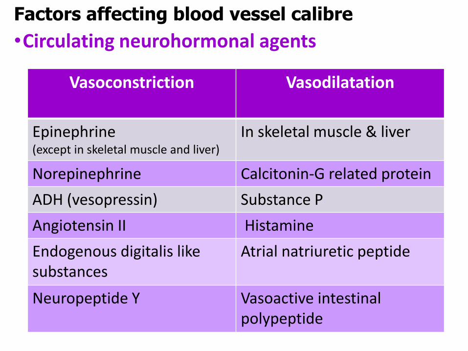
•Circulating neurohormonal agents
Factors affecting blood vessel calibre
Vasoconstriction Vasodilatation
Epinephrine(except in skeletal muscle and liver)
In skeletal muscle & liver
Norepinephrine Calcitonin-G related protein
ADH (vesopressin) Substance P
Angiotensin II Histamine
Endogenous digitalis like substances
Atrial natriuretic peptide
Neuropeptide Y Vasoactive intestinal polypeptide
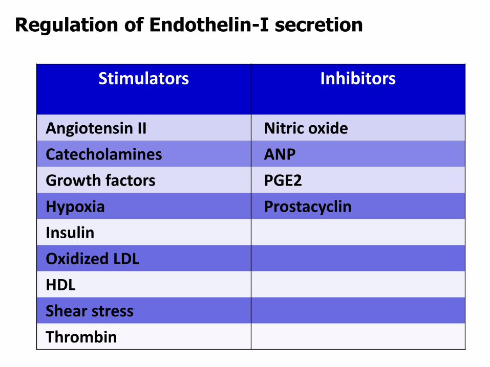
Stimulators Inhibitors
Angiotensin II Nitric oxide
Catecholamines ANP
Growth factors PGE2
Hypoxia Prostacyclin
Insulin
Oxidized LDL
HDL
Shear stress
Thrombin
Regulation of Endothelin-I secretion
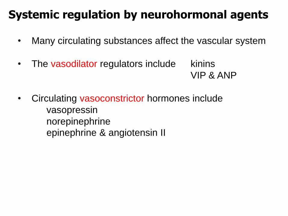
• Many circulating substances affect the vascular system
• The vasodilator regulators include kinins
VIP & ANP
• Circulating vasoconstrictor hormones include
vasopressin
norepinephrineepinephrine & angiotensin II
Systemic regulation by neurohormonal agents
C =
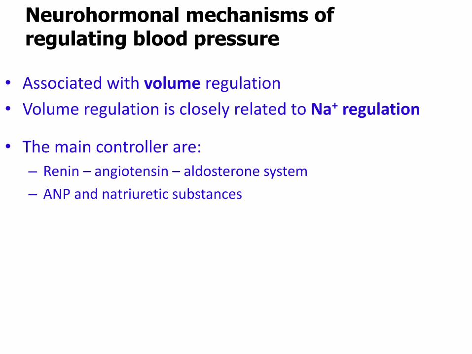
Neurohormonal mechanisms of regulating blood pressure
• Associated with volume regulation
• Volume regulation is closely related to Na+ regulation
• The main controller are:
– Renin – angiotensin – aldosterone system
– ANP and natriuretic substances
C =
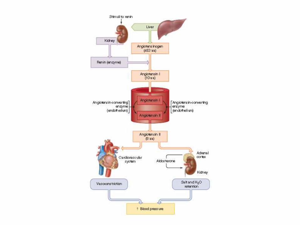
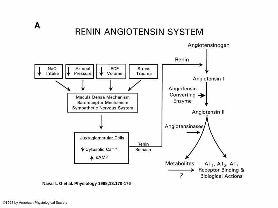
Renin – angiotensin – aldosterone system
Renin• Referred to as an enzyme / hormone• Synthesised as prorenin• Secreted from the JG cells of the kidney as • renin or prorenin• The active form is renin and only kidney can
produce this• Only known function is to cleave
angiotensinogen and form angiotensin-I
Angiotensinogen• Alpha-2 globulin • blood level increase by - glucocorticoids, thyroid
hormones, estrogens, several cytokines and angiotensin II.
C =Angiotensin Converting Enzyme & Angiotensin II
• ACE is formed by endothelial cells and happens in many parts of the body
• Conversion of Angiotensin I happens mainly when blood passes through the lungs
• Same ACE inactivates bradykinin
• Angiotensin-II has a very short half life of 1-2 min
• The active substance is Angiotensin-II
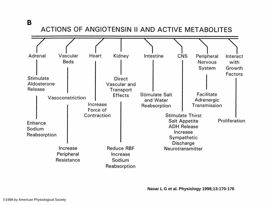
C =Actions of Angiotensin II1. Potent vasoconstrictor. Acts on AT1 receptors.
Constricts arterioles and elevate SBP & DBP2. Directly acts on adrenal cortex to increase
aldosterone secretion3. Facilitation of release of NE from sympathetic
postganglionic neurones4. Contraction of mesangial cells with a decrease in GFR 5. A direct effect on the renal tubules to increase Na+
reabsorption.
6. Acts on the brain to reduce the sensitivity of baroreflex
7. Increase thirst8. Increase ADH and ACTH secretion
C =Juxtaglomerular apparatus
• Comprise of JG cells, Lacis cells and macula densa
• Renin is produced by JG cells – located in the media of afferent arterioles
• Renin is also found in lacis cells that are located in the junction between the afferent & efferent arterioles – functional importance of this renin?
• Macula densa – modified efferent arteriolar cells in close proximity to JG cells
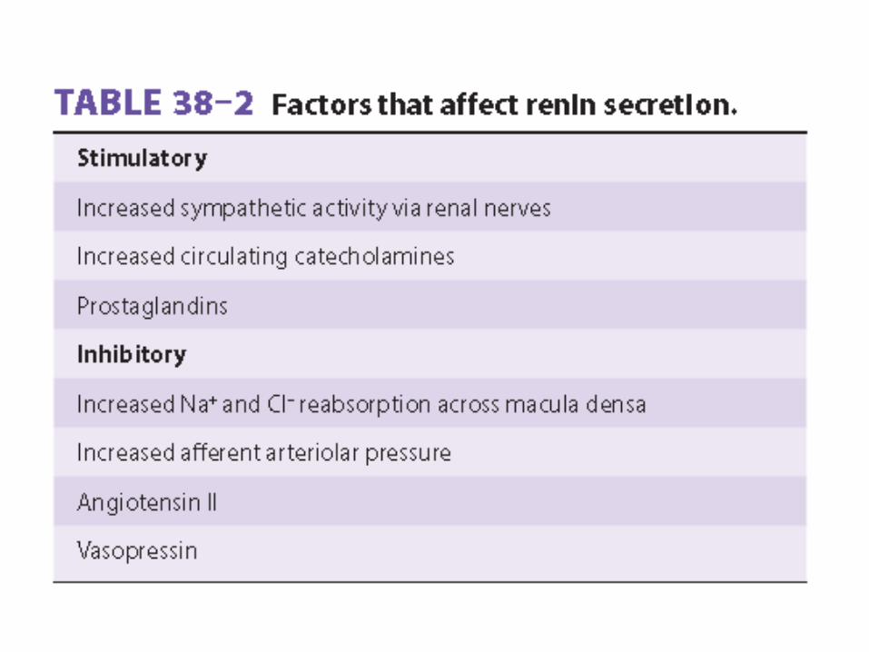
C =Regulation of renin secretion
Occur due to the balance of many factors
1. Intrarenal baroreceptor mechanism that decrease renin when pressure in the JG cells increase
2. Increased Na+ and Cl- amount delivered to the macula densa cells decrease renin secretion
3. Angiotensin-II has a direct feedback inhibition on JG cells
4. ADH also has an inhibitory effect on renin secretion
C =Regulation of renin secretion
5. Increased sympathetic activity
Increase renin secretion by
- increased circulating catecholamines acting on β1 receptors on the JG cells
- stimulation of renal sympathetic nerves
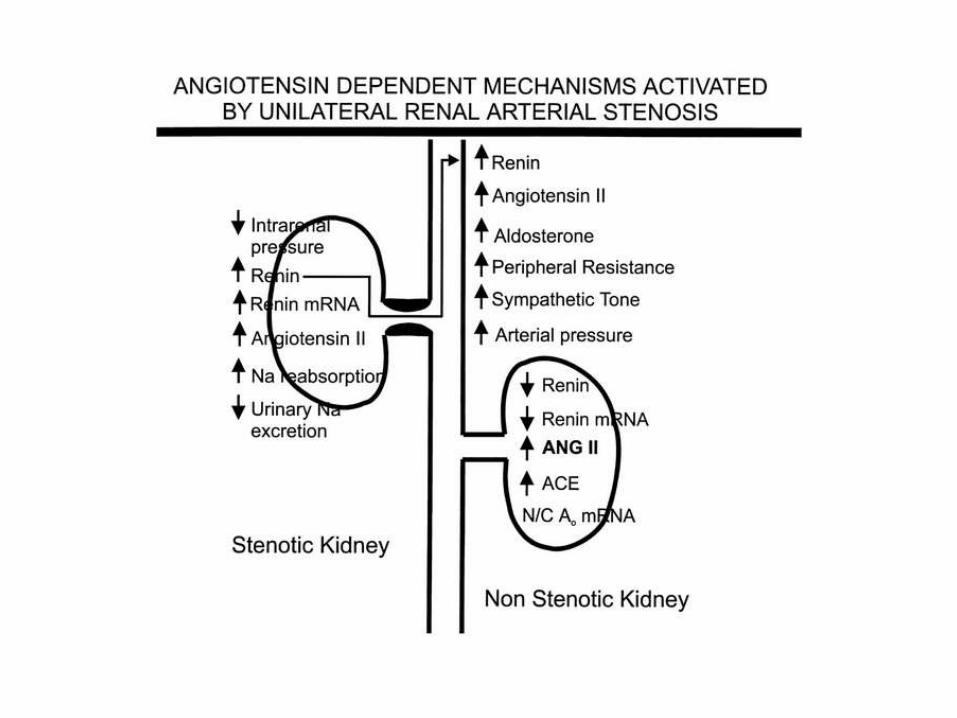
6. Reduced renal artery pressure (due to renal artery constriction or aorta) produce increased renal sympathetic nerve stimulation and that increase renin secretion
Navar L G et al. Physiology 1998;13:170-176
©1998 by American Physiological Society
Navar L G et al. Physiology 1998;13:170-176
©1998 by American Physiological Society
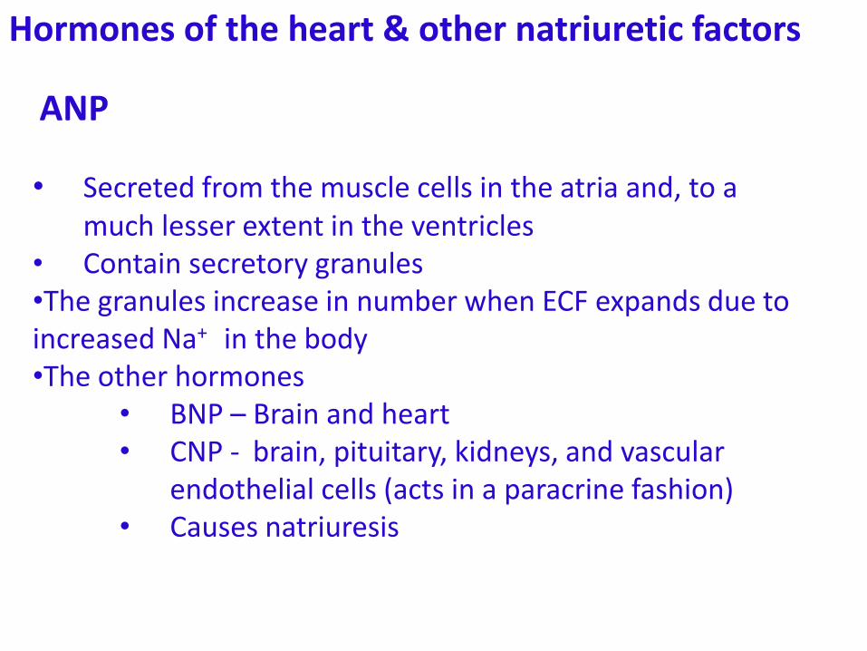
Hormones of the heart & other natriuretic factors
• Secreted from the muscle cells in the atria and, to a much lesser extent in the ventricles
• Contain secretory granules•The granules increase in number when ECF expands due to increased Na+ in the body•The other hormones
• BNP – Brain and heart• CNP - brain, pituitary, kidneys, and vascular
endothelial cells (acts in a paracrine fashion)• Causes natriuresis
ANP
Hormones of the heart & other natriuretic factors
Actions:
• Increase GFR by dilating afferent arteriole & relaxing mesangial cells
•Acts on the renal tubule to inhibit Na+ reabsorption
•An increase in capillary permeability, leading to extravasation of fluid and a decline in blood pressure.
•Relax vascular smooth muscle in arterioles and venules. CNP has a greater dilator effect on veins
• Inhibit renin secretion and
• Counteract the pressor effects of catecholamines
ANP
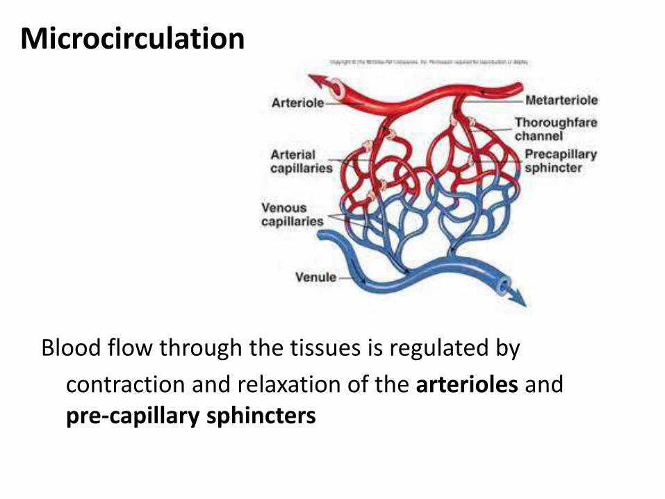
Microcirculation
Blood flow through the tissues is regulated by
contraction and relaxation of the arterioles and pre-capillary sphincters
C =
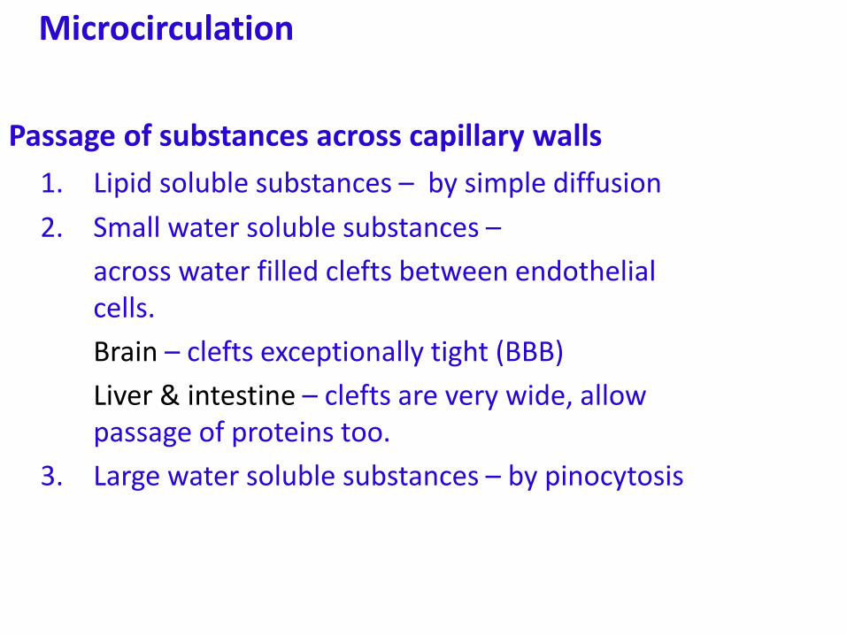
Microcirculation
Passage of substances across capillary walls
1. Lipid soluble substances – by simple diffusion
2. Small water soluble substances –
across water filled clefts between endothelial cells.
Brain – clefts exceptionally tight (BBB)
Liver & intestine – clefts are very wide, allow passage of proteins too.
3. Large water soluble substances – by pinocytosis
C =
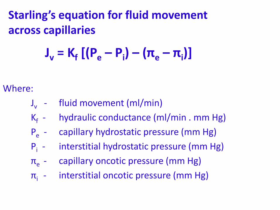
Starling’s equation for fluid movement across capillaries
Jv = Kf [(Pe – Pi) – (πe – πi)]
Where:
Jv - fluid movement (ml/min)
Kf - hydraulic conductance (ml/min . mm Hg)
Pe - capillary hydrostatic pressure (mm Hg)
Pi - interstitial hydrostatic pressure (mm Hg)
πe - capillary oncotic pressure (mm Hg)
πi - interstitial oncotic pressure (mm Hg)
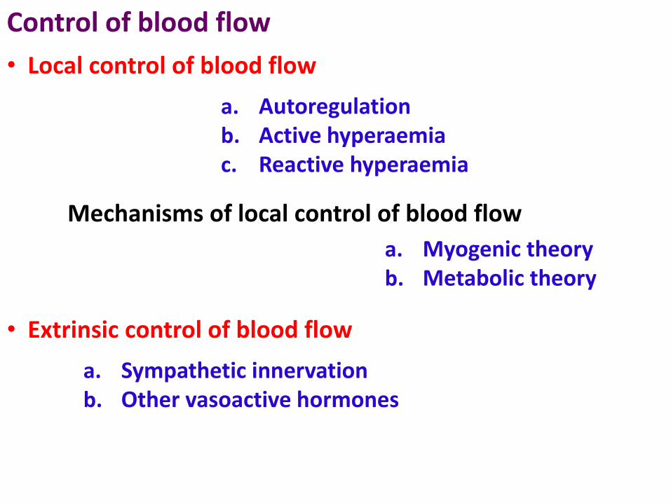
Control of blood flow
a. Autoregulationb. Active hyperaemiac. Reactive hyperaemia
• Local control of blood flow
Mechanisms of local control of blood flow
a. Myogenic theoryb. Metabolic theory
• Extrinsic control of blood flow
a. Sympathetic innervationb. Other vasoactive hormones
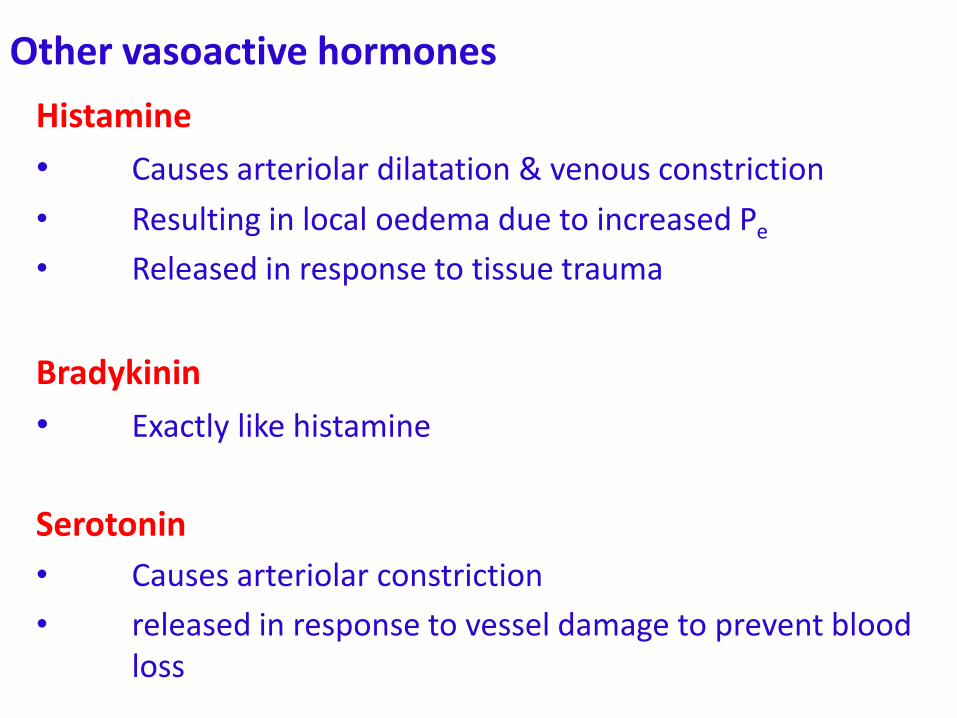
Other vasoactive hormones
Histamine
• Causes arteriolar dilatation & venous constriction
• Resulting in local oedema due to increased Pe
• Released in response to tissue trauma
Bradykinin
• Exactly like histamine
Serotonin
• Causes arteriolar constriction
• released in response to vessel damage to prevent blood loss

Other vasoactive hormones
Prostaglandins
• Prostacyclin is a vasodilator in several vascular beds
• E-series prostaglandins are vasodilators
• F-series prostaglandins are vasoconstrictors
• Thromboxane A2 is a vasoconstrictor
Special circulations
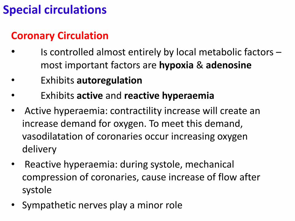
Coronary Circulation
• Is controlled almost entirely by local metabolic factors –most important factors are hypoxia & adenosine
• Exhibits autoregulation
• Exhibits active and reactive hyperaemia
• Active hyperaemia: contractility increase will create an increase demand for oxygen. To meet this demand, vasodilatation of coronaries occur increasing oxygen delivery
• Reactive hyperaemia: during systole, mechanical compression of coronaries, cause increase of flow after systole
• Sympathetic nerves play a minor role
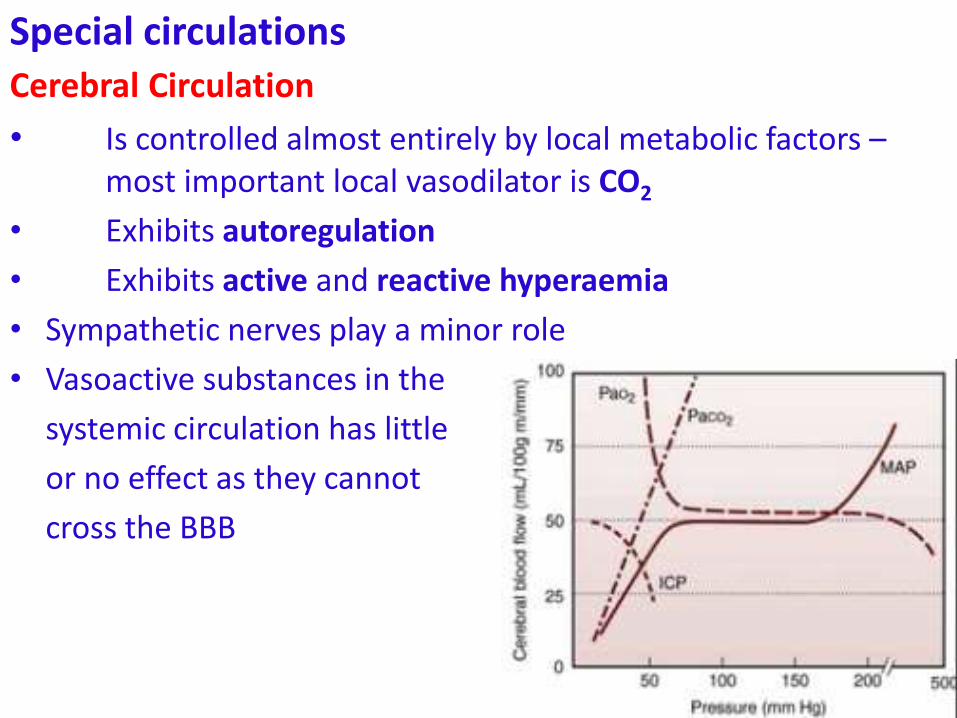
Special circulationsCerebral Circulation
• Is controlled almost entirely by local metabolic factors –most important local vasodilator is CO2
• Exhibits autoregulation
• Exhibits active and reactive hyperaemia
• Sympathetic nerves play a minor role
• Vasoactive substances in the
systemic circulation has little
or no effect as they cannot
cross the BBB
Special circulations
Skeletal muscle
• Is controlled by sympathetic nerves of blood vessels & by local metabolic factors
• Sympathetic innervation:
• Primary regulator of flow at rest
• There are both 1 and β2 receptors in vessels
• 1 – cause vasoconstriction
• β2 – cause vasodilatation
• Vasoconstriction of skeletal muscle vessels is the major contributor to TPR at rest
Special circulations
Skeletal muscle
• Local metabolic control:
• Exhibits autoregulation, active and reactive hyperaemia
• Local vasodilatory substances are lactate, adenosine and K+
• Mechnical occlusion during exercise can occlude arteries temporarily and cause an oxygen debt producing a reactive hyperaemia later
Special circulations
Skin
• Sympathetic nerves play a Major role
• Temperature regulation is the principal function of cutaneous sympathetics
• trauma produce the triple response with a red line, flare and a wheal
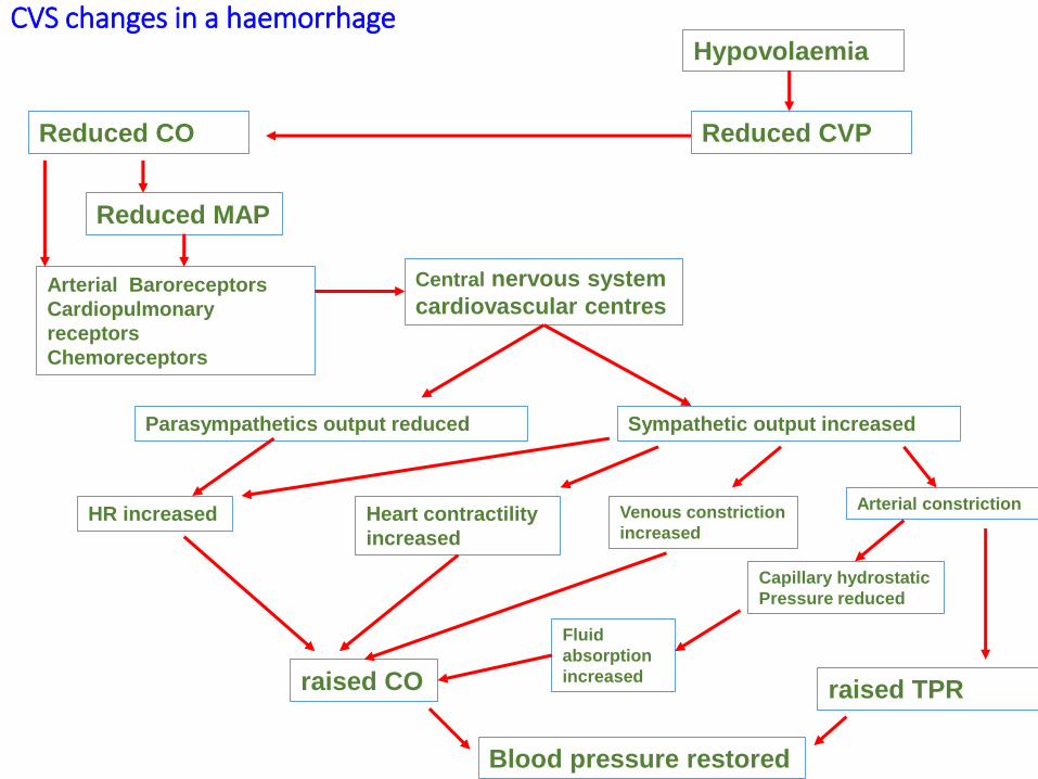
CVS changes in a haemorrhage
Arterial Baroreceptors
Cardiopulmonary
receptors
Chemoreceptors
Central nervous system
cardiovascular centres
Hypovolaemia
Sympathetic output increasedParasympathetics output reduced
Reduced CVPReduced CO
Reduced MAP
HR increased Heart contractility
increased
Arterial constrictionVenous constriction
increased
Fluid
absorption
increased
Capillary hydrostatic
Pressure reduced
raised CO raised TPR
Blood pressure restored
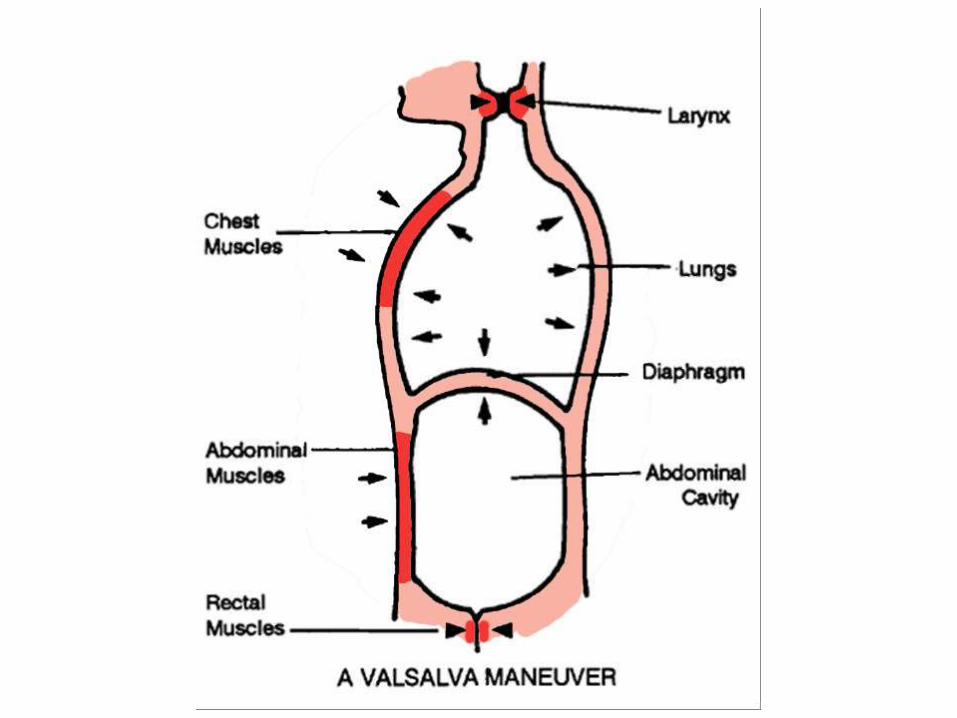
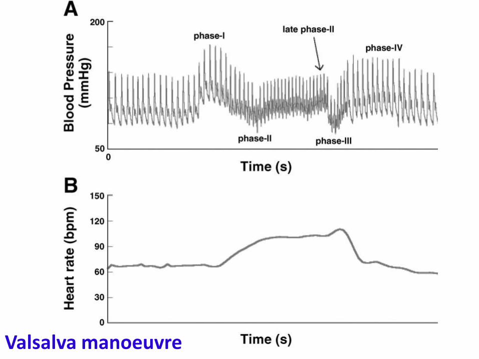
Valsalva manoeuvre
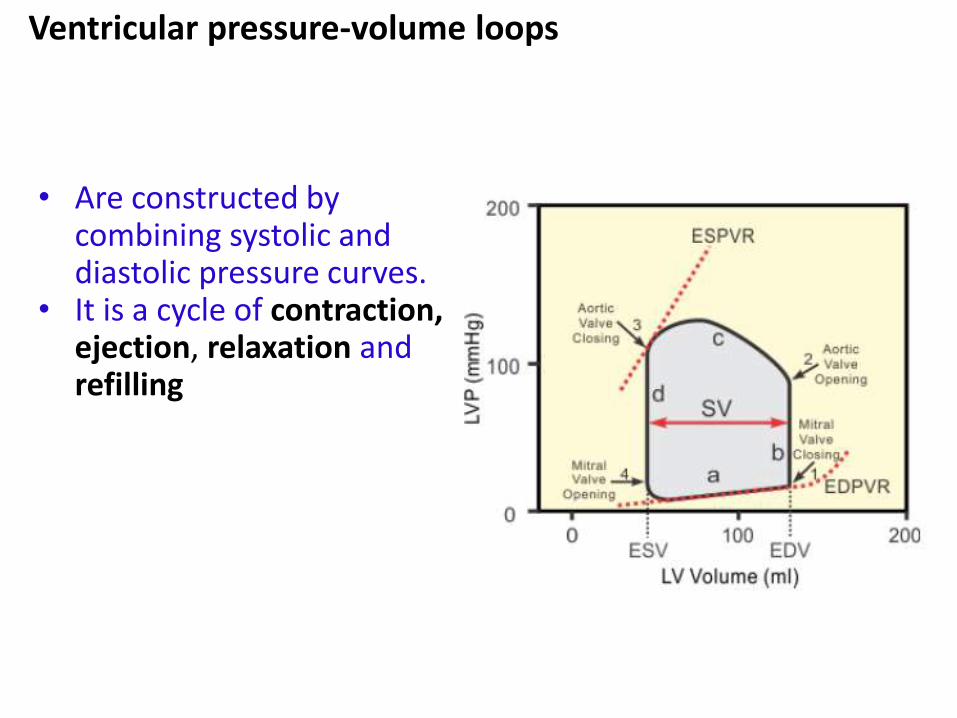
• Are constructed by combining systolic and diastolic pressure curves.
• It is a cycle of contraction, ejection, relaxation and refilling
Ventricular pressure-volume loops
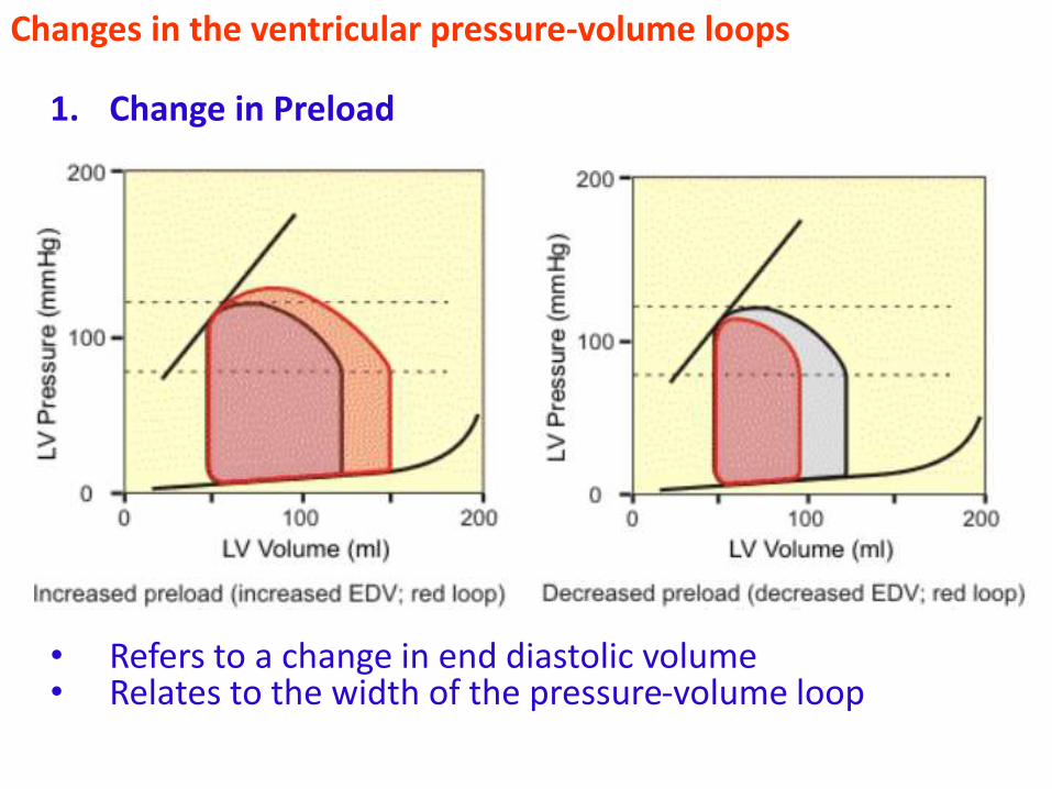
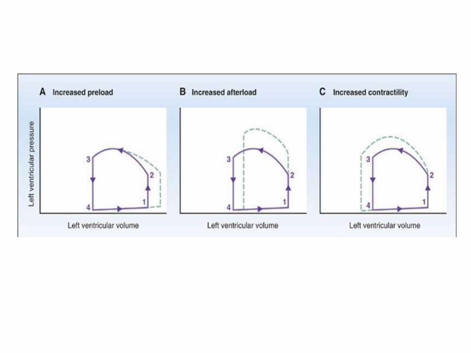
1. Change in Preload
• Refers to a change in end diastolic volume• Relates to the width of the pressure-volume loop
Changes in the ventricular pressure-volume loops
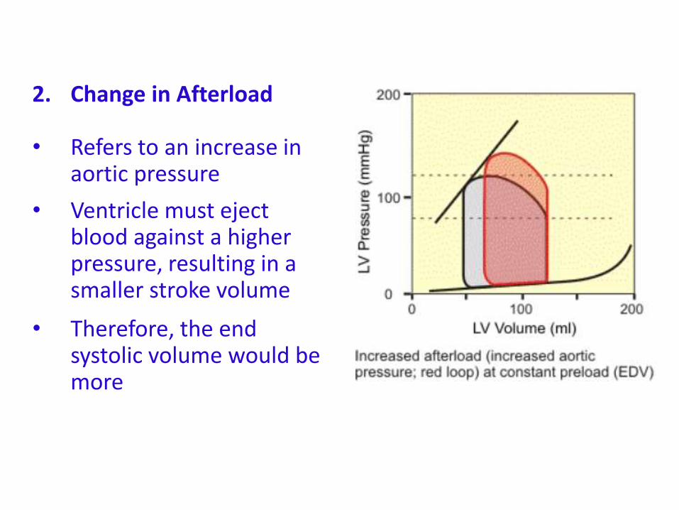
2. Change in Afterload
• Refers to an increase in aortic pressure
• Ventricle must eject blood against a higher pressure, resulting in a smaller stroke volume
• Therefore, the end systolic volume would be more
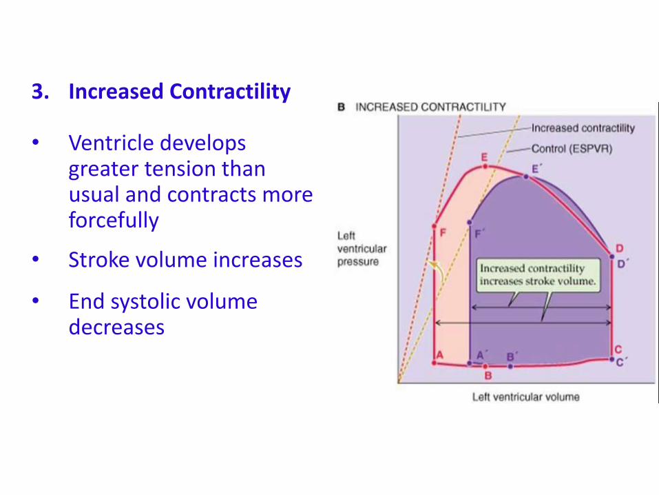
3. Increased Contractility
• Ventricle develops greater tension than usual and contracts more forcefully
• Stroke volume increases
• End systolic volume decreases
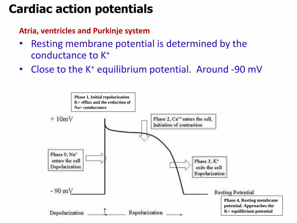
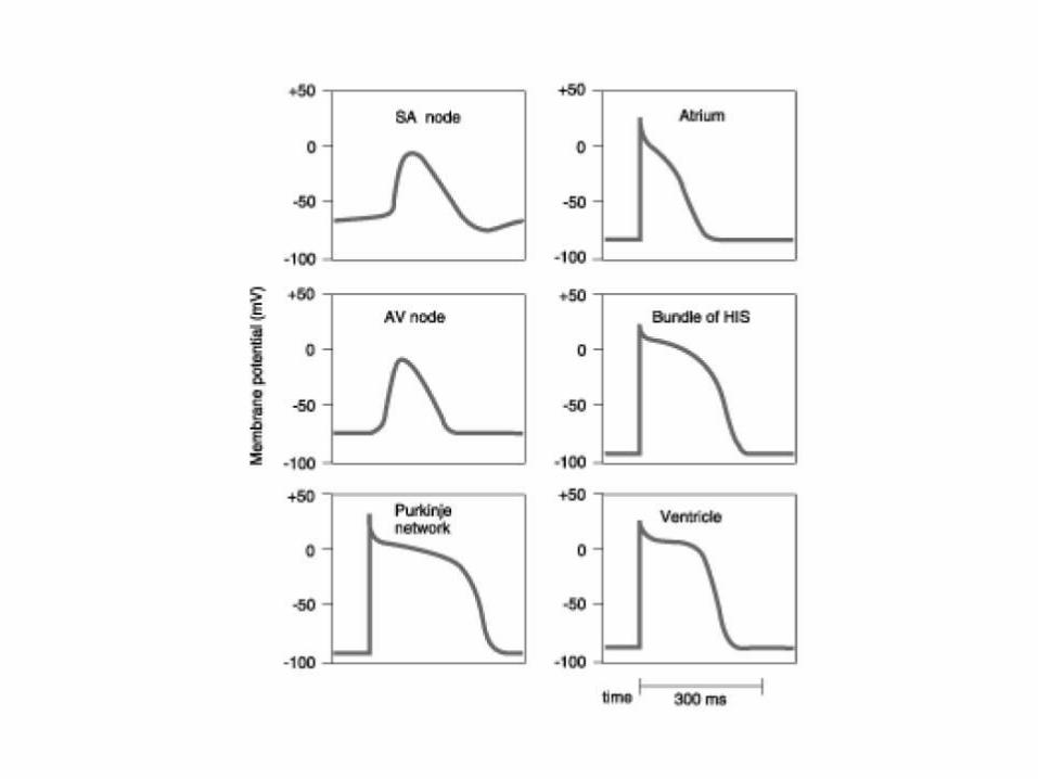
Atria, ventricles and Purkinje system
• Resting membrane potential is determined by the conductance to K+
• Close to the K+ equilibrium potential. Around -90 mV
Cardiac action potentials
Phase 1, Initial repolarization
K+ efflux and the reduction of
Na+ conductance
Phase 4, Resting membrane
potential. Approaches the
K+ equilibrium potential
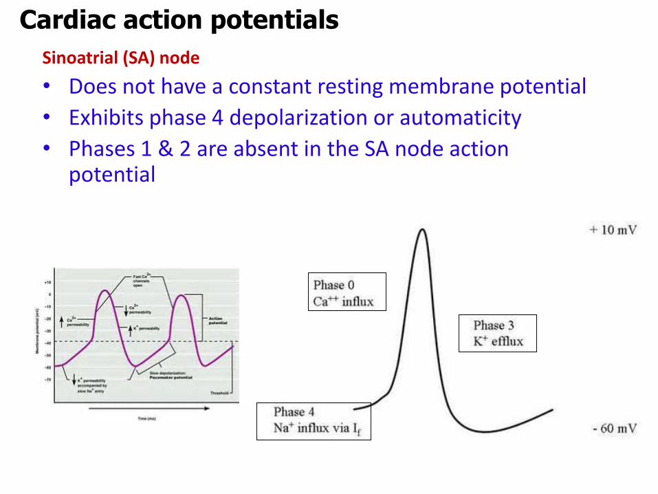
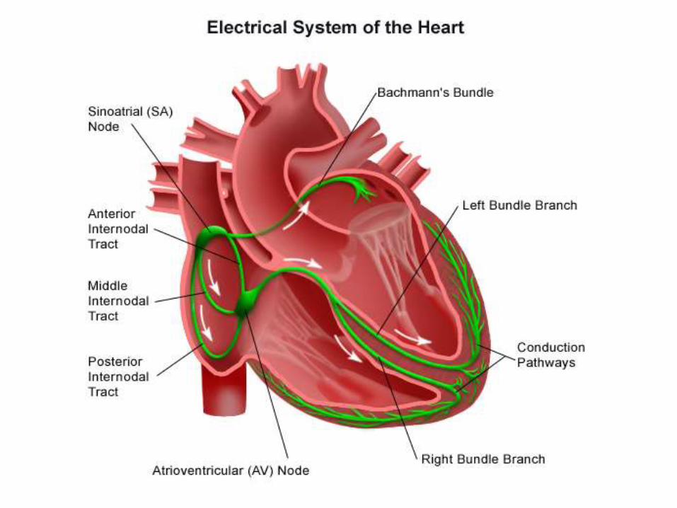
Sinoatrial (SA) node
• Does not have a constant resting membrane potential
• Exhibits phase 4 depolarization or automaticity
• Phases 1 & 2 are absent in the SA node action potential
Cardiac action potentials
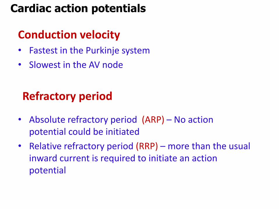
Conduction velocity • Fastest in the Purkinje system
• Slowest in the AV node
• Absolute refractory period (ARP) – No action potential could be initiated
• Relative refractory period (RRP) – more than the usual inward current is required to initiate an action potential
Cardiac action potentials
Refractory period
Autonomic effects on the heart & vessels
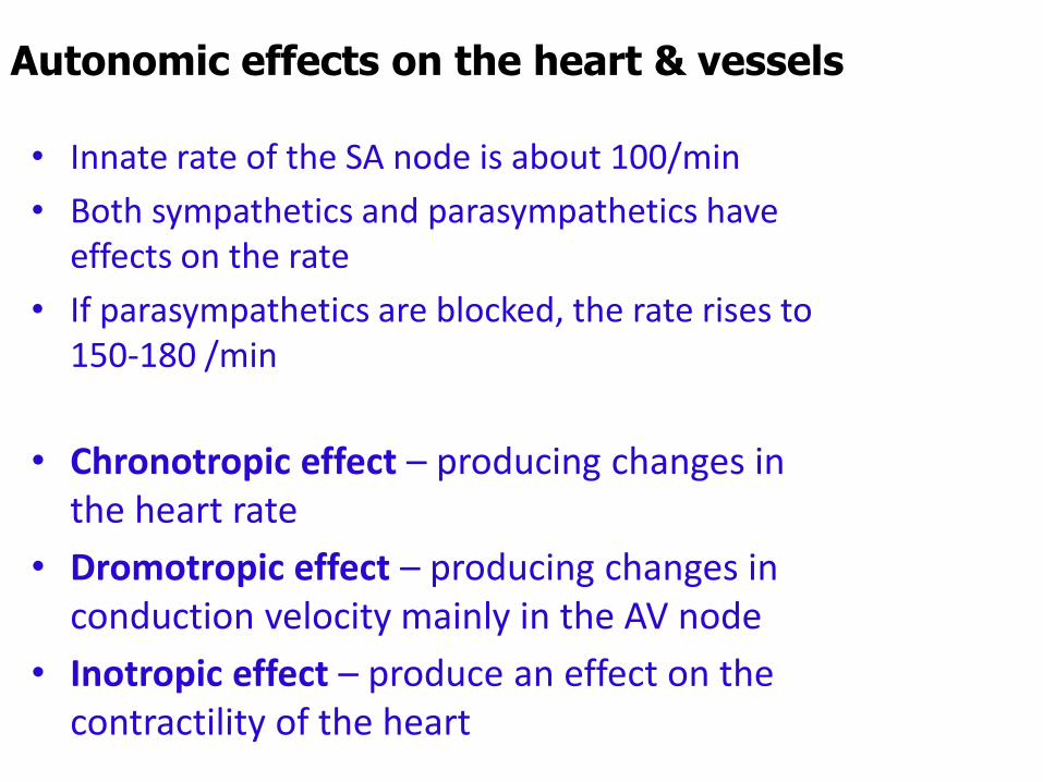
• Innate rate of the SA node is about 100/min
• Both sympathetics and parasympathetics have effects on the rate
• If parasympathetics are blocked, the rate rises to 150-180 /min
• Chronotropic effect – producing changes in the heart rate
• Dromotropic effect – producing changes in conduction velocity mainly in the AV node
• Inotropic effect – produce an effect on the contractility of the heart
Parasympathetic effect on heart
• SA node, atria and AV node has parasympathetic innervation
• Neurotransmitter is Ach. Acting on muscarinic receptors
• Effects are:
• Decreasing heart rate (threshold potential is reached
slowly)
• Decrease conduction velocity through the AV node
• Increase the PR interval (decreased inward Ca++
current)
Sympathetic effect on heart
• Neurotransmitter is Norepinephrine. Acting on β1
receptors
• Effects are:
• Positive chronotropic effect (threshold potential is reached faster
• Increase conduction velocity through the AV node
• Decrease the PR interval (increase inward Ca++ current)
• Positive inotropic effect
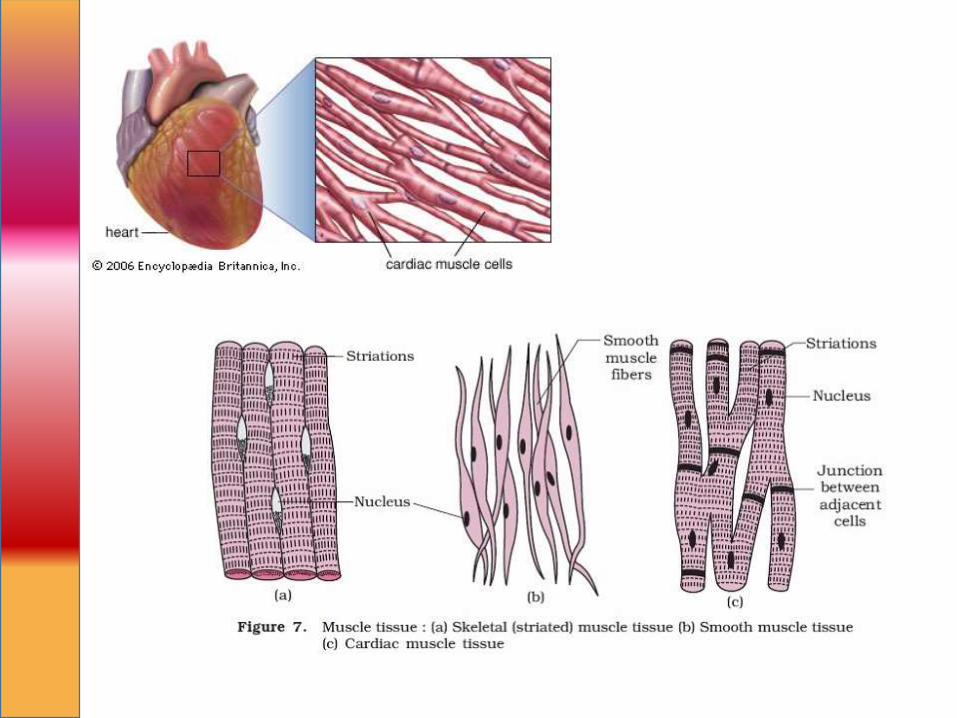
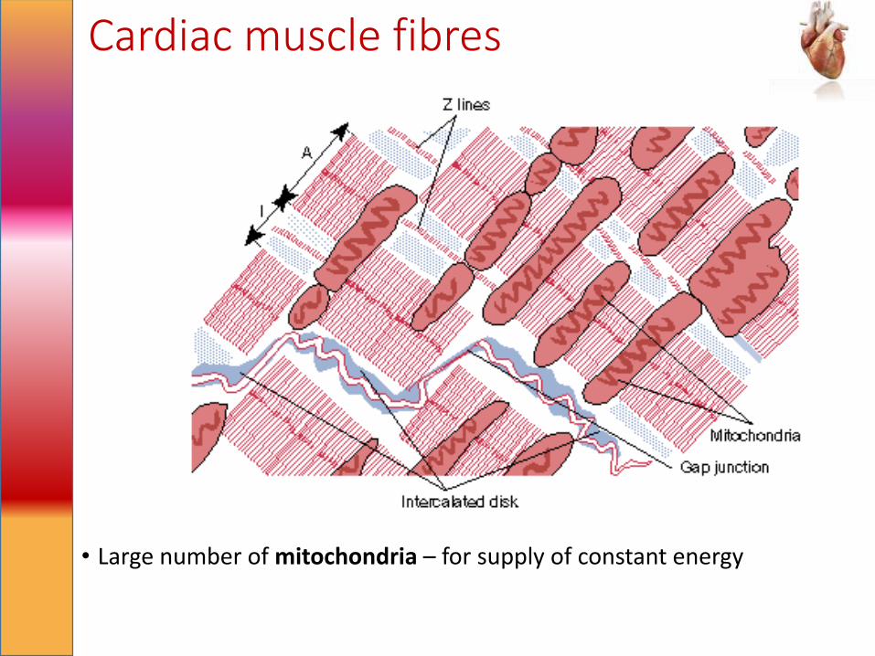
Cardiac muscle fibres
• Large number of mitochondria – for supply of constant energy
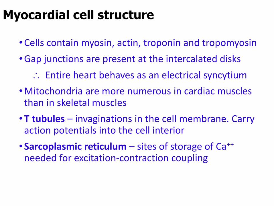
•Cells contain myosin, actin, troponin and tropomyosin
•Gap junctions are present at the intercalated disks
Entire heart behaves as an electrical syncytium
•Mitochondria are more numerous in cardiac muscles than in skeletal muscles
•T tubules – invaginations in the cell membrane. Carry action potentials into the cell interior
•Sarcoplasmic reticulum – sites of storage of Ca++
needed for excitation-contraction coupling
Myocardial cell structure
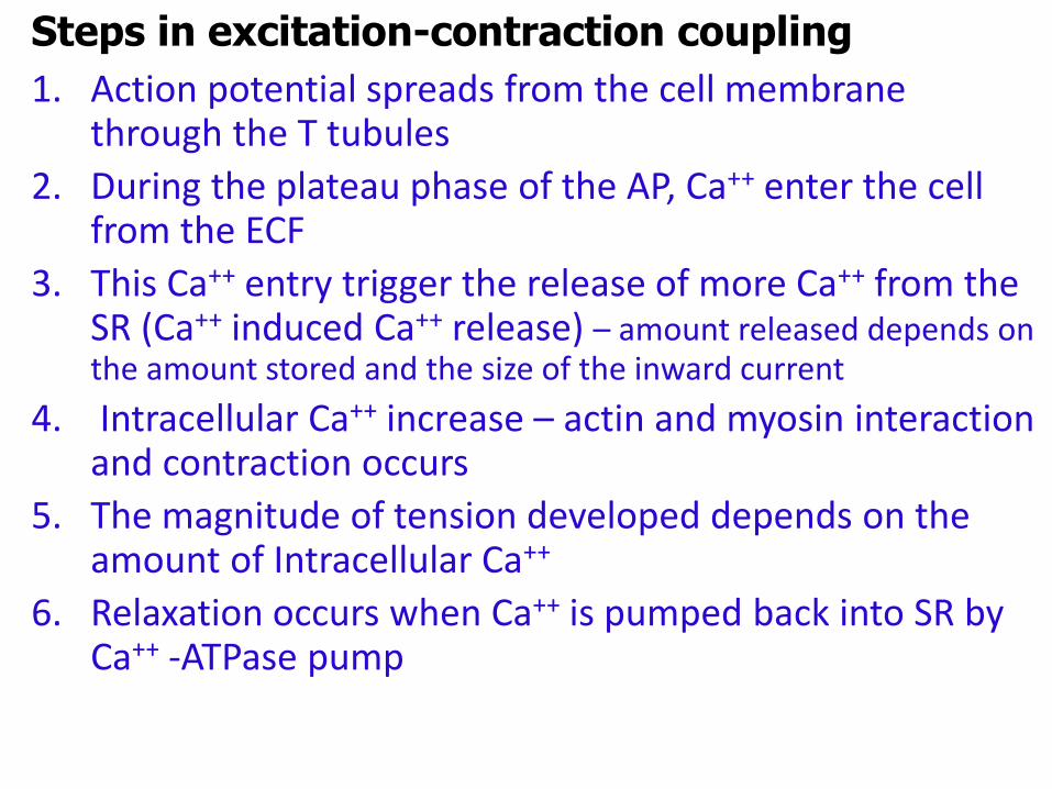
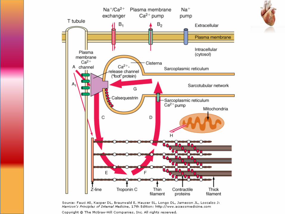
1. Action potential spreads from the cell membrane through the T tubules
2. During the plateau phase of the AP, Ca++ enter the cell from the ECF
3. This Ca++ entry trigger the release of more Ca++ from the SR (Ca++ induced Ca++ release) – amount released depends on the amount stored and the size of the inward current
4. Intracellular Ca++ increase – actin and myosin interaction and contraction occurs
5. The magnitude of tension developed depends on the amount of Intracellular Ca++
6. Relaxation occurs when Ca++ is pumped back into SR by Ca++ -ATPase pump
Steps in excitation-contraction coupling
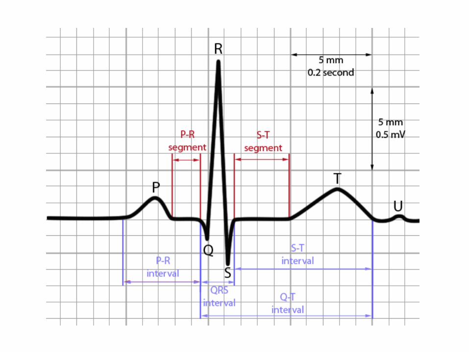
P wave
• Represents atrial depolarization
PR interval
• Is the interval between the beginning of P wave to beginning of Q wave
• Increases with problems in conduction velocity (heart blocks)
• Varies with heart rate.
QRS complex
• Represents ventricular depolarization
Electrocardiogram (ECG)
QT interval
• From beginning of QRS to end of T wave
• Represents entire ventricular depolarization and repolarisation
ST segment
• Is the segment from the end of S wave to the beginning of T wave
• Is isoelectric
• Represents the period when the ventricle is depolarized
T wave• Represents ventricular repolarisation
Electrocardiogram (ECG)

Atria, ventricles and Purkinje system
• Resting membrane potential is determined by the conductance to K+
• Close to the K+ equilibrium potential. Around -90 mV
Cardiac action potentials
Phase 1, Initial repolarization
K+ efflux and the reduction of
Na+ conductance
Phase 4, Resting membrane
potential. Approaches the
K+ equilibrium potential
1. Explain the physiological determinants of ejection fraction.
(40 % marks
1. Importance of Ca++ in cardiac muscle contraction. (30% marks)
2. Explain the physiological basis of the following :
4.2. Tachycardia in shock. (25 marks)
4.3. Low urine output in a patient who has lost IL of blood. (25 marks)
3. 3.1. Explain how variations in arteriolar resistance affect the
arterial blood flow. (50 marks)
1. Outline the factors that determine the blood flow to an organ (15 marks)
2. Explain the autoregulation of cerebral blood flow. (35 marks)
3. Describe the baroreceptor reflex regulation of blood pressure. (50 marks)
4. Give the physiological mechanisms that facilitate the venous return from
5. the extremities to the heart.
1. Explain the physiological basis of the following ,
1.1 A drop in systolic blood pressure when standing from supine position (30 marks)
1.2 Low urine output following a haemorrhage (40 marks)
1.1. What biophysical factors determine the blood pressure?
1.2. Explain with examples how blood pressure is increased when these factors are altered by diseases.