Embed Size (px)
Citation preview
THE CARDIAC CYCLETHE CARDIAC CYCLE
Dr G Bhanu PrakashDr G Bhanu Prakash
ATRIAL SYSTOLEATRIAL SYSTOLE
The end of diastole The end of diastole
ATRIAL SYSTOLE - HeartATRIAL SYSTOLE - Heart
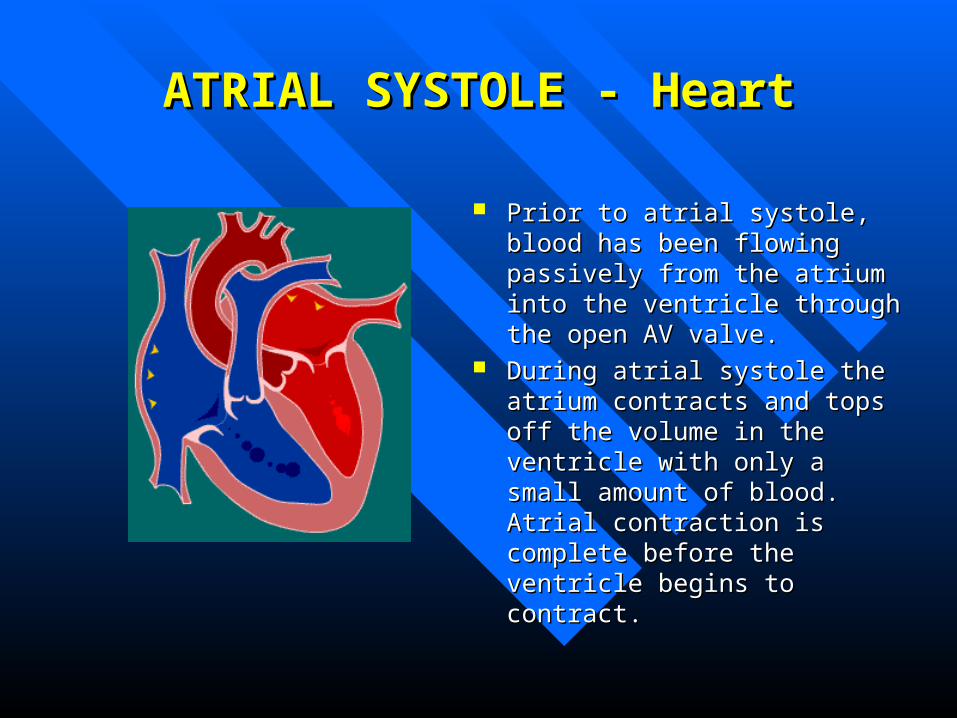
Prior to atrial systole, blood has been Prior to atrial systole, blood has been flowing passively from the atrium flowing passively from the atrium into the ventricle through the open into the ventricle through the open AV valve. AV valve.
During atrial systole the atrium During atrial systole the atrium contracts and tops off the volume in contracts and tops off the volume in the ventricle with only a small the ventricle with only a small amount of blood. Atrial contraction amount of blood. Atrial contraction is complete before the ventricle is complete before the ventricle begins to contract. begins to contract.
ATRIAL SYSTOLEATRIAL SYSTOLEPressures & VolumesPressures & Volumes
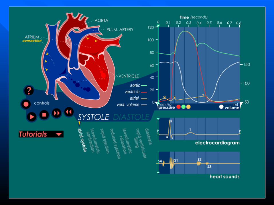
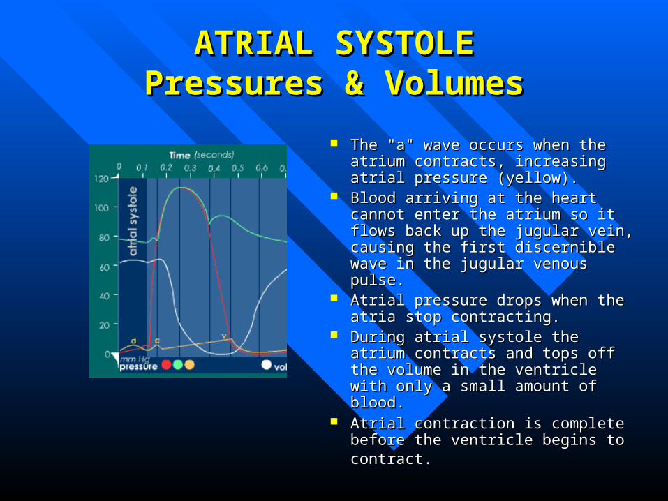
The "a" wave occurs when the atrium The "a" wave occurs when the atrium contracts, increasing atrial pressure contracts, increasing atrial pressure (yellow). (yellow).
Blood arriving at the heart cannot enter Blood arriving at the heart cannot enter the atrium so it flows back up the the atrium so it flows back up the jugular vein, causing the first jugular vein, causing the first discernible wave in the jugular venous discernible wave in the jugular venous pulse. pulse.
Atrial pressure drops when the atria Atrial pressure drops when the atria stop contracting. stop contracting.
During atrial systole the atrium During atrial systole the atrium contracts and tops off the volume in contracts and tops off the volume in the ventricle with only a small amount the ventricle with only a small amount of blood. of blood.
Atrial contraction is complete before Atrial contraction is complete before the ventricle begins to contract.the ventricle begins to contract.

ATRIAL SYSTOLEATRIAL SYSTOLEECGECG
An impulse arising from the SA node results in depolarization and An impulse arising from the SA node results in depolarization and contraction of the atria (the right atrium contracts slightly before the left contraction of the atria (the right atrium contracts slightly before the left atrium). atrium).
The P wave is due to this atrial depolarization.The P wave is due to this atrial depolarization. The PR segment is electrically quiet as the depolarization proceeds to the The PR segment is electrically quiet as the depolarization proceeds to the
AV node. AV node. This brief pause before contraction allows the ventricles to fill completely This brief pause before contraction allows the ventricles to fill completely
with blood. with blood.
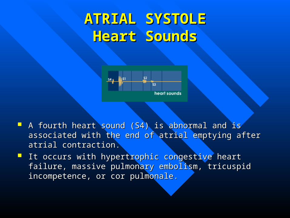
ATRIAL SYSTOLEATRIAL SYSTOLEHeart SoundsHeart Sounds
A fourth heart sound (S4) is abnormal and is associated with the end of A fourth heart sound (S4) is abnormal and is associated with the end of atrial emptying after atrial contraction. atrial emptying after atrial contraction.
It occurs with hypertrophic congestive heart failure, massive pulmonary It occurs with hypertrophic congestive heart failure, massive pulmonary embolism, tricuspid incompetence, or cor pulmonale. embolism, tricuspid incompetence, or cor pulmonale.
ISOVOLUMETRIC ISOVOLUMETRIC CONTRACTIONCONTRACTION
The Beginning of systoleThe Beginning of systole
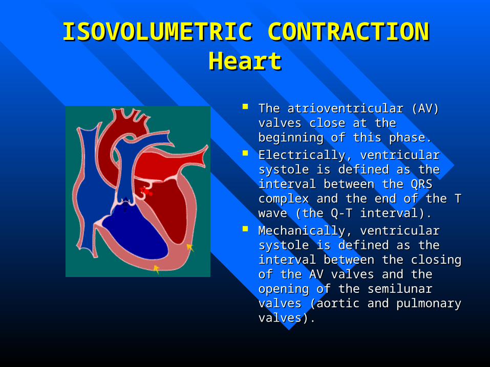
ISOVOLUMETRIC CONTRACTIONISOVOLUMETRIC CONTRACTIONHeartHeart
The atrioventricular (AV) valves The atrioventricular (AV) valves close at the beginning of this phase.close at the beginning of this phase.
Electrically, ventricular systole is Electrically, ventricular systole is defined as the interval between the defined as the interval between the QRS complex and the end of the T QRS complex and the end of the T wave (the Q-T interval).wave (the Q-T interval).
Mechanically, ventricular systole is Mechanically, ventricular systole is defined as the interval between the defined as the interval between the closing of the AV valves and the closing of the AV valves and the opening of the semilunar valves opening of the semilunar valves (aortic and pulmonary valves). (aortic and pulmonary valves).
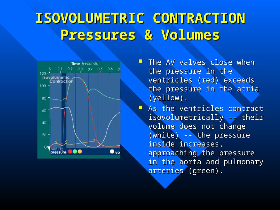
ISOVOLUMETRIC CONTRACTIONISOVOLUMETRIC CONTRACTIONPressures & VolumesPressures & Volumes
The AV valves close when the The AV valves close when the pressure in the ventricles (red) pressure in the ventricles (red) exceeds the pressure in the atria exceeds the pressure in the atria (yellow). (yellow).
As the ventricles contract As the ventricles contract isovolumetrically -- their volume isovolumetrically -- their volume does not change (white) -- the does not change (white) -- the pressure inside increases, pressure inside increases, approaching the pressure in the aorta approaching the pressure in the aorta and pulmonary arteries (green). and pulmonary arteries (green).
ISOVOLUMETRIC CONTRACTIONISOVOLUMETRIC CONTRACTIONECGECG
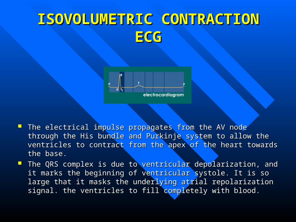
The electrical impulse propagates from the AV node through the His bundle The electrical impulse propagates from the AV node through the His bundle and Purkinje system to allow the ventricles to contract from the apex of the and Purkinje system to allow the ventricles to contract from the apex of the heart towards the base.heart towards the base.
The QRS complex is due to ventricular depolarization, and it marks the The QRS complex is due to ventricular depolarization, and it marks the beginning of ventricular systole. It is so large that it masks the underlying beginning of ventricular systole. It is so large that it masks the underlying atrial repolarization signal. the ventricles to fill completely with blood. atrial repolarization signal. the ventricles to fill completely with blood.
ISOVOLUMETRIC CONTRACTIONISOVOLUMETRIC CONTRACTIONHeart SoundsHeart Sounds
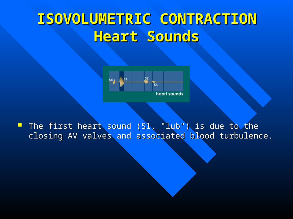
The first heart sound (S1, "lub") is due to the closing AV valves and The first heart sound (S1, "lub") is due to the closing AV valves and associated blood turbulence. associated blood turbulence.
RAPID EJECTIONRAPID EJECTION

RAPID EJECTIONRAPID EJECTIONHeartHeart
The semilunar (aortic and The semilunar (aortic and pulmonary) valves open at the pulmonary) valves open at the beginning of this phase. beginning of this phase.
RAPID EJECTIONRAPID EJECTIONPressures & VolumesPressures & Volumes
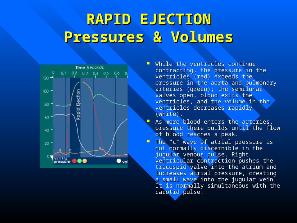
While the ventricles continue contracting, While the ventricles continue contracting, the pressure in the ventricles (red) exceeds the pressure in the ventricles (red) exceeds the pressure in the aorta and pulmonary the pressure in the aorta and pulmonary arteries (green); the semilunar valves open, arteries (green); the semilunar valves open, blood exits the ventricles, and the volume in blood exits the ventricles, and the volume in the ventricles decreases rapidly (white).the ventricles decreases rapidly (white).
As more blood enters the arteries, pressure As more blood enters the arteries, pressure there builds until the flow of blood reaches there builds until the flow of blood reaches a peak.a peak.
The "c" wave of atrial pressure is not The "c" wave of atrial pressure is not normally discernible in the jugular venous normally discernible in the jugular venous pulse. Right ventricular contraction pushes pulse. Right ventricular contraction pushes the tricuspid valve into the atrium and the tricuspid valve into the atrium and increases atrial pressure, creating a small increases atrial pressure, creating a small wave into the jugular vein. It is normally wave into the jugular vein. It is normally simultaneous with the carotid pulse. simultaneous with the carotid pulse.
RAPID EJECTIONRAPID EJECTIONECGECG
No DeflectionsNo Deflections
RAPID EJECTIONRAPID EJECTIONHeart SoundsHeart Sounds
NoneNone
REDUCED EJECTIONREDUCED EJECTION
The end of systole The end of systole
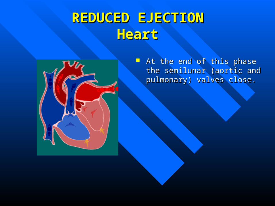
REDUCED EJECTIONREDUCED EJECTIONHeartHeart
At the end of this phase the At the end of this phase the semilunar (aortic and pulmonary) semilunar (aortic and pulmonary) valves close.valves close.
REDUCED EJECTIONREDUCED EJECTIONPressures & VolumesPressures & Volumes
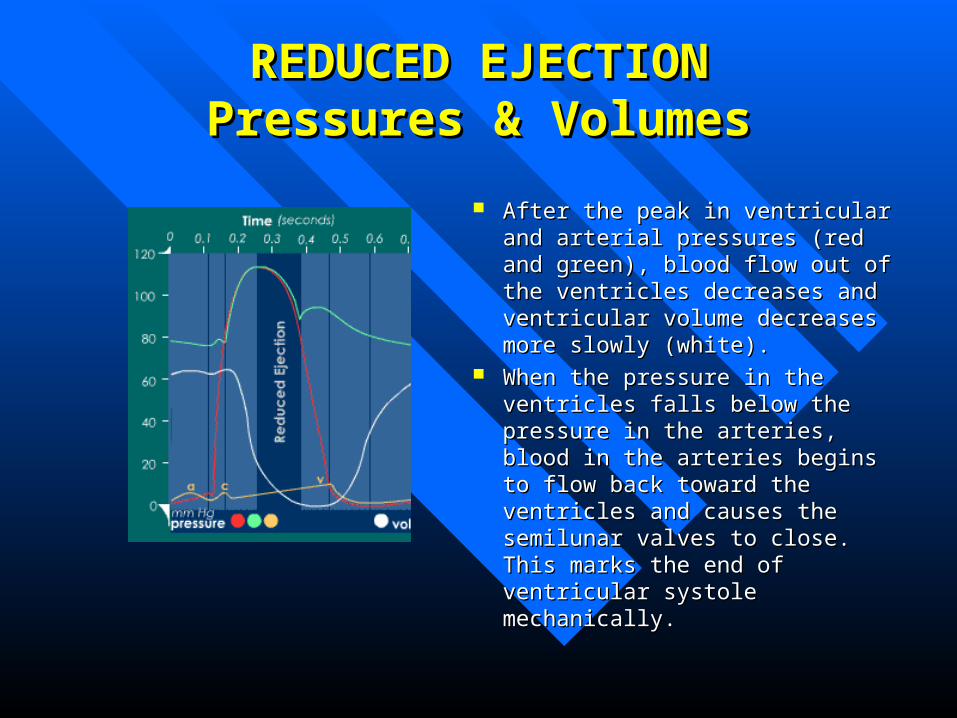
After the peak in ventricular and After the peak in ventricular and arterial pressures (red and green), arterial pressures (red and green), blood flow out of the ventricles blood flow out of the ventricles decreases and ventricular volume decreases and ventricular volume decreases more slowly (white).decreases more slowly (white).
When the pressure in the ventricles When the pressure in the ventricles falls below the pressure in the falls below the pressure in the arteries, blood in the arteries begins arteries, blood in the arteries begins to flow back toward the ventricles to flow back toward the ventricles and causes the semilunar valves to and causes the semilunar valves to close. This marks the end of close. This marks the end of ventricular systole mechanically.ventricular systole mechanically.
REDUCED EJECTIONREDUCED EJECTIONECGECG
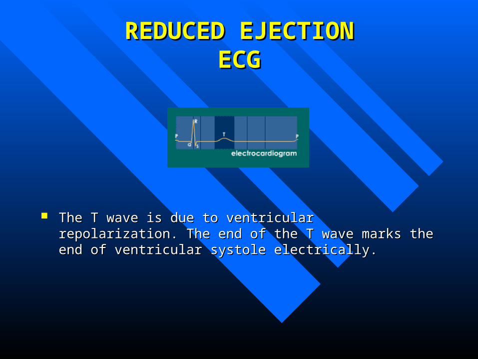
The T wave is due to ventricular repolarization. The end of the T wave The T wave is due to ventricular repolarization. The end of the T wave marks the end of ventricular systole electrically. marks the end of ventricular systole electrically.

REDUCED EJECTIONREDUCED EJECTIONHeart SoundsHeart Sounds
NoneNone
ISOVOLUMETRIC ISOVOLUMETRIC RELAXATIONRELAXATION
The beginning of Diastole The beginning of Diastole
ISOVOLUMETRIC RELAXATIONISOVOLUMETRIC RELAXATIONHeartHeart
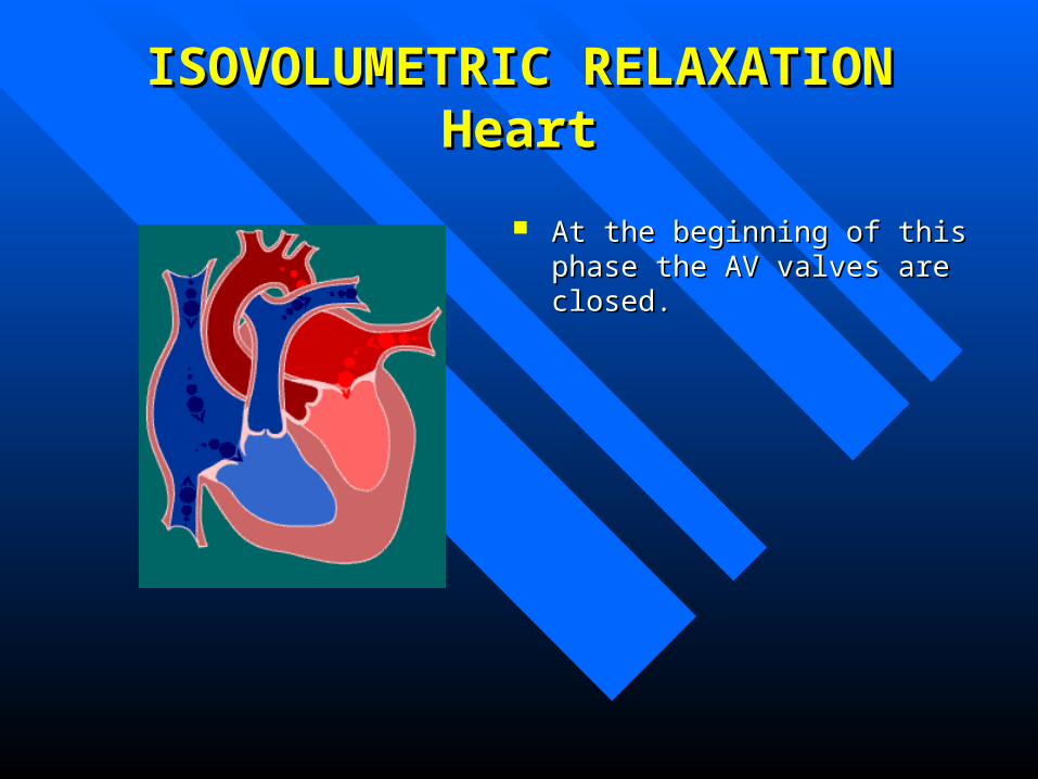
At the beginning of this phase the At the beginning of this phase the AV valves are closed. AV valves are closed.
ISOVOLUMETRIC RELAXATIONISOVOLUMETRIC RELAXATIONPressures & VolumesPressures & Volumes
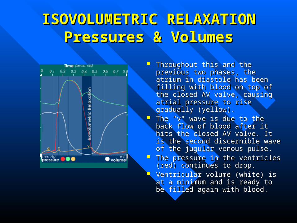
Throughout this and the previous Throughout this and the previous two phases, the atrium in diastole has two phases, the atrium in diastole has been filling with blood on top of the been filling with blood on top of the closed AV valve, causing atrial closed AV valve, causing atrial pressure to rise gradually (yellow).pressure to rise gradually (yellow).
The "v" wave is due to the back flow The "v" wave is due to the back flow of blood after it hits the closed AV of blood after it hits the closed AV valve. It is the second discernible valve. It is the second discernible wave of the jugular venous pulse.wave of the jugular venous pulse.
The pressure in the ventricles (red) The pressure in the ventricles (red) continues to drop. continues to drop.
Ventricular volume (white) is at a Ventricular volume (white) is at a minimum and is ready to be filled minimum and is ready to be filled again with blood. again with blood.

ISOVOLUMETRIC RELAXATIONISOVOLUMETRIC RELAXATIONECGECG
No Deflections No Deflections
ISOVOLUMETRIC RELAXATIONISOVOLUMETRIC RELAXATIONHeart SoundsHeart Sounds
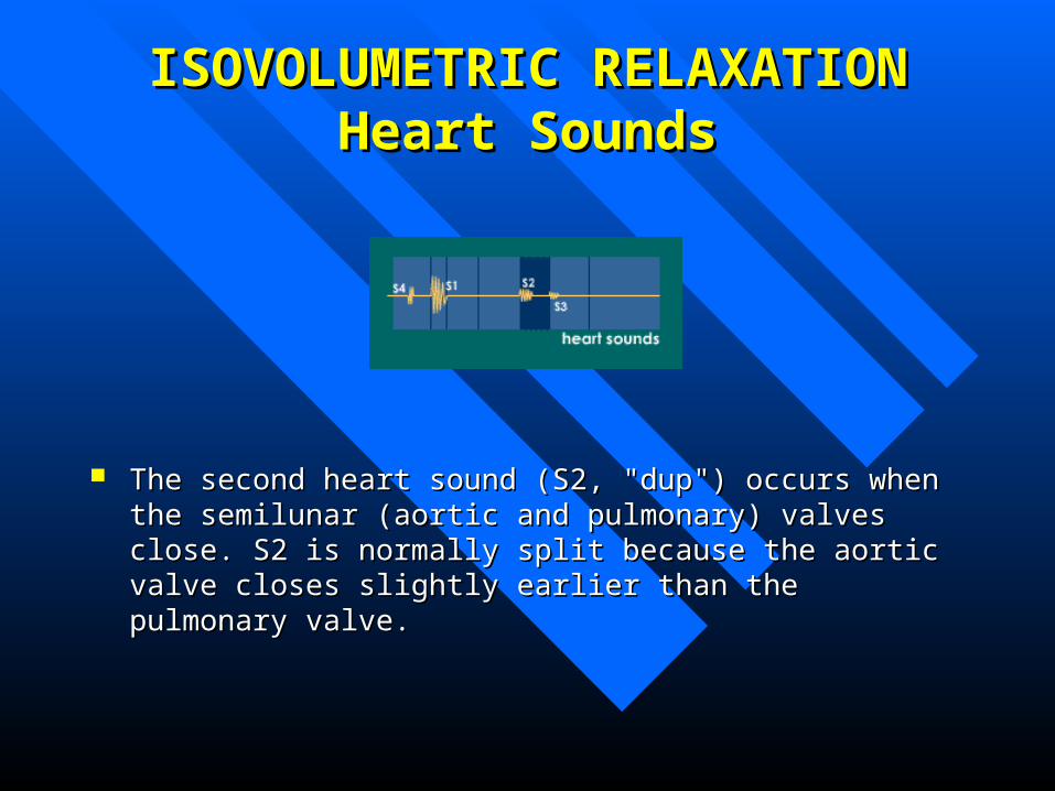
The second heart sound (S2, "dup") occurs when the semilunar (aortic The second heart sound (S2, "dup") occurs when the semilunar (aortic and pulmonary) valves close. S2 is normally split because the aortic and pulmonary) valves close. S2 is normally split because the aortic valve closes slightly earlier than the pulmonary valve. valve closes slightly earlier than the pulmonary valve.
RAPID VENTRICULAR RAPID VENTRICULAR FILLINGFILLING
RAPID VENTRICULAR FILLINGRAPID VENTRICULAR FILLINGHeartHeart
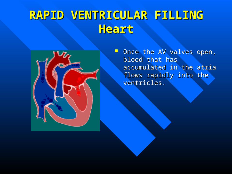
Once the AV valves open, blood that Once the AV valves open, blood that has accumulated in the atria flows has accumulated in the atria flows rapidly into the ventricles. rapidly into the ventricles.
RAPID VENTRICULAR FILLING RAPID VENTRICULAR FILLING Pressures & VolumesPressures & Volumes
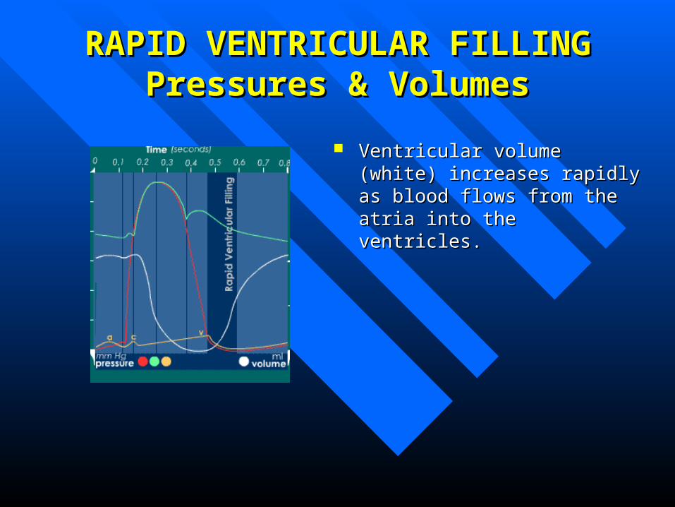
Ventricular volume (white) increases Ventricular volume (white) increases rapidly as blood flows from the atria rapidly as blood flows from the atria into the ventricles.into the ventricles.
RAPID VENTRICULAR FILLING RAPID VENTRICULAR FILLING ECGECG
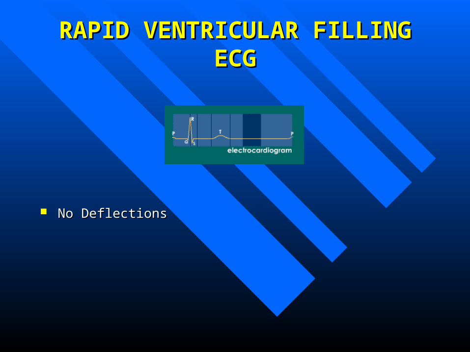
No Deflections No Deflections
RAPID VENTRICULAR FILLINGRAPID VENTRICULAR FILLINGHeart SoundsHeart Sounds
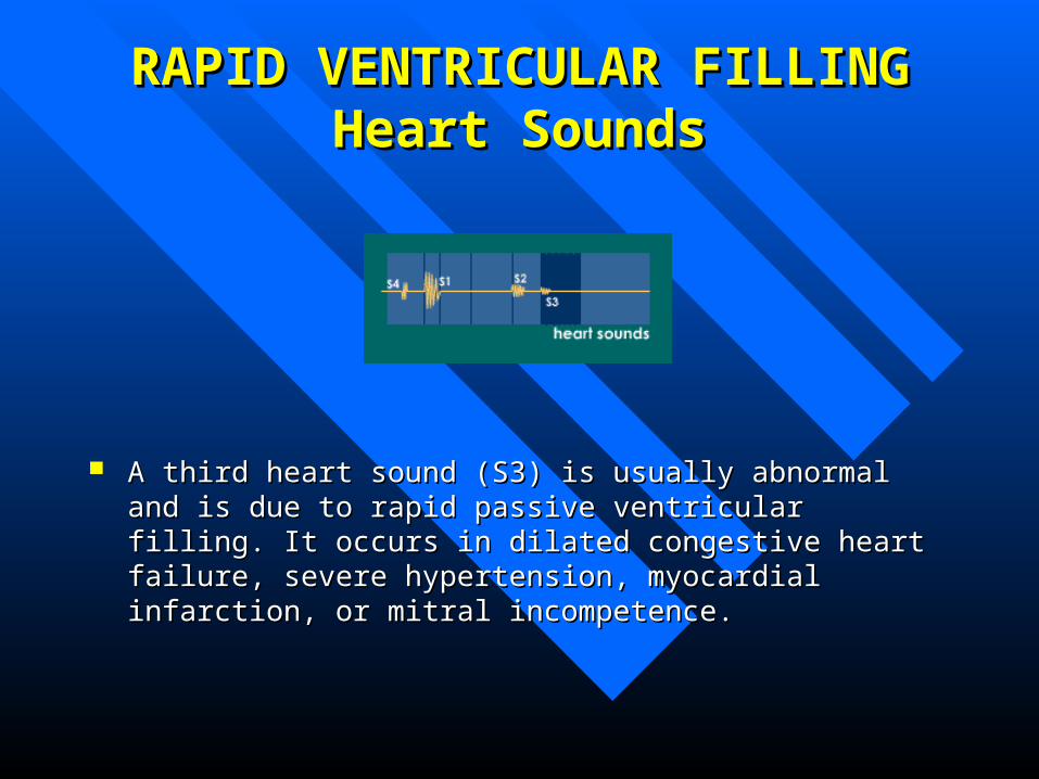
A third heart sound (S3) is usually abnormal and is due to rapid A third heart sound (S3) is usually abnormal and is due to rapid passive ventricular filling. It occurs in dilated congestive heart failure, passive ventricular filling. It occurs in dilated congestive heart failure, severe hypertension, myocardial infarction, or mitral incompetence.severe hypertension, myocardial infarction, or mitral incompetence.
REDUCED VENTRICULAR REDUCED VENTRICULAR FILLINGFILLING
(Diastasis) (Diastasis)
REDUCED VENTRICULAR FILLINGREDUCED VENTRICULAR FILLINGHeartHeart
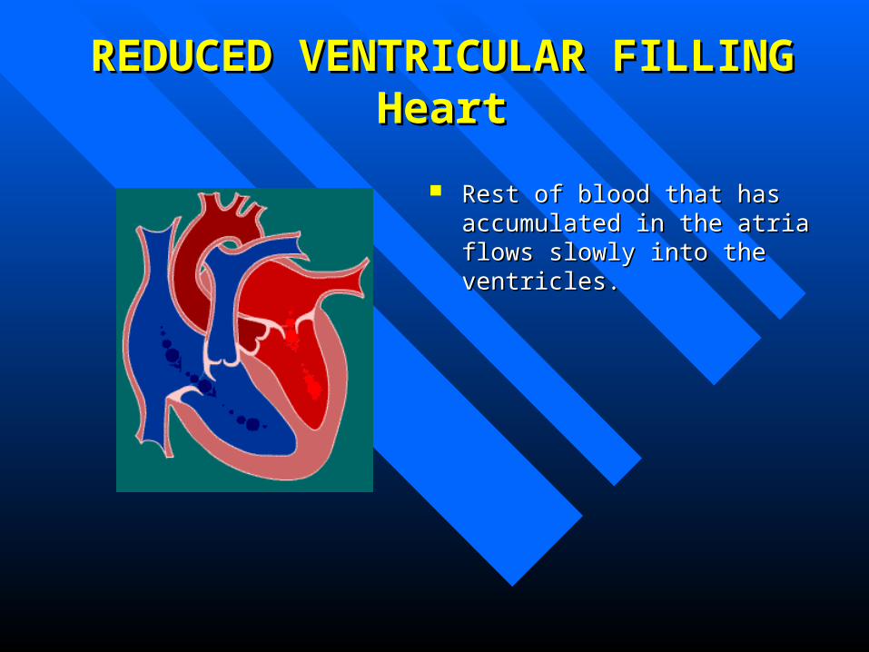
Rest of blood that has accumulated Rest of blood that has accumulated in the atria flows slowly into the in the atria flows slowly into the ventricles.ventricles.
REDUCED VENTRICULAR FILLING REDUCED VENTRICULAR FILLING Pressures & VolumesPressures & Volumes
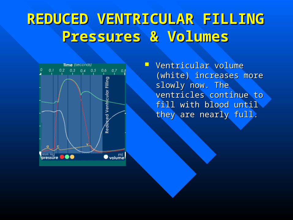
Ventricular volume (white) increases Ventricular volume (white) increases more slowly now. The ventricles more slowly now. The ventricles continue to fill with blood until they continue to fill with blood until they are nearly full.are nearly full.
REDUCED VENTRICULAR FILLING REDUCED VENTRICULAR FILLING ECGECG
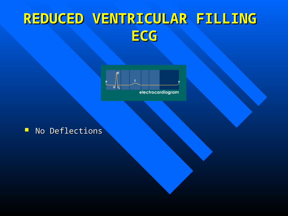
No Deflections No Deflections
REDUCED VENTRICULAR FILLING REDUCED VENTRICULAR FILLING Heart SoundsHeart Sounds
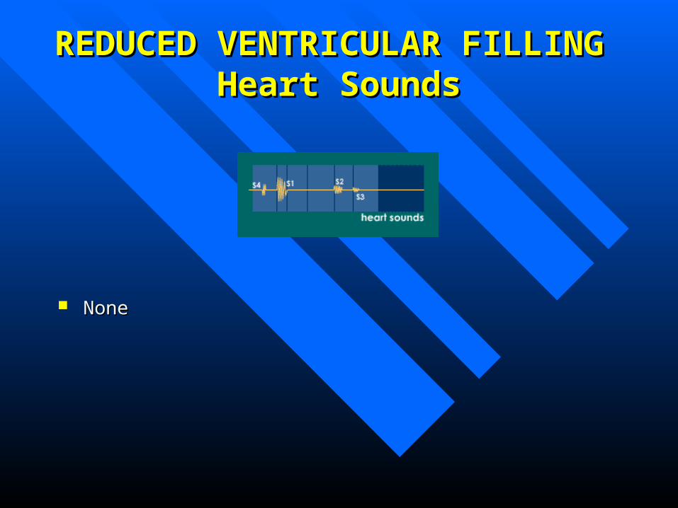
None None

THE COMPLETE PICTURETHE COMPLETE PICTURE