Embed Size (px)
Citation preview
http://www.die.upm.es/im/
BIOMEDICAL IMAGE TECHNOLOGIES LAB
Universidad Politécnica de Madrid
CIBER-BBN
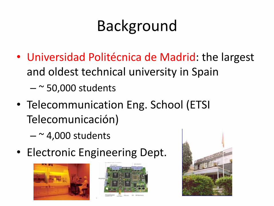
Background
• Universidad Politécnica de Madrid: the largest and oldest technical university in Spain
– ~ 50,000 students
• Telecommunication Eng. School (ETSI Telecomunicación)
– ~ 4,000 students
• Electronic Engineering Dept.
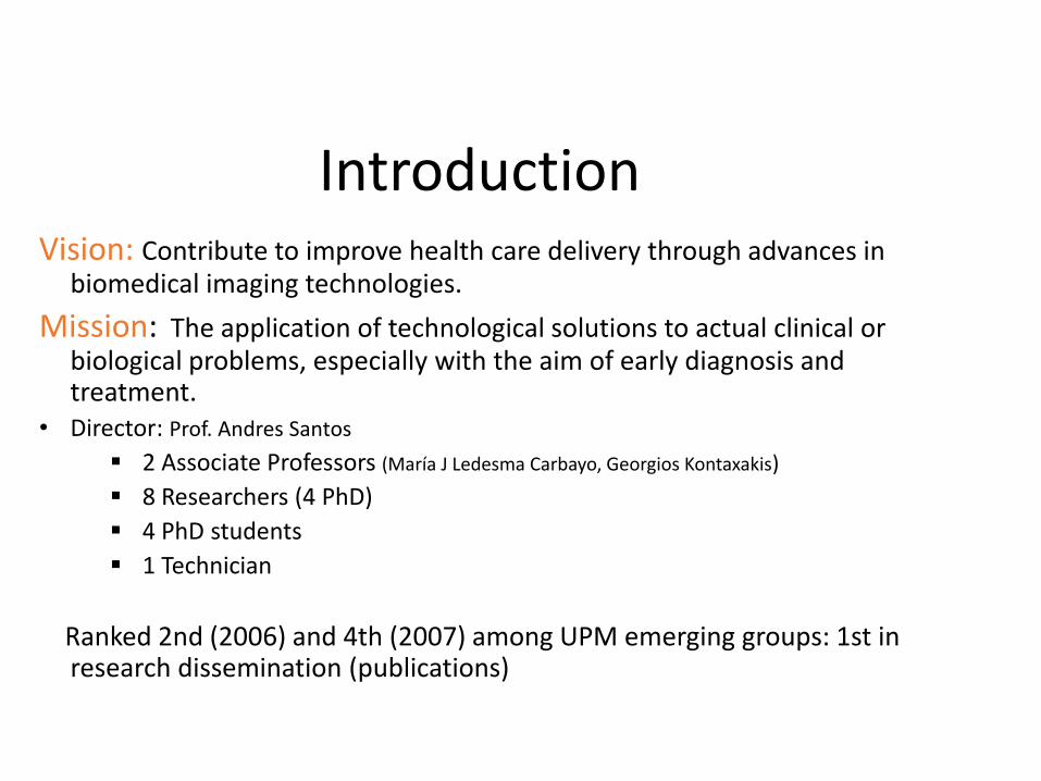
Introduction Vision: Contribute to improve health care delivery through advances in
biomedical imaging technologies.
Mission: The application of technological solutions to actual clinical or biological problems, especially with the aim of early diagnosis and treatment.
• Director: Prof. Andres Santos
2 Associate Professors (María J Ledesma Carbayo, Georgios Kontaxakis)
8 Researchers (4 PhD)
4 PhD students
1 Technician
Ranked 2nd (2006) and 4th (2007) among UPM emerging groups: 1st in research dissemination (publications)
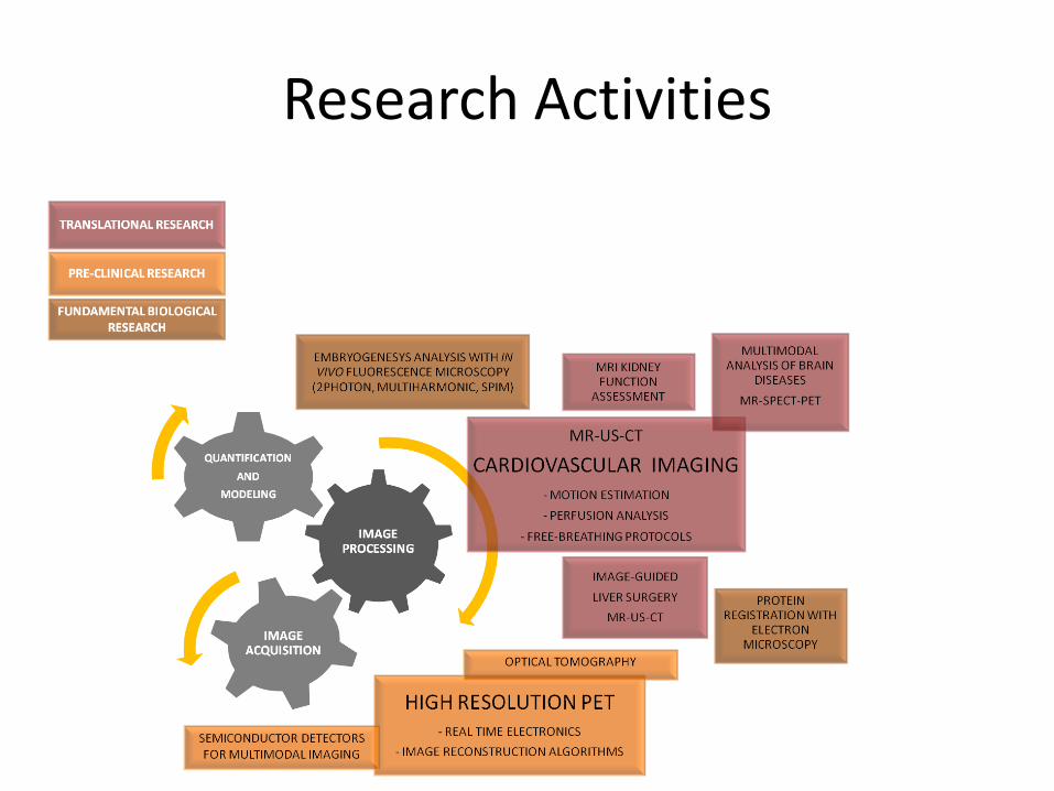
Research Activities
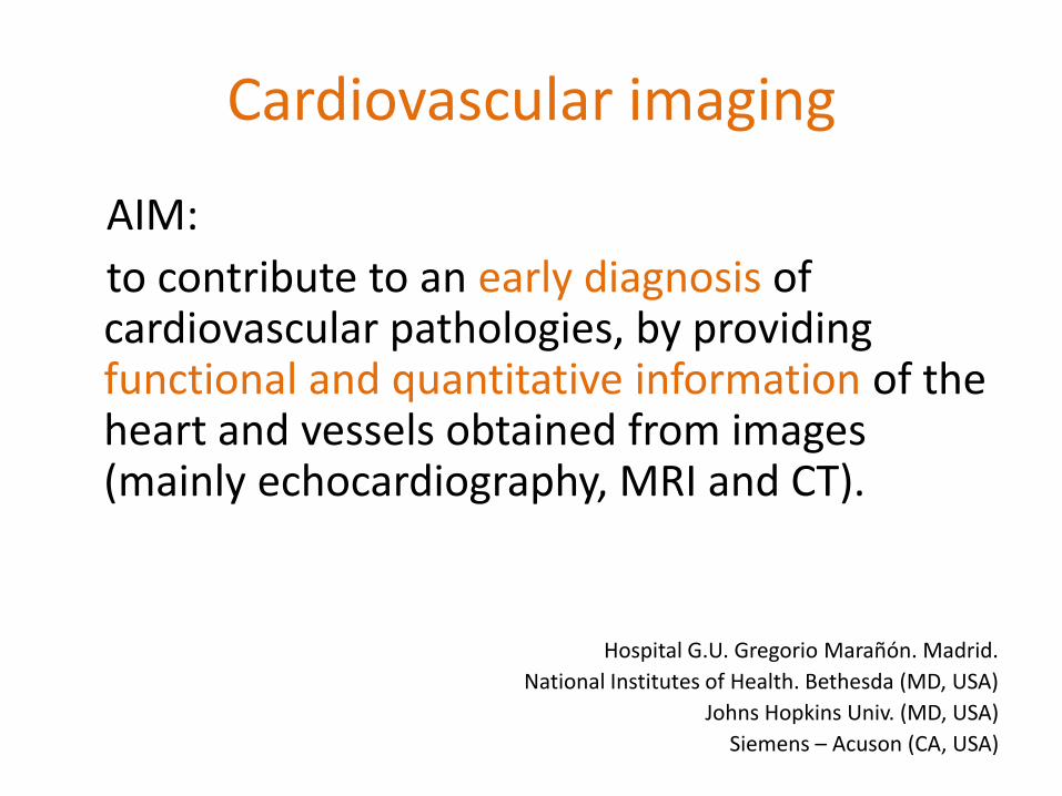
Cardiovascular imaging
AIM:
to contribute to an early diagnosis of cardiovascular pathologies, by providing functional and quantitative information of the heart and vessels obtained from images (mainly echocardiography, MRI and CT).
Hospital G.U. Gregorio Marañón. Madrid.
National Institutes of Health. Bethesda (MD, USA)
Johns Hopkins Univ. (MD, USA)
Siemens – Acuson (CA, USA)
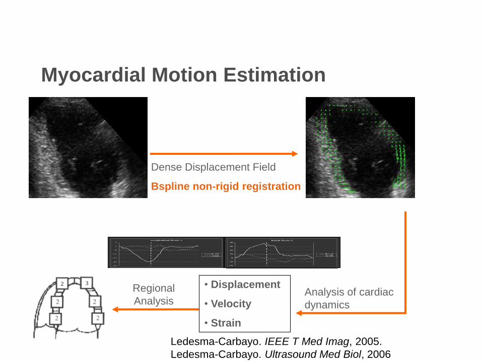
Myocardial Motion Estimation
Dense Displacement Field
Bspline non-rigid registration
2 3 Regional
Analysis
• Displacement
• Velocity
• Strain
Analysis of cardiac
dynamics
Ledesma-Carbayo. IEEE T Med Imag, 2005.
Ledesma-Carbayo. Ultrasound Med Biol, 2006
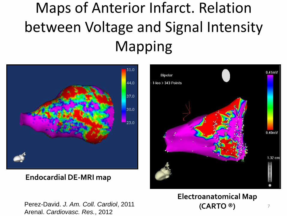
Maps of Anterior Infarct. Relation between Voltage and Signal Intensity
Mapping
7
Endocardial DE-MRI map
Electroanatomical Map (CARTO ®) Perez-David. J. Am. Coll. Cardiol, 2011
Arenal. Cardiovasc. Res., 2012
High Resolution Pre-clinical Imaging
AIM:
to contribute to the design and implementation of high resolution experimental Positron Emission Tomography (PET) scanners. A new research line has been started on Optical Tomography.
Hospital G.U. Gregorio Marañón. Madrid.
SEDECAL (SUINSA). Spain
Università di Pisa (Italy)
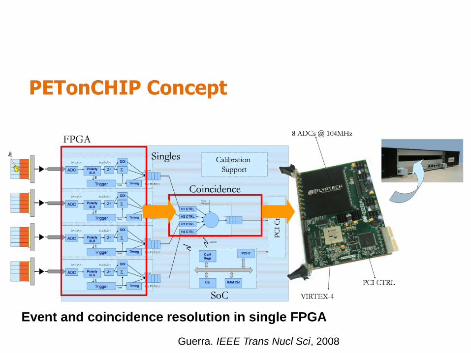
PETonCHIP Concept
Event and coincidence resolution in single FPGA
Guerra. IEEE Trans Nucl Sci, 2008
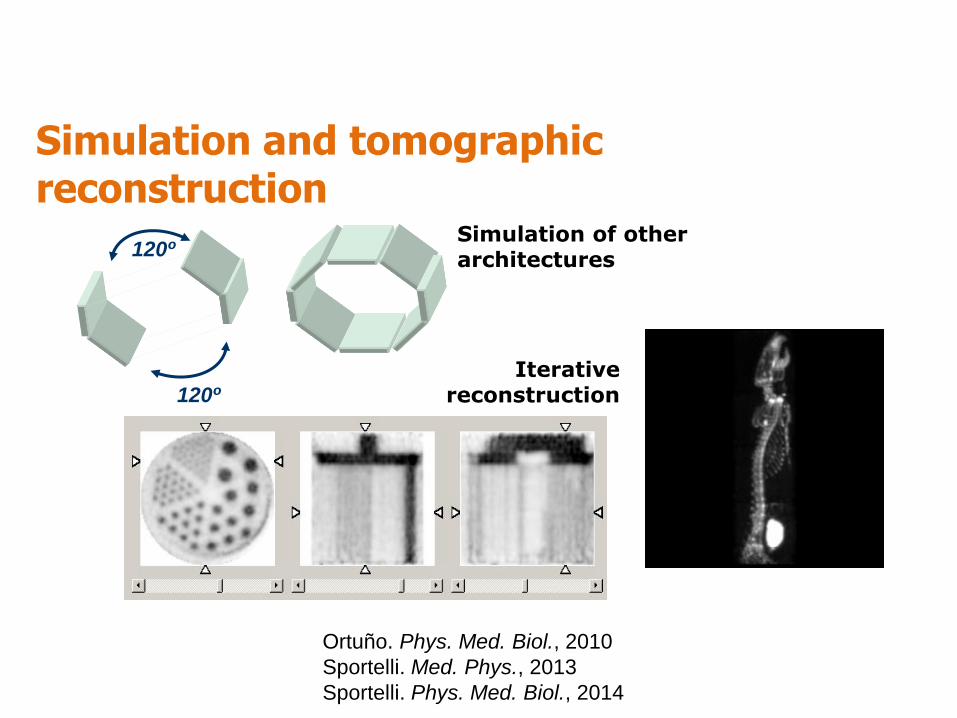
Iterative reconstruction 120º
120º Simulation of other architectures
Simulation and tomographic reconstruction
Ortuño. Phys. Med. Biol., 2010
Sportelli. Med. Phys., 2013
Sportelli. Phys. Med. Biol., 2014
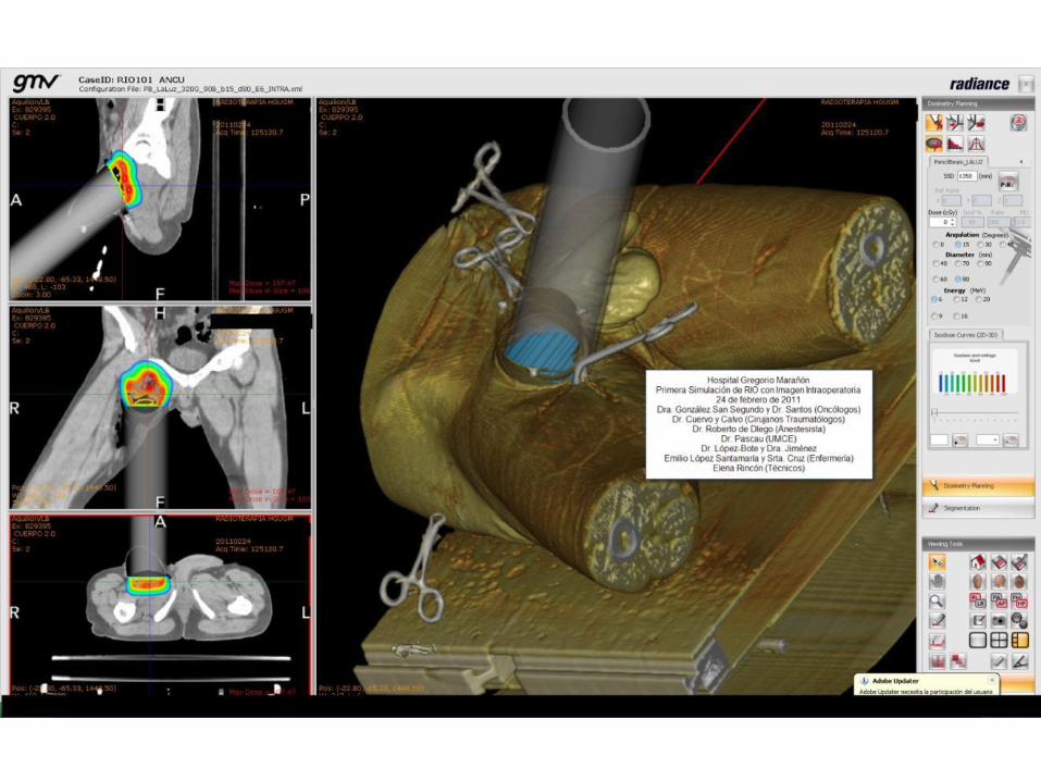
Image-guided radiotherapy and dosimetry
AIM:
to contribute to the radiotherapy planning, dosimetry and surgery planning by means of image registration, image segmentation and MC simulations.
GMV Soluciones Globales Internet. Spain
Hospital G.U. Gregorio Marañón. Madrid.
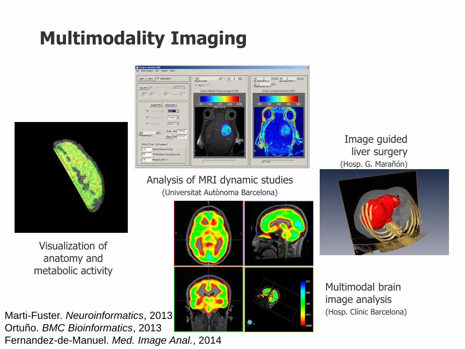
Multimodality Imaging
Image guided liver surgery
(Hosp. G. Marañón)
Visualization of anatomy and
metabolic activity
Analysis of MRI dynamic studies (Universitat Autònoma Barcelona)
Multimodal brain image analysis (Hosp. Clínic Barcelona) Marti-Fuster. Neuroinformatics, 2013
Ortuño. BMC Bioinformatics, 2013
Fernandez-de-Manuel. Med. Image Anal., 2014
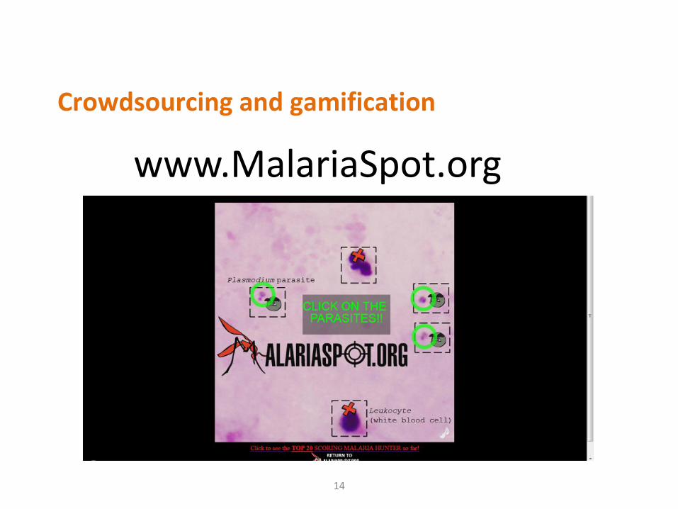
www.MalariaSpot.org
14
Crowdsourcing and gamification

Microscopy Image Analysis for Systems
Biology AIM: to develop image analysis methods to process data acquired with the most recent state-of-the-art microscopy technologies for biomedical understanding.
Multidimensional in-vivo observations of organism models such as zebrafish embryo need images analysis to solve new challenges in systems biology at the different genetic, proteomic, cellular, organic and individual levels.
Institut de Neurobiologie Alfred Fessard - CNRS, France
Complex Systems Institute of Paris (ISC-PIF), France
Spanish National Center of Biotechnology (CSIC-CNB), Spain
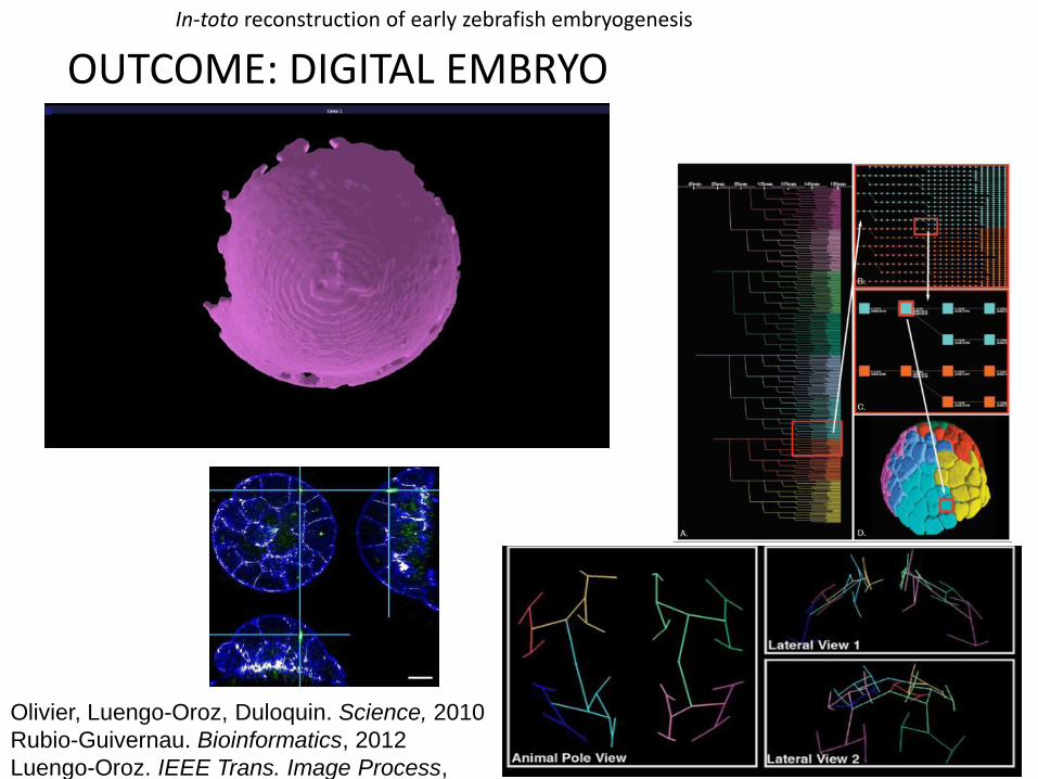
In-toto reconstruction of early zebrafish embryogenesis
OUTCOME: DIGITAL EMBRYO
Olivier, Luengo-Oroz, Duloquin. Science, 2010
Rubio-Guivernau. Bioinformatics, 2012
Luengo-Oroz. IEEE Trans. Image Process,
2012
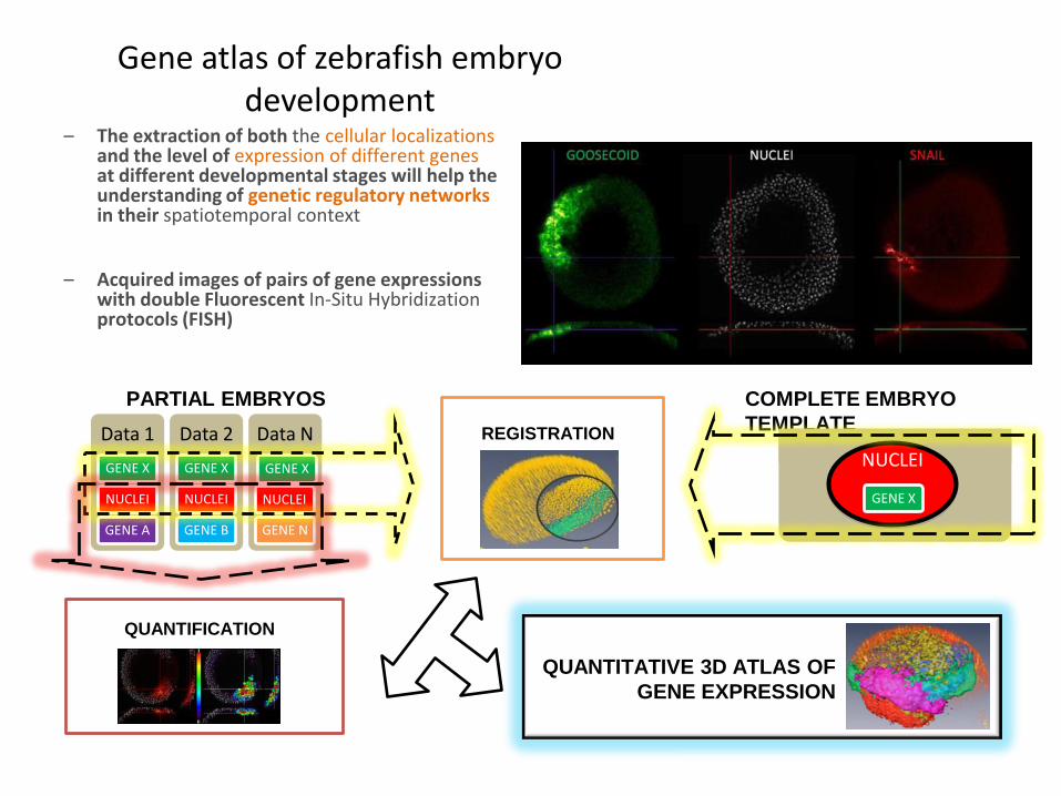
Gene atlas of zebrafish embryo development
Data 1
GENE X
NUCLEI
GENE A
Data 2
GENE X
NUCLEI
GENE B
Data N
GENE X
NUCLEI
GENE N
PARTIAL EMBRYOS COMPLETE EMBRYO
TEMPLATE
NUCLEI
GENE X
QUANTIFICATION
QUANTITATIVE 3D ATLAS OF
GENE EXPRESSION
– The extraction of both the cellular localizations and the level of expression of different genes at different developmental stages will help the understanding of genetic regulatory networks in their spatiotemporal context
– Acquired images of pairs of gene expressions with double Fluorescent In-Situ Hybridization protocols (FISH)
REGISTRATION
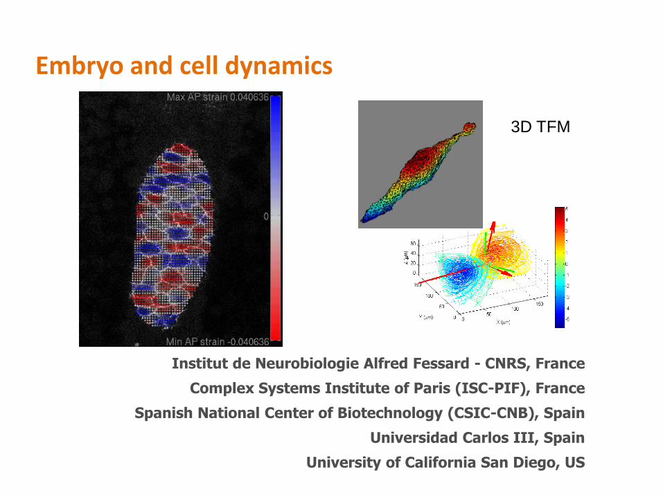
Embryo and cell dynamics
Institut de Neurobiologie Alfred Fessard - CNRS, France
Complex Systems Institute of Paris (ISC-PIF), France
Spanish National Center of Biotechnology (CSIC-CNB), Spain
Universidad Carlos III, Spain
University of California San Diego, US
3D TFM
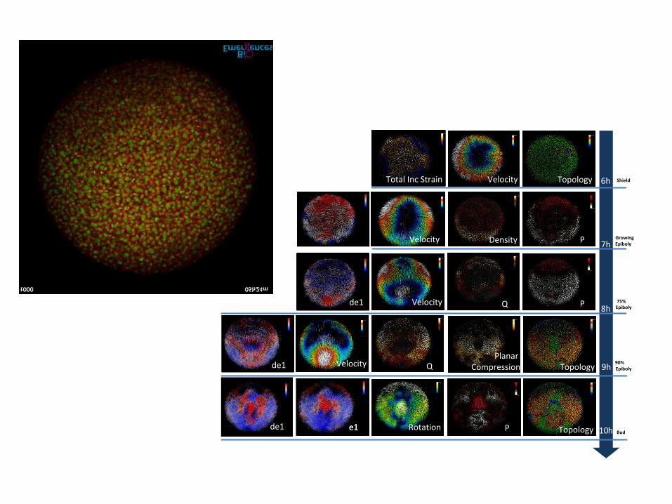
e1
90%Epiboly
Bud
Shield
GrowingEpiboly
75%EpibolyQ P
PDensity
TopologyVelocity 6h
7h
8h
9hQ TopologyPlanar
Compression
Velocity
Velocity
10hPRotation Topologye1
Total Inc Strain
e1
e1
de1
de1
de1
Velocity