Embed Size (px)
Citation preview
109
Methods of Isolation of Bacteria
MICROBIOLOGY
MODULEMicrobiology
Notes
10
METHODS OF ISOLATION OFBACTERIA
10.1 INTRODUCTION
We have learned in earlier chapters that there exist so many bacteria that causehuman disease.so now our task is to isolate these bacteria and identify them. Theidentification is required so as to cure the illness or the infection caused due tothese bacteria, using appropriate antibiotics. Identification also holds significancefor epidemiological purposes.
This chapter would focus on various methods used for isolation of bacteria.While in subsequent chapters we would learn about identification of bacteria andthe ways to contain the infections caused by them.
OBJECTIVES
After reading this chapter, you will be able to :
Expalin the steps involved in the isolation of bacteria.
describe the significance of Specimen collection.
describe the significance of Preservation and transportation of specimen.
explain the role of microscopy in isolation of bacteria.
explain various methods for isolation of bacteria.
10.2 ISOLATION OF BACTERIA
Isolation of bacteria forms a very significant step in the diagnosis andmanagement of the illness. Isolation of bacteria involves various steps –
Specimen collection
Preservation and transportation of specimen
MICROBIOLOGY
MODULE Methods of Isolation of Bacteria
Microbiology
110
Notes
Microscopic examination of sample
Various methods used for isolation of bacteria
Specimen collection
Many different specimens are sent for microbiological examination frompatients with suspected bacterial infection. Common specimens include urine,faeces, wound swabs, throat swabs, vaginal swabs, sputum, and blood. Lesscommon, but important specimens include cerebrospinal fluid, pleural fluid,joint aspirates, tissue, bone and prosthetic material (e.g. line tips).
Some types of specimen are normally sterile e.g. blood, CSF. These samples areusually obtained via a percutaneous route with needle and syringe, usingappropriate skin disinfection and an aseptic technique. The culture of bacteriafrom such specimens is usually indicative of definite infection except if they areskin contaminants (bacteria inhabitants of normal skin).
Fig. 10.1: Universal container.
In contrast, many microbiological specimens are obtained from non-sterile sitese.g. vaginal or throat swabs, urine sample, stool sample. Such samples oftencontain bacteria of no clinical relevance in addition to possible pathogens,making the interpretation of culture results more difficult. In general it ispreferable to send samples from sterile sites if available.
It is preferred to obtain the samples for bacteriological culture before antibiotictherapy is started. This maximizes the sensitivity of the investigations andreduces false-negative results. Similarly, samples of tissue or pus are preferredover swabs, to maximize the recovery of bacteria in the laboratory.
Specimens must be accurately labelled and accompanied by a properlycompleted requisition form, indicating the nature of the specimen, the date ofsample collection, relevant clinical information, the investigations required, anddetails of antibiotic therapy, if any.
111
Methods of Isolation of Bacteria
MICROBIOLOGY
MODULEMicrobiology
Notes
This allows the laboratory to perform the correct range of tests, and helps in theinterpretation of results and reporting. Along with clinical specimens, medicalmicrobiology laboratories also process samples of food, water and otherenvironmental samples (e.g. air sampling from operating theatres) as part ofinfection control procedures.
High-risk samples
Certain bacterial infections are a particular hazard to laboratory staff, andspecimens that might contain these pathogens should be labelled as ‘high risk’to allow for additional safety measures if necessary. For example - blood culturesfrom suspected typhoid (Salmonella typhi) or brucellosis (Brucella species), andsamples from suspected Mycobacterium tuberculosis.
Preservation and Transport of specimen
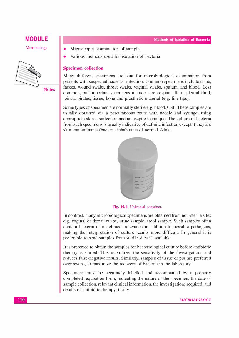
Most specimens are sent to the laboratory in sterile universal containers. Swabsare placed in a suitable transport medium (eg. charcoal medium) otherwise itleads to false negative reporting.
Fig. 10.2: Charcoal laden transport media
Specimens should be transported as soon as possible to the laboratory. In casea delay is anticipated the specimen should be stored at 4° C.
Immediate transport is necessary in order to:
(i) Preserve the viability of the ‘delicate’ bacteria, such as Streptococcuspneumoniae or Haemophilus influenzae (delays in processing can causefalse-negative culture results);
(ii) Minimize the multiplication of bacteria (e.g. coliforms) within specimensbefore they reach the laboratory. In particular urine and other specimensthat utilize a semiquantitative culture technique for thier detection, asdelays in transport can give rise to falsely high bacterial counts when thespecimen is processed.
Microscopy
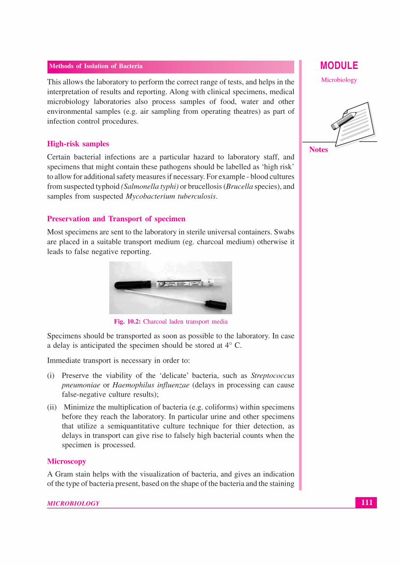
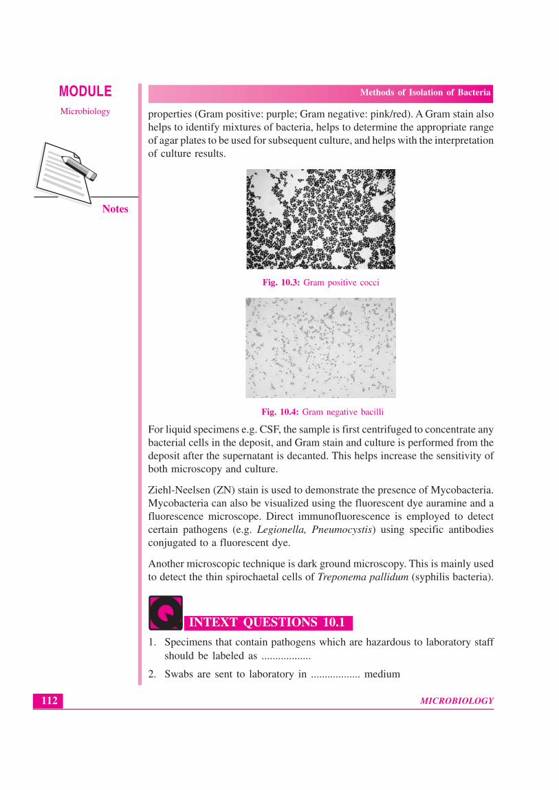
A Gram stain helps with the visualization of bacteria, and gives an indicationof the type of bacteria present, based on the shape of the bacteria and the staining
MICROBIOLOGY
MODULE Methods of Isolation of Bacteria
Microbiology
112
Notes
properties (Gram positive: purple; Gram negative: pink/red). A Gram stain alsohelps to identify mixtures of bacteria, helps to determine the appropriate rangeof agar plates to be used for subsequent culture, and helps with the interpretationof culture results.
Fig. 10.3: Gram positive cocci
Fig. 10.4: Gram negative bacilli
For liquid specimens e.g. CSF, the sample is first centrifuged to concentrate anybacterial cells in the deposit, and Gram stain and culture is performed from thedeposit after the supernatant is decanted. This helps increase the sensitivity ofboth microscopy and culture.
Ziehl-Neelsen (ZN) stain is used to demonstrate the presence of Mycobacteria.Mycobacteria can also be visualized using the fluorescent dye auramine and afluorescence microscope. Direct immunofluorescence is employed to detectcertain pathogens (e.g. Legionella, Pneumocystis) using specific antibodiesconjugated to a fluorescent dye.
Another microscopic technique is dark ground microscopy. This is mainly usedto detect the thin spirochaetal cells of Treponema pallidum (syphilis bacteria).
INTEXT QUESTIONS 10.1
1. Specimens that contain pathogens which are hazardous to laboratory staffshould be labeled as ..................
2. Swabs are sent to laboratory in .................. medium
113
Methods of Isolation of Bacteria
MICROBIOLOGY
MODULEMicrobiology
Notes
3. If delay is anticipated in transporting the specimen, it should be stored at.................. temperature
4. .................. gives an indication of bacteria present in the sample
5. .................. stain is used in demonstration of mycobacteria
6. .................. microscopy is used to detect syphilis organism
10.3 METHODS OF ISOLATION OF BACTERIA
Methods of isolation of bacteria can be broadly classified into two
Culture methods
On Solid media
On Liquid media
Automated systems
Non-culture methods
Culture methods
The specimens received in the laboratory are plated on the culture media. Theappropriate culture media is selected depending upon the bacteria suspected. Thefollowing precautions need to be taken into consideration when the culturemethods are processed
Optimal atmospheric conditions
Optimal temperature
Growth requirement of the bacteria
Atmospheric conditions:
Colonies of bacteria are usually large enough to identify after 18–24 hours ofincubation (usually at 37°C), but for some bacteria longer incubation times arerequired (from 2 days to several weeks). Culture plates are incubated (1) in air,(2) in air with added carbon dioxide (5%), (3) anaerobically (without oxygen)or (4) micro-aerophilically (a trace of oxygen) according to the requirements ofthe different types of bacteria that may be present in specimens.
In case of Mycobacteria especially the scotochromogen the culture bottles areplaced in dark or the bottles are covered with black paper and kept for incubationat 37°C.
Temperature:
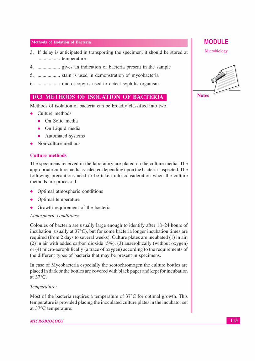
Most of the bacteria requires a temperature of 37°C for optimal growth. Thistemperature is provided placing the inoculated culture plates in the incubator setat 37°C temperature.
MICROBIOLOGY
MODULE Methods of Isolation of Bacteria
Microbiology
114
Notes
Fig. 10.5: Incubator
Growth requirement of the bacteria
Different bacteria have different growth requirements. For eg Streptococcuspneumoniae requires factor V and factor X for its growth, which are found inchocolate agar. Thus for sample suspected of S. pneumoniae the samples areplated on chocolate agar. Similarly depending upon the growth requirementsthe appropriate culture media are used.
INTEXT QUESTIONS 10.2
1. .................. & .................. methods are commonly used methods forbacterial isolation
2. Colonies of bacteria can be identified after .................. hours of incubation
3. The optimum temperature most bacteria require to grow are ..................
4. Chocolate agar has .................. & .................. which is used in the diagnosisof streptococci Pneumonia
115
Methods of Isolation of Bacteria
MICROBIOLOGY
MODULEMicrobiology
Notes
10.4 CULTURE ON SOLID MEDIA
The principal method for the detection of bacteria from clinical specimens is byculture on solid culture media. Bacteria grow on the surface of culture mediato produce distinct colonies.
Different bacteria produce different but characteristic colonies, allowing forearly presumptive identification and easy identification of mixed cultures. Thereare many different types of culture media. Agar is used as the gelling agent towhich is added a variety of nutrients (e.g. blood, peptone and sugars) and otherfactors (e.g. buffers, salts and indicators).
Some culture media are nonselective (e.g. blood agar, nutrient agar) and thesewill grow a wide variety of bacteria. While some e.g. MacConkey agar are moreselective (in this case through the addition of bile salts selecting for the ‘bile-tolerant’ bacteria found in the large intestine such as Escherichia coli andEnterococcus faecalis). MacConkey agar also contains lactose and an indicatorsystem that identifies lactose-fermenting coliforms (e.g. Escherichia coli,Klebsiella) from lactose-non fermenting coliforms (e.g. Morganella Salmonella).Media can be made even more selective by the addition of antibiotics or otherinhibitory substances, and sophisticated indicator systems can allow for the easydetection of defined bacteria from mixed populations.
Method of inoculating the solid culture media
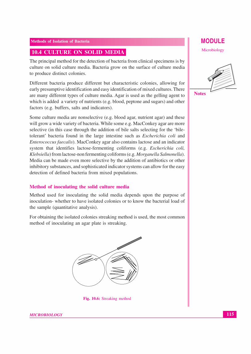
Method used for inoculating the solid media depends upon the purpose ofinoculation- whether to have isolated colonies or to know the bacterial load ofthe sample (quantitative analysis).
For obtaining the isolated colonies streaking method is used, the most commonmethod of inoculating an agar plate is streaking.
Fig. 10.6: Streaking method
MICROBIOLOGY
MODULE Methods of Isolation of Bacteria
Microbiology
116
Notes
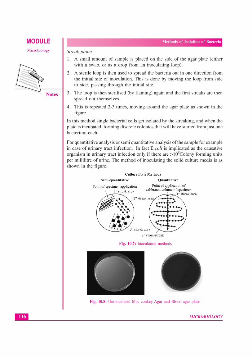
Streak plates
1. A small amount of sample is placed on the side of the agar plate (eitherwith a swab, or as a drop from an inoculating loop).
2. A sterile loop is then used to spread the bacteria out in one direction fromthe initial site of inoculation. This is done by moving the loop from sideto side, passing through the initial site.
3. The loop is then sterilised (by flaming) again and the first streaks are thenspread out themselves.
4. This is repeated 2-3 times, moving around the agar plate as shown in thefigure.
In this method single bacterial cells get isolated by the streaking, and when theplate is incubated, forming discrete colonies that will have started from just onebacterium each.
For quantitative analysis or semi quantitative analysis of the sample for examplein case of urinary tract infection. In fact E.coli is implicated as the causativeorganism in urinary tract infection only if there are >105Colony forming unitsper millilitre of urine. The method of inoculating the solid culture media is asshown in the figure.
Fig. 10.7: Inoculation methods
Fig. 10.8: Uninoculated Mac conkey Agar and Blood agar plate
117
Methods of Isolation of Bacteria
MICROBIOLOGY
MODULEMicrobiology
Notes
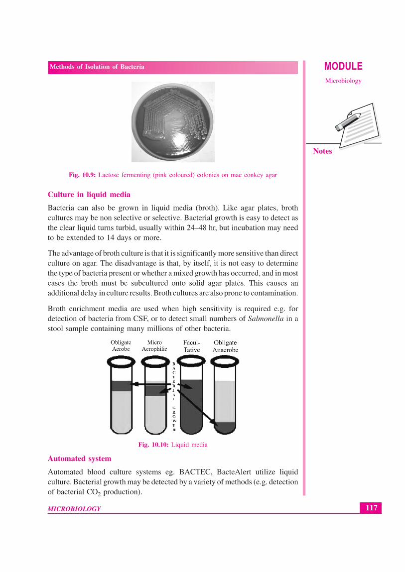
Fig. 10.9: Lactose fermenting (pink coloured) colonies on mac conkey agar
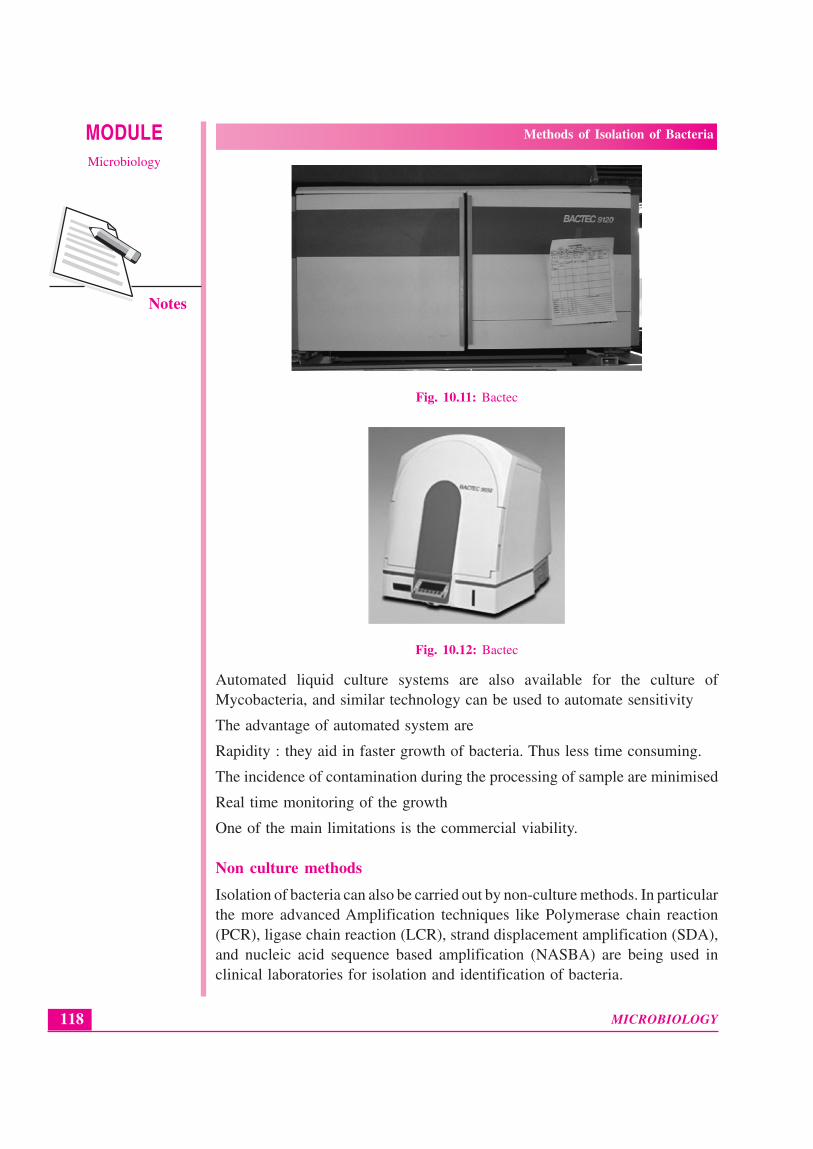
Culture in liquid media
Bacteria can also be grown in liquid media (broth). Like agar plates, brothcultures may be non selective or selective. Bacterial growth is easy to detect asthe clear liquid turns turbid, usually within 24–48 hr, but incubation may needto be extended to 14 days or more.
The advantage of broth culture is that it is significantly more sensitive than directculture on agar. The disadvantage is that, by itself, it is not easy to determinethe type of bacteria present or whether a mixed growth has occurred, and in mostcases the broth must be subcultured onto solid agar plates. This causes anadditional delay in culture results. Broth cultures are also prone to contamination.
Broth enrichment media are used when high sensitivity is required e.g. fordetection of bacteria from CSF, or to detect small numbers of Salmonella in astool sample containing many millions of other bacteria.
Fig. 10.10: Liquid media
Automated system
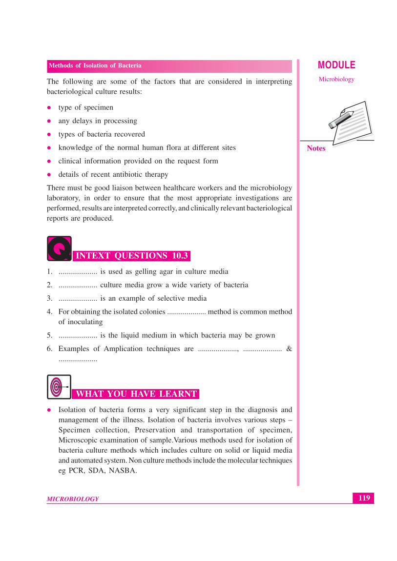
Automated blood culture systems eg. BACTEC, BacteAlert utilize liquidculture. Bacterial growth may be detected by a variety of methods (e.g. detectionof bacterial CO2 production).
MICROBIOLOGY
MODULE Methods of Isolation of Bacteria
Microbiology
118
Notes
Fig. 10.11: Bactec
Fig. 10.12: Bactec
Automated liquid culture systems are also available for the culture ofMycobacteria, and similar technology can be used to automate sensitivity
The advantage of automated system are
Rapidity : they aid in faster growth of bacteria. Thus less time consuming.
The incidence of contamination during the processing of sample are minimised
Real time monitoring of the growth
One of the main limitations is the commercial viability.
Non culture methods
Isolation of bacteria can also be carried out by non-culture methods. In particularthe more advanced Amplification techniques like Polymerase chain reaction(PCR), ligase chain reaction (LCR), strand displacement amplification (SDA),and nucleic acid sequence based amplification (NASBA) are being used inclinical laboratories for isolation and identification of bacteria.
119
Methods of Isolation of Bacteria
MICROBIOLOGY
MODULEMicrobiology
Notes
The following are some of the factors that are considered in interpretingbacteriological culture results:
type of specimen
any delays in processing
types of bacteria recovered
knowledge of the normal human flora at different sites
clinical information provided on the request form
details of recent antibiotic therapy
There must be good liaison between healthcare workers and the microbiologylaboratory, in order to ensure that the most appropriate investigations areperformed, results are interpreted correctly, and clinically relevant bacteriologicalreports are produced.
INTEXT QUESTIONS 10.3
1. .................... is used as gelling agar in culture media
2. .................... culture media grow a wide variety of bacteria
3. .................... is an example of selective media
4. For obtaining the isolated colonies .................... method is common methodof inoculating
5. .................... is the liquid medium in which bacteria may be grown
6. Examples of Amplication techniques are ...................., .................... &....................
WHAT YOU HAVE LEARNT
Isolation of bacteria forms a very significant step in the diagnosis andmanagement of the illness. Isolation of bacteria involves various steps –Specimen collection, Preservation and transportation of specimen,Microscopic examination of sample.Various methods used for isolation ofbacteria culture methods which includes culture on solid or liquid mediaand automated system. Non culture methods include the molecular techniqueseg PCR, SDA, NASBA.

MICROBIOLOGY
MODULE Methods of Isolation of Bacteria
Microbiology
120
Notes
TERMINAL QUESTIONS
1. What is the need for isolation of bacteria?
2. Describe in brief various steps involved in the isolation of bacteria.
3. What is difference between blood agar and chocolate agar
4. Explain the term selective and non selective media with proper examples.
5. Draw a labeled diagram of inoculation of solid culture media for isolationof bacteria.
6. Draw a labeled diagram for inoculation of solid media for processing theurine sample of a patient suspected of urinary tract infection.
7. Describe in brief the advantages and the limitation of use of liquid culturemedia for isolation of bacteria.
8. Mention the advantages and the disadvantages of automated system forisolation of bacteria.
9. Name some non culture methods for isolation of bacteria
ANSWERS TO INTEXT QUESTIONS
10.1
1. High-risk
2. Charcoal
3. 4oC
4. Gram stain
5. Ziehl-Neelson
6. Dark Ground
10.2
1. Direct culture & Non-culture
2. 18-24
3. 37oC
4. Factor V & Factor X
121
Methods of Isolation of Bacteria
MICROBIOLOGY
MODULEMicrobiology
Notes
10.3
1. Agar
2. Non-selective
3. MacConkey
4. Streaking
5. Broth
6. Polymerase Chain Reaction, Ligase Chain Reaction, Nucleic Acid SequenceBased Amplification