Embed Size (px)
Citation preview
Presented by Dr. Hemant Nargawe
Introduction
The complete blood count (CBC) is the most commonly ordered blood test in a healthcare set up. The fully automated sophisticated cell counters give us very accurate and precise blood counts rapidly along with a number of additional newer parameters which give valuable extra information added to the routine CBS. The origin of the modern cell counters date back to 1953, when Wallace H Coulter, an American engineer by training, obtained a patent for a principle for counting and sizing microparticles suspended in a fluid. The principle was named the 'coulter principle' and is the basis of the technology of most cell counters even today.
A basic CBC comprises of the following parameters Hemoglobin (Hb) Hematocrit (Hct)
Red Blood Cells (RBCs) and indices Total Red Cell count (TRBC) Mean corpuscular volume (MCV) Mean corpuscular hemoglobin (MCH) Mean Corpuscular hemoglobin concentration (MCHC) Red cell distribution width (RDW)
White blood cells (WBCs) Total while cell count (WBC) White cell count differential (DC) Absolute white cell counts
Platelets Platelet count (PC) Mean platelet volume (MPV) Platelet distribution width (PDW)
The Hb and the Hct
The haemoglobin is measured after the RBCs are lysed and the Hb converted to a stable colored compound which is measured spectrophotometrically. Hence, like lipemia and hyperbilirubinemia can interfere with the results.
The Hct can be used synonymously with the Hb to classify a sample as anaemic or polycythemic. The Hct is the given volume of packed RBCs as compared to the plasma and can be measured by a microhaematocrit centrifuge (as in Sysmex analyzers) or calculated (as a Beckman Coulter instruments) by taking into account the Hb and the MCV.
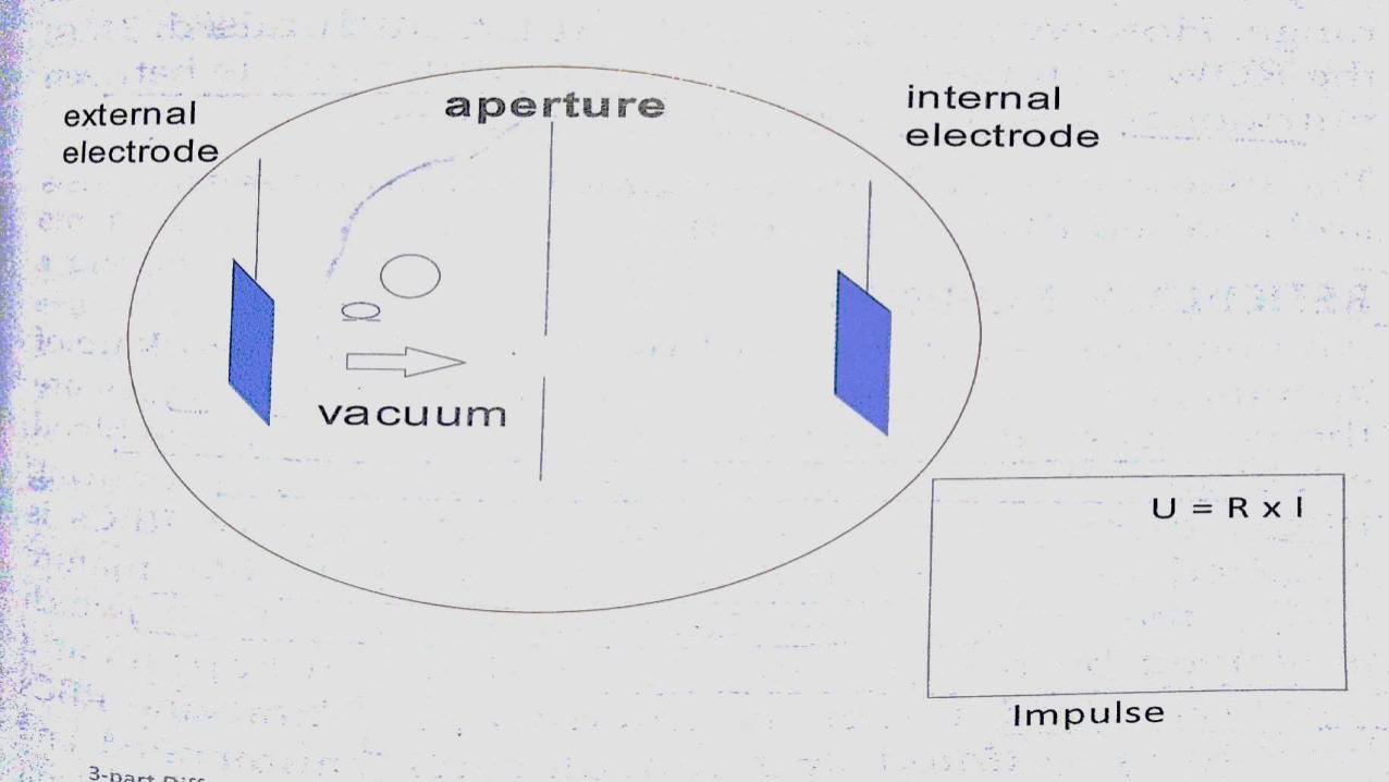
TRBC and MCV
The most common technology for counting and sizing the blood cells is the impedance technology based on the coulter principles. The basic counting chamber comprises of two electrodes with a partition between them. The partition has an aperture through which electrolyte flows and maintains flow of electrical current between the electrodes. The TRBC and the platelet counts are obtained in this manner.
The height / size of a pulse generated depends on the size of the cell causing it. In the case of RBCs, the average height of the pulses gives us the MCV. Besides numerical values, the instruments also give RBC volume distribution histograms.
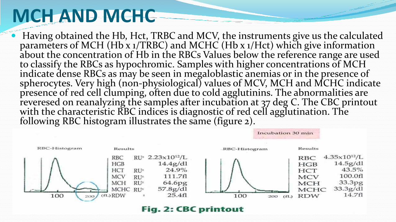
MCH AND MCHC Having obtained the Hb, Hct, TRBC and MCV, the instruments give us the calculated
parameters of MCH (Hb x 1/TRBC) and MCHC (Hb x 1/Hct) which give information about the concentration of Hb in the RBCs Values below the reference range are used to classify the RBCs as hypochromic. Samples with higher concentrations of MCH indicate dense RBCs as may be seen in megaloblastic anemias or in the presence of spherocytes. Very high (non-physiological) values of MCV, MCH and MCHC indicate presence of red cell clumping, often due to cold agglutinins. The abnormalities are reveresed on reanalyzing the samples after incubation at 37 deg C. The CBC printout with the characteristic RBC indices is diagnostic of red cell agglutination. The following RBC histogram illustrates the same (figure 2).
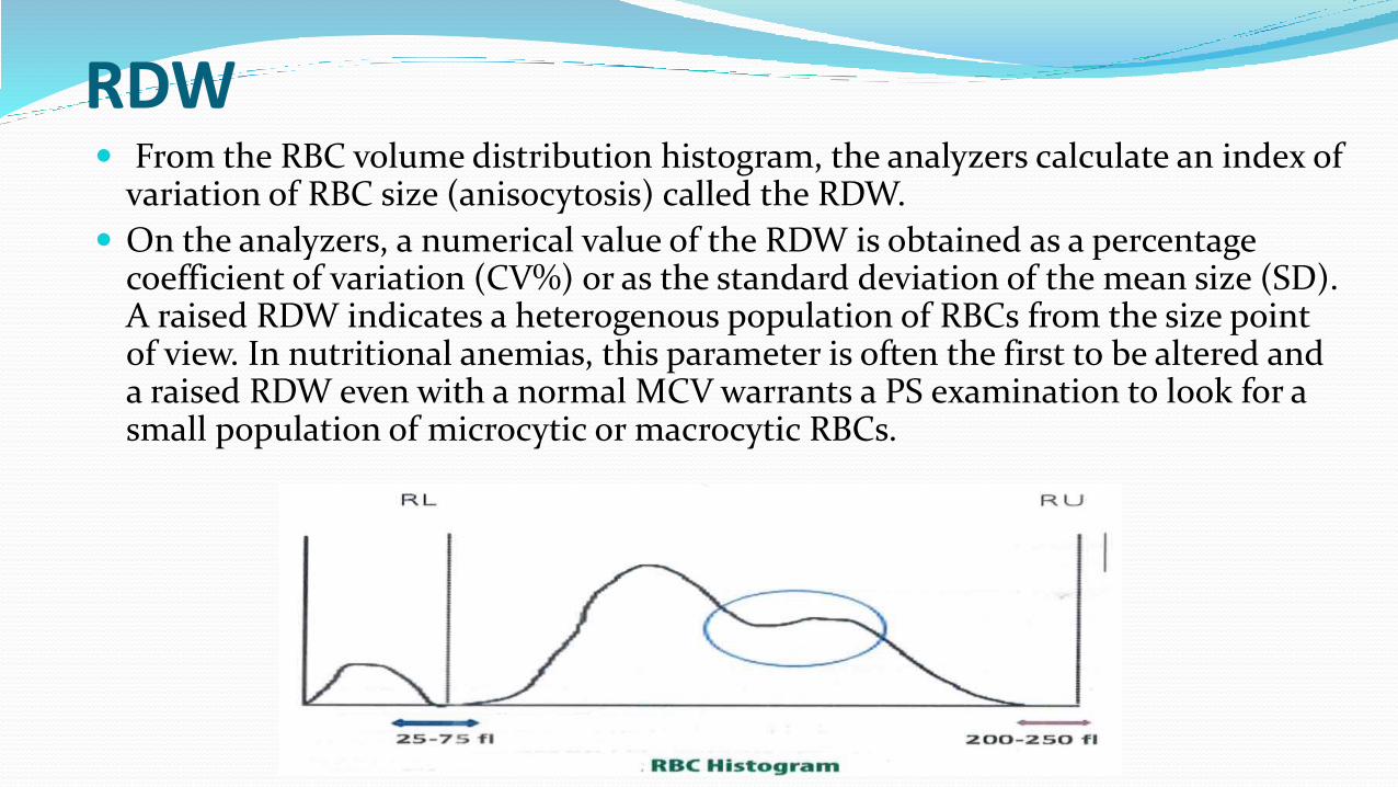
RDW From the RBC volume distribution histogram, the analyzers calculate an index of
variation of RBC size (anisocytosis) called the RDW.
On the analyzers, a numerical value of the RDW is obtained as a percentage coefficient of variation (CV%) or as the standard deviation of the mean size (SD). A raised RDW indicates a heterogenous population of RBCs from the size point of view. In nutritional anemias, this parameter is often the first to be altered and a raised RDW even with a normal MCV warrants a PS examination to look for a small population of microcytic or macrocytic RBCs.
RETICULOCYTE COUNTS
An under utilised but useful parameter in the work up of anemias is the reticulocyte count. As reticulocytes are the youngest forms of RBCs, there presence in the blood establishes the fact that the bone marrow is producing RBCs. Increased bone marrow production of RBCs is reflected in a raised reticulocyte count. Unlike mature RBCs, the reticulocytes contain remnant RNA which is stained by a supravital dye and which appears as a blue reticulum under the microscope.
Today's cell counter provide automated reticulocyte counts on the basis of their larger volumes and presence of RNA.
For ease and familiarity, the reticulocytes, continue to be expressed as a percentage of the RBCs which may lead to falsely raised values when the absolute number of RBCs is low as seen following hemolysis or blood loss. A better way is the 'correct' the reticulocyte count for the patient's Hb or Hct. A simple formula is Corrected reticulocyte count = % of reticulocyte x patient's Hct/45.
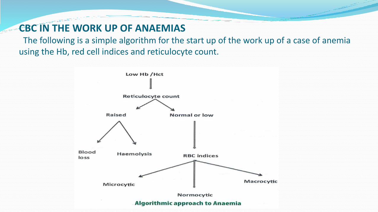
CBC IN THE WORK UP OF ANAEMIASThe following is a simple algorithm for the start up of the work up of a case of anemia
using the Hb, red cell indices and reticulocyte count.
If the reticulocyte count is raised and blood loss is ruled out, few routine biochemical tests like serum bilirubin and fractions, haptoglobin, lactate dehydrogenase (LDH), etc, may establish ongoing hemolysis. Added to these a peripheral smear examination may help to clinch the exact diagnosis / cause of hemolysis. For example, finding a spherocyte would suggest and autoimmune haemolytic anemia or hereditary spehyrocytosis while a bite cell or blister cell would suggest oxidant damage as seen in G6PD enzyme deficiency.
Alternatively, one may look at the RBC indices. Macrocytic anaemias may be due to B12/folate deficiencies but may be also seen with use of anti-retroviral drugs, liver disease, hypothyroidism. Macrocytic anaemia as a part of generalized pancytopenia may be seen in megaloblastic anaemia or aplastic anaemia. A unique type of myelodysplastic syndrome, associated with loss of the long arm of chromosome 5 (5q- syndrome) is characterized by macrocytic anemia and thrombocytosis. Increased reticulocytes seen in haemolytic anaemias may lead to slight macrocytois. Normocytic indices may be seen in ACD, haemolytic anaemias and even combined nutritional anaemias.
In the presence of microcytosis, the TRBC (raised in thalassemia) and RDW (increased in IDA) can help differentiate between iron deficiency and beta thalassemia trait. Indices like the Mentzer's index have been put forward to make this differentiation more objective. If the quotient of the MCV (in fl) divided by the TRBC (in millions / cubic mm) is less than 13, thalassemia is more likely. If the result is greater than 13, iron deficiency is more likely. Co-existing iron deficiency and between thalassemia trait make these values redundant. Beta thalassemia carriers have microcytic hypochromic RBC indices with a normal RDW as the red cells are uniformly small, i.e. homogenously microcytic. On the other hand, iron deficiency shows microcytosis along with an altered RDW.
CHr / Ret – He(reticulocyte Hb content/ reticulocyte Hb equivevalent)
A close differential diagnosis of iron deficiency anaemia (IDA) is anaemia of chronic disease (ACD), characterized by functional iron deficiency (FID) where iron is not incorporated into developing erythroid cells in spite of adequate bone marrow iron stores. It is now well established that release of hepcidin from liver cells under the influence of inflammatory cytokines like interleukin - 6 is responsible for blockage iron iron movement from iron storing macrophages into the developing red cells. This block also occurs at the level of the intestinal cells where in absorbed iron cannot enter the blood stream. Over weeks, this lead to formation of RBCs with low MCV and low MCH. The traditional biochemical tests like serum iron, transferrin and ferritin levels fail to convincingly distinguish between IDA and ACD. An improvement over these tests is the soluble transferrin receptor level (sTfr) which is raised only in IDA and remains low in ACD. This parameter is limited by its complexity of testing and its relative unavailability.
Changes in the RBC parameters as described occur over the life span of RBCs (120 days) while changes occurring in reticulocytes occur quickly i.e. within 2-3 days (the life) span of the reticulocytes. CHr provided by instruments manufactured by Siemens and Ret-He provided by Sysmex analyzers measure reticulocyte hemoglobin content or its equivalent directly. Both are helpful parameters to detect iron insufficient erythropoiesis, either due to pure and simple iron deficiency or due to FID at a very early phase. Effective treatment of these conditions brings about a rapid change or increase in the CHr or Ret-He as these are measurements on the reticulocytes. Moreover, this information is obtained at the cost and at turnaround time of a routine CBC. However, it should be noted that the Ret-He is a size based assay and will be useless in the presence of an underlying beta thalassemia or in the presence of concomitant megaloblastic anemia.
It has been shown that using a combination of sTfr, serum ferritin and the Ret-He, it is possible to accurately diagnose and provide guidelines for appropriate management of all microcytic hypochromic anaemias (Thomas plot, 11). Both Ret-He and CHr have been incorporated into guidelines for management of anemia in chronic renal disease patients and FID.
A high hematocrit which is inexplicable (no history of living at high altitude, no smoking, no chronic obstructive airway disease, etc.) should prompt one to work up for a primary hematological disorder like polycythemia vera (PV). A the Jak2 mutation performed on peripheral blood is warranted.
WBCS
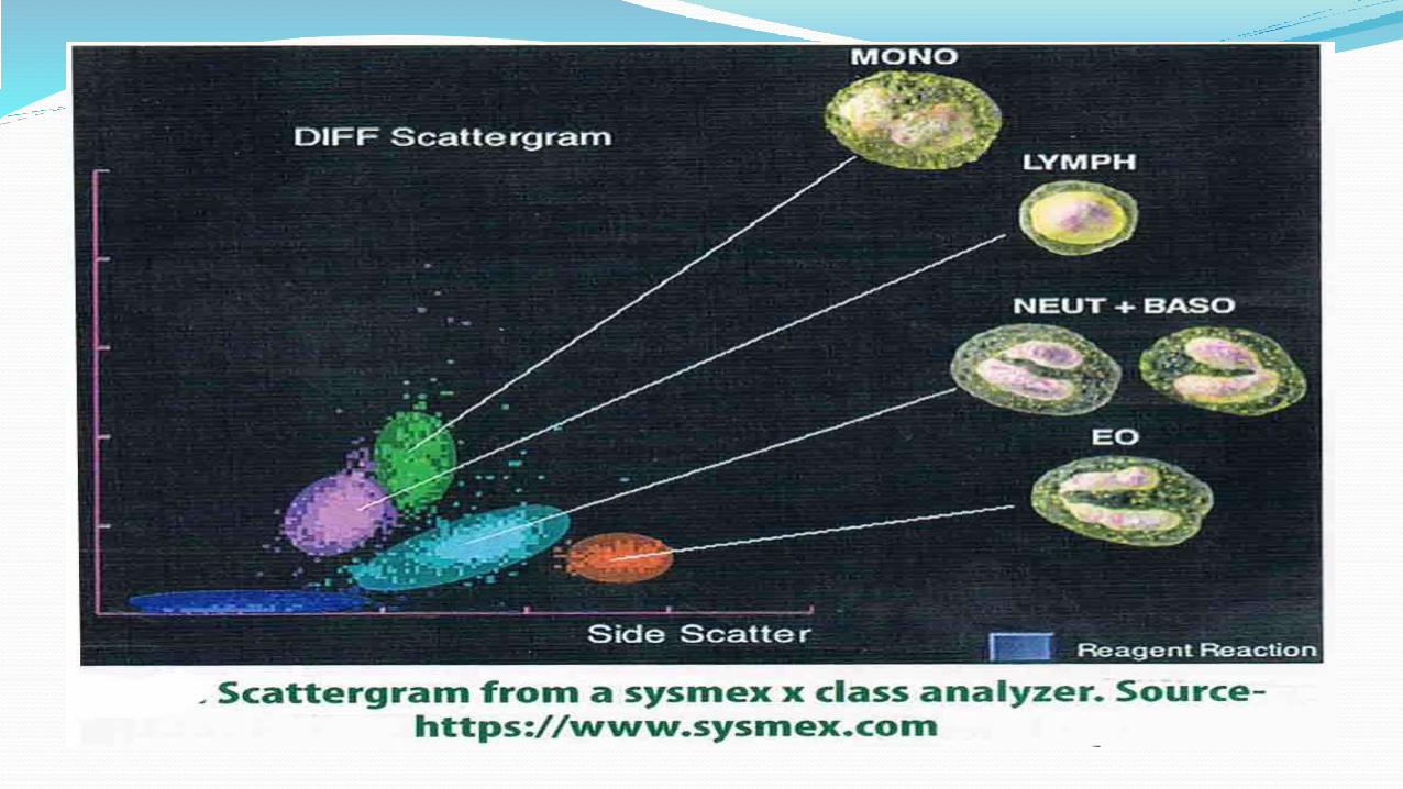
Popular technologies for enumeration and differentiation of WBCs include the Volume Conductivity and Scatter (VCS) used by the Beckman Coulter instruments (from Beckman Coulter Inc, USA) and the Fluorescence Flowcytometry (FFC) principle used by the Sysmex analyzers (from Sysmex Corporation, Japan). The VCS principle differentiates cells based on their volume measured by impedance, conductivity of radio frequencies which give information about the internal structure of cells and scatter of light which tells us about surface structure and granularity. In the FFC technology, cells are hit by a laser beam which gets scattered various directions. The forward scattered light is a measure of the cell volume while the side scattered light is a measure of cell granularity. When a fluorescent dye is used, side fluorescence gives information about the nucleic acids in the cells. Thus, monocytes have the highest forward scatter owing to their large size while eosinophils have the highest side scatter owing to the presence of the prominent granules, immature granulocytes have high fluorescence. Thus, various scatter plots are generated and the total WBC and the differential counts obtained.
Leucocyte positional parameters - Numerical data coordinates generated by VCS technology are available as research parameters on LH Beckman Coulter analyzers. Volume, conductivity and light scatter for each WBC type are characteristic. Any deviation from these as given by the mean value and standard deviation of each of these parameters reflect changes in size and complexity for a particular cell type, which in turn may be reflective of specific diseases. These positional parameters have been used to differentiate between chronic lymphoproliferative disorders (CLPD) like chronic lymphocytic leukemia (CLL) vis a vis reactive lymphocytosis as in former, the cells are uniformly small. Changes in the parameters of neutrophils have been found to indicate sepsis and bacterial infections. Changes involving lymphocytes and monocytes have also been used to flag for presence of malaria. As of now, these are still research parameters and yet to be incorporated into routine clinical practice.
The CBC will indicate if there is leucocytosis or leucopenia. The type of cell involved suggests the possible differential diagnoses. For example, increase neutrophils could suggest a bacterial infection, and inflammatory state steroids use, etc. An extremely high count with left shift suggests a chronic myeloid leukaemia (CML), or a leukamoid reaction. A bimodal peak of myelocytes and neutrophils along with presence of basophils point towards CML and the molecular test for the diagnostic fusion gene, BCR : ABL1 should be asked for.Similarly, finding sustained absolute lymphocytosis without any chronic infection would warrant a hematological work up for a CLPD like CLL. Lymphoma spillover into blood may also cause a lymphocytosis. Peripheral smear may reveal characteristic morphology but immunophenotyping using flowcytometry is the main stay of diagnosis.
Eosinophilia is most often secondary to allergic conditions, parasitic infestations, drugs, vasculitis. If no such cause is found after reasonable investigations and if eosinophilia is sustained, a primary eosinophilia (clonal or hyper eosinophilic syndrome) needs to be considered. Molecular studies for F1P1L1-PDGFR mutation are important to determine response to a tyrosine kinase inhibitor like Imatinib.
Monocytosis may be seen in some chronic infections or during recovery of counts following chemotherapy or drugs. However, sustained absolute monocytosis of at least 1000/ul in absence of chronic infections warrant investigating for a hematological disorder like chronic myelomonocyticleukemia in an adult and juvenile myelomonocytic leukaemia in children. In the presence of leucopenia, it is prudent to look at the absolute cell counts to see if any particular cell line in predominantly suppressed. Neutropenia is most commonly encountered due to drugs and severe infections which would be apparent from the patient's clinical condition. When neutropenia occurs along with anaemia and thrombocytompenia, a serious bone marrow disorder like a plastic anaemia should be thought of. Inherited conditions of neutropenia are rare but known (e.g. Kostman syndrome) Acquired lymphopenia may be seen following use of monoclonal anti CD20 antibodies like Rituximab. Absolute moncytopenia is a feature of Hairy cell leukaemia while eosinopenia is a feature of sepsis.
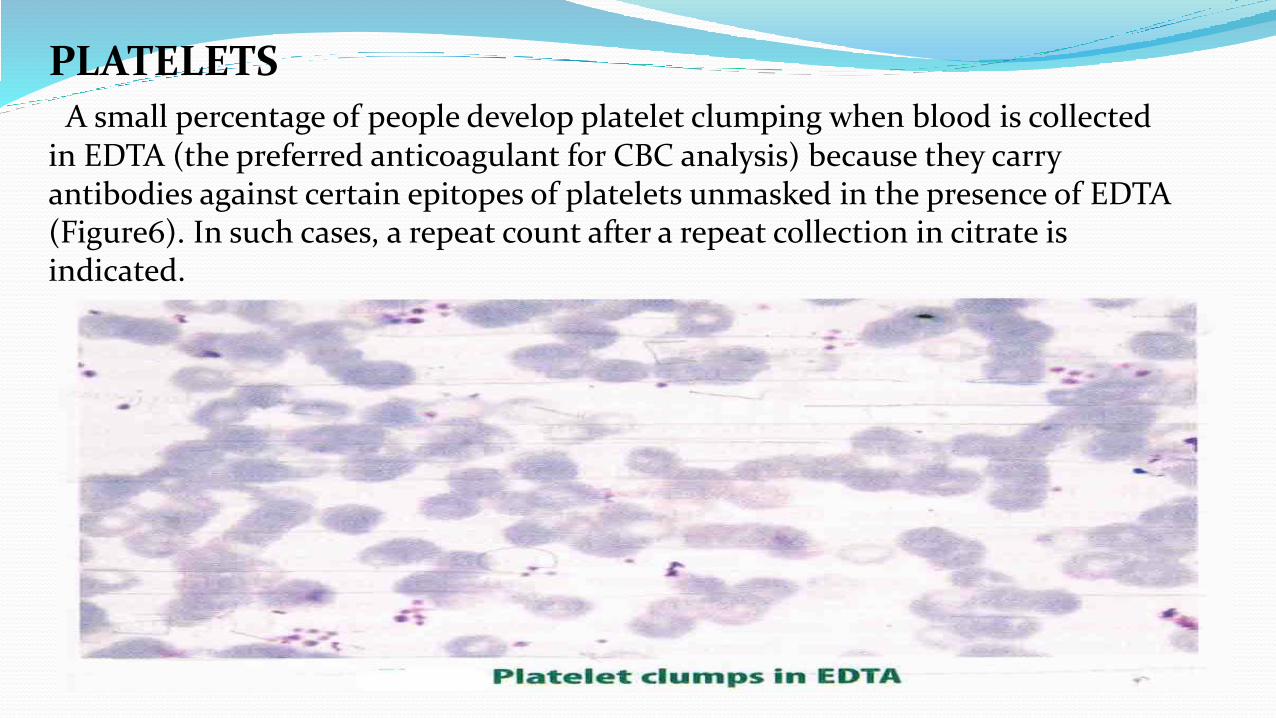
PLATELETSA small percentage of people develop platelet clumping when blood is collected
in EDTA (the preferred anticoagulant for CBC analysis) because they carry antibodies against certain epitopes of platelets unmasked in the presence of EDTA (Figure6). In such cases, a repeat count after a repeat collection in citrate is indicated.
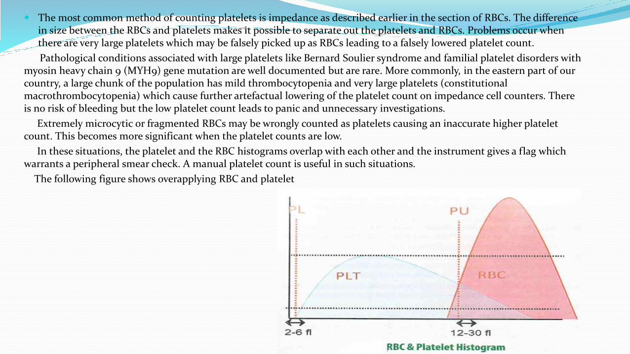
The most common method of counting platelets is impedance as described earlier in the section of RBCs. The difference in size between the RBCs and platelets makes it possible to separate out the platelets and RBCs. Problems occur when there are very large platelets which may be falsely picked up as RBCs leading to a falsely lowered platelet count.
Pathological conditions associated with large platelets like Bernard Soulier syndrome and familial platelet disorders with myosin heavy chain 9 (MYH9) gene mutation are well documented but are rare. More commonly, in the eastern part of our country, a large chunk of the population has mild thrombocytopenia and very large platelets (constitutional macrothrombocytopenia) which cause further artefactual lowering of the platelet count on impedance cell counters. There is no risk of bleeding but the low platelet count leads to panic and unnecessary investigations.
Extremely microcytic or fragmented RBCs may be wrongly counted as platelets causing an inaccurate higher platelet count. This becomes more significant when the platelet counts are low.
In these situations, the platelet and the RBC histograms overlap with each other and the instrument gives a flag which warrants a peripheral smear check. A manual platelet count is useful in such situations.
The following figure shows overapplying RBC and platelet
As manual platelet counts are difficult and time consuming, there have been attempts to innovate technology to overcome the problems with impedance counts like optical counts based on light scatter, use of monoclonal antiplatelet antibodies (Abbott), fluorescent platelet counts (platelet-F, Sysmex) etc.
Platelet - F involves use of a fluorescent dye which is highly specific for platelet membrane and counts obtained by this method have been found to be very accurate.
Besides the counts, platelet indices like the MPV and PDW have also been useful reflecting the average platelet size and platelet anisocytosis respectively.
A PS confirmation of thrombocytopenia is a must to rule out EDTA induced agglutination, presence of large platelets, fibrin strands etc.
Infections and sepsis along with drugs are the commonest causes of thrombocytopenia encountered in a hospital setup wherein the clinical history is indicative of the cause. Thrombocytopenia associated with abnormalities in other cell lines may indicate a bone marrow disorder like an acute leukaemia. Isolated thrombocytopenia in the absence of any other cytopenia (except perhaps for an IDA secondary to bleeding) may be due to an immune thrombocytopenia (ITP) which may be primary or secondary to an associated immunological disorder. ITP is a diagnosis of exclusion. It is crucial to rule out a thrombotic thrombocytopenia purpura (TTP) in the appropriate clinical setting by looking for fragmented cells (schistocytes) in the PS and analyzing the serum LDH as TTP has a 90% mortality if plasmapheresis is not started early. Another life-threatening situation is Heparin induced thrombocytopenia which needs to be considered in patients exposed to unfractionated heparin.
Thrombocytosis (>450,000/ul) is seen in inflammatory states, infections and IDA. However, persistent thrombocytosis in the absence of these conditions may indicate an underlying chronic myeloproliferative neoplasm like CML, essential thrombocytosis, idiopathic myelofibrosis, etc. In such situations, bone marrow trephine biopsy and molecular tests for BCR; ABL1, JAK2 mutation, CALR mutations are warranted.
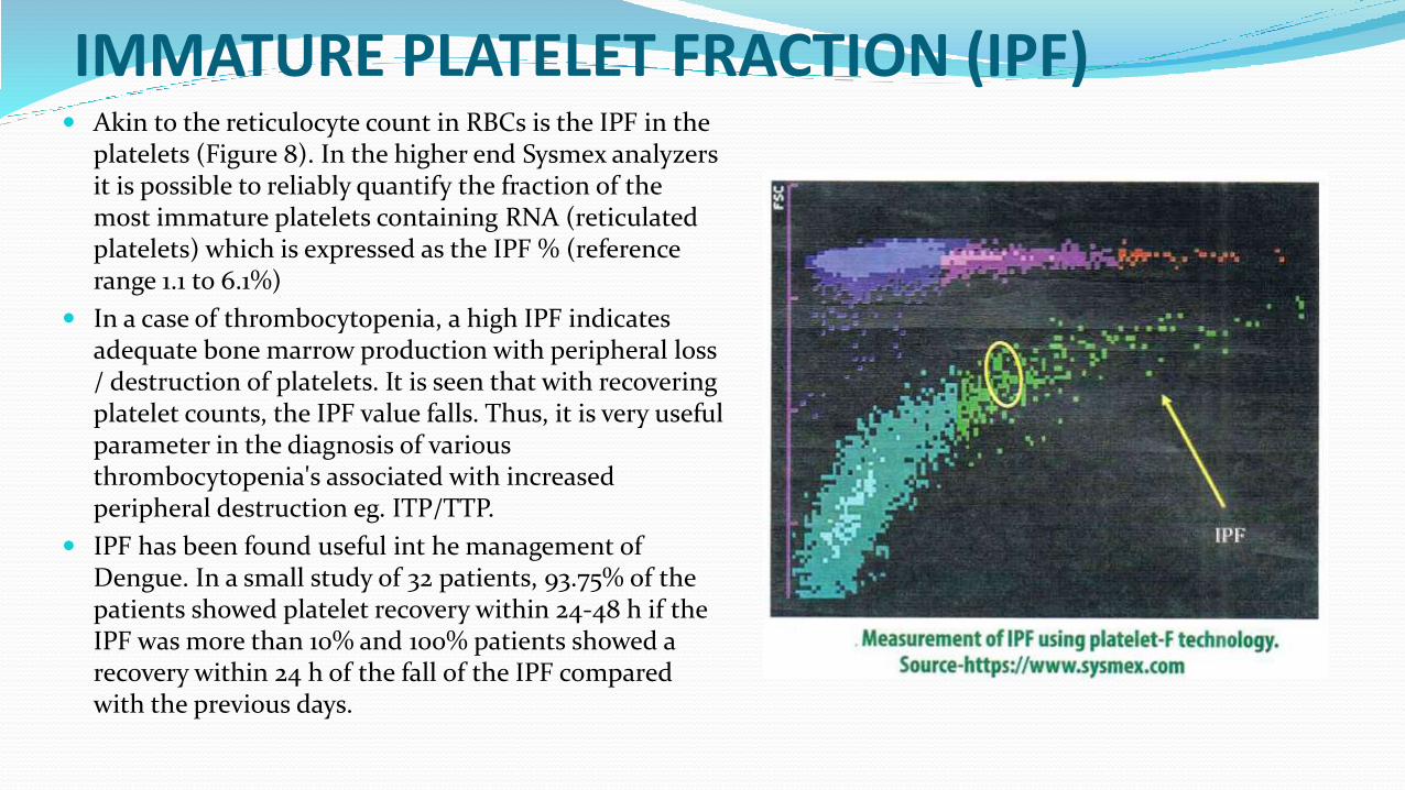
IMMATURE PLATELET FRACTION (IPF) Akin to the reticulocyte count in RBCs is the IPF in the
platelets (Figure 8). In the higher end Sysmex analyzers it is possible to reliably quantify the fraction of the most immature platelets containing RNA (reticulated platelets) which is expressed as the IPF % (reference range 1.1 to 6.1%)
In a case of thrombocytopenia, a high IPF indicates adequate bone marrow production with peripheral loss / destruction of platelets. It is seen that with recovering platelet counts, the IPF value falls. Thus, it is very useful parameter in the diagnosis of various thrombocytopenia's associated with increased peripheral destruction eg. ITP/TTP.
IPF has been found useful int he management of Dengue. In a small study of 32 patients, 93.75% of the patients showed platelet recovery within 24-48 h if the IPF was more than 10% and 100% patients showed a recovery within 24 h of the fall of the IPF compared with the previous days.
SUMMARY The CBS is often the starting point in the investigation of a sick patient and important clinical
decisions are made based on the findings. Today, the CBC is obtained from automated analyzers which give fast, accurate and reproducible results. Parameters like the RDW are obtained only from the automated counters. Newer parameters like Ret-He have been incorporated into international guidelines for investigation of FID. A basic understanding of the technologies makes one aware of the pitfalls and possible problems. While interpreting and normal CBC, it is important to take the patient's clinical status into account and to review previous reports if any. For eg. neutrophilia in a hospitalized patient occurring over 2-3 days is likely to be due to an acute stress / infection while neutrophilia sustained over months in the absence of any apparent illness may be due to a myeloproliferative neoplasm like chronic neutrophilic leukaemia. Platelets are the most difficult cells to count due to the inherent problems associated with impedance technology, Methods like platelet-F give highly accurate counts. Newer parameters like IPF have been found to be in management of dengue and are being incorporated into the routine CBC.
Referances Volume 1 progress in medicine 2017 volume 27.2017