Embed Size (px)
Citation preview
7 Sellar and Juxtasellar Masses on Computed Tomography
CLINICAL IMAGAGINGAN ATLAS OF DIFFERENTIAL DAIGNOSIS
EISENBERG
DR. Muhammad Bin Zulfiqar PGR-FCPS III SIMS/SHL
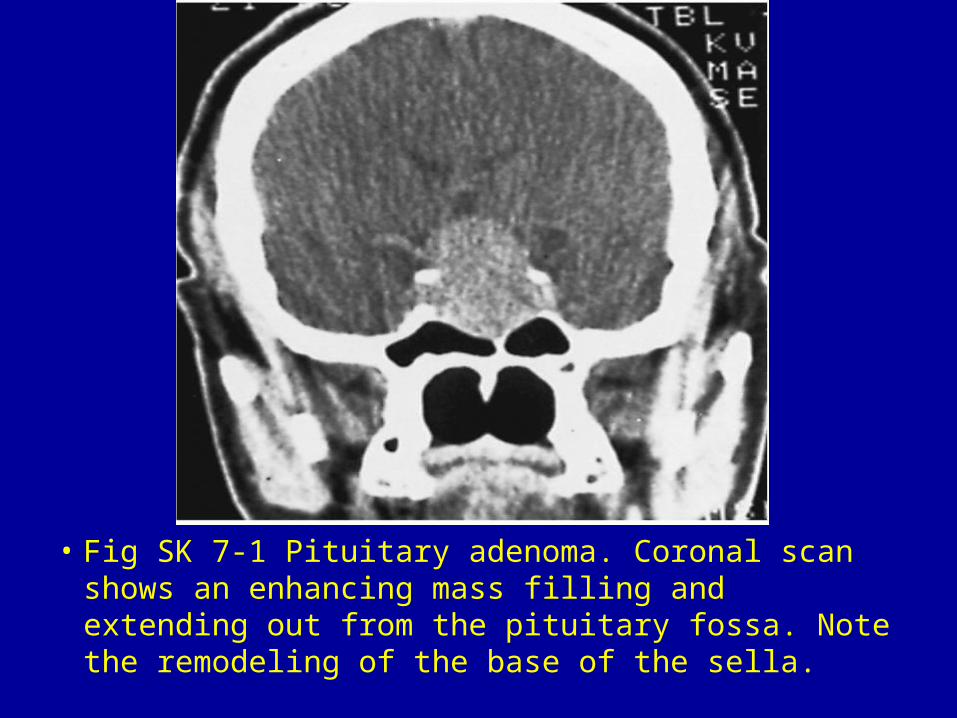
• Fig SK 7-1 Pituitary adenoma. Coronal scan shows an enhancing mass filling and extending out from the pituitary fossa. Note the remodeling of the base of the sella.
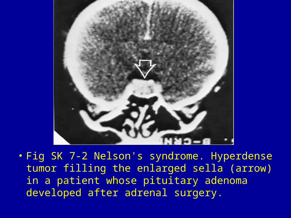
• Fig SK 7-2 Nelson's syndrome. Hyperdense tumor filling the enlarged sella (arrow) in a patient whose pituitary adenoma developed after adrenal surgery.
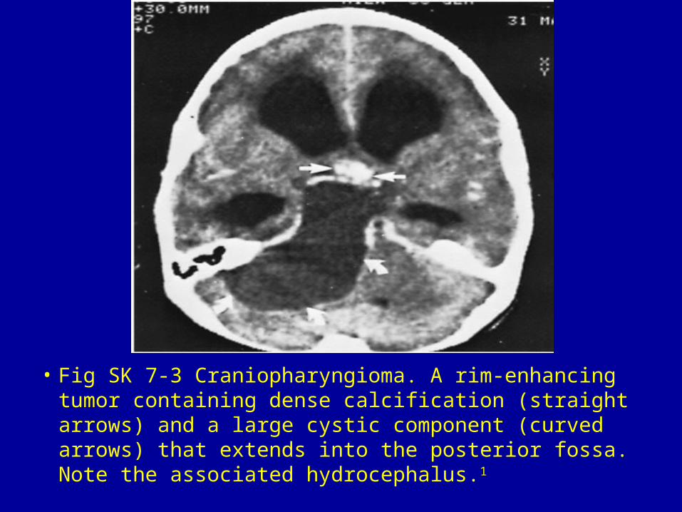
• Fig SK 7-3 Craniopharyngioma. A rim-enhancing tumor containing dense calcification (straight arrows) and a large cystic component (curved arrows) that extends into the posterior fossa. Note the associated hydrocephalus.1
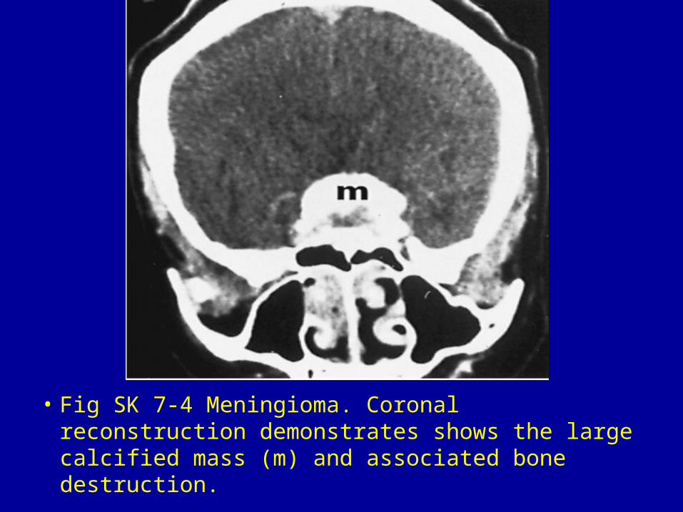
• Fig SK 7-4 Meningioma. Coronal reconstruction demonstrates shows the large calcified mass (m) and associated bone destruction.
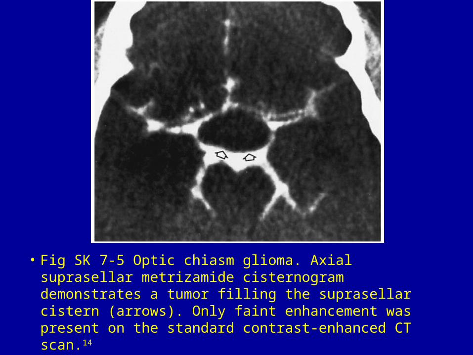
• Fig SK 7-5 Optic chiasm glioma. Axial suprasellar metrizamide cisternogram demonstrates a tumor filling the suprasellar cistern (arrows). Only faint enhancement was present on the standard contrast-enhanced CT scan.14
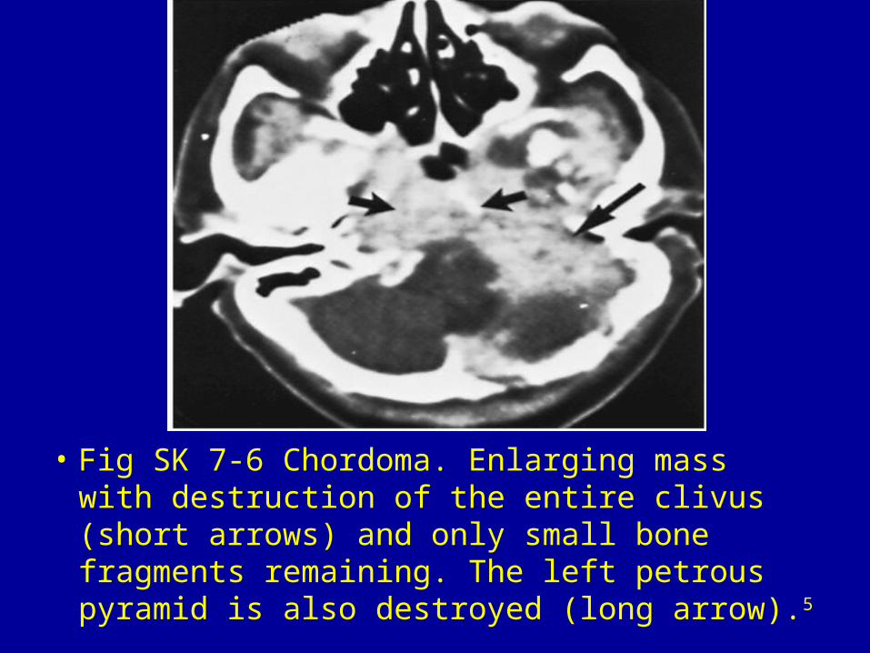
• Fig SK 7-6 Chordoma. Enlarging mass with destruction of the entire clivus (short arrows) and only small bone fragments remaining. The left petrous pyramid is also destroyed (long arrow).5
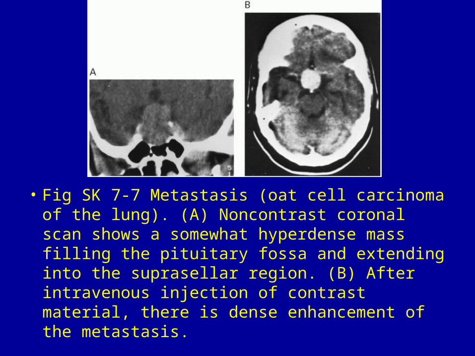
• Fig SK 7-7 Metastasis (oat cell carcinoma of the lung). (A) Noncontrast coronal scan shows a somewhat hyperdense mass filling the pituitary fossa and extending into the suprasellar region. (B) After intravenous injection of contrast material, there is dense enhancement of the metastasis.
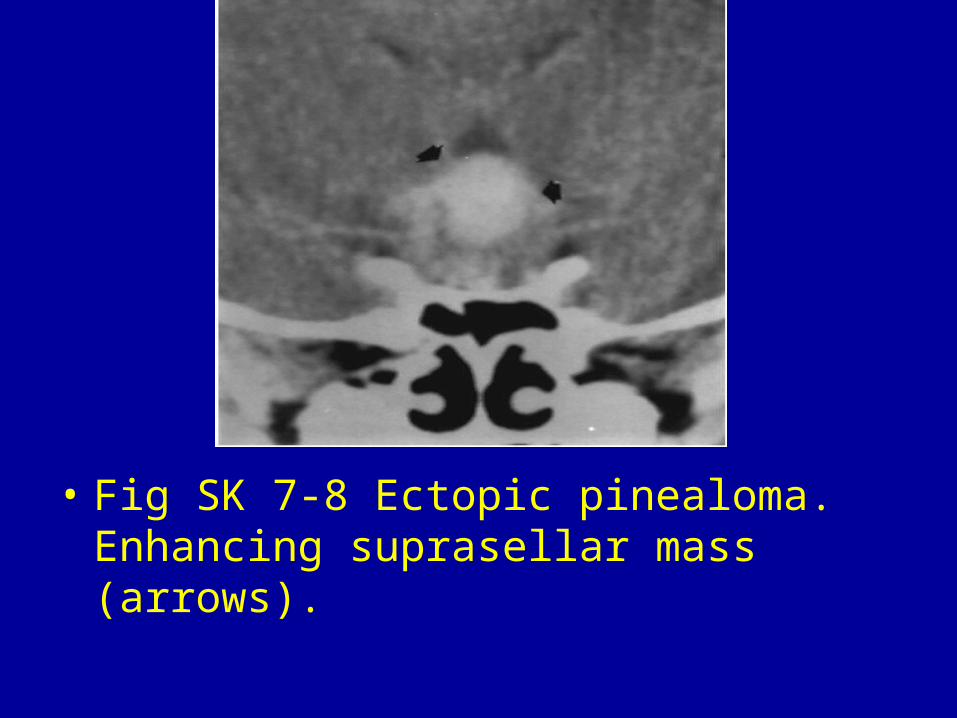
• Fig SK 7-8 Ectopic pinealoma. Enhancing suprasellar mass (arrows).
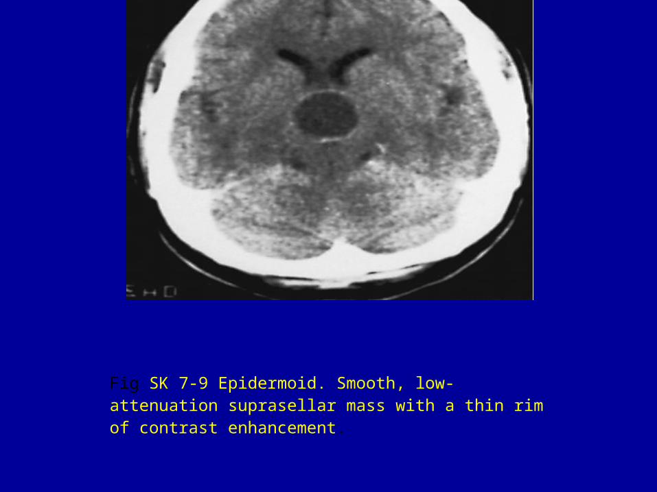
Fig SK 7-9 Epidermoid. Smooth, low-attenuation suprasellar mass with a thin rim of contrast enhancement.

• Fig SK 7-10 Hamartoma of the tuber cinereum. Intrathecally enhanced coronal scan shows a small mass (arrow) that was isodense and nonenhancing on initial CT scans.
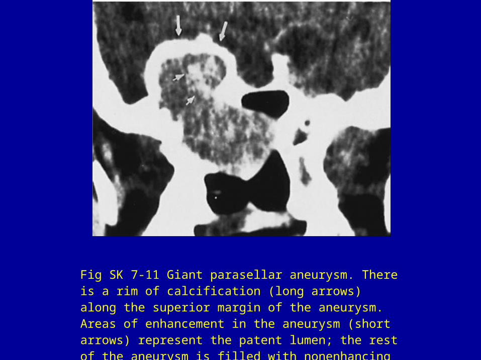
Fig SK 7-11 Giant parasellar aneurysm. There is a rim of calcification (long arrows) along the superior margin of the aneurysm. Areas of enhancement in the aneurysm (short arrows) represent the patent lumen; the rest of the aneurysm is filled with nonenhancing thrombus.
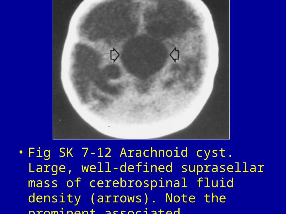
• Fig SK 7-12 Arachnoid cyst. Large, well-defined suprasellar mass of cerebrospinal fluid density (arrows). Note the prominent associated hydrocephalus.