Embed Size (px)
Citation preview

40 Bladder Masses on Computed Tomography and
Magnetic Resonance Imaging
CLINICAL IMAGAGINGAN ATLAS OF DIFFERENTIAL DAIGNOSIS
EISENBERG
DR. Muhammad Bin Zulfiqar PGR-FCPS III SIMS/SHL

• Fig GU 40-1 Transitional cell carcinoma. (A) CT scan shows a large, lobular mass within the bladder. (B) In another patient, the tumor appears as an enhancing area of focal wall thickening (arrow). (C) In a third patient, a large bladder mass is associated with irregular soft-tissue stranding (arrows) from tumor invasion into the perivesical fat.51

• Fig GU 40-2 Transitional cell carcinoma. (A) Coronal T2-weighted MR image shows an intermediate-signal-intensity mass (arrow) within the bright urine-filled bladder. The hyperintense bladder wall is intact. (B) Axial contrast-enhanced T1-weighted image in another patient shows tumor invasion into the perivesical fat (arrows).51

• Fig GU 40-3 Squamous cell carcinoma. CT cystogram shows an irregular mass involving the lateral wall of the bladder. Note the loss of trabecular structure in the bones and fatty infiltration of the muscles in this paraplegic patient.51
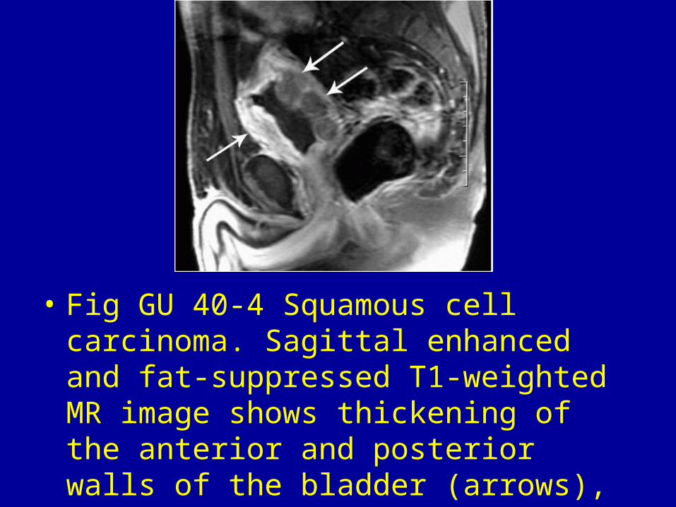
• Fig GU 40-4 Squamous cell carcinoma. Sagittal enhanced and fat-suppressed T1-weighted MR image shows thickening of the anterior and posterior walls of the bladder (arrows), which represented chronic inflammatory changes with diffuse invasive malignancy.51

• Fig GU 40-5 Urachal adenocarcinoma. CT scan shows a large, predominantly solid, midline mass with peripheral calcifications (arrowheads). Within the mass are scattered low-attenuation areas (arrows), which represent mucin.51
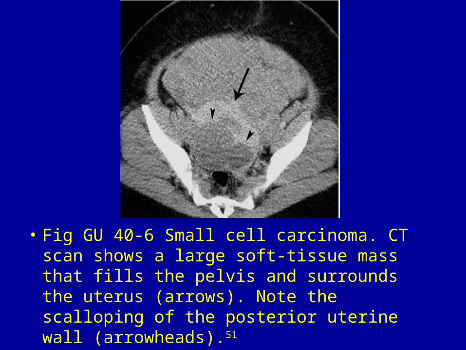
• Fig GU 40-6 Small cell carcinoma. CT scan shows a large soft-tissue mass that fills the pelvis and surrounds the uterus (arrows). Note the scalloping of the posterior uterine wall (arrowheads).51

Fig GU 40-7 Carcinoid. Contrast CT demonstrates a nonspecific solid, polypoid, enhancing mass (arrow) in the bladder.51
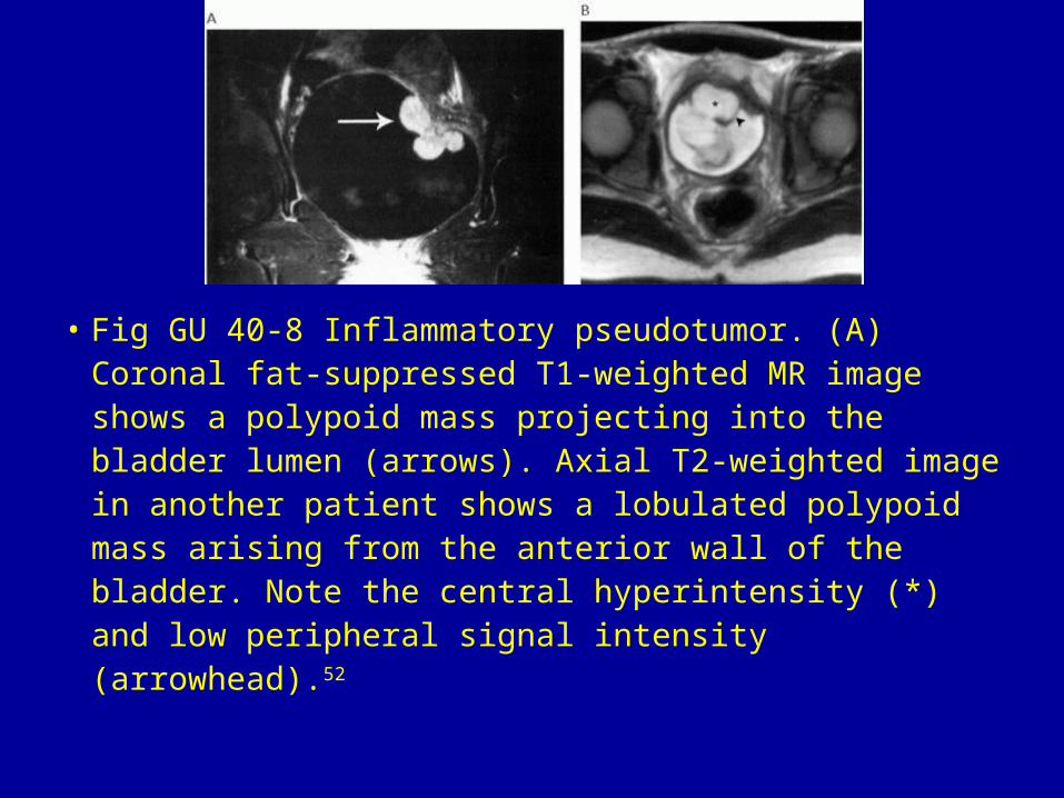
• Fig GU 40-8 Inflammatory pseudotumor. (A) Coronal fat-suppressed T1-weighted MR image shows a polypoid mass projecting into the bladder lumen (arrows). Axial T2-weighted image in another patient shows a lobulated polypoid mass arising from the anterior wall of the bladder. Note the central hyperintensity (*) and low peripheral signal intensity (arrowhead).52

• Fig GU 40-9 Endometriosis. Fat-suppressed T1-weighted MR image shows high-signal-intensity foci (arrow) within a soft-tissue mass in the vesicouterine space, projecting into the bladder lumen posteriorly.52

• Fig GU 40-10 Malacoplakia. Contrast CT scan through the upper pelvis shows a large, irregularly enhancing mass (arrows) that is contiguous with the bladder.52

• Fig GU 40-11 Schistosomiasis. CT scan demonstrates curvilinear calcification in the bladder wall (arrowheads), which also extends to the distal left ureter (arrow).52

• Fig GU 40-12 Crohn's disease. Contrast coronal CT reformation shows thickening of the wall of the distal small bowel (straight arrow) and the adjacent bladder (curved arrow). A small bowel series (not shown) showed a fistula connecting the abnormal segment of ileum to the bladder.52

• Fig GU 40-13 Diverticulitis. Coronal CT reformation shows a thick-walled, gas-filled abscess (arrowhead) immediately adjacent to the bladder. There is diffuse thickening of the bladder wall (arrow).52

• Fig GU 40-14 Chemotherapy cystitis. CT scan shows marked thickening and increased mucosal enhancement of the bladder after cyclophosphamide therapy following bone marrow transplantation in a child with acute lymphoblastic leukemia.53

• Fig GU 40-15 Radiation cystitis (chronic). CT scan shows focal thickening and calcification of the right wall of the bladder (straight arrow). There is subtle widening of the presacral space (curved arrow) and fatty infiltration of the pelvic musculature (arrowheads).52

• Fig GU 40-16 Benign prostatic hypertrophy. CT scan shows a large, smooth, lobular mass bulging into the base of the bladder (arrow). Despite its large size, this “mass” proved to be benign hypertrophy of the prostate.52

• Fig GU 40-17 Urahcal adenocarcinoma. CY shows a cystic mass (arrows) at the anterior dome of the bladder.54