Embed Size (px)
Citation preview
05/03/2305/03/23 Ossama El-ShallOssama El-Shall 11
Dental RadiographicDental Radiographic anatomy & Interpretation
Part II Dr. Ossama EL-ShallChairman of Oral Medicine, Chairman of Oral Medicine,
Periodontology, Diagnosis & Periodontology, Diagnosis & Radiology Department, Faculty of Radiology Department, Faculty of Dental Medicine for girls, Al-Azhar Dental Medicine for girls, Al-Azhar
University, Cairo Egypt.University, Cairo Egypt.E.mail address: [email protected] address: [email protected]

05/03/2305/03/23 Ossama El-ShallOssama El-Shall 22
Radiographically, lesions may Radiographically, lesions may classified into:classified into:
I-I- Radiolucent lesions Radiolucent lesions
II-II- Radioopaque lesions Radioopaque lesions
III-III- Combination of RL + RO. Combination of RL + RO. lesions.lesions.

05/03/2305/03/23 Ossama El-ShallOssama El-Shall 33
Radiolucent lesionsRadiolucent lesions

05/03/2305/03/23 Ossama El-ShallOssama El-Shall 44
Radiolucent lesionsRadiolucent lesions1-1-Lesions related to tooth apexLesions related to tooth apex2-2-Lesions related to side of rootsLesions related to side of roots3-3-Lesions related to crown of unerupted or Lesions related to crown of unerupted or
impacted toothimpacted tooth4-4-Unilocular lesions in midline of maxilla.Unilocular lesions in midline of maxilla.5-5-Unilocular lesions lateral to midline of Unilocular lesions lateral to midline of
maxilla.maxilla.6-6-Solitary RL lesion with either well or ill- Solitary RL lesion with either well or ill-
defined margins.defined margins.7-7-Multilocular RL lesion with either well or ill-Multilocular RL lesion with either well or ill-
defined margins.defined margins.8-8-Multiple but separate RL with well-defined Multiple but separate RL with well-defined
or punched out marginsor punched out margins

05/03/2305/03/23 Ossama El-ShallOssama El-Shall 55
1-1- Radiolucent lesions related Radiolucent lesions related to tooth apex:to tooth apex:
A- Periapical cystA- Periapical cystB- Periapical granulomaB- Periapical granulomaC- Periapical abscessC- Periapical abscessD- Periapical periodontitisD- Periapical periodontitisE- Periapical cementoma. (early E- Periapical cementoma. (early
stage)stage)F- Periapical scarF- Periapical scar

05/03/2305/03/23 Ossama El-ShallOssama El-Shall 66
2-2-Radiolucent lesions related to Radiolucent lesions related to sides of roots:sides of roots:
1- Lateral periodontal cyst1- Lateral periodontal cyst
2- Periodontal abscess.2- Periodontal abscess.

05/03/2305/03/23 Ossama El-ShallOssama El-Shall 77
3-3-Lesions related to crown of Lesions related to crown of unerupted or impacted toothunerupted or impacted tooth
Pericoronal R.L, uni or multilocularPericoronal R.L, uni or multilocular
A-Pericoronal or follicular space.A-Pericoronal or follicular space.B-Dentigerous cyst. B-Dentigerous cyst. C-AmeloblastomaC-AmeloblastomaD-Odontogenic keratocyst.D-Odontogenic keratocyst.E-Odontogenic fibroma. E-Odontogenic fibroma. F-Odontogenic myxoma.F-Odontogenic myxoma.
05/03/2305/03/23 Ossama El-ShallOssama El-Shall 88
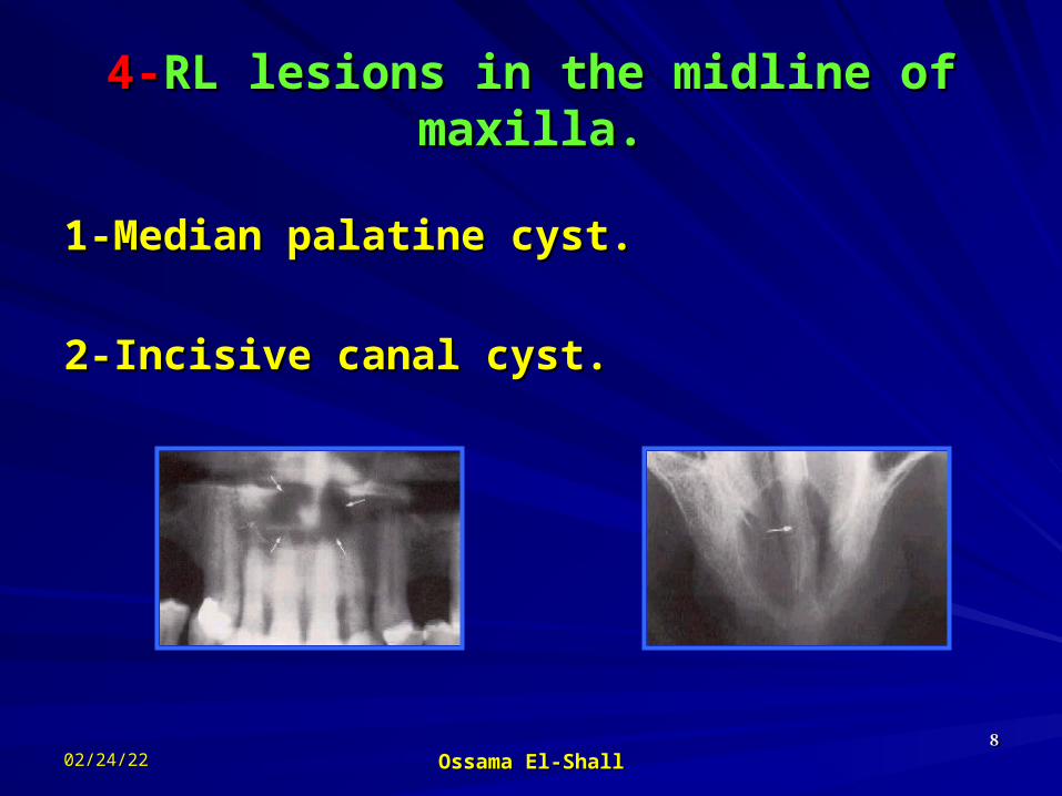
4-4-RL lesions in the midline of RL lesions in the midline of maxilla.maxilla.
1-Median palatine cyst.1-Median palatine cyst.
2-Incisive canal cyst.2-Incisive canal cyst.

05/03/2305/03/23 Ossama El-ShallOssama El-Shall 99
5-5-RL lesions lateral to midline of RL lesions lateral to midline of maxilla.maxilla.
1- Globulomaxillary 1- Globulomaxillary cystcyst
2- Residual cyst2- Residual cyst

05/03/2305/03/23 Ossama El-ShallOssama El-Shall 1010
6-6-Solitary R.L lesions with either Solitary R.L lesions with either well or ill-defined margins and not well or ill-defined margins and not
necessarily containing teeth.necessarily containing teeth.Well-defined marginWell-defined margin
1-Residual cyst 1-Residual cyst 2-Traumatic bone cyst2-Traumatic bone cyst3-Primordial cyst3-Primordial cyst4-Odontogenic 4-Odontogenic
keratocystkeratocyst5- Ameloblastoma5- Ameloblastoma6-Central giant cell 6-Central giant cell
granulomagranuloma7-Central odontogenic 7-Central odontogenic
fibromafibroma
Ill-defined marginIll-defined margin1-Residual infection1-Residual infection2-bone loss due to PD2-bone loss due to PD3-Myloma3-Myloma4-carcinoma4-carcinoma5-Ameloplastoma5-Ameloplastoma6-metastasis6-metastasis7-osteomylitis7-osteomylitis8-odontoenic fibroma8-odontoenic fibroma

05/03/2305/03/23 Ossama El-ShallOssama El-Shall 1111
77-Multilocular RL lesion with -Multilocular RL lesion with either well or ill-defined margins.either well or ill-defined margins.
Well-definedWell-defined1-Ameloblastoma 1-Ameloblastoma 2-Odontogenic keratocyst2-Odontogenic keratocyst3-Central g. cell granuloma3-Central g. cell granuloma4-Odontogenic myxoma4-Odontogenic myxoma5-Central hemangioma5-Central hemangioma6-Fibrous dysplasia6-Fibrous dysplasia7-Cherubism7-Cherubism8-Anneyrsmal bone cyst8-Anneyrsmal bone cyst9-Central fibroma9-Central fibroma10-Traumatic bone cyst10-Traumatic bone cyst
Ill-definedIll-defined1-Ameloblastoma (late stage)1-Ameloblastoma (late stage)
2-Central myxoma (late 2-Central myxoma (late stage)stage)
3-Fibrous dysplasia3-Fibrous dysplasia4-Cherubism4-Cherubism
05/03/2305/03/23 Ossama El-ShallOssama El-Shall 1212
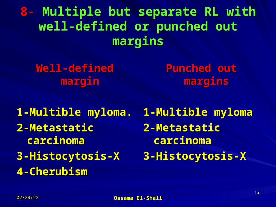
8- 8- Multiple but separate RL with Multiple but separate RL with well-defined or punched out well-defined or punched out
marginsmargins
Well-defined marginWell-defined margin
1-Multible myloma.1-Multible myloma.2-Metastatic carcinoma2-Metastatic carcinoma3-Histocytosis-X3-Histocytosis-X4-Cherubism4-Cherubism
Punched out marginsPunched out margins
1-Multible myloma1-Multible myloma2-Metastatic carcinoma2-Metastatic carcinoma3-Histocytosis-X3-Histocytosis-X

05/03/2305/03/23 Ossama El-ShallOssama El-Shall 1313
Radio-opaque lesionsRadio-opaque lesions

05/03/2305/03/23 Ossama El-ShallOssama El-Shall 1414
Radio-opaque lesionsRadio-opaque lesions1-1-Periapical solitary radio-opaque lesionsPeriapical solitary radio-opaque lesions
2-2-Solitary R.O lesions not contacting Solitary R.O lesions not contacting teethteeth
3-3-Multiple separate radio-opacities.Multiple separate radio-opacities.
4-4-Generalized radio-opacities.Generalized radio-opacities.

05/03/2305/03/23 Ossama El-ShallOssama El-Shall 1515
1-1-Periapical solitary radio-Periapical solitary radio-opaque lesionsopaque lesions
1-Condesing osteitis1-Condesing osteitis2-Sclerosing bone2-Sclerosing bone3-Periapical cementoma (late stage)3-Periapical cementoma (late stage)4-Odontoma4-Odontoma5-supernumerary unerupted tooth5-supernumerary unerupted tooth6-Hypercementosis6-Hypercementosis7-forign body.7-forign body.

05/03/2305/03/23 Ossama El-ShallOssama El-Shall 1616
2-2-SolitarySolitary R.O lesions not R.O lesions not contacting teethcontacting teeth
1- All the above item (Periapical R.O)1- All the above item (Periapical R.O)8-Osteoma8-Osteoma9-Salivary gland stone9-Salivary gland stone10-Osteomlitis10-Osteomlitis11-Remeaning root11-Remeaning root12-Unerupted tooth12-Unerupted tooth

05/03/2305/03/23 Ossama El-ShallOssama El-Shall 1717
3-3-MultipleMultiple separate radio-separate radio-opacities.opacities.
1-All the first item1-All the first item8-Paget’s disease8-Paget’s disease9-osteogenic sarcoma9-osteogenic sarcoma

05/03/2305/03/23 Ossama El-ShallOssama El-Shall 1818
4-4-Generalized radio-opacitiesGeneralized radio-opacities
1-Paget’s disease1-Paget’s disease2-osteopetrosis2-osteopetrosis

05/03/2305/03/23 Ossama El-ShallOssama El-Shall 1919
Combined RL & RO Combined RL & RO lesionslesions

05/03/2305/03/23 Ossama El-ShallOssama El-Shall 2020
Combined RL & RO Combined RL & RO lesionslesions
1-1-Mixed Periapical lesionsMixed Periapical lesions
2-2-Mixed lesions not necessarily Mixed lesions not necessarily contacting a tooth.contacting a tooth.
3-3-Pericoronal mixed lesions.Pericoronal mixed lesions.

05/03/2305/03/23 Ossama El-ShallOssama El-Shall 2121
1-1-Mixed Periapical lesionsMixed Periapical lesions
1-Cementoma1-Cementoma2-Sclerosing osteitis2-Sclerosing osteitis3-Odontoma3-Odontoma4-Ossifying or cementifying fibroma4-Ossifying or cementifying fibroma
05/03/2305/03/23 Ossama El-ShallOssama El-Shall 2222
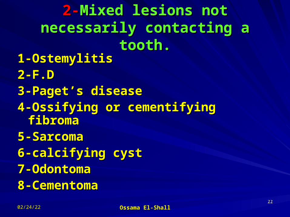
2-2-Mixed lesions not Mixed lesions not necessarily contacting a tooth.necessarily contacting a tooth.1-Ostemylitis1-Ostemylitis2-F.D2-F.D3-Paget’s disease3-Paget’s disease4-Ossifying or cementifying fibroma4-Ossifying or cementifying fibroma5-Sarcoma5-Sarcoma6-calcifying cyst6-calcifying cyst7-Odontoma7-Odontoma8-Cementoma8-Cementoma

05/03/2305/03/23 Ossama El-ShallOssama El-Shall 2323
3-3-Pericoronal mixed lesions.Pericoronal mixed lesions.
1-Odontoma1-Odontoma2-Calcifying Odontogenic tumor2-Calcifying Odontogenic tumor3-calcifying cyst3-calcifying cyst4-Odontogenic fibroma4-Odontogenic fibroma

05/03/2305/03/23 Ossama El-ShallOssama El-Shall 2424
Most common lesions as seen in Most common lesions as seen in dental radiographs dental radiographs
1-1- Inflammatory lesions. Inflammatory lesions. 2-2-Cysts and pseudocystsCysts and pseudocysts3-3- Odontogenic tumors. Odontogenic tumors.4-4- Non Odontogenic tumors. Non Odontogenic tumors.5-5- Developmental anomalies of teeth. Developmental anomalies of teeth.6-6- Foreign bodies. Foreign bodies.

05/03/2305/03/23 Ossama El-ShallOssama El-Shall 2525
Periapical inflammatory Periapical inflammatory lesions.lesions.
Ill defined RL area, widening of PM space, loss of LD
05/03/2305/03/23 Ossama El-ShallOssama El-Shall 2626
Periapical Inflamatory Periapical Inflamatory LesionsLesions
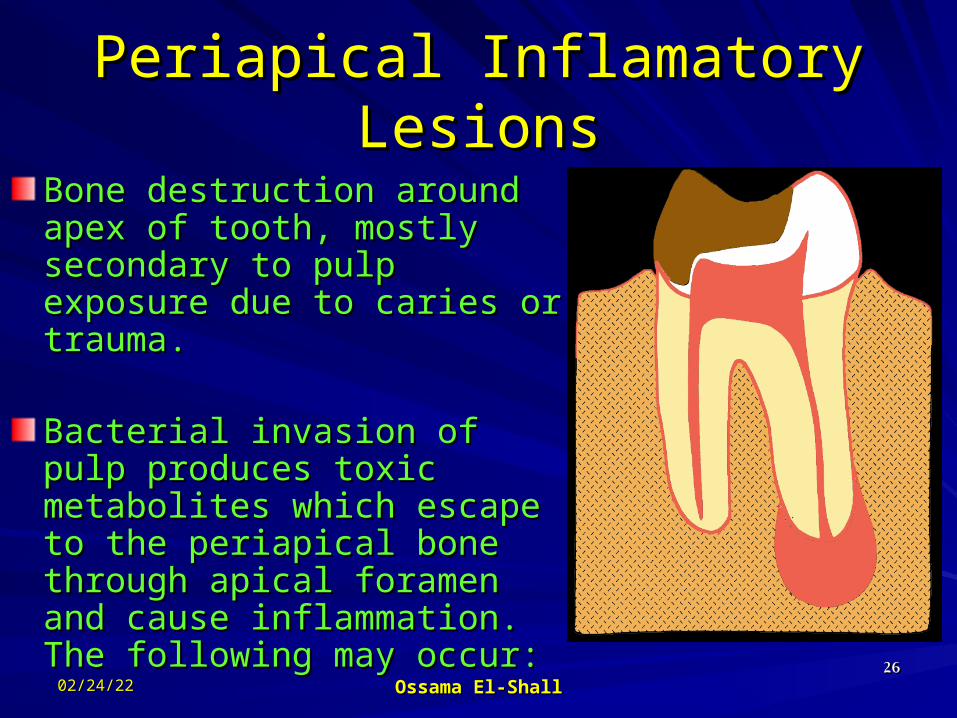
Bone destruction around Bone destruction around apex of tooth, mostly apex of tooth, mostly secondary to pulp exposure secondary to pulp exposure due to caries or trauma.due to caries or trauma.
Bacterial invasion of pulp Bacterial invasion of pulp produces toxic metabolites produces toxic metabolites which escape to the which escape to the periapical bone through periapical bone through apical foramen and cause apical foramen and cause inflammation. The following inflammation. The following may occur: may occur:

05/03/2305/03/23 Ossama El-ShallOssama El-Shall 2727
Periapical Inflamatory LesionsPeriapical Inflamatory Lesions
Periapical Periapical granuloma:granuloma: Localized Localized mass of chronic mass of chronic granulation tissue granulation tissue containing PMN’s, containing PMN’s, lymphocytes, plasma lymphocytes, plasma cells. cells.

05/03/2305/03/23 Ossama El-ShallOssama El-Shall 2828
Periapical GranulomaPeriapical Granuloma
Radiographically, Radiographically, widening of PDL widening of PDL or variable size of or variable size of periapical periapical radiolucency may radiolucency may be presentbe present

05/03/2305/03/23 Ossama El-ShallOssama El-Shall 2929
Periapical GranulomaPeriapical Granuloma

05/03/2305/03/23 Ossama El-ShallOssama El-Shall 3030
Periapical Granuloma??Periapical Granuloma??

05/03/2305/03/23 Ossama El-ShallOssama El-Shall 3131
Apical periodontitis.Apical periodontitis.

05/03/2305/03/23 Ossama El-ShallOssama El-Shall

05/03/2305/03/23 Ossama El-ShallOssama El-Shall 3333

05/03/2305/03/23 Ossama El-ShallOssama El-Shall 3434

05/03/2305/03/23 Ossama El-ShallOssama El-Shall 3535
Cysts affecting oral cavityCysts affecting oral cavity
Cyst;Cyst; is a pathological cavity contains fluid or is a pathological cavity contains fluid or semi-solid materials semi-solid materials
Cysts can be true or pseudo according its lining Cysts can be true or pseudo according its lining tissues:tissues:
True cysts:True cysts: cysts which lined with epithelium cysts which lined with epitheliumPseudo-cysts:Pseudo-cysts: cysts which cysts which notnot lined with lined with
epithelium but lined with connective tissue epithelium but lined with connective tissue membrane membrane

05/03/2305/03/23 Ossama El-ShallOssama El-Shall 3636
Classification of true Cysts of interest to Classification of true Cysts of interest to the dentist the dentist
I- Odontogenic cystsI- Odontogenic cysts 1-1-Radicular cysts….Radicular cysts….a-a- Apical….. Apical…..b-b- Lateral Lateral 2-2-Periodontal cysts Periodontal cysts 3-3-Primordial cysts (Before formation of hard tooth Primordial cysts (Before formation of hard tooth
structures)structures) 4-4-Keratocysts.Keratocysts. 5-5-DentigerousDentigerous cystcyst -Follicular cyst -Follicular cyst - Eruption cysts.- Eruption cysts. - Coronal cysts.- Coronal cysts. -Lateral cysts.-Lateral cysts. 6-6-Residual cysts of all typesResidual cysts of all types..

05/03/2305/03/23 Ossama El-ShallOssama El-Shall 3737
II- Non-odontogenic cysts.II- Non-odontogenic cysts.
Fissural cysts Fissural cysts
1- Median palatine cyst.1- Median palatine cyst.2- Nasoalveolar cyst2- Nasoalveolar cyst3- Globulomaxillary cyst3- Globulomaxillary cyst4- Median mandibular 4- Median mandibular
cyst.cyst.
Non-fissural cystsNon-fissural cysts
1- Nasopalatine cysts1- Nasopalatine cysts2- Median alveolar cyst2- Median alveolar cyst

05/03/2305/03/23 Ossama El-ShallOssama El-Shall 3838
Pseudocysts Pseudocysts (not lining with epithelial)(not lining with epithelial)
Solitary bone cyst.Solitary bone cyst. Aneurysmal bone cyst.Aneurysmal bone cyst. Latent bone cyst.Latent bone cyst.

05/03/2305/03/23 Ossama El-ShallOssama El-Shall 3939
Radicular cystsRadicular cystsIt developed around apex It developed around apex
of a diseases tooth or of a diseases tooth or around an accessory around an accessory canal from the pulp canal from the pulp (lateral radicular cyst)(lateral radicular cyst)
The radiographic The radiographic appearance of a appearance of a clinically symptom-free clinically symptom-free cyst reveals a clear, RO cyst reveals a clear, RO borders that surround borders that surround the radiolucency. the radiolucency.

05/03/2305/03/23 Ossama El-ShallOssama El-Shall 4040
Radicular cystsRadicular cysts
Rounded RL with RO Rounded RL with RO margin at periapical margin at periapical region.region.Apex of the tooth is Apex of the tooth is within the cystic cavity.within the cystic cavity.Adjacent teeth and Adjacent teeth and structures are displaced.structures are displaced.Infected cysts exhibits Infected cysts exhibits poorly demarcated poorly demarcated bordersborders
Small, clinically symptom-Small, clinically symptom-free radicular cyst that is free radicular cyst that is expanding towards the floor expanding towards the floor of maxillary sinusof maxillary sinus

05/03/2305/03/23 Ossama El-ShallOssama El-Shall 4141
Small, symptom Small, symptom free radicular cyst free radicular cyst
with typical RO with typical RO boundariesboundaries
Infected radicular cyst, has Infected radicular cyst, has lost its typical radiographic lost its typical radiographic signs as a result of serous signs as a result of serous
infiltration of the infiltration of the surrounding tissue.surrounding tissue.
05/03/2305/03/23 Ossama El-ShallOssama El-Shall 4242
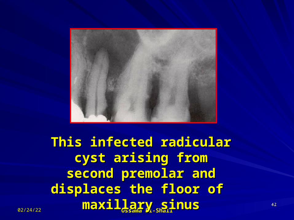
This infected radicular cyst This infected radicular cyst arising from second premolar arising from second premolar
and displaces the floor of and displaces the floor of maxillary sinusmaxillary sinus

05/03/2305/03/23 Ossama El-ShallOssama El-Shall 4343
Typical manifestation Typical manifestation of radiclar cystof radiclar cyst
Infected radiclar cystInfected radiclar cyst

05/03/2305/03/23 Ossama El-ShallOssama El-Shall 4444
Atypical manifestation of a radicular cyst
-This cyst emanates from the remaining root of lower canine
-The radiograph showing a multi-locular pattern
-This picture may misdiagnosed as ameloblastoma or keratocyst

05/03/2305/03/23 Ossama El-ShallOssama El-Shall 4545
Radicular maxillary cyst extending from central and lateral incisors.
The cyst expanded in horizontal plane, which is clear in the occlusal view.
From the panoramic view we can notice its relation to max.sinus.

05/03/2305/03/23 Ossama El-ShallOssama El-Shall 4646
Radicular residual cyst
Notes the relation to max. sinus

05/03/2305/03/23 Ossama El-ShallOssama El-Shall 4747
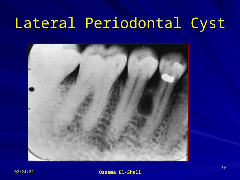
Lateral Periodontal CystLateral Periodontal CystArises directly from Arises directly from epithelial cells in epithelial cells in PDL on lateral PDL on lateral aspect of tooth. aspect of tooth. Origin: cell rests of Origin: cell rests of Mallasez or Mallasez or remnants of dental remnants of dental lamina.lamina.Tooth is VITAL.Tooth is VITAL.

05/03/2305/03/23 Ossama El-ShallOssama El-Shall 4848
Lateral Periodontal CystLateral Periodontal CystHow do you How do you differentiate this cyst differentiate this cyst from radicular cyst from radicular cyst which may develop in which may develop in this location?this location?Seen as a unilocular, Seen as a unilocular, well-defined well-defined radiolucency on radiolucency on lateral aspect of a lateral aspect of a vital tooth.vital tooth.
05/03/2305/03/23 Ossama El-ShallOssama El-Shall 4949
Lateral Periodontal CystLateral Periodontal Cyst

05/03/2305/03/23 Ossama El-ShallOssama El-Shall 5050
Odontogenic KeratocystOdontogenic KeratocystOriginate before tooth development from a remnants Originate before tooth development from a remnants of epithelium has the capacity to produce keratin.of epithelium has the capacity to produce keratin.it appears as it appears as multilocularmultilocular well-defined RL lesion with well-defined RL lesion with an ability for root divergence and cortical expansion. an ability for root divergence and cortical expansion.

05/03/2305/03/23 Ossama El-ShallOssama El-Shall 5151
Odontogenic Keratocyst Odontogenic Keratocyst (OKC)(OKC)
Can cause severe bone destruction.Can cause severe bone destruction.Can displace teeth and cause root resorption.Can displace teeth and cause root resorption.Should be followed for recurrence for 5-10 years.Should be followed for recurrence for 5-10 years.

05/03/2305/03/23 Ossama El-ShallOssama El-Shall 5252
Odontogenic Keratocyst (OKC)Odontogenic Keratocyst (OKC)

05/03/2305/03/23 Ossama El-ShallOssama El-Shall 5353
Dentigerous Cyst (Follicular Dentigerous Cyst (Follicular Cyst)Cyst)
Always associated Always associated with crown of an with crown of an impacted or impacted or unerupted (normal or unerupted (normal or supernumerary) supernumerary) tooth.tooth.Due to accumulation Due to accumulation of fluid between of fluid between layers of reduced layers of reduced enamel epithelium or enamel epithelium or between epithelium between epithelium and crown.and crown.

05/03/2305/03/23 Ossama El-ShallOssama El-Shall 5454
Dentigerous cyst Dentigerous cyst
Most common site, Most common site, around the third molar around the third molar and the midline of the and the midline of the maxilla maxilla Radiographically it Radiographically it appears as well appears as well demarcated unilocular, demarcated unilocular, radiolucent area, radiolucent area, surrounding a crown of surrounding a crown of unerupted tooth. unerupted tooth.

05/03/2305/03/23 Ossama El-ShallOssama El-Shall 5555
Lateral Dentigerous cyst on lower third molar

05/03/2305/03/23 Ossama El-ShallOssama El-Shall 5656
Coronal Dentigerous cyst surrounding lateral Coronal Dentigerous cyst surrounding lateral incisor with displaced of canine and retention of incisor with displaced of canine and retention of deciduous canine.deciduous canine.Tooth 22 appears enlarged and overexposed.Tooth 22 appears enlarged and overexposed.Tooth 23 is displaced in the vistibular direction.Tooth 23 is displaced in the vistibular direction.

05/03/2305/03/23 Ossama El-ShallOssama El-Shall 5757
Enlarged Dentigerous cyst Enlarged Dentigerous cyst

05/03/2305/03/23 Ossama El-ShallOssama El-Shall 5858
Dentigerous Cyst (Follicular Dentigerous Cyst (Follicular Cyst)Cyst)

05/03/2305/03/23 Ossama El-ShallOssama El-Shall 5959
Dentigerous Cyst (Follicular Dentigerous Cyst (Follicular Cyst)Cyst)

05/03/2305/03/23 Ossama El-ShallOssama El-Shall 6060
Eruption cyst on upper 8 as seen in Periapical film
It is a type of Dentigerous cysts developed after the formation of dental hard tissues from the enamel epithelium

05/03/2305/03/23 Ossama El-ShallOssama El-Shall 6161
Nonodontogenic cystsNonodontogenic cystsNasopalatine cyst
Median palatal cystMedian palatal cyst
Globulomaxillary cystGlobulomaxillary cyst

05/03/2305/03/23 Ossama El-ShallOssama El-Shall 6262
Or incisive canal cyst, Or anterior maxillary cystIt forms in incisive canal, causing swelling of incisive papillaIt may enlarge and extend posteriorly, where it called Median palatine cystIt may extend anteriorly, between central incisors, diverge them and destroy the labial cortical plate, (median alveolar cyst)
Nasopalatine cyst

05/03/2305/03/23 Ossama El-ShallOssama El-Shall 6363
Nasopalatine cystNasopalatine cyst
Nasopalatine cyst in an early Nasopalatine cyst in an early stage stage
It developed between the roots It developed between the roots of two central incisors, forcing of two central incisors, forcing
them apart.them apart.

05/03/2305/03/23 Ossama El-ShallOssama El-Shall 6464
Typical nasopalatine cyst as seen in a panoramic Typical nasopalatine cyst as seen in a panoramic radiograph.radiograph.It appears as a typical heart-shape without It appears as a typical heart-shape without displacement of roots of central incisorsdisplacement of roots of central incisors

05/03/2305/03/23 Ossama El-ShallOssama El-Shall 6565
Nasopalatine cyst

Differential diagnosis of periapical inflammatory Differential diagnosis of periapical inflammatory lesionlesion
05/03/2305/03/23 Ossama El-ShallOssama El-Shall 6666
Periapical cemental dysplasiaPeriapical cemental dysplasiaPeriapical scarPeriapical scarTraumatic bone cystTraumatic bone cystCentral giant cell granulomaCentral giant cell granuloma

05/03/2305/03/23 Ossama El-ShallOssama El-Shall 6767
Failure apicectomy successful apicectomy

2- Pericoronitis2- Pericoronitis
Inflammation of the Inflammation of the gingival tissues around gingival tissues around the crown of the tooththe crown of the toothAssociated with third Associated with third molarmolarNo radiographic No radiographic changes, but may be changes, but may be found in sever caseafound in sever casea
05/03/2305/03/23 Ossama El-ShallOssama El-Shall 6868

3- Osteomyelitis3- OsteomyelitisThe word “osteomyelitis” originates from the ancient Greek The word “osteomyelitis” originates from the ancient Greek words osteon (bone) and muelinos (marrow) and means infection words osteon (bone) and muelinos (marrow) and means infection of medullary portion of the bone.of medullary portion of the bone.
It is an acute & chronic inflammatory process in the medullary It is an acute & chronic inflammatory process in the medullary spaces or cortical surfaces of bone that extends away from the spaces or cortical surfaces of bone that extends away from the initial site of involvement.initial site of involvement. It is the inflammation of the bone as a result of spread of It is the inflammation of the bone as a result of spread of inflammatory process to involve bone marrow, cortex cancellous inflammatory process to involve bone marrow, cortex cancellous parts and periosteumparts and periosteum
05/03/2305/03/23 Ossama El-ShallOssama El-Shall 6969

Factors Factors predisposingpredisposing to to osteomyelitisosteomyelitis
LOCAL FACTORS
(decreased vascularity/vitality of
bone)Trauma.Radiation injury.Paget’s disease.Osteoporosis.Major vessel disease.
SYSTEMIC FACTORS
(impaired host defense)
Immunedeficiency states.ImmunosuppressionDiabetes mellitus.Malnutrition.Extremes of age.

types of osteomyelitistypes of osteomyelitis

05/03/2305/03/23 Ossama El-ShallOssama El-Shall 7272
SUPPURATIVE OSTEOMYELITIS

ONSET OF DISEASE 4 WEEKS
Acute suppurative osteomyelitis
Chronic suppurative osteomyelitis
Onset of disease:Deep bacterial invasion into medullary & cortical bone
Suppurative osteomyelitisSuppurative osteomyelitis

Suppurative osteomyelitisSuppurative osteomyelitisSource of infection is usually an adjacent focus of infection associated with teeth or with local trauma.
It is a polymicrobial infection, predominating anaerobes such as Bacteriods, Porphyromonas or Provetella.
Staphylococci may be a cause when an open fracture is involved.
Mandible is more prone than maxilla as vascular supply is readily compromised.
panoramic radiograph of suppurative
osteomyelitis at the right side of mandible.
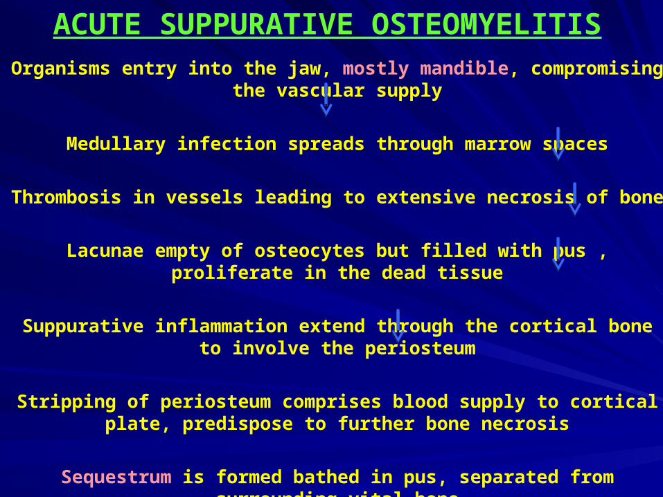
ACUTE SUPPURATIVE OSTEOMYELITISOrganisms entry into the jaw, mostly mandible, compromising the vascular supply
Medullary infection spreads through marrow spaces
Thrombosis in vessels leading to extensive necrosis of bone
Lacunae empty of osteocytes but filled with pus , proliferate in the dead tissue
Suppurative inflammation extend through the cortical bone to involve the periosteum
Stripping of periosteum comprises blood supply to cortical plate, predispose to further bone necrosis
Sequestrum is formed bathed in pus, separated from surrounding vital bone

Acute suppurative osteomyelitis
CLINICAL FEATURESEARLY :
Severe throbbing, deep- seated pain.Swelling due to inflammatory edema.Gingiva appears red, swollen & tender.
LATE :Distension of periosteum with pus.
FINAL: Subperiosteal bone formation cause swelling to become firm.

Acute suppurative osteomyelitisAcute suppurative osteomyelitisRadiographic featuers
May be normal in early stages of disease .Do not appear until after at least 10 days.
After sufficient bone resorption irregular, mot-
eaten areas of radiolucency may appear.
Radiograph may demonstrate ill-defined radiolucency.

05/03/2305/03/23 Ossama El-ShallOssama El-Shall 7878
CHRONIC SUPPURATIVE OSTEOMYELITIS

CHRONIC SUPPURATIVE OSTEOMYELITISInadequate treatment of acute osteomyelitis
Periodontal diseases, Pulpal infections, Extraction wounds Infected fractures
Infection in the medulllary spaces spread and form granulation tissue
Granulation tissue forms dense scar to wall off the infected area
Encircled dead space acts as a reserviour for bacteria & antibiotics have great difficulty reaching the site

CHRONIC SUPPURATIVE OSTEOMYELITIS
CLINICAL FEATURESSwellingIntermittent PainSinus formationPurulent dischargeSequestrum formationTooth lossPathologic fracture

CHRONIC SUPPURATIVE OSTEOMYELITIS
RADIOLOGYPatchy, ragged & ill defined radiolucency.Often contains radiopaque sequestra.
• Sequestra lying close to the peripheral sclerosis & lower border.
• New bone formation is evident below lower border.

05/03/2305/03/23 Ossama El-ShallOssama El-Shall 8282
CHRONIC SUPPURATIVE OSTEOMYELITIS
Sequestra

05/03/2305/03/23 Ossama El-ShallOssama El-Shall 8383
FOCAL SCLEROSING OSTEOMYELITIS
FOCAL SCLEROSING OSTEOMYELITIS
Also known as “Condensing osteitis”.
Localized areas of bone sclerosis.
Bony reaction to low-grade peri-apical infection or unusually strong host defensive response.
Association with an area of inflammation is critical.

FOCAL SCLEROSING OSTEOMYELITISFOCAL SCLEROSING OSTEOMYELITIS
CLINICAL FEATURES
Children & young adults are affected.
In mandible, premolar & molar regions are affected.
Bone sclerosis is associated with non-vital or vital tooth.
No expansion of the jaw.

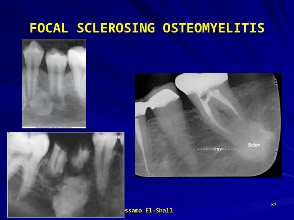
RADIOLOGYLocalized but uniform increased RO related to tooth. Widened periodontal ligament space or peri-apical area.Sometimes an adjacent radiolucent inflammatory lesion may be present.
FOCAL SCLEROSING OSTEOMYELITISFOCAL SCLEROSING OSTEOMYELITIS
Increased areas of radiodensity surrounding
apices of nonvital mandibular first
molar
05/03/2305/03/23 Ossama El-ShallOssama El-Shall 8787
FOCAL SCLEROSING OSTEOMYELITISFOCAL SCLEROSING OSTEOMYELITIS

05/03/2305/03/23 Ossama El-ShallOssama El-Shall 8888
FOCAL SCLEROSING OSTEOMYELITISFOCAL SCLEROSING OSTEOMYELITIS
05/03/2305/03/23 Ossama El-ShallOssama El-Shall 8989
DIFFUSE SCLEROSING OSTEOMYELITIS

DIFFUSE SCLEROSING OSTEOMYELITIS
It is an ill-defined, highly controversial type of osteomyelitis.
Bone metabolism tipped toward increased bone formation.
Chronic intraosseous bacterial infection creates a mass of chronically inflammed granulation tissue.

RADIOLOGY
Increased radiodensity may be seen surrounding areas of lesion.
DIFFUSE SCLEROSING OSTEOMYELITIS
Diffuse area of increased
radiodensity of Rt. Side of
mandible

05/03/2305/03/23 Ossama El-ShallOssama El-Shall 9292
DIFFUSE SCLEROSING OSTEOMYELITIS

05/03/2305/03/23 Ossama El-ShallOssama El-Shall 9393
DIFFUSE SCLEROSING OSTEOMYELITIS

05/03/2305/03/23 Ossama El-ShallOssama El-Shall 9494
Proliferative periosteitis “ Periostitis ossificans” “Garee’s osteomyelitis”.

Proliferative periosteitis Also known as “ Periostitis ossificans” & “Garee’s osteomyelitis”.
It represents a periosteal reaction to the presence of inflammation.
Affected periosteum forms several rows of reactive vital bone that parallel each other & expand surface of altered bone.
More common at mandibular first molar area, in young persons

“ “Garee’s osteomyelitis”.Garee’s osteomyelitis”.
CLINICAL FEATURESAffected patients are primarily children & young adults.Incidence is mean age of 13 years.No sex predominance is noted.Most cases arise in the premolar & molar area of mandible.Hyperplasia is located most commonly along lower border of mandible.Most cases are uni-focal, multiple quadrants may be affected.

PROLIFERATIVEPROLIFERATIVE PERIOSTITIS
RADIOLOGYRadiopaque laminations of bone roughly parallel each other & underlying cortical surface.
Laminations may vary from 1-12 in number.Best seen by occlusal film
Radiolucent separations often are present between new bone & original cortex.

05/03/2305/03/23 Ossama El-ShallOssama El-Shall 9898
PROLIFERATIVE PERIOSTITIS

OsteoradionecrosisOsteoradionecrosis
05/03/2305/03/23 Ossama El-ShallOssama El-Shall 9999
-It is the inflammatory condition occurs in bone after the bone has been exposed to therapeutic dose of radiation
- Infection or trauma are necessary- mandible more affected

05/03/2305/03/23 Ossama El-ShallOssama El-Shall 100100
OsteoradionecrosisOsteoradionecrosis

05/03/2305/03/23 Ossama El-ShallOssama El-Shall 101101

05/03/2305/03/23 Ossama El-ShallOssama El-Shall 102102
Odontogenic TumorsOdontogenic TumorsThey develops as neoplasias from the dental lamina. They are usually benign but several of them have the tendency towards malignant transformation.
Because growth occurs only slowly, asymptomatically and without any changes in mucosal appearance, the existence of such lesions in their early stages is usually detected only by chance, or after the development of some structural deformation.

05/03/2305/03/23 Ossama El-ShallOssama El-Shall 103103
AmeloblastomaAmeloblastomaBenign but locally invasive neoplasm.Benign but locally invasive neoplasm.Arises from epithelial remnants of dental Arises from epithelial remnants of dental lamina or dental organ.lamina or dental organ.Cells do not differentiate enough to form Cells do not differentiate enough to form enamel.enamel.Extreme expansion of bone, Extreme expansion of bone, Resorption of adjoining roots. Resorption of adjoining roots. May cause perforation of cortical bone.May cause perforation of cortical bone.Average age at discovery: 35-40 years.Average age at discovery: 35-40 years.

05/03/2305/03/23 Ossama El-ShallOssama El-Shall 104104
Most common sites of ameloblastoma
80%
20%

05/03/2305/03/23 Ossama El-ShallOssama El-Shall 105105
Ameloblastoma (Cont.)Ameloblastoma (Cont.)Occasionally develops in the wall of Occasionally develops in the wall of dentigerous cyst (mural dentigerous cyst (mural Ameloblatoma).Ameloblatoma).80% in mandible. ¾ of these in 80% in mandible. ¾ of these in molar-ramus area.molar-ramus area.Pain and paresthesia not common.Pain and paresthesia not common.Extremely high recurrence rate.Extremely high recurrence rate.
05/03/2305/03/23 Ossama El-ShallOssama El-Shall 106106
Ameloblastoma (Cont.)Ameloblastoma (Cont.)
Most often a well-corticated Most often a well-corticated multilocular radiolucency. multilocular radiolucency. ““Honey-comb”, “soap-bubble” or Honey-comb”, “soap-bubble” or “tennis-racket” appearance.“tennis-racket” appearance.May be a well-corticated unilocular May be a well-corticated unilocular lesion resembling a cyst.lesion resembling a cyst.
05/03/2305/03/23 Ossama El-ShallOssama El-Shall 107107
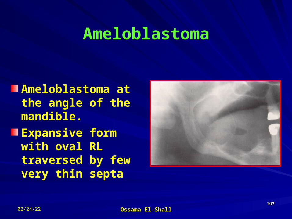
Ameloblastoma
Ameloblastoma at the angle of the mandible.Expansive form with oval RL traversed by few very thin septa

05/03/2305/03/23 Ossama El-ShallOssama El-Shall 108108
Ameloblastoma
Soap-like form of ameloblastoma of the molar region.

05/03/2305/03/23 Ossama El-ShallOssama El-Shall 109109
Ameloblastoma Large multilocular soap bubble appearance. Typically located in the molar region, angle of the
mandible and ascending ramus Thin not penetrated cortical plate. Impacted or neighboring teeth are displaced with
roots often resorped.
05/03/2305/03/23 Ossama El-ShallOssama El-Shall 110110
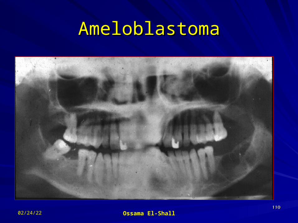
AmeloblastomaAmeloblastoma

05/03/2305/03/23 Ossama El-ShallOssama El-Shall 111111
Large ameloblastoma in the right ascending ramus of the mandible

05/03/2305/03/23 Ossama El-ShallOssama El-Shall 112112
Honeycomb-like small ameloblastoma at Honeycomb-like small ameloblastoma at early stage with evidence of root resorption.early stage with evidence of root resorption.

05/03/2305/03/23 Ossama El-ShallOssama El-Shall 113113
AmeloblastomaAmeloblastoma

05/03/2305/03/23 Ossama El-ShallOssama El-Shall 114114
Ameloblastic fibromaAmeloblastic fibroma
Appears as a follecular Appears as a follecular cystic cavity cystic cavity surrounding a crown surrounding a crown of a tooth.of a tooth.In early stages appears In early stages appears as a hat upon the as a hat upon the occlusal surface of occlusal surface of affected toothaffected tooth

05/03/2305/03/23 Ossama El-ShallOssama El-Shall 115115
More advanced case of ameloblastic fibroma demonstrates how the follicular sac is opened.Note also the displacement of the tooth bud of lower 8 in the ascending ramus.

05/03/2305/03/23 Ossama El-ShallOssama El-Shall 116116
Odontogenic myxomaOdontogenic myxoma
It is a benign, mucous-It is a benign, mucous-containing tumor that containing tumor that originates from the originates from the tooth bud.tooth bud.It appears as a soap It appears as a soap bubble-like bubble-like appearance.appearance.

05/03/2305/03/23 Ossama El-ShallOssama El-Shall 117117
CementomaCementomaUsually appears at lower Usually appears at lower anterior area.anterior area.First appears as fibrous First appears as fibrous tissue stage, which may tissue stage, which may confused with a confused with a granuloma (vitality test).granuloma (vitality test).The second stage is The second stage is characterized with characterized with accumulation of calcified accumulation of calcified materials.materials.The third stage consists of The third stage consists of radio-opaque materials. radio-opaque materials. Early stageEarly stage

05/03/2305/03/23 Ossama El-ShallOssama El-Shall 118118
R.LR.L R.L+R.OR.L+R.O

05/03/2305/03/23 Ossama El-ShallOssama El-Shall 119119
Periapical cemental dysplasia

05/03/2305/03/23 Ossama El-ShallOssama El-Shall 120120
CementoblastomaCementoblastoma( True Cementoma )( True Cementoma )
Slow growing Slow growing neoplasm composed neoplasm composed of cementum.of cementum.Usually solitary Usually solitary lesion seen as a lesion seen as a growth on root of growth on root of tooth. Most common tooth. Most common in mandible, in mandible, premolar or 1premolar or 1stst molar (80%).molar (80%).

05/03/2305/03/23 Ossama El-ShallOssama El-Shall 121121
Cementoblastoma
It not removed after tooth extractionRemarks the RL related to canine and second premolar, it is another cementoblastoma in the fibrous stage.

05/03/2305/03/23 Ossama El-ShallOssama El-Shall 122122
Cementoblastoma
Another case remaining after tooth extraction.It surrounded by the radiographic signs of chronic inflammation.Periapical cemental dysplasia related to 4 tooth

05/03/2305/03/23 Ossama El-ShallOssama El-Shall 123123
Odontoma
Most common sites

05/03/2305/03/23 Ossama El-ShallOssama El-Shall 124124
OdontomaOdontoma
Complex typeComplex type

05/03/2305/03/23 Ossama El-ShallOssama El-Shall 125125
OdontomaOdontoma
Intermediate typeIntermediate type

05/03/2305/03/23 Ossama El-ShallOssama El-Shall 126126
Compound Composite Compound Composite OdontomaOdontoma

05/03/2305/03/23 Ossama El-ShallOssama El-Shall 127127
Non-Odontogenic Tumors, Granulomatous and
Osteofibrous lesions of the jaws.

05/03/2305/03/23 Ossama El-ShallOssama El-Shall 128128
Central giant cell granuloma.Ossifying fibromaFibrous dysplasia.Osteoma.Osteoblastoma.Exostosis & Enostosis.Osteoporosis.Osteogenesis imperfecta.Eosinophilic granuloma.Hemangioma.
05/03/2305/03/23 Ossama El-ShallOssama El-Shall 129129
Central giant cell granuloma
Or Central Reparative Giant cell Granuloma, this type of granuloma grows expansively within the bone and occurs more frequently in female under age of 25 than in males.
It characterized by asymptomatic swelling of the affected jaw that is manifested by facial asymmetry

05/03/2305/03/23 Ossama El-ShallOssama El-Shall 130130
Radiographically appears as isolated or multilocular radiolucences that are sharply demarcated & exhibit soap-bubble like structures with lobulated margin. It may cause thinning of cortical plate
DD: 1- Ameloblastoma. It difficult to differentiate. 2- Eosinopilic granuloma. 3- Odontogenic cyst. 4- Aneurysmal & solitary bone cyst.

05/03/2305/03/23 Ossama El-ShallOssama El-Shall 131131
Most common sites of central giant cell granuloma (dark) and peripheral giant cell granuloma (Light)

05/03/2305/03/23 Ossama El-ShallOssama El-Shall 132132
Central Giant Cell Granuloma With its characteristic appearance of soap-bubble
appearance which can confused as ameloblastoma Note that the Periapical view cannot provide an
overview of the lesion extension

05/03/2305/03/23 Ossama El-ShallOssama El-Shall 133133
The impacted ankylosed canine and displacement of the teeth may indicate a signs of follicular cyst.
The fine septa and soap-bubble like contour resemble Ameloblastoma
Central Giant Cell Granuloma

05/03/2305/03/23 Ossama El-ShallOssama El-Shall 134134
Central HemangiomaCentral HemangiomaTumor characterized Tumor characterized by proliferation of by proliferation of blood vessels.blood vessels.Central hemangiomas Central hemangiomas of jaws uncommon.of jaws uncommon.50% occur in children 50% occur in children and teens.and teens.More common in More common in females and mandible.females and mandible.Well-defined or ill-Well-defined or ill-defined, unilocular or defined, unilocular or multilocular multilocular radiolucency.radiolucency.

05/03/2305/03/23 Ossama El-ShallOssama El-Shall 135135
Central Hemangioma Central Hemangioma (Cont.)(Cont.)
May cause expansion of bone May cause expansion of bone and resorption of teeth.and resorption of teeth.Early treatment is desirable in Early treatment is desirable in order to avoid profuse bleeding order to avoid profuse bleeding due to accidental trauma. due to accidental trauma. Aspiration prior to surgical Aspiration prior to surgical procedure is advised.procedure is advised.

05/03/2305/03/23 Ossama El-ShallOssama El-Shall 136136
Central Hemangioma Central Hemangioma (Cont.)(Cont.)

05/03/2305/03/23 Ossama El-ShallOssama El-Shall 137137
Central Hemangioma Central Hemangioma (Cont.)(Cont.)

05/03/2305/03/23 Ossama El-ShallOssama El-Shall 138138
Central Hemangioma Central Hemangioma (Cont.)(Cont.)

05/03/2305/03/23 Ossama El-ShallOssama El-Shall 139139
Malignant tumors
SarcomaCarcinoma.Metastasis.

05/03/2305/03/23 Ossama El-ShallOssama El-Shall 140140
SarcomaThis tumor, which affects males twice
as females, exhibit a predilection for the mandible.
Radiographically, bone destruction as well as new bone formation and osteolysis can be observed, along with perforation of the compact bone with spicules (sunrays effect), where the lesion borders on the soft tissues

05/03/2305/03/23 Ossama El-ShallOssama El-Shall 141141
Thank you all for listening
Dr. Ossama El-ShallChairman of Oral Medicine &
Periodontology department, Faculty of Dental Medicine for girls, Al-Azhar
University, Cairo, Egypt.
E-mail address: [email protected]