Embed Size (px)
Citation preview

Cell Biology
S. Rahgozar,PhD
University of Isfahan
Faculty of Science
1. An overview of cells and cell research
1.2. General methods of Cell Biology
1392-93

Robert Hooke
(1665)
The cellular
structure of cork
Light microscope
1670s: Antony van Leeuwenhoek
observed sperm
RBCs
Bacteria
1838: Schleiden & Theodor
proposed the “cell theory”
All organisms are composed of cells
Cells are not formed de novo
Microscope

Magnification
Main concerns
Resolution
1000
0.22 µm
λ = Wavelength (fo visible light: 0.4-0.7μm)
NA= Numerical Aperture
(The light gathering power)
= ŋ sinα
ŋ= refractive index of the medium
through which light travels
α= The angle corresponding to half the
width of the cone of light collected
by the lens
(1< ŋ <1.4)
Different types of light microscopy
Bright field microscopy
Contrast resulting from the absorption of visible light by cell components
Staining: Dye may react with proteins/nucleic acids
and increase the contrast
Bright field micrograph of stained
tissue
o Requires fixation which kills the
cells
Probing with:
Nucleic acid probes
Antibodies
Labeled with a variety of tags
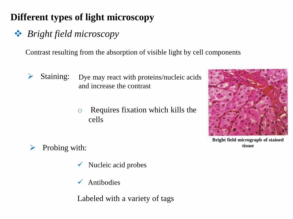
Phase contrast microscopy
Bright-field microscopy
Phase contrast microscopy
Converts difference in density between
different parts of the cell to difference in
contrast using optical system.
Difference in density difference in phase
difference in contrast
Differential interference -
contrast microscopy
Differential interfrence contrast microscopy
Video enhanced differential
interference - contrast microscopy

Fluorescence microscopy
Studying the intracellular distribution of molecules
For fixed or living cells
Fluorescent micrograph of a lung cell
in which the DNA is stained blue and
microtubules green.

The use of green fluorescent protein (GFP)
A mouse neuron in which microtubule-associated
protein is fused to GFP and visualized by fluorescence
microscopy.
GFP (from jelly fish) is fused to a protein of
interest using recombinant DNA. The GFP –
tagged protein can then be expressed in cell
and detected .
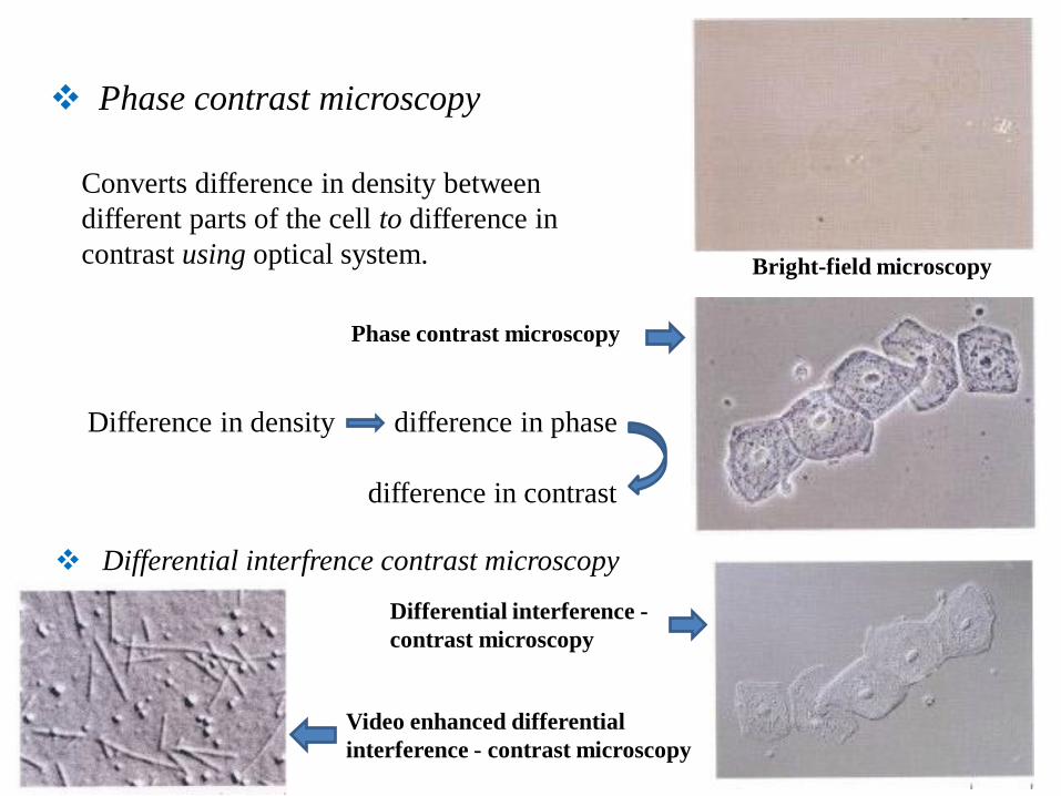
Fluorescence recovery after
photobleaching (FRAP)
To follow the movement of GFP-labeled
proteins within living cells
Fluorescence resonance energy
transfer (FRET)

Confocal microscopy
A human cell in which microtubules and actin filaments are
stained with red and green fluorescent dyes Only in-focus light emitted from the chosen
depth of the specimen is detected
A series of images obtained at different depths can
be used to reconstruct a three-dimensional image
of the sample

Electron microscopy
A Positively stained WBC
Negatively stained actin filaments
Greater resolution (2-0.2nm)
shorter wavelength of electrons ie: 0.004nm
Transmission electron microscopy
Fixation, Staining with salts of heavy metals providing contrast by
scattering electrons.
Isolated organelle is deposited on a film and
metal stains around its surface. Specimens are cut into thin sections

Metal shadowing of actin/myosin
o Specimen is coated with a thin
layer of evaporated metal.
Surfaces that face the metal are
coated more heavily.
o Freeze fracture splits the lipid
bilayer, leaving proteins
embedded in the membrane
associated with one of the two
membrane halves
Freeze fracture Specimens are frozen at -196 ̊C, fractured with a
knife blade, then shadowed with platinum

Scanning electron microscopy
The surface of the cell is coated
with a heavy metal, and a beam of
electrons is used to scan across the
specimen.
Electrons that are scattered from
the sample surface are collected to
generate a 3 dimensional image

Subcellular Fractionation
o Sonication
o Grinding in a mechanical homogenizer
o Treatment with a high speed blender
Differential centrifugation
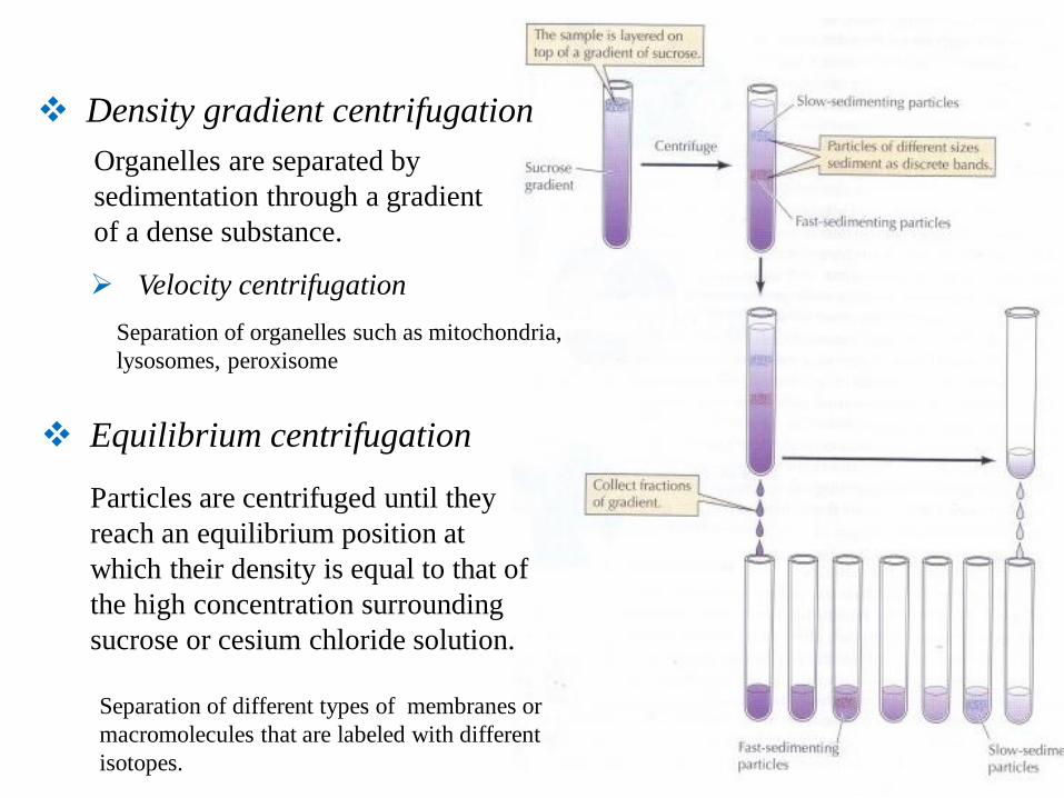
Density gradient centrifugation
Organelles are separated by
sedimentation through a gradient
of a dense substance.
Velocity centrifugation
Separation of organelles such as mitochondria,
lysosomes, peroxisome
Equilibrium centrifugation
Particles are centrifuged until they
reach an equilibrium position at
which their density is equal to that of
the high concentration surrounding
sucrose or cesium chloride solution.
Separation of different types of membranes or
macromolecules that are labeled with different
isotopes.
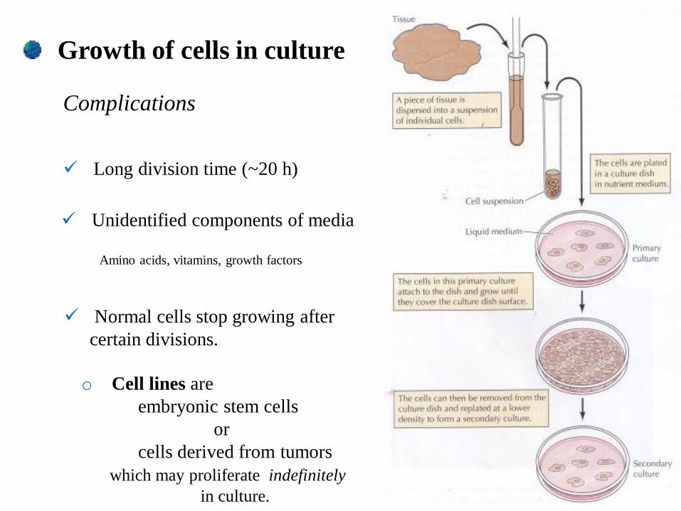
Growth of cells in culture
Complications
Long division time (~20 h)
Unidentified components of media
Amino acids, vitamins, growth factors
Normal cells stop growing after
certain divisions.
o Cell lines are
embryonic stem cells
or
cells derived from tumors
which may proliferate indefinitely
in culture.

Cultures of Animal and Plant cells
o Growth factors in animal cultures are polypeptides,
however,
growth factors in plant cells are small molecules which
may pass through the plant cell wall
o Embryonic stem cells in animal cultures
and
Callus in plant cultures
are undifferentiated cells which are capable of
forming any of the different cell types
Callus

Viruses
Human papilomavirus particles
Simple systems that can be used to
investigate the functions of cells