Embed Size (px)
Citation preview

Prof. Dr. Sudibyo Martono, M.S., Apt.
ANALISIS
FISIKO KIMIA I

Spektra Massa

SPECTRA MS
Mass Spectrometry
The basic principles
Applications
3

SPECTRA MS
THE MAIN USE OF MS IN ORGANIC CHEMISTRY IS:
• DETERMINE THE MOLECULAR MASS OF ORGANIC COMPOUNDS
• DETERMINE THE MOLECULAR FORMULA OF ORGANIC COMPOUNDS
Mass Spectrometry
4

SPECTRA MS
Mass Spectrometry
• Molecular weight can be obtained from a very small sample.
• It does not involve the absorption or emission of light.
• A beam of high-energy electrons breaks the molecule apart.
• The masses of the fragments and their relative abundance reveal information about the structure of the molecule.
5

SPECTRA MS
What is a mass spectrometer ?
A mass spectrometer is an instrument which produces charged particles (ions) from chemical substances under analysis.
It then uses magnetic and/or electric fields to separate those ions and to measure their mass.
6

SPECTRA MS 7

SPECTRA MS 8

SPECTRA MS 9

SPECTRA MS 10

SPECTRA MS
Ion Generation
~70 Volts
+
_
+_
e- e-e-
++ ++++
_
Electron Collector (Trap)
Repeller
ExtractionPlate
Filament
To Analyzer
Inlet
Electrons
NeutralMolecules Positive Ions
11

SPECTRA MS 12

APCIAPCI
SPECTRA MS 13

Electrospray Ionization• Sample dissolved in polar, volatile buffer
(no salts) and pumped through a stainless steel capillary (70 - 150 µm) at a rate of 10-100 µL/min
• Strong voltage (3-4 kV) applied at tip along with flow of nebulizing gas causes the sample to “nebulize” or aerosolize
• Aerosol is directed through regions of higher vacuum until droplets evaporate to near atomic size (still carrying charges)
SPECTRA MS 14

SPECTRA MS 15

ESIESI
SPECTRA MS 16
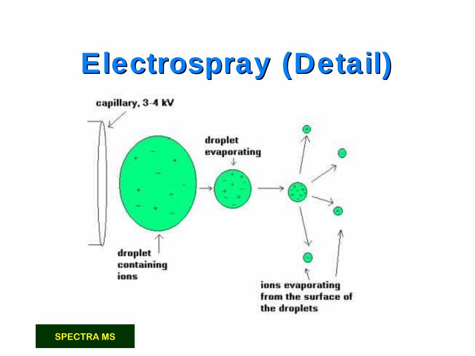
Electrospray (Detail)Electrospray (Detail)
SPECTRA MS 17

Electrospray Ionization
• Can be modified to “nanospray” system with flow < 1 µL/min
• Very sensitive technique, requires less than a picomole of material
• Strongly affected by salts & detergents
• Positive ion mode measures (M + H) + (add formic acid to solvent)
• Negative ion mode measures (M - H) - (add ammonia to solvent)
SPECTRA MS 18

SPECTRA MS
ELECTRON IMPACT
H-C:HH
H+ e H-C
H
HH + 2e
RADICAL CATION
CH4
H-CH
HH
H-CH
H+ + H
ONLY CATIONS ARE CARRIED TO DETECTOR
H-CH
HH++
CATIONRADICALBOND-BREAKING
19

SPECTRA MS
Electron Impact Ionization
A high-energy electron can dislodge an electron from a bond, creating a radical cation (a positive ion with an unpaired e-).
e- + H C
H
H
C
H
H
H
H C
H
H
C
H
H
H
H C
H
H
C
H
H
+ H
H C
H
H
C
H
H
H
+=>20

SPECTRA MS
SEPARATION OF IONS
• Only the cations are deflected by the magnetic field.
• Amount of deflection depends on m/z.
• The detector signal is proportional to the number of ions hitting it.
• By varying the magnetic field, ions of all masses are collected and counted. =>
21

Positive or Negative Ion Mode ?
• If the sample has functional groups that readily accept H+ (such as amide and amino groups found in peptides and proteins) then positive ion detection is used-PROTEINS
• If a sample has functional groups that readily lose a proton (such as carboxylic acids and hydroxyls as found in nucleic acids and sugars) then negative ion detection is used-DNA
SPECTRA MS 22

SPECTRA MS
Mass Spectrometer Schematic
IonSource
Mass Analyzer
IonDetector
Inlet DataSystem
VacuumPumps
SampleIntroduction
DataOutput
23

SPECTRA MS
The magnetic field exerts a force on these fast-moving ions and causes them to move in a circular path, the radius of which is dependent upon their
mass to charge ratio (m/z) and speed.
24

SPECTRA MS
Magnetic Mass Separation
ion not detectedm/z too large
ion not detected m/z too small
Correct m/z ratioion detected
IonSource
Detector
S
N
Electromagnet
25

SPECTRA MS
Fragments appear due to bond cleavage next to C=O (alkoxy group loss, -OR) and hydrogen rearrangements.
10080
60
40
200
100806040200mass / charge (m/z)
rela
tive
ab
un
dan
ce
-OCH2CH3
-C2H3
43
45 8861
CH3
O
O CH2CH3
61
43 45
HH
m/z(parent ion) = 88
26

SPECTRA MS
Typical Mass Spectrum
100%
50%
43
58
71114
m/e
base peak given 100% abundance
molecular ion
Isotope peaks - P+1, P+2, etc
Alarm Pheromone of Honey Bee
Note: 114-71 = 43
Note: 43 = mass of radical
27

SPECTRA MS
CH3CH2CH2CHCH3
CH3
m/z 86
+•
Facile
Facile
More difficult
CH3CH2CH2CHCH3
CH3
+• +
m/z 43
CH3 CH3CH2CH2CH
CH3
m/z 71
+• +
+• +
m/z 57
Fragmentation of 2-Methylpentane
CH3CH2 CH2CHCH3
CH3
28

SPECTRA MS
Mass Spectrum of 2-MethylpentaneIn
t ens
ity
m/z
20
40
60
80
100
10 20 30 40 50 60 70 80 90
43
57
71
86, M+
CH3CH2CH2CHCH3
CH3
29

SPECTRA MS
Mass Spectrum of PentaneIn
t ens
ity
20
40
60
80
100
10 20 30 40 50 60 70
43
57
72, M+
CH3CH2CH2CH2CH3
42
41
3927
29
m/z 30

SPECTRA MS
Int e
nsit
y
20
40
60
80
100
20 40 60 80 100 120 140
94
156107
160
94
107
m/z
2-Chloroethylphenyl ether
O
Cl H
31
SPECTRA MS
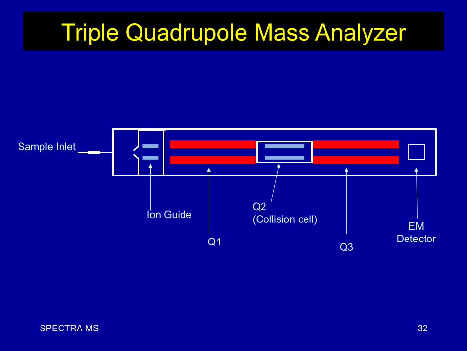
Triple Quadrupole Mass Analyzer
Sample Inlet
Q2(Collision cell)
Q3
Ion Guide
Q1
EMDetector
32

SPECTRA MS
LC-MS-MSLC-MS-MS
33

SPECTRA MS
MRM
34

SPECTRA MS
LCcolumn
Q1 (227) Q2 collision cell
Fragment parent ion
Q3 (181)
Set on mass ofparent ion
Transmit only diagnosticproduct ion
highest duty cycle triple quadrupole scan type!
OH
NH3+
NO2
O OH
OH
NH2+
NO2
Multiple Reaction Monitoring in Triple Quadrupole
35
SPECTRA MS
Applications
- Chemical analysis (Chemical Research
- Environmental analysis - Analysis of petroleum products
- Trace metals
- Biological materials
Mass spectrometers are used for all kinds of chemical analyses:
36

SPECTRA MS 38