Embed Size (px)
Citation preview

DAVID SUTTON
2 THE MEDIASTINUM
DAVID SUTTON PICTURES
DR. Muhammad Bin Zulfiqar PGR-FCPS III SIMS/SHL
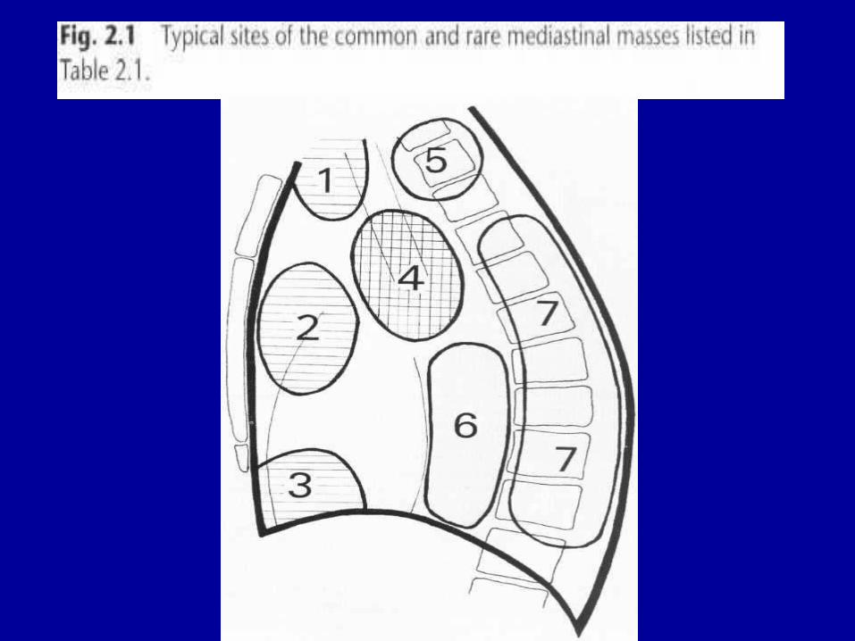
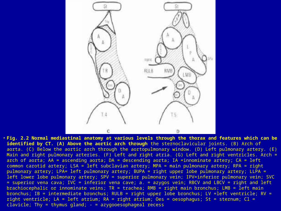
• Fig. 2.2 Normal mediastinal anatomy at various levels through the thorax and features which can be identified by CT. (A) Above the aortic arch through the sternoclavicular joints. (B) Arch of aorta. (C) Below the aortic arch through the aortopulmonary window. (D) Left pulmonary artery. (E) Main and right pulmonary arteries. (F) Left and right atria. (G) Left and right ventricles. Arch = arch of aorta; AA = ascending aorta; DA = descending aorta; IA =innominate artery; CA = left common carotid artery; LSA = left subclavian artery; MPA = main pulmonary artery; RPA = right pulmonary artery; LPA= left pulmonary artery; BUPA = right upper lobe pulmonary artery; LLPA = left lower lobe pulmonary artery; SPV = superior pulmonary vein; IPV=inferior pulmonary vein; SVC = superior vena cava; IVC = inferior vena cave; a. = azygos vein; RBCV and LBCV = right and left brachiocephalic or innominate veins; TR = trachea; RMB = right main bronchus; LMB = left main bronchus; IB = intermediate bronchus; RULB = right upper lobe bronchus; LV =left ventricle; RV = right ventricle; LA = left atrium; RA = right atrium; Oes = oesophagus; St = sternum; Cl = clavicle; Thy = thymus gland; - = azygooesophageal recess
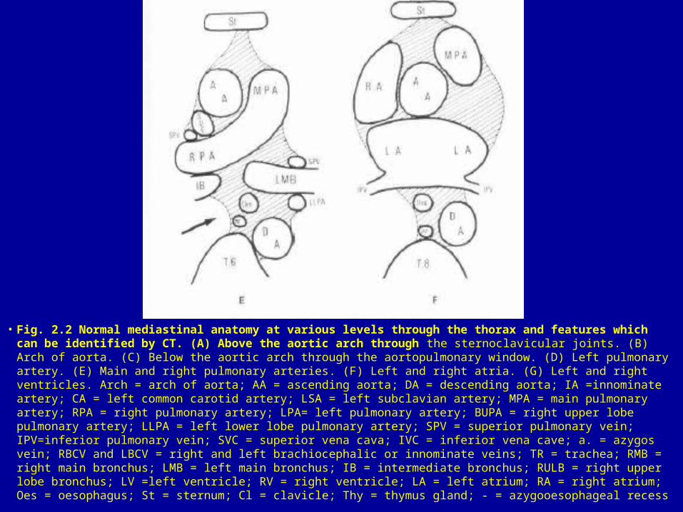
• Fig. 2.2 Normal mediastinal anatomy at various levels through the thorax and features which can be identified by CT. (A) Above the aortic arch through the sternoclavicular joints. (B) Arch of aorta. (C) Below the aortic arch through the aortopulmonary window. (D) Left pulmonary artery. (E) Main and right pulmonary arteries. (F) Left and right atria. (G) Left and right ventricles. Arch = arch of aorta; AA = ascending aorta; DA = descending aorta; IA =innominate artery; CA = left common carotid artery; LSA = left subclavian artery; MPA = main pulmonary artery; RPA = right pulmonary artery; LPA= left pulmonary artery; BUPA = right upper lobe pulmonary artery; LLPA = left lower lobe pulmonary artery; SPV = superior pulmonary vein; IPV=inferior pulmonary vein; SVC = superior vena cava; IVC = inferior vena cave; a. = azygos vein; RBCV and LBCV = right and left brachiocephalic or innominate veins; TR = trachea; RMB = right main bronchus; LMB = left main bronchus; IB = intermediate bronchus; RULB = right upper lobe bronchus; LV =left ventricle; RV = right ventricle; LA = left atrium; RA = right atrium; Oes = oesophagus; St = sternum; Cl = clavicle; Thy = thymus gland; - = azygooesophageal recess
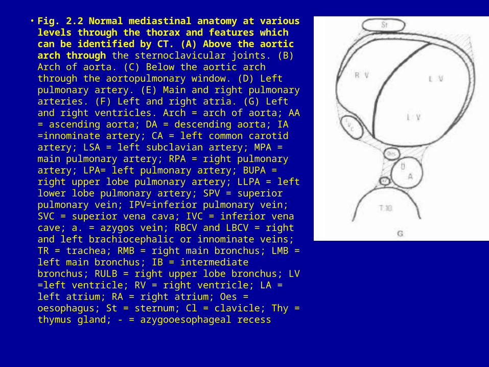
• Fig. 2.2 Normal mediastinal anatomy at various levels through the thorax and features which can be identified by CT. (A) Above the aortic arch through the sternoclavicular joints. (B) Arch of aorta. (C) Below the aortic arch through the aortopulmonary window. (D) Left pulmonary artery. (E) Main and right pulmonary arteries. (F) Left and right atria. (G) Left and right ventricles. Arch = arch of aorta; AA = ascending aorta; DA = descending aorta; IA =innominate artery; CA = left common carotid artery; LSA = left subclavian artery; MPA = main pulmonary artery; RPA = right pulmonary artery; LPA= left pulmonary artery; BUPA = right upper lobe pulmonary artery; LLPA = left lower lobe pulmonary artery; SPV = superior pulmonary vein; IPV=inferior pulmonary vein; SVC = superior vena cava; IVC = inferior vena cave; a. = azygos vein; RBCV and LBCV = right and left brachiocephalic or innominate veins; TR = trachea; RMB = right main bronchus; LMB = left main bronchus; IB = intermediate bronchus; RULB = right upper lobe bronchus; LV =left ventricle; RV = right ventricle; LA = left atrium; RA = right atrium; Oes = oesophagus; St = sternum; Cl = clavicle; Thy = thymus gland; - = azygooesophageal recess
• Fig. 2.2 Normal mediastinal anatomy at various levels through the thorax and features which can be identified by CT. (A) Above the aortic arch through the sternoclavicular joints. (B) Arch of aorta. (C) Below the aortic arch through the aortopulmonary window. (D) Left pulmonary artery. (E) Main and right pulmonary arteries. (F) Left and right atria. (G) Left and right ventricles. Arch = arch of aorta; AA = ascending aorta; DA = descending aorta; IA =innominate artery; CA = left common carotid artery; LSA = left subclavian artery; MPA = main pulmonary artery; RPA = right pulmonary artery; LPA= left pulmonary artery; BUPA = right upper lobe pulmonary artery; LLPA = left lower lobe pulmonary artery; SPV = superior pulmonary vein; IPV=inferior pulmonary vein; SVC = superior vena cava; IVC = inferior vena cave; a. = azygos vein; RBCV and LBCV = right and left brachiocephalic or innominate veins; TR = trachea; RMB = right main bronchus; LMB = left main bronchus; IB = intermediate bronchus; RULB = right upper lobe bronchus; LV =left ventricle; RV = right ventricle; LA = left atrium; RA = right atrium; Oes = oesophagus; St = sternum; Cl = clavicle; Thy = thymus gland; - = azygooesophageal recess
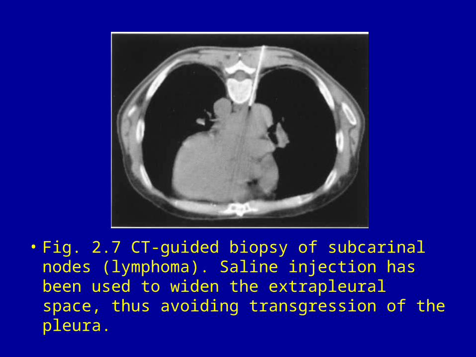
• Fig. 2.7 CT-guided biopsy of subcarinal nodes (lymphoma). Saline injection has been used to widen the extrapleural space, thus avoiding transgression of the pleura.
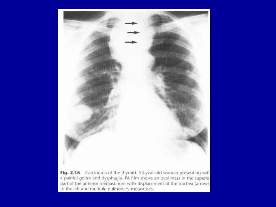
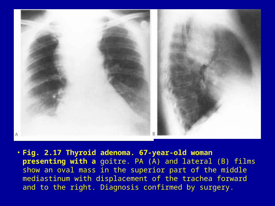
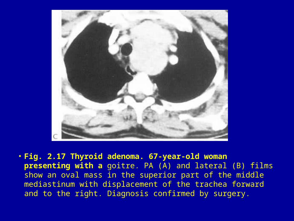
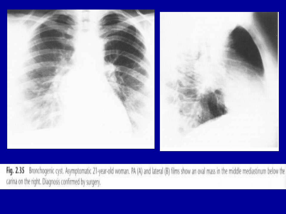
• Fig. 2.17 Thyroid adenoma. 67-year-old woman presenting with a goitre. PA (A) and lateral (B) films show an oval mass in the superior part of the middle mediastinum with displacement of the trachea forward and to the right. Diagnosis confirmed by surgery.
• Fig. 2.17 Thyroid adenoma. 67-year-old woman presenting with a goitre. PA (A) and lateral (B) films show an oval mass in the superior part of the middle mediastinum with displacement of the trachea forward and to the right. Diagnosis confirmed by surgery.
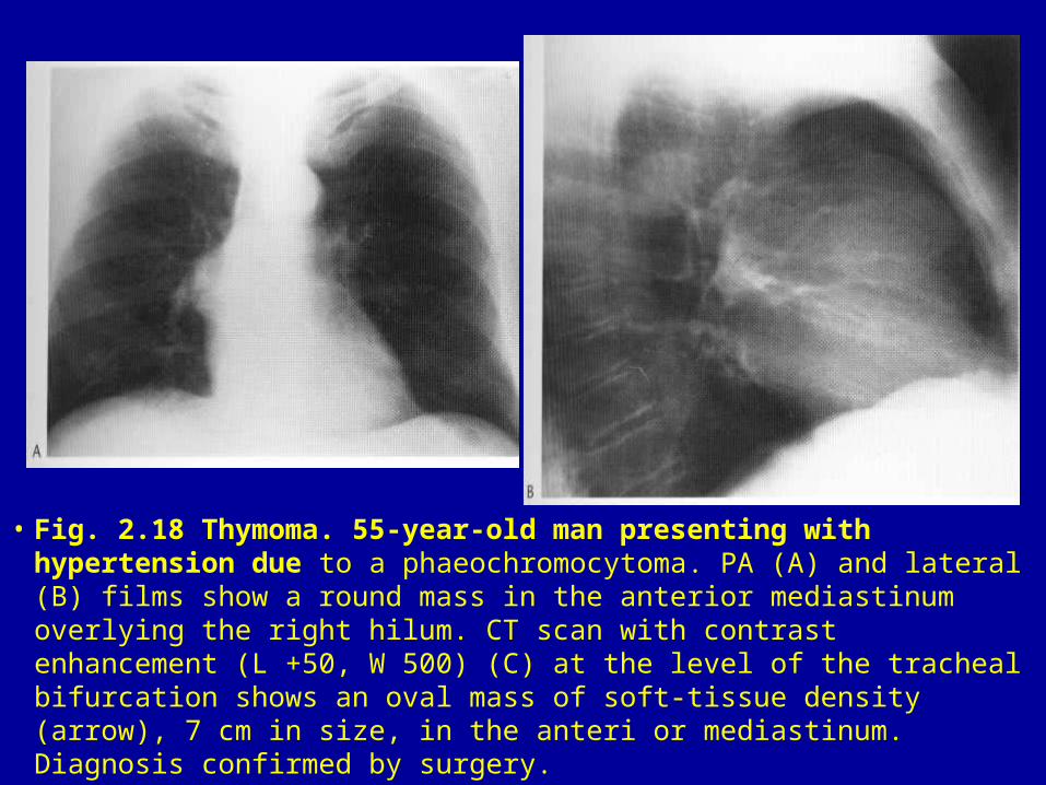
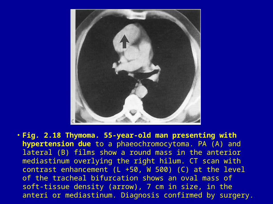
• Fig. 2.18 Thymoma. 55-year-old man presenting with hypertension due to a phaeochromocytoma. PA (A) and lateral (B) films show a round mass in the anterior mediastinum overlying the right hilum. CT scan with contrast enhancement (L +50, W 500) (C) at the level of the tracheal bifurcation shows an oval mass of soft-tissue density (arrow), 7 cm in size, in the anteri or mediastinum. Diagnosis confirmed by surgery.
• Fig. 2.18 Thymoma. 55-year-old man presenting with hypertension due to a phaeochromocytoma. PA (A) and lateral (B) films show a round mass in the anterior mediastinum overlying the right hilum. CT scan with contrast enhancement (L +50, W 500) (C) at the level of the tracheal bifurcation shows an oval mass of soft-tissue density (arrow), 7 cm in size, in the anteri or mediastinum. Diagnosis confirmed by surgery.
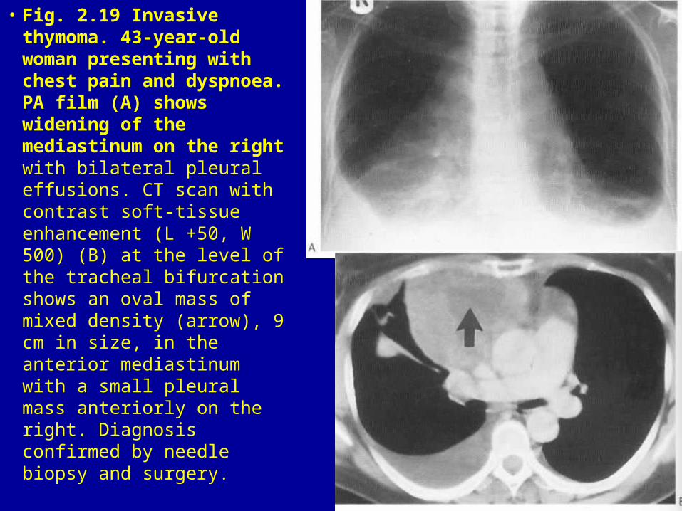
• Fig. 2.19 Invasive thymoma. 43-year-old woman presenting with chest pain and dyspnoea. PA film (A) shows widening of the mediastinum on the right with bilateral pleural effusions. CT scan with contrast soft-tissue enhancement (L +50, W 500) (B) at the level of the tracheal bifurcation shows an oval mass of mixed density (arrow), 9 cm in size, in the anterior mediastinum with a small pleural mass anteriorly on the right. Diagnosis confirmed by needle biopsy and surgery.
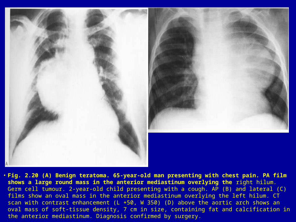
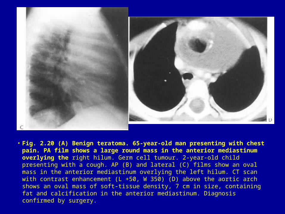
• Fig. 2.20 (A) Benign teratoma. 65-year-old man presenting with chest pain. PA film shows a large round mass in the anterior mediastinum overlying the right hilum. Germ cell tumour. 2-year-old child presenting with a cough. AP (B) and lateral (C) films show an oval mass in the anterior mediastinum overlying the left hilum. CT scan with contrast enhancement (L +50, W 350) (D) above the aortic arch shows an oval mass of soft-tissue density, 7 cm in size, containing fat and calcification in the anterior mediastinum. Diagnosis confirmed by surgery.
• Fig. 2.20 (A) Benign teratoma. 65-year-old man presenting with chest pain. PA film shows a large round mass in the anterior mediastinum overlying the right hilum. Germ cell tumour. 2-year-old child presenting with a cough. AP (B) and lateral (C) films show an oval mass in the anterior mediastinum overlying the left hilum. CT scan with contrast enhancement (L +50, W 350) (D) above the aortic arch shows an oval mass of soft-tissue density, 7 cm in size, containing fat and calcification in the anterior mediastinum. Diagnosis confirmed by surgery.
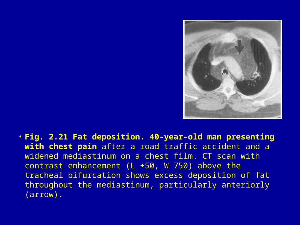
• Fig. 2.21 Fat deposition. 40-year-old man presenting with chest pain after a road traffic accident and a widened mediastinum on a chest film. CT scan with contrast enhancement (L +50, W 750) above the tracheal bifurcation shows excess deposition of fat throughout the mediastinum, particularly anteriorly (arrow).
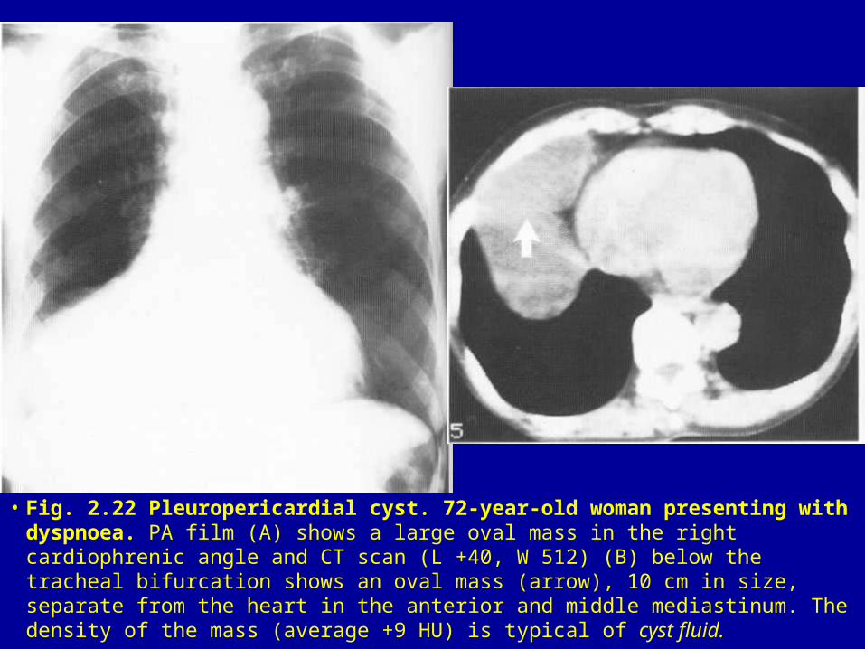
• Fig. 2.22 Pleuropericardial cyst. 72-year-old woman presenting with dyspnoea. PA film (A) shows a large oval mass in the right cardiophrenic angle and CT scan (L +40, W 512) (B) below the tracheal bifurcation shows an oval mass (arrow), 10 cm in size, separate from the heart in the anterior and middle mediastinum. The density of the mass (average +9 HU) is typical of cyst fluid.
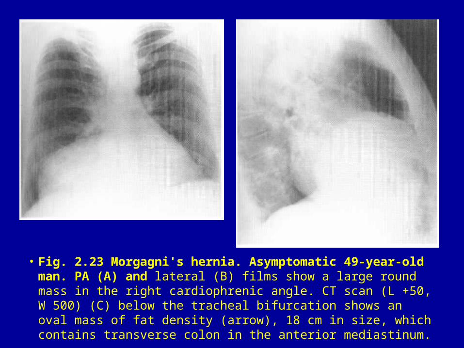
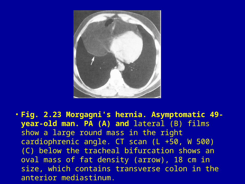
• Fig. 2.23 Morgagni's hernia. Asymptomatic 49-year-old man. PA (A) and lateral (B) films show a large round mass in the right cardiophrenic angle. CT scan (L +50, W 500) (C) below the tracheal bifurcation shows an oval mass of fat density (arrow), 18 cm in size, which contains transverse colon in the anterior mediastinum.
• Fig. 2.23 Morgagni's hernia. Asymptomatic 49-year-old man. PA (A) and lateral (B) films show a large round mass in the right cardiophrenic angle. CT scan (L +50, W 500) (C) below the tracheal bifurcation shows an oval mass of fat density (arrow), 18 cm in size, which contains transverse colon in the anterior mediastinum.
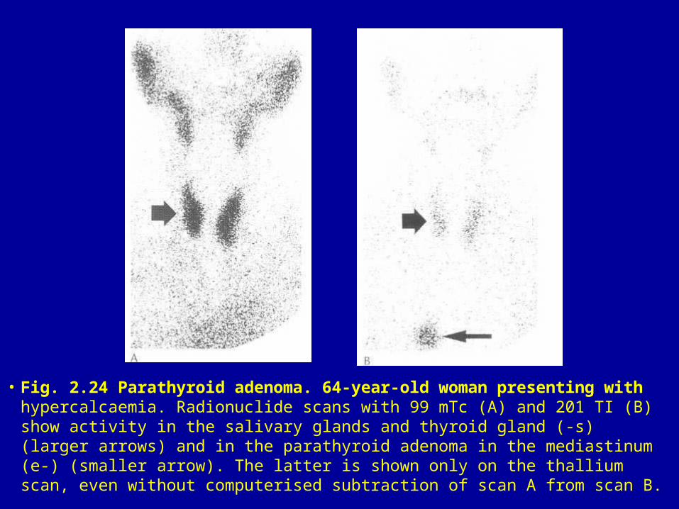
• Fig. 2.24 Parathyroid adenoma. 64-year-old woman presenting with hypercalcaemia. Radionuclide scans with 99 mTc (A) and 201 TI (B) show activity in the salivary glands and thyroid gland (-s) (larger arrows) and in the parathyroid adenoma in the mediastinum (e-) (smaller arrow). The latter is shown only on the thallium scan, even without computerised subtraction of scan A from scan B.
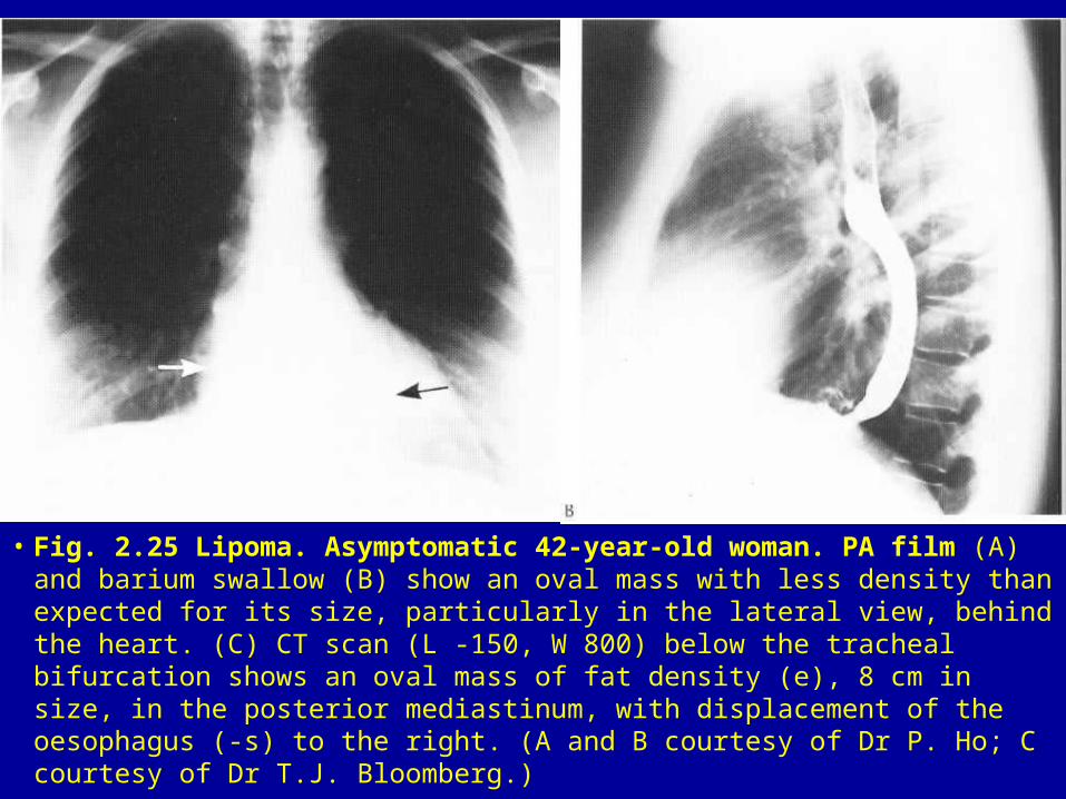
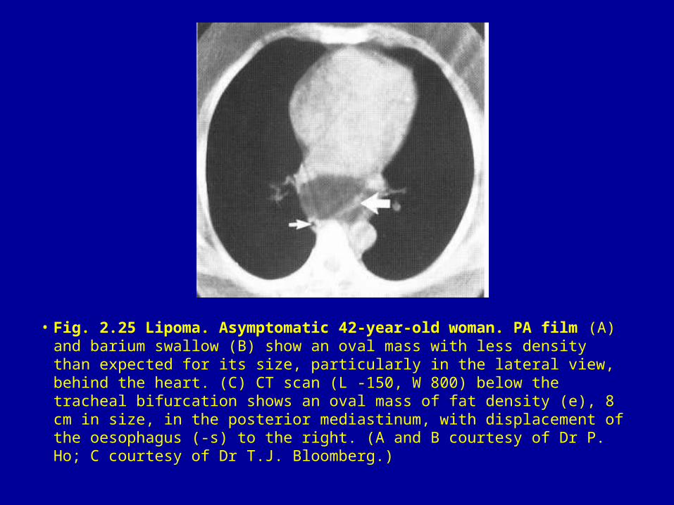
• Fig. 2.25 Lipoma. Asymptomatic 42-year-old woman. PA film (A) and barium swallow (B) show an oval mass with less density than expected for its size, particularly in the lateral view, behind the heart. (C) CT scan (L -150, W 800) below the tracheal bifurcation shows an oval mass of fat density (e), 8 cm in size, in the posterior mediastinum, with displacement of the oesophagus (-s) to the right. (A and B courtesy of Dr P. Ho; C courtesy of Dr T.J. Bloomberg.)
• Fig. 2.25 Lipoma. Asymptomatic 42-year-old woman. PA film (A) and barium swallow (B) show an oval mass with less density than expected for its size, particularly in the lateral view, behind the heart. (C) CT scan (L -150, W 800) below the tracheal bifurcation shows an oval mass of fat density (e), 8 cm in size, in the posterior mediastinum, with displacement of the oesophagus (-s) to the right. (A and B courtesy of Dr P. Ho; C courtesy of Dr T.J. Bloomberg.)
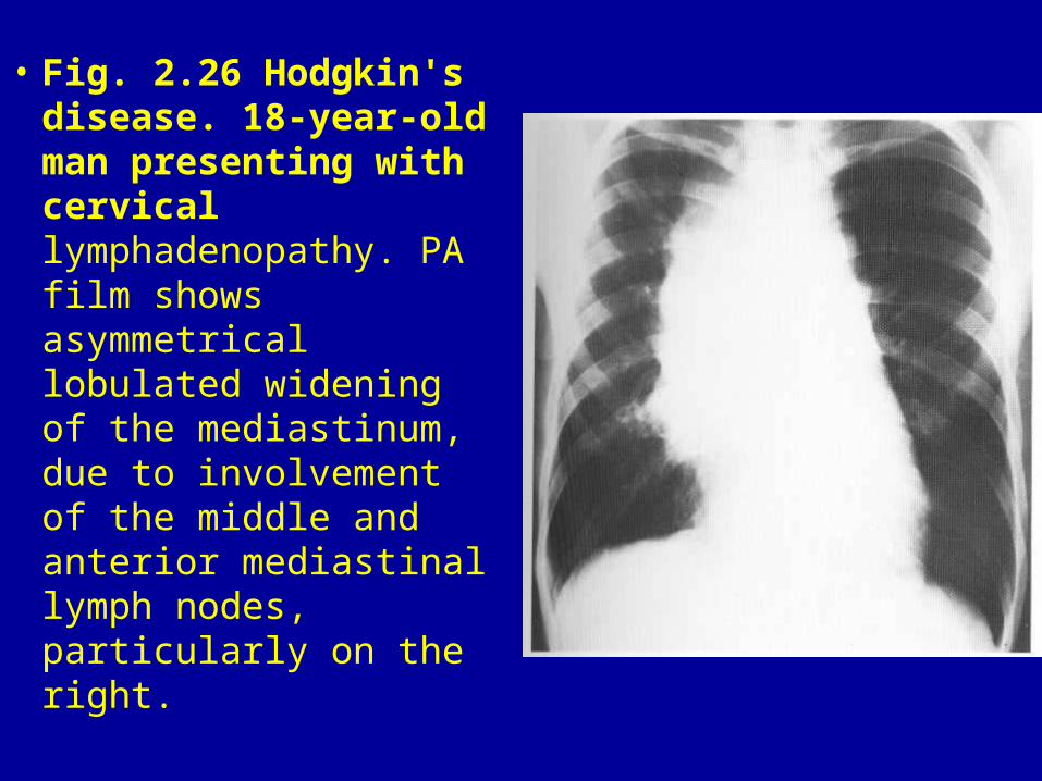
• Fig. 2.26 Hodgkin's disease. 18-year-old man presenting with cervical lymphadenopathy. PA film shows asymmetrical lobulated widening of the mediastinum, due to involvement of the middle and anterior mediastinal lymph nodes, particularly on the right.
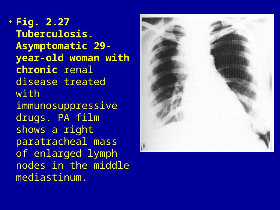
• Fig. 2.27 Tuberculosis. Asymptomatic 29-year-old woman with chronic renal disease treated with immunosuppressive drugs. PA film shows a right paratracheal mass of enlarged lymph nodes in the middle mediastinum.
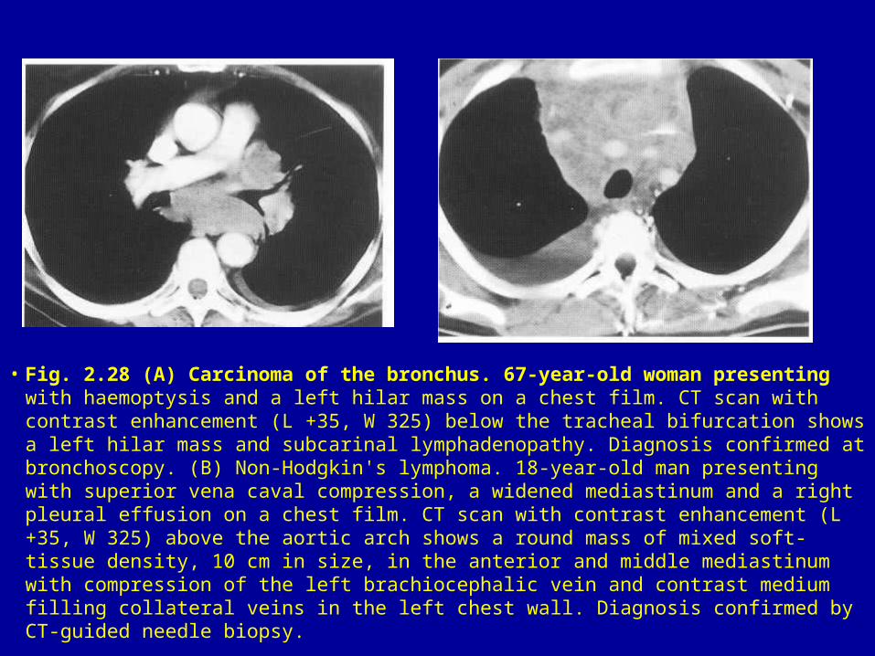
• Fig. 2.28 (A) Carcinoma of the bronchus. 67-year-old woman presenting with haemoptysis and a left hilar mass on a chest film. CT scan with contrast enhancement (L +35, W 325) below the tracheal bifurcation shows a left hilar mass and subcarinal lymphadenopathy. Diagnosis confirmed at bronchoscopy. (B) Non-Hodgkin's lymphoma. 18-year-old man presenting with superior vena caval compression, a widened mediastinum and a right pleural effusion on a chest film. CT scan with contrast enhancement (L +35, W 325) above the aortic arch shows a round mass of mixed soft-tissue density, 10 cm in size, in the anterior and middle mediastinum with compression of the left brachiocephalic vein and contrast medium filling collateral veins in the left chest wall. Diagnosis confirmed by CT-guided needle biopsy.
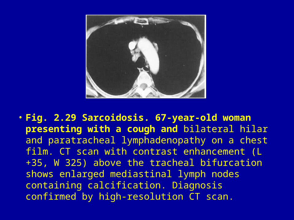
• Fig. 2.29 Sarcoidosis. 67-year-old woman presenting with a cough and bilateral hilar and paratracheal lymphadenopathy on a chest film. CT scan with contrast enhancement (L +35, W 325) above the tracheal bifurcation shows enlarged mediastinal lymph nodes containing calcification. Diagnosis confirmed by high-resolution CT scan.
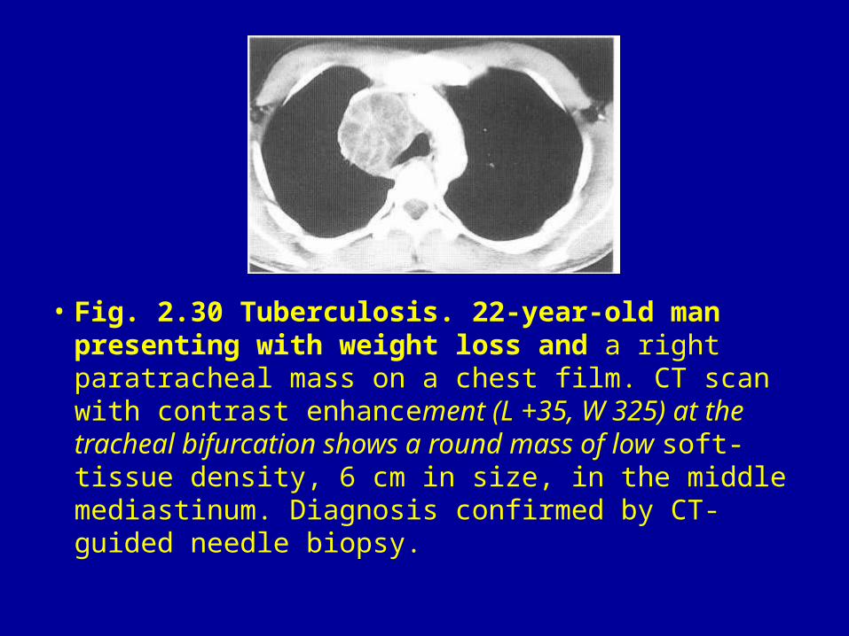
• Fig. 2.30 Tuberculosis. 22-year-old man presenting with weight loss and a right paratracheal mass on a chest film. CT scan with contrast enhancement (L +35, W 325) at the tracheal bifurcation shows a round mass of low soft-tissue density, 6 cm in size, in the middle mediastinum. Diagnosis confirmed by CT-guided needle biopsy.
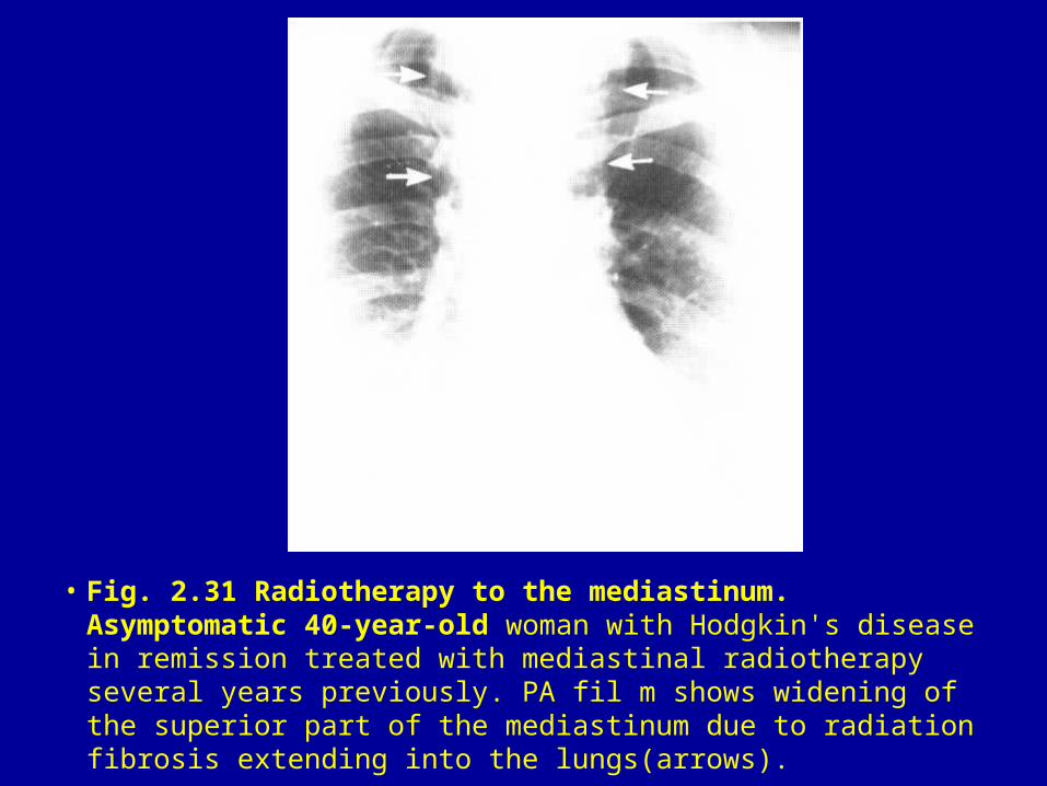
• Fig. 2.31 Radiotherapy to the mediastinum. Asymptomatic 40-year-old woman with Hodgkin's disease in remission treated with mediastinal radiotherapy several years previously. PA fil m shows widening of the superior part of the mediastinum due to radiation fibrosis extending into the lungs(arrows).
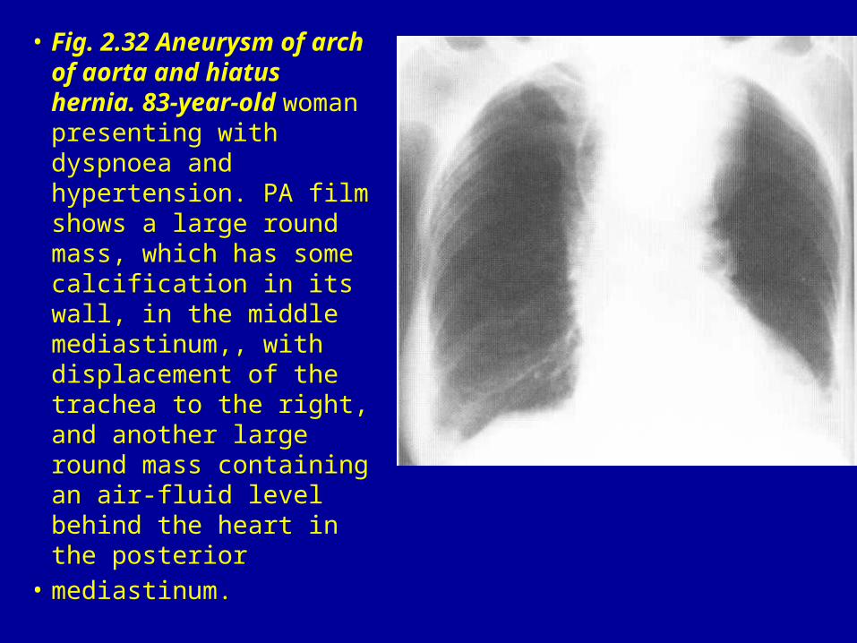
• Fig. 2.32 Aneurysm of arch of aorta and hiatus hernia. 83-year-old woman presenting with dyspnoea and hypertension. PA film shows a large round mass, which has some calcification in its wall, in the middle mediastinum,, with displacement of the trachea to the right, and another large round mass containing an air-fluid level behind the heart in the posterior
• mediastinum.
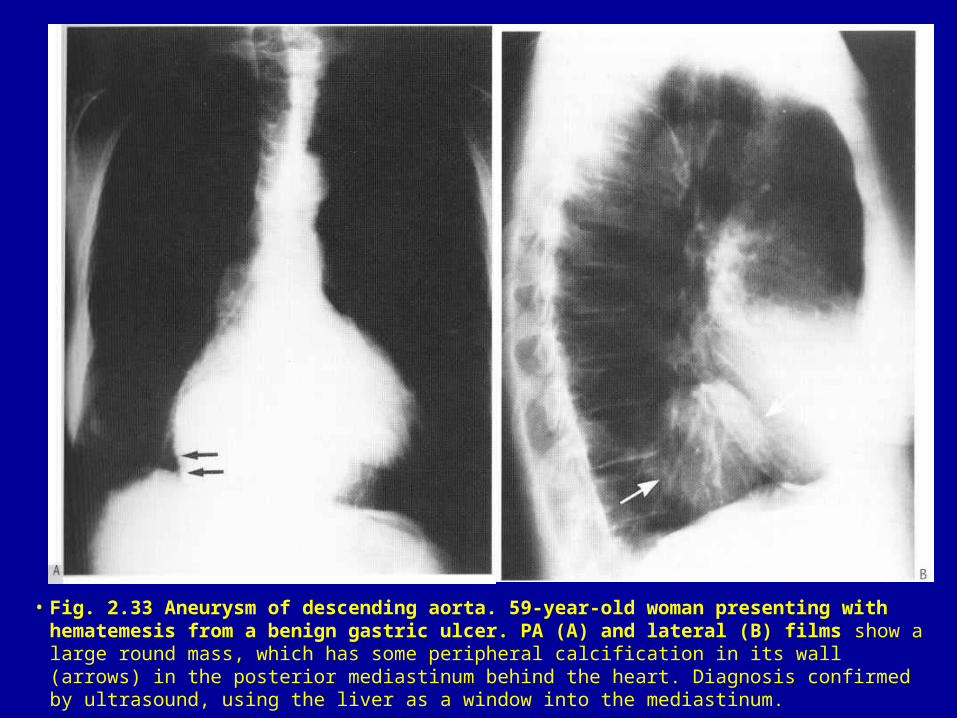
• Fig. 2.33 Aneurysm of descending aorta. 59-year-old woman presenting with hematemesis from a benign gastric ulcer. PA (A) and lateral (B) films show a large round mass, which has some peripheral calcification in its wall (arrows) in the posterior mediastinum behind the heart. Diagnosis confirmed by ultrasound, using the liver as a window into the mediastinum.
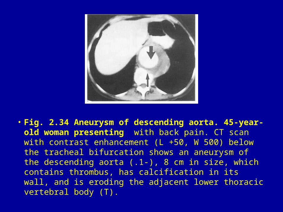
• Fig. 2.34 Aneurysm of descending aorta. 45-year-old woman presenting with back pain. CT scan with contrast enhancement (L +50, W 500) below the tracheal bifurcation shows an aneurysm of the descending aorta (.1-), 8 cm in size, which contains thrombus, has calcification in its wall, and is eroding the adjacent lower thoracic vertebral body (T).
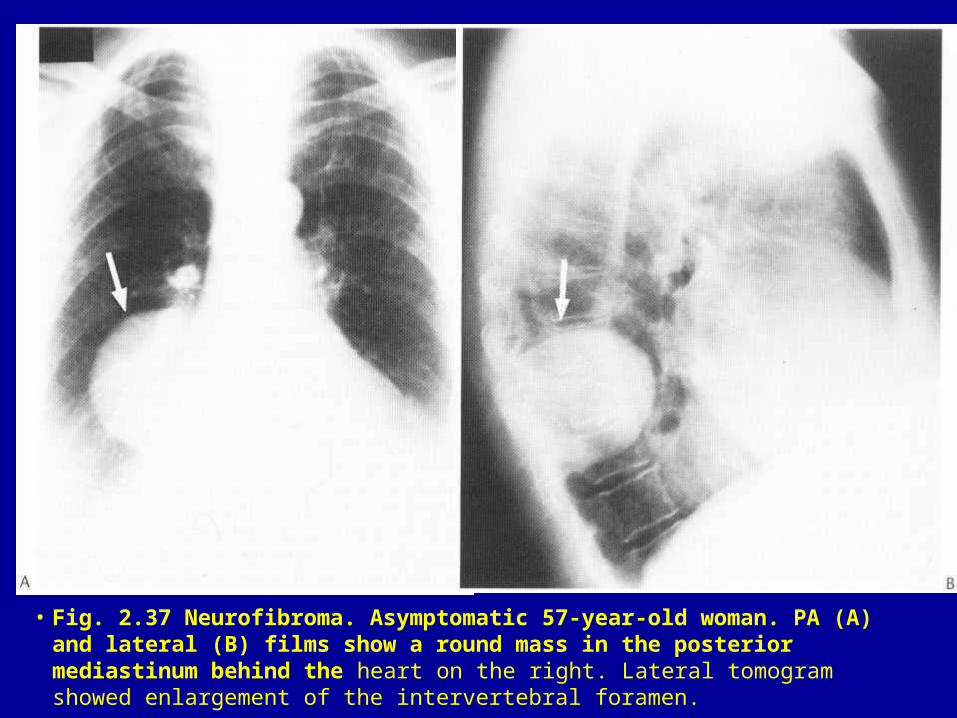
• Fig. 2.37 Neurofibroma. Asymptomatic 57-year-old woman. PA (A) and lateral (B) films show a round mass in the posterior mediastinum behind the heart on the right. Lateral tomogram showed enlargement of the intervertebral foramen.
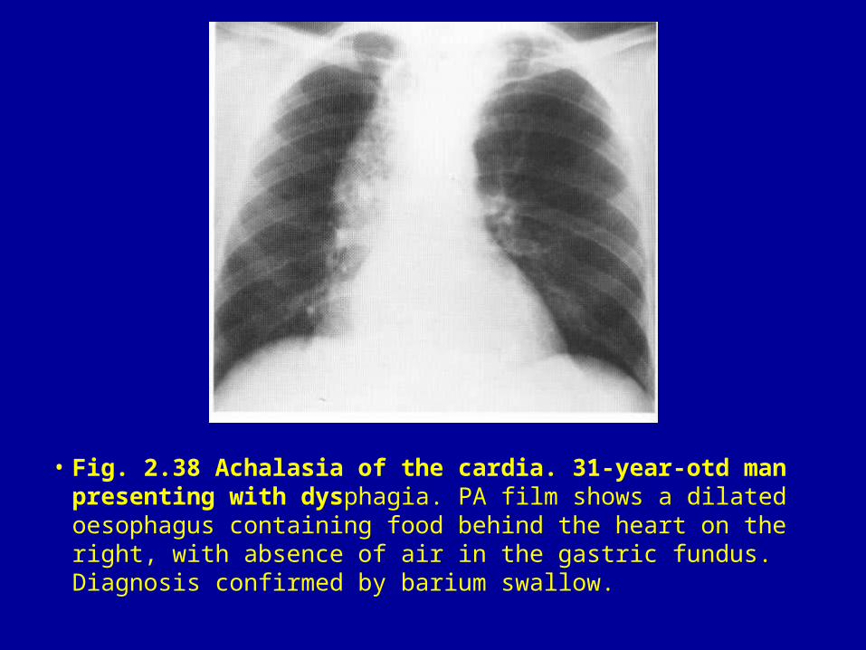
• Fig. 2.38 Achalasia of the cardia. 31-year-otd man presenting with dysphagia. PA film shows a dilated oesophagus containing food behind the heart on the right, with absence of air in the gastric fundus. Diagnosis confirmed by barium swallow.
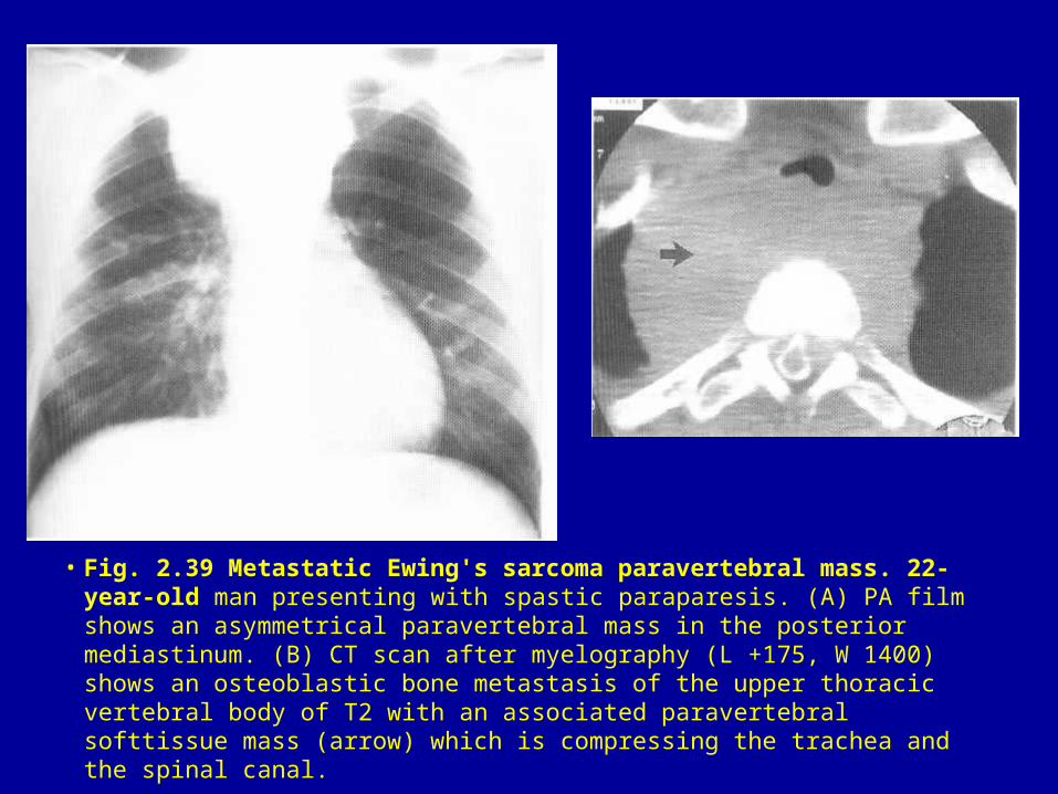
• Fig. 2.39 Metastatic Ewing's sarcoma paravertebral mass. 22-year-old man presenting with spastic paraparesis. (A) PA film shows an asymmetrical paravertebral mass in the posterior mediastinum. (B) CT scan after myelography (L +175, W 1400) shows an osteoblastic bone metastasis of the upper thoracic vertebral body of T2 with an associated paravertebral softtissue mass (arrow) which is compressing the trachea and the spinal canal.
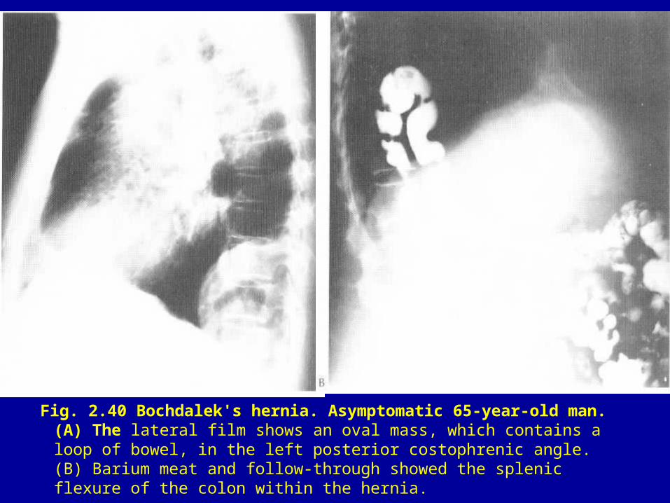
Fig. 2.40 Bochdalek's hernia. Asymptomatic 65-year-old man. (A) The lateral film shows an oval mass, which contains a loop of bowel, in the left posterior costophrenic angle. (B) Barium meat and follow-through showed the splenic flexure of the colon within the hernia.
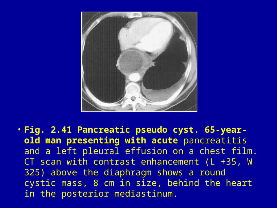
• Fig. 2.41 Pancreatic pseudo cyst. 65-year-old man presenting with acute pancreatitis and a left pleural effusion on a chest film. CT scan with contrast enhancement (L +35, W 325) above the diaphragm shows a round cystic mass, 8 cm in size, behind the heart in the posterior mediastinum.
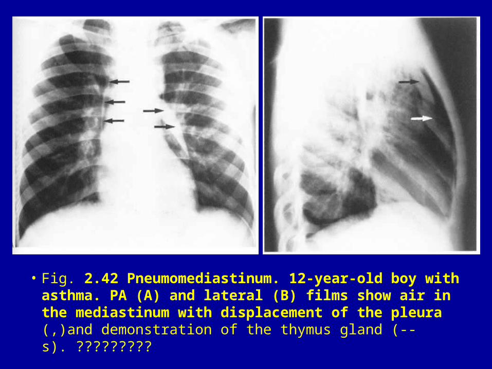
• Fig. 2.42 Pneumomediastinum. 12-year-old boy with asthma. PA (A) and lateral (B) films show air in the mediastinum with displacement of the pleura (,)and demonstration of the thymus gland (--s). ?????????
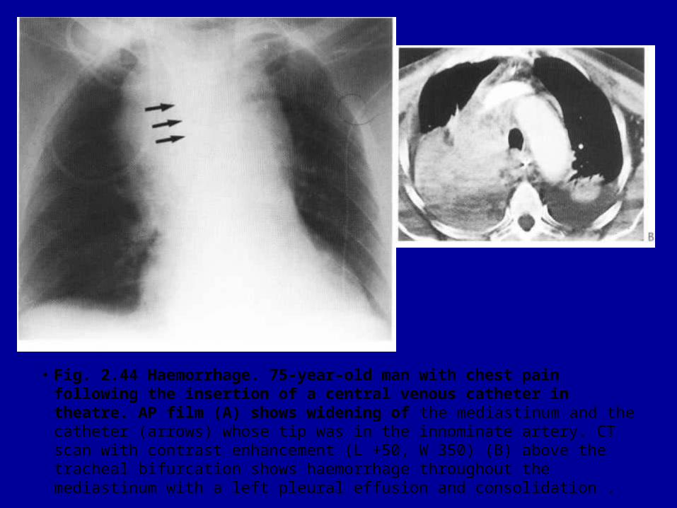
• Fig. 2.44 Haemorrhage. 75-year-old man with chest pain following the insertion of a central venous catheter in theatre. AP film (A) shows widening of the mediastinum and the catheter (arrows) whose tip was in the innominate artery. CT scan with contrast enhancement (L +50, W 350) (B) above the tracheal bifurcation shows haemorrhage throughout the mediastinum with a left pleural effusion and consolidation .
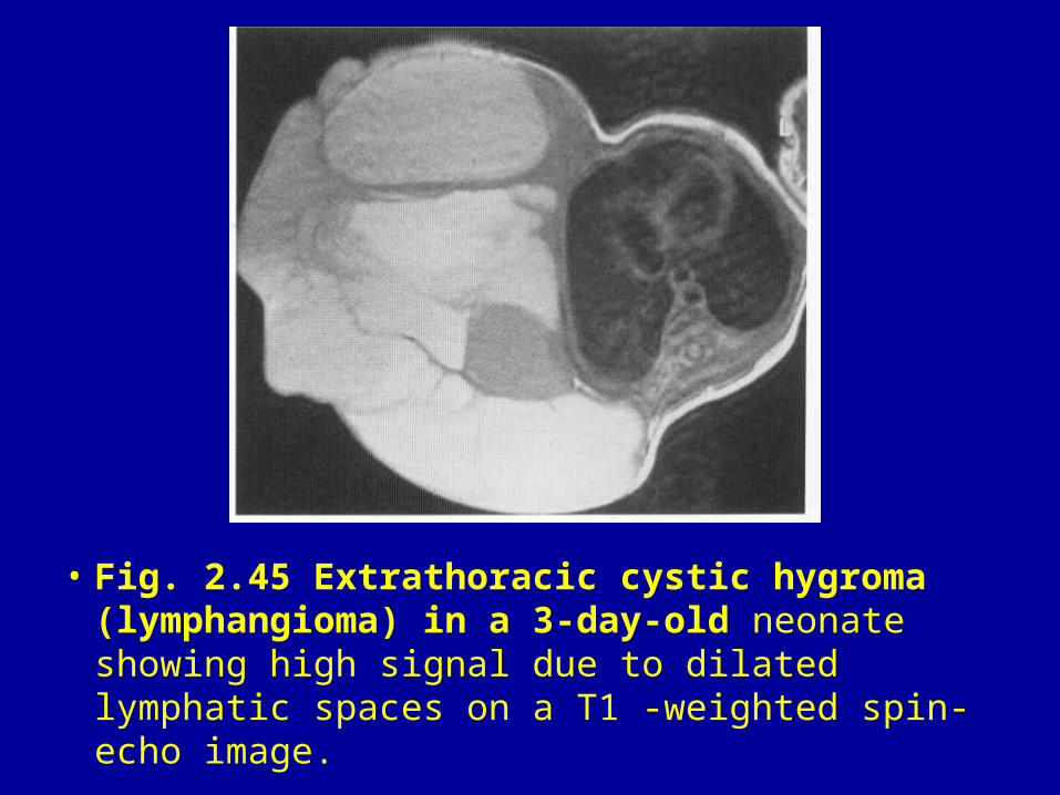
• Fig. 2.45 Extrathoracic cystic hygroma (lymphangioma) in a 3-day-old neonate showing high signal due to dilated lymphatic spaces on a T1 -weighted spin-echo image.
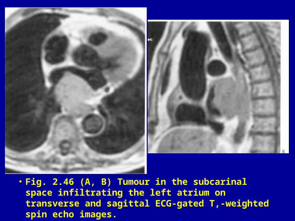
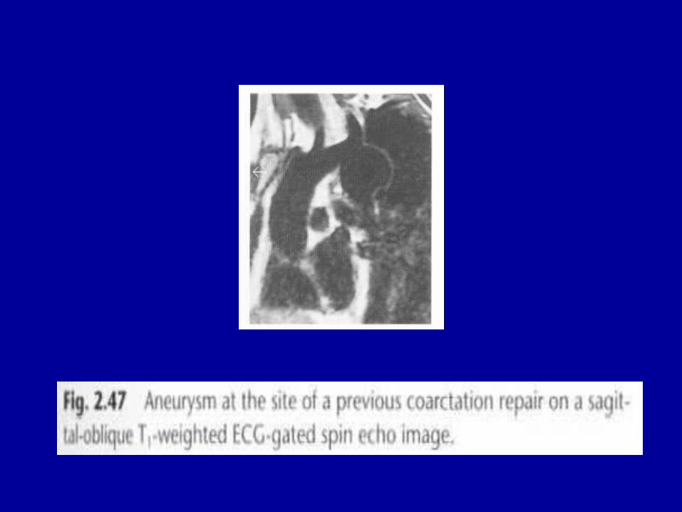
• Fig. 2.46 (A, B) Tumour in the subcarinal space infiltrating the left atrium on transverse and sagittal ECG-gated T,-weighted spin echo images.
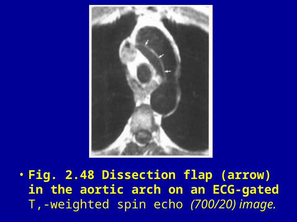
• Fig. 2.48 Dissection flap (arrow) in the aortic arch on an ECG-gated T,-weighted spin echo (700/20) image.
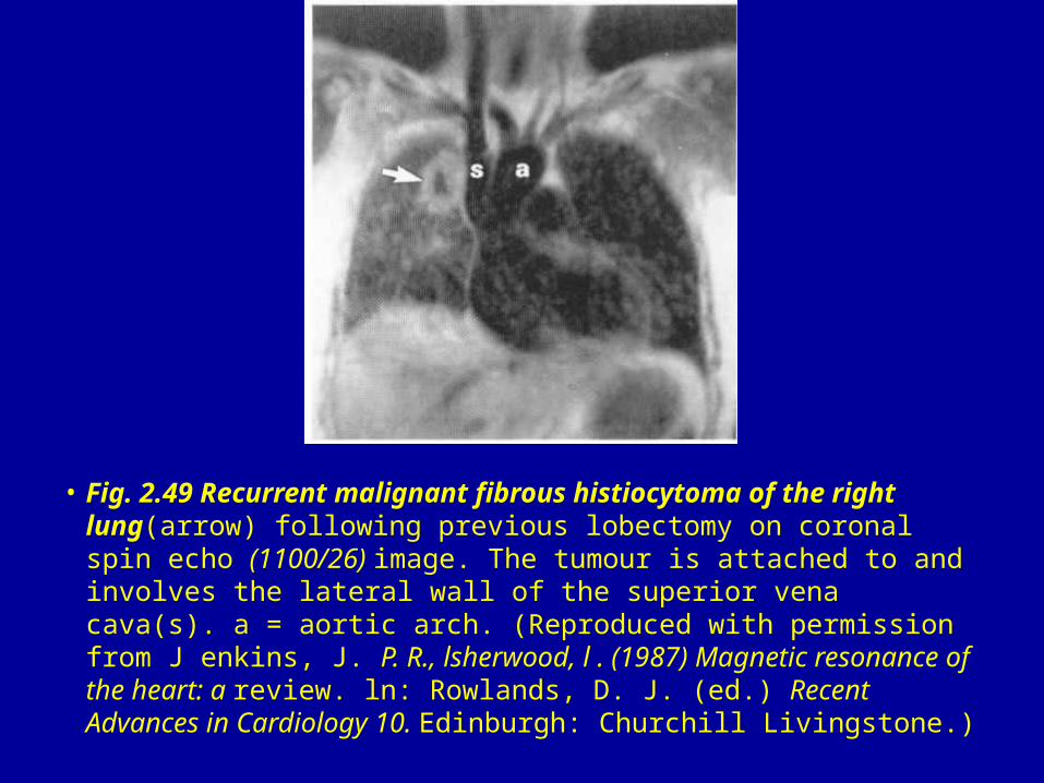
• Fig. 2.49 Recurrent malignant fibrous histiocytoma of the right lung(arrow) following previous lobectomy on coronal spin echo (1100/26) image. The tumour is attached to and involves the lateral wall of the superior vena cava(s). a = aortic arch. (Reproduced with permission from J enkins, J. P. R., lsherwood, l . (1987) Magnetic resonance of the heart: a review. ln: Rowlands, D. J. (ed.) Recent Advances in Cardiology 10. Edinburgh: Churchill Livingstone.)
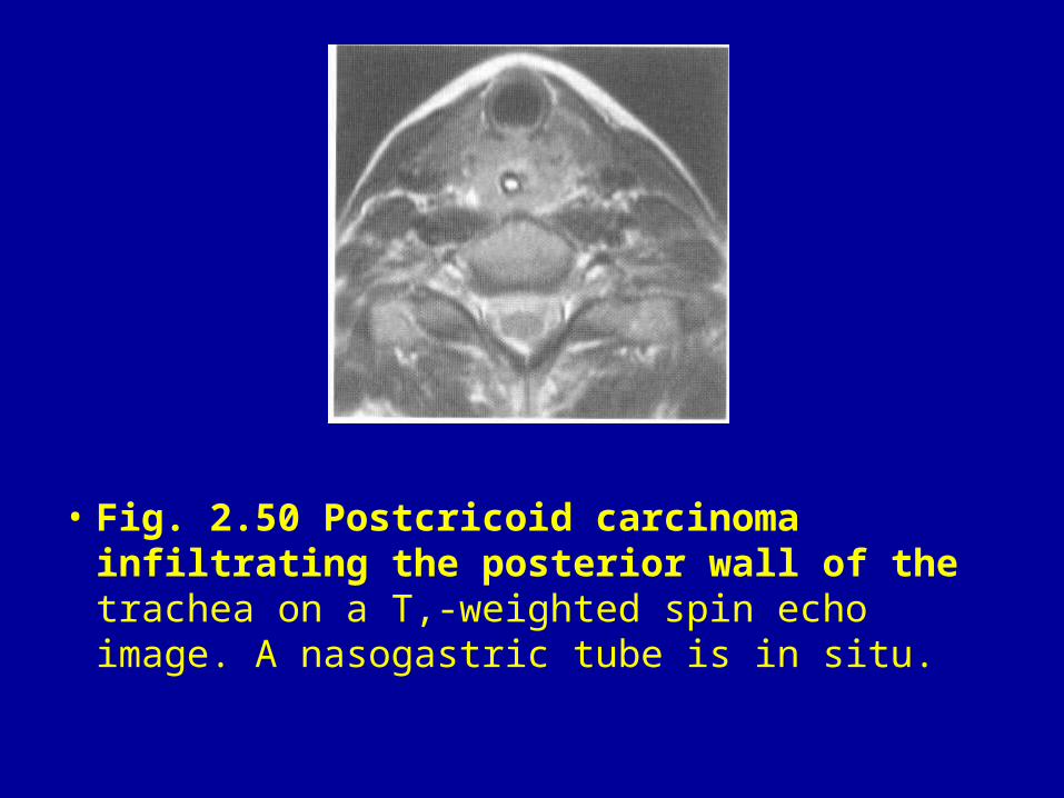
• Fig. 2.50 Postcricoid carcinoma infiltrating the posterior wall of the trachea on a T,-weighted spin echo image. A nasogastric tube is in situ.
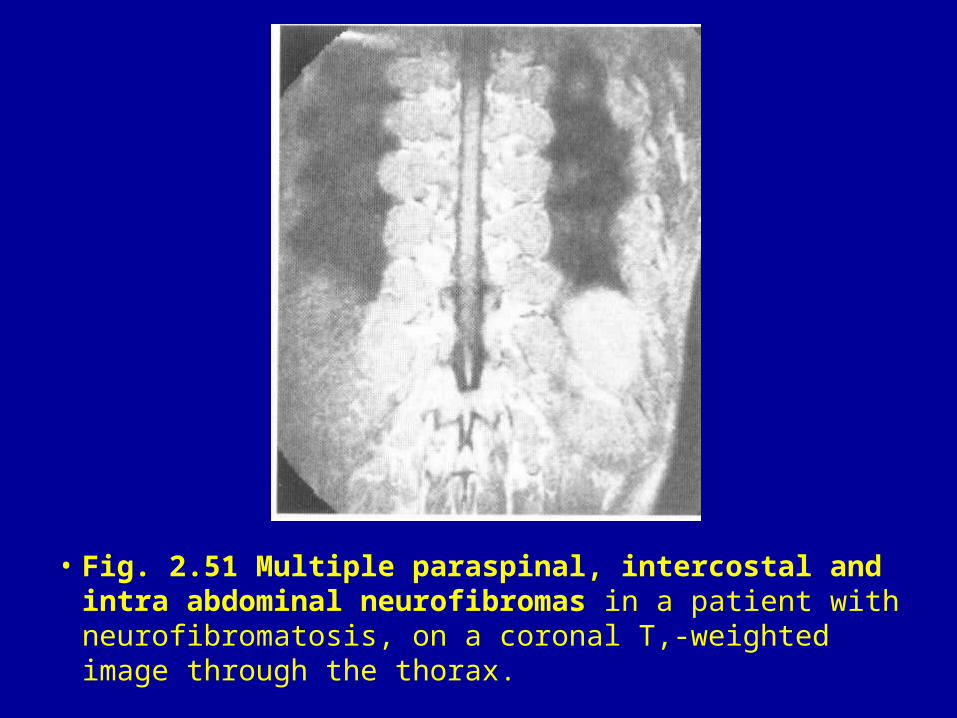
• Fig. 2.51 Multiple paraspinal, intercostal and intra abdominal neurofibromas in a patient with neurofibromatosis, on a coronal T,-weighted image through the thorax.
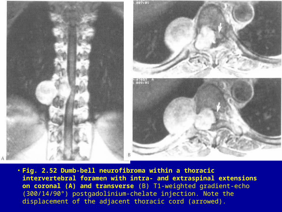
• Fig. 2.52 Dumb-bell neurofibroma within a thoracic intervertebral foramen with intra- and extraspinal extensions on coronal (A) and transverse (B) T1-weighted gradient-echo (300/14/90°) postgadolinium-chelate injection. Note the displacement of the adjacent thoracic cord (arrowed).