ORIGINAL ARTICLE
Virus-induced gene silencing (VIGS)-mediated functionalcharacterization of two genes involved in lignocellulosic secondarycell wall formation
Shashank K. Pandey1• Akula Nookaraju1,2,4
• Takeshi Fujino1• Sivakumar Pattathil3 •
Chandrashekhar P. Joshi1,2
Received: 11 April 2016 / Accepted: 9 August 2016
� Springer-Verlag Berlin Heidelberg 2016
Abstract
Key message Functional characterization of two
tobacco genes, one involved in xylan synthesis and the
other, a positive regulator of secondary cell wall for-
mation, is reported.
Abstract Lignocellulosic secondary cell walls (SCW)
provide essential plant materials for the production of
second-generation bioethanol. Therefore, thorough under-
standing of the process of SCW formation in plants is
beneficial for efficient bioethanol production. Recently, we
provided the first proof-of-concept for using virus-induced
gene silencing (VIGS) approach for rapid functional
characterization of nine genes involved in cellulose,
hemicellulose and lignin synthesis during SCW formation.
Here, we report VIGS-mediated functional characterization
of two tobacco genes involved in SCW formation. Stems of
VIGS plants silenced for both selected genes showed
increased amount of xylem formation but thinner cell walls
than controls. These results were further confirmed by
production of stable transgenic tobacco plants manipulated
in expression of these genes. Stems of stable transgenic
tobacco plants silenced for these two genes showed
increased xylem proliferation with thinner walls, whereas
transgenic tobacco plants overexpressing these two genes
showed increased fiber cell wall thickness but no change in
xylem proliferation. These two selected genes were later
identified as possible members of DUF579 family involved
in xylan synthesis and KNAT7 transcription factor family
involved in positive regulation of SCW formation,
respectively. Glycome analyses of cell walls showed
increased polysaccharide extractability in 1 M KOH
extracts of both VIGS-NbDUF579 and VIGS-NbKNAT7
lines suggestive of cell wall loosening. Also, VIGS-
NbDUF579 and VIGS-NbKNAT7 lines showed increased
saccharification rates (74.5 and 40 % higher than controls,
respectively). All these properties are highly desirable for
producing higher quantities of bioethanol from lignocel-
lulosic materials of bioenergy plants.
Keywords Secondary cell wall (SCW) � Saccharification �Bioethanol production � Transcriptional regulation � Virus-induced gene silencing (VIGS) � Xylan synthesis
Introduction
Plant cells are delimited by cell walls. The primary cell
wall mainly consists of cellulose, hemicellulose, pectin and
glycoproteins, whereas some specialized cells like xylem
vessels, tracheids and fibers contain an additional wall, the
secondary cell wall (SCW) that is largely composed of
Communicated by E. Guiderdoni.
S. K. Pandey and A. Nookaraju contributed equally to this work.
Electronic supplementary material The online version of thisarticle (doi:10.1007/s00299-016-2039-2) contains supplementarymaterial, which is available to authorized users.
& Chandrashekhar P. Joshi
1 Department of Bioenergy Science and Technology, Chonnam
National University, Gwangju 500-757, South Korea
2 Department of Biological Sciences and School of Forest
Resources and Environmental Science, Michigan
Technological University, Houghton, MI 49931, USA
3 Complex Carbohydrate Research Center, University of
Georgia, 31, Riverbend Road, Athens, GA 30602, USA
4 Present Address: Kaveri Seed Company Ltd., Minerva
Complex, Secunderabad 500003, India
123
Plant Cell Rep
DOI 10.1007/s00299-016-2039-2

cellulose, hemicellulose and lignin. The middle lamella,
rich in pectins, joins and holds adjacent plant cells together.
Cell walls play many important roles in plant’s life: helping
in cell adhesion and expansion, acting as a barrier to attack
by pests and pathogens, and determining the physical
properties of the plant (Braam 1999; Jones and Takemoto
2004; Scheible and Pauly 2004; Vorwerk et al. 2004).
Apart from providing dietary fiber for human and animal
consumption and raw materials for textile, paper and pulp
industries, plant cell walls also play an important role in
human lives by providing raw materials for the production
of biofuels (Nookaraju et al. 2013). Yong et al. (2005)
estimated that more than 1000 genes encoding cell wall-
related proteins could be involved in cell wall formation,
and that an equal number of unannotated genes might be
functional in cell wall assembly and disassembly in plants.
Virus-induced gene silencing (VIGS) is a simple, rapid and
highly efficient approach for quick assessment of plant
gene functions. It is a transient RNAi-mediated gene
silencing method that facilitates fast and easy assessment
of gene function that could be later confirmed by gener-
ating stable transgenics (Burch-Smith et al. 2004). VIGS
system has been successfully used for addressing biological
questions related to plant defense, development, and
metabolism in many plant species (Lu et al. 2003; Burch-
Smith et al. 2004; Robertson 2004). Previously, we suc-
cessfully used this system for the first time to silence Ni-
cotiana benthamiana (N. benthamiana) homologues of
nine Arabidopsis cell wall genes (three genes each
involved in cellulose, xylan and lignin pathways) and
demonstrated that VIGS plants exhibited the expected
morphological phenotypes, altered anatomy and cell wall
chemistry (Zhu et al. 2010). In the present study, we
employed the same tobacco rattle virus (TRV)-based VIGS
system for further understanding the function of two genes
involved in SCW formation. Furthermore, we also char-
acterized the functions of these two genes by generating
stable RNAi and overexpression transgenic lines.
By surveying the literature on xylem development in
various plant species, we first identified 38 gene targets for
VIGS screening. This list included genes encoding various
unknown function proteins and some transcription factors.
Among these, two genes when silenced using VIGS tech-
nique showed interesting phenotype of massive xylem
proliferation and were selected for further functional
studies. These two genes were later identified as members
of DUF579 family and KNAT7 transcription factor family,
respectively, based on their homology with Arabidopsis
genes. While this work was in progress, the functions of
these two genes were reported in Arabidopsis (Brown et al.
2011; Jensen et al. 2011; Li et al. 2012). Based on these
reports, the DUF579 gene appears to be involved in xylan
synthesis and KNAT7 gene is likely to be a negative
regulator of SCWdeposition. However, the phenomenon of
xylem proliferation that we observed in tobacco was not
observed by silencing either of these genes in Arabidopsis.
In the present study, we observed that gene NbDUF579 is
involved in xylan synthesis butNbKNAT7 gene is a positive
regulator of SCW formation in tobacco. Furthermore,
downregulation of these two genes in transgenic tobacco,
through VIGS and RNAi system, resulted in decreased cell
wall thickness and increased xylem proliferation with
overall alteration in cell wall extractability as observed by
glycome analysis and increase in sugar release (sacchari-
fication) efficiency. Whereas overexpression lines of
NbDUF579 and NbKNAT7 genes showed increased cell
wall thickness compared to control plants, but no alteration
in xylem proliferation was observed. All these observations
have direct implications in efficient bioethanol production
(Nookaraju et al. 2013).
Materials and methods
VIGS vector construction
Tobacco (N. benthamiana) orthologs of Arabidopsis
DUF579 and KNAT7 genes were PCR-amplified using
gene-specific primers (Table S1). Resulting gene fragments
were cloned into VIGS vector pTRV2-LIC, and transferred
to Agrobacterium strain GV2260 using freeze–thaw
method (Dong et al. 2007).
Plant transformation and growth conditions
For VIGS experiment, N. benthamiana plants were grown
under continuous light at 25 ± 1 �C for 3 weeks.
Agrobacterium strain GV2260 containing pTRV1, pTRV2-
LIC (control), and modified pTRV2-LIC vectors for
silencing gene NbDUF579 and NbKNAT7 was grown in
Luria broth (LB) medium containing kanamycin
(50 mg L-1) and rifampicin (25 mg L-1) at 28 �C for
overnight. Next day, cells were pelleted and re-suspended
in the infiltration medium (10 mM MES, 10 mM MgCl2,
200 lM acetosyringone) to a final OD600 of 1.0. Prior to
infiltration, Agrobacterium cell suspensions were incubated
at room temperature for 3 h (Zhu and Dinesh-Kumar
2008). Then, tobacco leaves were infiltrated with 1:1 cell
suspension of Agrobacterium carrying pTRV1 and modified
pTRV2-LIC vector with a needleless syringe through leaf
abaxial surface as described earlier (Zhu et al. 2010).
Control plants were infiltrated with 1:1 mixture of pTRV1
and pTRV2-LIC control vector. Infiltrated plants were
grown at 25 ± 1 �C under continuous light. The experi-
ment was repeated at least three times with four plants at
each time.
Plant Cell Rep
123

Generation of stable transgenic lines
For RNAi study, NbDUF579 and NbKNAT7 gene fragments
were PCR-amplified from N. benthamiana cDNA with
gene-specific primers (Table S1) that contain XhoI ? XbaI
and KpnI ? ClaI restriction sites on either sides, respec-
tively, and cloned into the pTOP Blunt V2 vector (Enzy-
nomics, Korea). After sequence confirmation, both the
gene fragments were digested and sub-cloned into sense
and antisense direction in pHANNIBAL vector as described
earlier (Wang et al. 2004). Resulting pHANNIBAL con-
structs were digested with NotI, and RNAi cassettes were
cloned into pART27 binary vector used for plant transfor-
mation. Plasmid constructs of pART-NbDUF579 (pART27
vector with gene NbDUF579) and pART-NbKNAT7
(pART27 vector with gene NbKNAT7) were transformed
into Agrobacterium tumefaciens strain GV2260 by freeze–
thaw method.
For overexpression, full-length N. benthamiana cDNAs
for NbDUF579 and NbKNAT7 genes were PCR-amplified
using gene-specific primers (Table S1) that contain XbaI
and SacI restriction sites on either sides and sub-cloned
into pTOP Blunt V2 vector (Enzynomics, Korea). The
plasmids were digested with XbaI and SacI to obtain full-
length cDNAs of NbDUF579 and NbKNAT7 genes, and
were cloned into pBIN121 binary vector replacing b-glu-curonidase (GUS) driven by CaMV 35S promoter. Plant
transformation of tobacco (N. benthamiana) was carried
out using leaf disks as explants by Agrobacterium-medi-
ated gene transfer method (Pogrebnyak et al. 2005).
Transgenic shoots were selected on kanamycin
(50 lg mL-1) and were rooted on half-strength MS med-
ium containing kanamycin (50 lg mL-1). The rooted
shoots were then transferred to commercial potting mixture
(3:2:1, peat:soil:perlite; Heung Nung Seeds, Korea), and
the hardened plants were established in a growth room
maintained at 25 ± 1 �C temperature and 50 % relative
humidity.
RNA extraction and RT-PCR analysis
Total RNA was extracted from the stems (1–7 internodes
counted from the base in plants with seven internodes)
using RNeasy plant mini kit (Qiagen). First-strand cDNA
was synthesized from 1 lg of total RNA using SuperScript
reverse transcriptase kit (Invitrogen). qRT-PCR was per-
formed using SYBR Green kit (Takara, Japan). The tran-
script abundance of all the genes was examined using gene-
specific primers shown in Table S1.
Microscopic image analysis
For light microscopy, stems of VIGS plants were cut at the
third internode from the base of plants with seven intern-
odes and were fixed in 4 % PFA (paraformaldehyde)
solution overnight. After fixation, internodes were dehy-
drated through ethanol and t-butanol series, and finally
embedded in wax. Five-micrometer-thick sections were cut
with a microtome, mounted on a slide and de-waxed. De-
waxed sections were stained with Toluidine Blue O (TBO)
and photographed using a light microscope. For measuring
cell wall thickness, stem tissues were prepared in LR-white
resin (London Resin Co., England) and sectioned at 0.5 lmusing ultramicrotome and photographed.
Immunolocalization of xylan and xyloglucan were car-
ried out using LM10 and LM15 monoclonal antibodies
(Plant Probes) that recognize unsubstituted and relatively
low substituted xylans (McCartney et al. 2005) and the
XXXG motif of xyloglucan (Marcus et al. 2008), respec-
tively, followed by incubation with fluorescein isothio-
cyanate (FITC)-conjugated secondary antibody (Sigma).
The FITC-labeled sections were observed and pho-
tographed under a Zeiss Axiovert 200M inverted micro-
scope equipped with an AxioCam fluorescence camera.
Glycosyl compositional analysis by GC–MS of TMS
derivatives of methyl glycosides
Glycosyl compositional analysis was performed by com-
bined gas chromatography/mass spectrometry (GC/MS) of
the per-O-trimethylsilyl (TMS) derivatives of the
monosaccharide methyl glycosides produced from the wood
sample (stems consisting of seven internodes from the base)
by acidic methanolysis as described earlier (Merkle and
Poppe 1994; York et al. 1986). The sample (400–500 lgeach) was placed in a separate tube with 20 lg of inositol asinternal standard. Dry samples were hydrolyzed with 2 M
TFA at 120 �C for 1 h. Hydrolysis products were dried by
adding isopropanol. Methyl glycosides were then prepared
from the dry sample by methanolysis with 1 M HCl
methanol at 80 �C (16 h), followed by re-N-acetylation with
pyridine and acetic anhydride in methanol (for detection of
amino sugars). The samples were then per-O-trimethylsily-
lated by treatment with Tri-Sil (Pierce) at 80 �C (30 min).
GC/MS analysis of the TMS methyl glycosides was per-
formed on an Agilent 7890A GC interfaced to a 5975C
MSD, using a Supelco EC-1 fused-silica capillary column
(30 m 9 0.25 mm ID). Data were collected and processed
by Agilent ChemStation software.
Plant Cell Rep
123
Sequential extraction and glycome profiling
Sequential extractions of cell walls and glycome profiling
of plants were carried out according to the procedure
described earlier (Pattathil et al. 2012; Zhu et al. 2010;
DeMartini et al. 2011). Tobacco stem samples consisting of
seven internodes from base were used for the analysis.
Plant glycan-directed monoclonal antibodies (McAbs)
were obtained from laboratory stocks (CCRC, JIM and
MAC series) at the Complex Carbohydrate Research
Center (available through CarboSource Services; http://
www.carbosource.net) and BioSupplies (Australia) (BG1,
LAMP). A detailed list of all McAbs used in this study has
been described earlier (Pattathil et al. 2015).
Lignin analysis
Tobacco stem samples consisting of seven internodes from
base were used for the analysis. Tobacco stem tissue
powder samples were weighed (5 mg) in a small sample
cup; pyrolyzed using a Pyrolyzer at 500 �C, and the resi-
dues were analyzed using molecular beam mass spec-
trometer (Evans and Milne 1987). Each sample was
analyzed in duplicates. The uncorrected lignin content was
calculated using multivariate analysis (principal compo-
nent analysis) and corrected with NIST 8492
(lignin = 26.2 %).
Cellulose estimation and sugar release
Tobacco stem samples consisting of seven internodes from
base were used for the analysis. Tobacco stem tissue
powders were treated with acetonitrile solution (acetic
acid:nitric acid:water 8:2:1) at 100 �C for 30 min (Upde-
graff 1969). Then, the tissue powders were washed three
times with sterile water and dried. The dried residue con-
taining mostly cellulose was weighed and expressed in
percentage. Sugar release was calculated using phenol–
sulfuric acid method by monitoring the reactants’ solution
absorbance at 490 nm (Dubois et al. 1956). A glucose
standard curve was prepared using 2–22 lg lL-1, and
glucose yields of tissue samples were calculated.
Phylogenetic analysis
Protein sequences of Arabidopsis for the two genes were
obtained from The Arabidopsis Information Resource
(TAIR; http://www.arabidopsis.org/), based on TAIR gene
family annotations, and by BLASTP searches using
KNAT7 and DUF579 as the queries. N. benthamiana
protein sequences were retrieved by N.benthamiana Gen-
ome v1.0.1 Contigs (solgenomics.net/tools/blast/in-
dex.pl?db_id = 196). The draft genome sequence of
Nicotiana benthamiana has been published (Bombarely
et al. 2012). Protein sequences were aligned with Clustal
Omega, and phylogenetic trees were generated using
MEGA7 software. The evolutionary history was inferred
using the neighbor-joining method. The percentage of
replicate trees in which the associated taxa clustered
together in the bootstrap test (500 replicates) is shown next
to the branches. The tree is drawn to scale, with branch
lengths in the same units as those of the evolutionary dis-
tances used to infer the phylogenetic tree. The evolutionary
distances were computed using the Poisson correction
method and are in the units of the number of amino acid
substitutions per site.
Statistical analysis
Data from all experiments were analyzed statistically using
one-way analysis of variance (ANOVA) followed by
Scheffe’s test for multiple comparisons (Scheffe 1959).
Results and discussion
Gene selection and study of expression of selected
genes in tobacco
Among the 38 genes selected initially for functional
screening using VIGS, two genes NbDUF579 and
NbKNAT7 showed interesting phenotype of thinner fiber
walls and increased xylem formation. Both the genes
showed differential expression in various tissues of tobacco
plants (Fig. S1). NbDUF579 gene showed higher expres-
sion in older stems (OS) followed by younger stems (YS).
The young leaf (YL), old leaf (OL) and root samples
showed moderate expression, while flower (F) and petiole
(P) samples showed very low expression (Fig. S1A). The
NbKNAT7 gene showed higher expression in old leaf (OL)
followed by old stem (OS), young leaf (YL) and root
(R) (Fig. S1B). Amino acid-based alignment of Ara-
bidopsis and tobacco DUF579 genes indicates that the two
close members in tobacco, DUF579 and DUF579-L are
closely related to two Arabidopsis DUF579 gene family
members, irx15 and irx15-L (Fig. S2A). Similarly, tobacco
NbKNAT7 gene is placed very close to its Arabidopsis
counterpart (Fig. S2B).
Silencing of genes NbDUF579 and NbKNAT7 showed
increased xylem formation and reduced wall
thickness
To understand the functions of these genes, VIGS experi-
ments were independently repeated followed by reconfir-
mation of those results by stable transformation of tobacco
Plant Cell Rep
123

plants with RNAi and overexpression constructs. The VIGS
plants silenced for expression of genes, NbDUF579 and
NbKNAT7 did not show any morphological variations and
were highly similar to control plants in terms of plant
height, internode number and stem girth (Fig. 1a, b). This
is a highly desirable trait since transgenic plants altered in
cell wall properties sometimes show negative effects on
overall plant growth (e.g., Joshi et al. 2011). However, as
expected, qRT-PCR expression results showed a significant
downregulation of these genes in VIGS plants as compared
to control (Fig. 1c). To examine whether similar to initial
screening, these VIGS plants also have any changes in their
xylem phenotype over control plants, the stems were hand-
sectioned at third internode from base and examined under
light microscope. We confirmed increased xylem formation
(increased number of cell layers and higher proportion of
xylem area) in stems of VIGS plants as compared to con-
trol plants, similar to what was observed in our preliminary
studies, and the increase in xylem area was statistically
significant (Fig. 2a). There were about 55 and 49 %
(C)2.5
0.0
0.5
1.0
1.5
2.0
0.0
0.5
1.0
1.5
2.0
Rel
ativ
e m
RN
A e
xpre
ssio
n
(A)
pTRV2-LIC V_NbDUF579-1 V_NbDUF579-2
Pla
nt h
eigh
t (cm
)
0
10
20
30
40
Ste
m g
irth
(mm
)
0
5
10
15
20N
umbe
r of i
nter
node
s
0
2
4
6
8
10(B)
pTRV2-LIC V_NbKNAT7-1 V_NbKNAT7-2
V_NbDUF579 V_NbKNAT7
a
b
c bcc
a
c
b
cc
Fig. 1 Morphological analysis of tobacco plants silenced for
NbDUF579 and NbKNAT7 genes through VIGS system. a Phenotype
of VIGS-silenced plants, b analysis of plant height, stem girth and
number of internodes of V_NbDUF579 (VIGS_NbDUF579) and
V_NbKNAT7 (VIGS_NbKNAT7) and vector control (pTRV2-LIC)
plants. c Relative mRNA expression levels of genes NbDUF579
(a) and NbKNAT7 (b) in stems of VIGS plants at 30 days after
infiltration (DAI) indicating the reduction in transcript level. Error
bars represent standard error (SE) of three independent experiments.
Gene expression levels were compared with actin control. Bars
denoted by the same letter are not significantly different (p = 0.01,
ANOVA, post hoc Scheffe’s test)
Plant Cell Rep
123

(A)V_NbDUF579pTRV2-LIC
V_NbKNAT7
Pro
porti
on o
f xyl
em a
rea
a a
b
0
20
40
60
80
(B) pTRV2-LIC
bb
a
0
0.5
1.0
1.5
2.5
2.0
Fibe
r wal
l thi
ckne
ss (µ
m)
V_NbDUF579
V_NbKNAT7
Fig. 2 Anatomical analysis of VIGS-silenced vs. vector control
plants. a TBO-stained transverse sections (TS) of stems at third
internode from base and proportion of xylem area in V_NbDUF579
(VIGS_NbDUF579) and V_NbKNAT7 (VIGS_NbKNAT7) plants as
compared to control. Scale bars 200 lm. Bars are mean ± SE of 12
samples. Bars denoted by the same letter are not significantly
different (p = 0.01, ANOVA, post hoc Scheffe’s test). b Fiber wall
thickness in stems of 6-week-old control (pTRV2-LIC) and VIGS-
silenced NbDUF579 and NbKNAT7 plants. Scale bars 5 lm. Bars are
mean ± SE of 60 observations. Bars denoted by the same letter are
not significantly different (p = 0.01, ANOVA, post hoc Scheffe’s
test)
Plant Cell Rep
123
increases in xylem area in stems of VIGS plants silenced
for genes NbDUF579 and NbKNAT7, respectively, over
control stems. At the same time, xylem fiber wall thickness
was reduced by 35 and 42 % in VIGS lines silenced for the
genes NbDUF579 and NbKNAT7, respectively, as com-
pared to control plants (Fig. 2b). Increased xylem prolif-
eration in gene-silenced lines could be partially attributed
as a compensatory mechanism to reduced fiber cell wall
thickness. Similar reduction in fiber cell wall thickness and
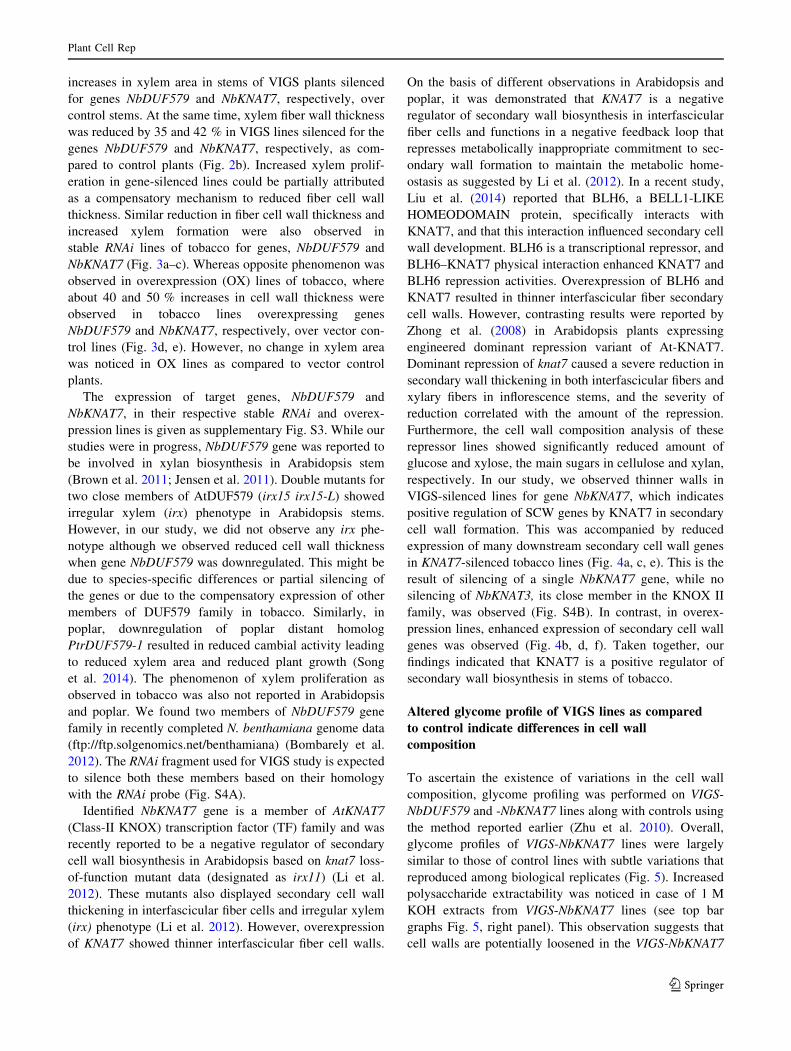
increased xylem formation were also observed in
stable RNAi lines of tobacco for genes, NbDUF579 and
NbKNAT7 (Fig. 3a–c). Whereas opposite phenomenon was
observed in overexpression (OX) lines of tobacco, where
about 40 and 50 % increases in cell wall thickness were
observed in tobacco lines overexpressing genes
NbDUF579 and NbKNAT7, respectively, over vector con-
trol lines (Fig. 3d, e). However, no change in xylem area
was noticed in OX lines as compared to vector control
plants.
The expression of target genes, NbDUF579 and
NbKNAT7, in their respective stable RNAi and overex-
pression lines is given as supplementary Fig. S3. While our
studies were in progress, NbDUF579 gene was reported to
be involved in xylan biosynthesis in Arabidopsis stem
(Brown et al. 2011; Jensen et al. 2011). Double mutants for
two close members of AtDUF579 (irx15 irx15-L) showed
irregular xylem (irx) phenotype in Arabidopsis stems.
However, in our study, we did not observe any irx phe-
notype although we observed reduced cell wall thickness
when gene NbDUF579 was downregulated. This might be
due to species-specific differences or partial silencing of
the genes or due to the compensatory expression of other
members of DUF579 family in tobacco. Similarly, in
poplar, downregulation of poplar distant homolog
PtrDUF579-1 resulted in reduced cambial activity leading
to reduced xylem area and reduced plant growth (Song
et al. 2014). The phenomenon of xylem proliferation as
observed in tobacco was also not reported in Arabidopsis
and poplar. We found two members of NbDUF579 gene
family in recently completed N. benthamiana genome data
(ftp://ftp.solgenomics.net/benthamiana) (Bombarely et al.
2012). The RNAi fragment used for VIGS study is expected
to silence both these members based on their homology
with the RNAi probe (Fig. S4A).
Identified NbKNAT7 gene is a member of AtKNAT7
(Class-II KNOX) transcription factor (TF) family and was
recently reported to be a negative regulator of secondary
cell wall biosynthesis in Arabidopsis based on knat7 loss-
of-function mutant data (designated as irx11) (Li et al.
2012). These mutants also displayed secondary cell wall
thickening in interfascicular fiber cells and irregular xylem
(irx) phenotype (Li et al. 2012). However, overexpression
of KNAT7 showed thinner interfascicular fiber cell walls.
On the basis of different observations in Arabidopsis and
poplar, it was demonstrated that KNAT7 is a negative
regulator of secondary wall biosynthesis in interfascicular
fiber cells and functions in a negative feedback loop that
represses metabolically inappropriate commitment to sec-
ondary wall formation to maintain the metabolic home-
ostasis as suggested by Li et al. (2012). In a recent study,
Liu et al. (2014) reported that BLH6, a BELL1-LIKE
HOMEODOMAIN protein, specifically interacts with
KNAT7, and that this interaction influenced secondary cell
wall development. BLH6 is a transcriptional repressor, and
BLH6–KNAT7 physical interaction enhanced KNAT7 and
BLH6 repression activities. Overexpression of BLH6 and
KNAT7 resulted in thinner interfascicular fiber secondary
cell walls. However, contrasting results were reported by
Zhong et al. (2008) in Arabidopsis plants expressing
engineered dominant repression variant of At-KNAT7.
Dominant repression of knat7 caused a severe reduction in
secondary wall thickening in both interfascicular fibers and
xylary fibers in inflorescence stems, and the severity of
reduction correlated with the amount of the repression.
Furthermore, the cell wall composition analysis of these
repressor lines showed significantly reduced amount of
glucose and xylose, the main sugars in cellulose and xylan,
respectively. In our study, we observed thinner walls in
VIGS-silenced lines for gene NbKNAT7, which indicates
positive regulation of SCW genes by KNAT7 in secondary
cell wall formation. This was accompanied by reduced
expression of many downstream secondary cell wall genes
in KNAT7-silenced tobacco lines (Fig. 4a, c, e). This is the
result of silencing of a single NbKNAT7 gene, while no
silencing of NbKNAT3, its close member in the KNOX II
family, was observed (Fig. S4B). In contrast, in overex-
pression lines, enhanced expression of secondary cell wall
genes was observed (Fig. 4b, d, f). Taken together, our
findings indicated that KNAT7 is a positive regulator of
secondary wall biosynthesis in stems of tobacco.
Altered glycome profile of VIGS lines as compared
to control indicate differences in cell wall
composition
To ascertain the existence of variations in the cell wall
composition, glycome profiling was performed on VIGS-
NbDUF579 and -NbKNAT7 lines along with controls using
the method reported earlier (Zhu et al. 2010). Overall,
glycome profiles of VIGS-NbKNAT7 lines were largely
similar to those of control lines with subtle variations that
reproduced among biological replicates (Fig. 5). Increased
polysaccharide extractability was noticed in case of 1 M
KOH extracts from VIGS-NbKNAT7 lines (see top bar
graphs Fig. 5, right panel). This observation suggests that
cell walls are potentially loosened in the VIGS-NbKNAT7
Plant Cell Rep
123

(B)
VC-RNAi NbDUF579-RNAi
Pro
porti
on o
f xyl
em (%
) a a
c
bbbcb
c
0
20
40
60
80a a
bbc
bcbcbcc
0
0.5
1.0
1.5
2.5
2.0
3.0
Fibe
r wal
l thi
ckne
ss (µ
m)
pBI121 NbDUF579-OX
0
0.5
1.0
1.5
2.5
2.0
3.0
3.5
4.0
Fibe
r wal
l thi
ckne
ss (µ
m)
(C)
(D)
(E)
(A)
NbKNAT7-RNAi
NbDUF579-RNAi NbKNAT7-RNAi NbDUF579-RNAi NbKNAT7-RNAi
NbKNAT7-OX
NbDUF579-OX NbKNAT7-OX
a ab
bc
ab
abab
dcd
Fig. 3 Anatomical analysis of stable RNAi and OX transgenic plants.
a Comparative anatomy of 6-week-old stems of vector control (VC)
and stable RNAi lines silenced for NbDUF579 and NbKNAT7 genes.
Scale bars 10 lm. b, c Fiber wall thickness in stems of control and
RNAi lines. d Comparative anatomy of 6-week-old stems of vector
control (pBI-VC) and overexpression (OX) lines for DUF579 and
KNAT7 genes. Stem sections of vector control (pBI), NbDUF579OX
and NbKNAT7 lines of tobacco. Scale bars 5 lm. e Fiber wall
thickness in stems of control and OX lines. Bars are mean ± SE of 60
observations. Bars denoted by the same letter are not significantly
different (p = 0.01, ANOVA, post hoc Scheffe’s test). Sections were
taken at third internode from base (dia. 5.2 mm) and sections were
stained with toluidine blue
Plant Cell Rep
123

lines as a result of the gene KNAT7 knockdown. A reduced
binding intensity of xylan-5 through seven groups of
antibodies (McAbs) was observed in the carbonate extracts
of VIGS-NbKNAT7 lines when compared to the controls. In
VIGS-NbKNAT7 lines, the binding patterns of homogalac-
turonan backbone-1 (HG backbone-1) group of McAbs
significantly changed in chlorite and post-chlorite 4 M
KOH extracts. Again, in these lines, the overall binding of
VC
#4
NbK
NA
T7#5
VC
#3
NbK
NA
T7#6
VC
#4
NbK
NA
T7#5
VC
#3
NbK
NA
T7#6
VC
#4
NbK
NA
T7#5
VC
#3
NbK
NA
T7#6
4CL CCR COMT
0.0
0.2
0.4
0.6
0.8
1.0
1.2
1.4
Rel
ativ
e m
RN
A E
xpre
ssio
n
VC
#4
NbK
NA
T7#2
VC
#3
NbK
NA
T7#5
VC
#4
NbK
NA
T7#2
VC
#3
NbK
NA
T7#5
VC
#4
NbK
NA
T7#2
VC
#3
NbK
NA
T7#5
4CL CCR COMT
(F)(E)
0.00.20.40.60.81.01.21.41.61.82.0
Rel
ativ
e m
RN
A E
xpre
ssio
n
(D)(C)
0.0
0.5
1.0
1.5
2.0
2.5
VC
#4
NbK
NA
T7#2
VC
#3
NbK
NA
T7#5
VC
#4
NbK
NA
T7#2
VC
#3
NbK
NA
T7#5
VC
#4
NbK
NA
T7#2
VC
#3
NbK
NA
T7#5
VC
#4
NbK
NA
T7#5
VC
#3
NbK
NA
T7#6
VC
#4
NbK
NA
T7#5
VC
#3
NbK
NA
T7#6
VC
#4
NbK
NA
T7#5
VC
#3
NbK
NA
T7#6
IRX8 IRX9 IRX14 IRX8 IRX9 IRX14
0.0
0.5
1.0
1.5
2.0
2.5
3.0
Rel
ativ
e m
RN
A E
xpre
ssio
n
(B)(A)
0.0
0.2
0.4
0.6
0.8
1.0
1.2
1.4
1.6
VC
#4
NbK
NA
T7#2
VC
#3
NbK
NA
T7#5
VC
#4
NbK
NA
T7#2
VC
#3
NbK
NA
T7#5
VC
#4
NbK
NA
T7#2
VC
#3
NbK
NA
T7#5
NbKNAT7-RNAi NbKNAT7-OX
VC
#4
NbK
NA
T7#5
VC
#3
NbK
NA
T7#6
VC
#4
NbK
NA
T7#5
VC
#3
NbK
NA
T7#6
VC
#4
NbK
NA
T7#5
VC
#3
NbK
NA
T7#6
CESA4 CESA7 CESA8 CESA4 CESA7 CESA8
bbc
aab
cb
ab
b
c
a
b
c c
a bb b
a a
b
b
a a
b b
a a
c c
a
b
cc
a
b
d
c
a
b
d
c
ab
c c
ab
b b
aa
b b
a a
bb
a
a
bb
a a
c c
b
a
bb
a
a
0.0
0.2
0.4
0.6
0.8
1.0
1.2
1.4
Fig. 4 Relative mRNA expression levels of secondary cell wall
cellulose (a, b), hemicellulose (c, d) and lignin (e, f) genes in stems of
tobacco RNAi (left side) and overexpression (right side) lines for gene
NbKNAT7 compared to vector control (VC) plants. Error bars
represent SE of three independent experiments. Transgene expression
levels were compared with actin control
Plant Cell Rep
123

pectic arabinogalactan-directed McAbs was notably
reduced in chlorite and post-chlorite 4 M KOH extracts
when compared to control lines. Together, these results
show that virus-induced NbKNAT7 gene silencing caused
alterations in the extractability of glycan epitopes.
Enhanced extractability of glycans in 1 M KOH fraction
indicates reduced recalcitrance of these lines in terms of
saccharification efficiency.
Similarly, glycome profiles of VIGS-NbDUF579 lines
were different than those of the control lines indicating
Fig. 5 Glycome profiling of
cell walls from stems of control
and VIGS lines. Sequential
extracts of cell walls were made
from stem tissues of N.
benthamiana control and VIGS
lines using ammonium oxalate,
sodium carbonate and
potassium hydroxide (1 M KOH
and 4 M KOH), chlorite and
post-chlorite 4MKOH as
explained in methods
section. The sequential extracts
were subsequently screened
with 155 mAbs directed against
most major plant cell wall
glycans. The ELISA binding
response data are denoted as
heat maps with white-yellow-
red-purple-blue-black scale
indicating the strength of the
ELISA signal (white, red and
black colors depict strong,
medium, and no binding,
respectively). The groupings of
McAbs are based on their
specificity to various cell wall
glycans as shown in the panel at
right hand side of the figure.
The amounts of carbohydrate
materials recovered out at each
extraction are depicted as bar
graph at the top of heat maps
Plant Cell Rep
123

altered cell wall composition in these lines (Fig. 5). As
observed in VIGS-NbKNAT7 lines, the extractability of
glycans by 1 M KOH was significantly higher in these lines
than the controls. Notably reduced binding intensity of
xylan-5 through seven groups of antibodies (McAbs) was
observed in the carbonate extracts of VIGS-NbDUF579
lines when compared to controls. In VIGS-NbDUF579
lines also, the binding patterns of homogalacturonan
backbone-1 (HG backbone-1) group of McAbs signifi-
cantly changed in the case of chlorite and post-chlorite 4 M
KOH extracts. Also, the overall presence of rhamno-
galacturonan backbone-1 (RG backbone-1) epitopes was
drastically reduced in carbonate extracts of VIGS-
NbDUF579 lines. The abundance of pectic arabinogalactan
epitopes in oxalate and carbonate extracts of VIGS-
NbDUF579 lines significantly dropped in comparison to
both control and VIGS-NbDUF579 lines. Overall, these
results indicate that virus-induced silencing of NbDUF579
(DUF579 family member) caused alteration in the cell
walls and, hence, changes in the extractability of wall
glycan epitopes in tobacco. Significantly enhanced
extractability of glycans in 1 M KOH indicates a reduced
recalcitrance of these lines similar to VIGS-NbKNAT7
lines.
Altered glycosyl composition and total carbohydrate
contents
Individual monosaccharides were estimated from non-cel-
lulosic cell wall fraction by TFA hydrolysis and
methanolysis. The total yield of monosaccharides increased
due to TFA hydrolysis conducted prior to methanolysis of
the sample as compared to methanolysis alone. The com-
position of sugar monomers differed significantly between
control and VIGS-NbDUF579 lines (Table 1). Particularly,
the amount of xylose significantly reduced in VIGS-
NbDUF579 lines indicating decreased polymerization of
xylan backbone. At the same time, the levels of
galacturonic acid (GalA), galactose, rhamnose and arabi-
nose sugars increased which indicates increased xylan
substitutions. Our results confirm the earlier findings in
Arabidopsis xylan double mutant, irx15 irx15-L (Jensen
et al. 2011; Brown et al. 2011). However, no irx phenotype
was observed in tobacco stems. Apart from this, a sub-
stantial increase in glucose yield ([5 fold increase) was
recorded in VIGS-NbDUF579 lines as compared to vector
control lines. This increase in glucose monomer yield
indicates higher proportion of xyloglucan chains, which is
a characteristic of primary walls. This shows substantial
increase in the proportion of primary wall xylan in VIGS
lines, which is evident from the increased amount of xylem
area with higher number of fiber cells. Another possible
reason for increased glucose yield could be the presence of
more non-crystalline cellulose. However, Arabidopsis
xylan double mutant, irx15 irx15-L did not show any
reduction in crystalline cellulose content (Jensen et al.
2011).
In contrast to VIGS-NbDUF579 lines, VIGS-NbKNAT7
lines did not show significant variation in the glycosyl
composition of non-cellulosic fraction. Only a minor
decrease in glucose monomer yield was observed in VIGS-
NbKNAT7 lines as compared to control lines. At the same
time, a slight increase in the proportion of galacturonic acid
(GalA) was observed which indicates increased xylan
substitutions.
Immunodetection of hemicellulose and lignin
deposition in transgenic tobacco stems
To understand the deposition of xylan and xyloglucan in
the stems of VIGS plants, we used specific antibodies
targeted to different cell wall components as described
earlier (Zhu et al. 2010). To detect xylan deposition, we
used LM10 monoclonal antibody that is known to bind
less branched (1-4)-b-D-xylans (McCartney et al. 2005).
We observed LM10-labeled xylan only in secondary cell
Table 1 Glycosyl composition analysis of stems of control and VIGS lines for genes NbDUF579 and NbKNAT7
Sample Glycosyl residue (mol%)A
Arabinose
(Ara)
Ribose
(Rib)
Rhamnose
(Rha)
Fucose
(Fuc)
Xylose
(Xyl)
OMe-
Glucuronic Acid
(OMe-GlcA)
Galacturonic
acid (GalA)
Mannose
(Man)
Galactose
(Gal)
Glucose
(Glc)
VIGS control – – 1.4c – 88.0a 1.2a 1.7c 2.0a 0.7b 5.1b
VIGS-
NbDUF579
0.8 – 2.1a – 59.9b 0.8b 5.4a 1.9a 2.3a 27.0a
VIGS-
NbKNAT7
0.7 – 1.9b – 87.8a 0.6b 3.1b 1.3b 1.0b 3.9b
Means within column followed by the same letters are not significantly different (p = 0.01, ANOVA, post hoc Scheffe’s test)
– Not detectedA Values are expressed as mole percent of total carbohydrate
Plant Cell Rep
123

wall in developing xylem of control plants. Relatively
strong fluorescence signal was observed in VIGS control
plants, whereas weak signal was observed in VIGS-
NbDUF579 plants (Fig. 6a), indicating a reduction of
xylan deposition in cell walls of VIGS-NbDUF579 plants;
whereas VIGS-NbKNAT7 stems showed slight decrease in
LM10 fluorescence as compared to control stems. Similar
decrease in FITC fluorescence signal was observed in
RNAi lines of tobacco downregulated for genes
NbDUF579 and NbKNAT7 (Fig. 6b). In overexpression
lines, moderate increase in FITC fluorescence signal was
observed in tobacco lines overexpressing gene
NbDUF579, indicating increased xylan deposition in cell
walls, while no significant increase in LM10 fluorescence
was observed in tobacco lines overexpressing gene
NbKNAT7 (Fig. 6c).
To monitor xyloglucan deposition, stem sections were
labeled with LM15, which binds strongly to non-fucosy-
lated xyloglucan (Marcus et al. 2008). A strong FITC
signal was observed in cortex region of VIGS control
plants (Fig. 7a). A slight decrease in fluorescence signal
was observed in VIGS-NbDUF579 and VIGS-NbKNAT7
plants, indicating a reduced xyloglucan deposition in these
lines. Similar decrease in FITC fluorescence signal was
observed in RNAi lines of tobacco for gene NbDUF579 and
NbKNAT7 (Fig. 7b). Similar to our study, Jensen et al.
(2011) observed reduced deposition of xyloglucan in
Arabidopsis double mutant for DUF579, irx15 irx15-L.
While increased FITC fluorescence signal was observed in
tobacco lines overexpressing NbDUF579 and NbKNAT7
genes indicating increased xyloglucan deposition in fiber
walls (Fig. 7c).
pTRV2-LIC V_NbKNAT7V_NbDUF579
VC-RNAi NbKNAT7-RNAiNbDUF579-RNAi
pBI121
(A)
(B)
(C)
NbKNAT7-OXNbDUF579-OX
Fig. 6 FITC fluorescence from LM10-labeled stem sections of
control and transgenic lines of tobacco. FITC signal indicates xylan
deposition in cell walls. a FITC fluorescence from LM10-labeled
stem sections of control (pTRV2-LIC), V_NbDUF579 (VIG-
S_NbDUF579) and V_NbKNAT7 (VIGS_NbKNAT7) plants; b FITC
fluorescence from LM10-labeled stem sections of vector control
(VC), NbDUF579-RNAi and NbKNAT7-RNAi lines of tobacco;
c FITC fluorescence from LM10-labeled stem sections of control
(pBI121), NbDUF579-OX and NbKNAT7-OX lines of tobacco.
Sections were taken from third internode from base (5.2 mm dia.).
Bar 100 lm
Plant Cell Rep
123

To monitor lignin deposition, we observed UV-in-
duced autofluorescence. A strong and uniform aut-
ofluorescence signal was observed in the secondary
xylem tissues of UV-illuminated sections of control
lines, while the signal was uneven in VIGS-NbDUF579
and -NbKNAT7 plants indicating disturbed lignin
deposition in secondary cell wall leading to thin walls
(Fig. 7d).
Cell wall composition and glucose yield
There was no significant difference in the amount of lignin
in the cell walls of VIGS-NbDUF579 and -NbKNAT7 lines
with respect to vector control lines (Table 2). The amount
of cellulose did not change in VIGS-NbKNAT7 lines as
compared to control (Table 2), while VIGS-NbDUF579
lines showed increased proportions of cellulose (27 %
pTRV2-LIC
(A)
(B)
(C)
(D)
V_NbKNAT7V_NbDUF579
VC-RNAi NbKNAT7-RNAiNbDUF579-RNAi
pBI121 NbKNAT7-OXNbDUF579-OX
pTRV2-LIC V_NbKNAT7V_NbDUF579
Fig. 7 FITC fluorescence from LM15-labeled stem sections of
control and transgenic lines of tobacco. FITC signal indicates
xyloglucan deposition in cell wall. a FITC fluorescence from
LM15-labeled stem sections of control (pTRV2-LIC), V_NbDUF579
(VIGS_NbDUF579) and V_NbKNAT7 (VIGS_NbKNAT7) plants;
b FITC fluorescence from LM15-labeled stem sections of vector
control (VC), NbDUF579-RNAi and NbKNAT7-RNAi lines of
tobacco; c FITC fluorescence from LM15-labeled stem sections of
control (pBI121), NbDUF579-OX and NbKNAT7-OX lines of
tobacco; d Lignin deposition in stems of control (pTRV2-LIC),
V_NbDUF579 (VIGS_NbDUF579) and V_NbKNAT7 (VIG-
S_NbKNAT7) plants. The level of lignin deposition in secondary
xylem cells is indicated by the intensity of blue autofluorescence
signals from UV-illuminated sections. Bar 100 lm
Plant Cell Rep
123

increase) as compared to that of control plants. Contrary to
our findings, a slight reduction in cellulose content was
observed in double mutants of Arabidopsis (irx15 irx15-L)
(Brown et al. 2011).
Cell wall sugar release from stems of control and VIGS
lines was calculated using phenol–sulfuric acid method as
reported earlier (DuBois et al. 1956). The amount of glu-
cose release was significantly higher from stems of both
types of VIGS lines as compared to control lines (Fig. 8).
Significantly higher glucose release was observed in VIGS-
NbDUF579 lines (74.5 % increase over control). Similarly,
VIGS-NbKNAT7 lines showed a 40 % increase in glucose
release from cell wall residue as compared to control. The
increase in cell wall sugar release in both types of VIGS
lines could be attributed to decreased secondary cell wall
thickening and an overall decrease in xylan content.
Increase in sugar release is a highly desired property of cell
walls for efficient bioethanol production. Similar to our
findings, Brown et al. (2011) earlier reported a 46 %
increase in sugar release in Arabidopsis single (irx15) and
double mutants (irx15 irx15-L) with reduced xylan and
xyloglucan deposition. Economically feasible production
of bioethanol from lignocellulosic feedstocks depends on
their efficiency for saccharification, requiring minimum
energy, intensive mechanical and/or chemical agents dur-
ing pretreatments (Nookaraju et al. 2013). Similar to pre-
sent study, downregulation of xylan synthesis and
substitutions also showed a potential for increased sugar
production or saccharification.
Conclusion
The two selected genes NbDUF579 and NbKNAT7 when
downregulated through transient VIGS system resulted in
increased xylem proliferation with thin-walled cells. To
confirm the results obtained from VIGS system, we gen-
erated stable RNAi and overexpression lines of tobacco for
both NbDUF579 and NbKNAT7 genes. Consistent with the
VIGS results, the silencing of NbDUF579 and NbKNAT7
genes through RNA interference did not result in any
adverse phenotypic variations in terms of plant height,
internodes number and stem girth when compared to con-
trol plants. However, analyses of xylem area and cell wall
thickness showed same phenotypic pattern as we observed
in VIGS lines. At the same time, we observed opposite
phenotypes for both NbDUF579 and NbKNAT7 overex-
pression lines, where all the tested lines showed significant
increase in xylem wall thickness compared to vector con-
trol plants (Fig. 3). This observation is in support of the
function of DUF579 gene in secondary wall biosynthesis
(Jensen et al. 2011; Brown et al. 2011). Contrary to earlier
reports on KNAT7 function in Arabidopsis (Li et al.,
2012), we report it as a positive regulator of secondary cell
wall formation in tobacco as indicated by thinner cell walls
in KNAT7-silenced lines. Also, expression profiling of key
genes involved in secondary wall cellulose (CesA4, 7, 8),
hemicellulose (IRX8, IRX9, IRX14) and lignin (4CL, CCR,
COMT) synthesis showed reduced and enhanced expres-
sion of these genes in RNAi lines and overexpression lines,
respectively, over vector control plants.
Author contribution statement SKP and AN conducted
most of the experiments, analyzed data, and wrote the
article; TF did specific experiments and analyzed the
microscopy data; SP did glycome profiling. CPJ planned
the experiments, interpreted results, wrote and edited the
article, provided guidance, and financial support for this
research.
Table 2 Lignin and cellulose
contents in stems of control and
VIGS lines of tobacco
Sample Ave. corrected lignin (%) Ave. cellulose content (%)
Control 22.5 30.7c
VIGS-NbDUF579 21.6 39.1a
VIGS-NbKNAT7 20.7 32.8b
Means within column followed by the same letters are not significantly different (p = 0.01, ANOVA, post
hoc Scheffe’s test)
a
b
c
0
1.0
2.0
3.0
4.0
3.5
2.5
1.5
0.5Glu
cose
yie
ld (m
g)/m
g tis
sue
Fig. 8 Glucose release from acetonitrile-washed cell wall residues of
control (pTRV2-LIC), V_NbDUF579 (VIGS_NbDUF579) and
V_NbKNAT7 (VIGS_NbKNAT7) tobacco plants. Bars are mean ± SE
of 12 observations. Bars denoted by the same letter are not
significantly different (p = 0.01, ANOVA, post hoc Scheffe’s test)
Plant Cell Rep
123

Acknowledgments This work was partially supported by the World
Class University project of the Ministry of Science and Technology of
South Korea (R31-2009-000-20025-0) and the National Science
Foundation, USA to ‘‘Wood- to-Wheels’’ (W2 W) program’s ‘‘Sus-
tainable Forest-Based Biofuel Pathways to Hydrocarbon Transporta-
tion Fuels’’ project at Michigan Technological University (grant
number # 1230803). We wish to thank Dr. Xiaohong Zhu who per-
formed initial VIGS screening. Glycome profiling studies were sup-
ported by BioEnergy Science Center (BESC) administered by Oak
Ridge National Laboratory and funded by a grant (DE-AC05-
00OR22725) from the Office of Biological and Environmental
Research, Office of Science, United States, Department of Energy.
The development of various CCRC series of cell wall glycan-directed
monoclonal antibodies was supported by the NSF Plant Genome
Program (DBI-0421683 and IOS-0923992). The authors declare no
conflict of interests.
References
Bombarely A, Rosli HG, Vrebalov J, Moffett P, Mueller LA, Martin
GB (2012) A draft genome sequence of Nicotiana benthamiana
to enhance molecular plant-microbe biology research. Mol Plant
Microbe Interact 25:1523–1530
Braam J (1999) If walls could talk. Curr Opin Plant Biol 2:521–524
Brown DM, Wightman R, Zhang Z, Atanassov I, Bukowski JP,
Tryfona T, Dupree P, Turner SR (2011) Arabidopsis genes
IRREGULAR XYLEM (IRX15) and IRX15L encode DUF579
containing proteins that are essential for normal xylan deposition
in the secondary cell wall. Plant J 66:387–400
Burch-Smith TM, Anderson JC, Martin GB, Dinesh-Kumar SP (2004)
Applications and advantages of virus-induced gene silencing for
gene function studies in plants. Plant J 39:734–746
DeMartini JD, Pattathil S, Avci U, Szekalski K, Mazumder K et al
(2011) Application of monoclonal antibodies to investigate plant
cell wall deconstruction for biofuels production. Energ Environ
Sci 4(10):4332–4339
Dong Y, Burch-Smith TM, Liu Y, Mamillapalli P, Dinesh-Kumar SP
(2007) A ligation-independent cloning tobacco rattle virus vector
for high-throughput virus-induced gene silencing identifies roles
for NbMADS4-1 and -2 in floral development. Plant Physiol
145:1161–1170
DuBois M, Gilles K, Hamilton J, Rebers P, Smith F (1956)
Colorimetric method for determination of sugars and related
substances. Anal Chem 28(3):350–356
Evans RJ, Milne TA (1987) Molecular characterization of the
pyrolysis of biomass. 1. Fundamentals. Energy Fuels
1(2):123–137
Jensen JK, Kim H, Cocuron JC, Orler R, Ralph J, Wilkerson CG
(2011) The DUF579 domain containing proteins IRX15 and
IRX15-L affect xylan synthesis in Arabidopsis. Plant J
66:387–400
Jones DA, Takemoto D (2004) Plant innate immunity: direct and
indirect recognition of general and specific pathogen-associated
molecules. Curr Opin Immunol 16:48–62
Joshi CP, Thammannagowda S, Fujino T, Gou JQ, Avci U, Haigler
CH, McDonnell LM, Mansfield SD, Mengesha B, Carpita NC,
Harris D, Debolt S, Peter GF (2011) Perturbation of wood
cellulose synthesis causes pleiotropic effects in transgenic aspen.
Mol Plant 4(2):331–345
Li E, Bhargava A, Qiang WY, Friedmann MC, Forneris N, Savidge
RA, Johnson LA, Mansfield SD, Ellis BE, Douglas CJ (2012)
The class II KNOX gene KNAT7 negatively regulates secondary
wall formation in Arabidopsis and is functionally conserved in
Populus. New Phytol 194:102–115
Liu Y, You S, Taylor-Teeples M, Li WHL, Schuetz M, Brady SM,
Douglas CJ (2014) BEL1-LIKE HOMEODOMAIN6 and
KNOTTED ARABIDOPSIS THALIANA7 interact and regulate
secondary cell wall formation via repression of REVOLUTA.
Plant Cell 26:4843–4861
Lu R, Martin-Hernandez AM, Peart JR, Malcuit I, Baulcombe DC
(2003) Virus-induced gene silencing in plants. Methods
30:296–303
Marcus S, Verhertbruggen Y, Herve C, Ordaz-Ortiz J, Farkas V,
Pedersen H, Willats W, Knox JP (2008) Pectic homogalactur-
onan masks abundant sets of xyloglucan epitopes in plant cell
walls. BMC Plant Biol 8:60
McCartney L, Marcus SE, Knox JP (2005) Monoclonal antibodies to
plant cell wall xylans and arabinoxylans. J Histochem Cytochem
53:543–546
Merkle R, Poppe I (1994) Carbohydrate composition analysis of
glycoconjugates by gas-liquid chromatography/mass spectrom-
etry. Methods Enzymol 230:1–15
Nookaraju A, Pandey SK, Bae HJ, Joshi CP (2013) Designing cell
walls for improved bioenergy production. Mol Plant 6:8–13
Pattathil S, Avci U, Hahn MG (2012) Immunological approaches to
plant cell wall and biomass characterization: glycome profiling.
Methods Mol Biol 908:61–72
Pattathil S, Hahn MG, Dale BE, Chundawat SPS (2015) Insights into
plant cell wall structure, architecture, and integrity using
glycome profiling of native and AFEXTM-pre-treated biomass.
J Exp Bot 66:4279–4294
Pogrebnyak N, Golovkin M, Andrianov V, Spitsin S, Smirnov Y,
Egolf R, Koprowski H (2005) Severe acute respiratory syndrome
(SARS) S protein production in plants: development of recom-
binant vaccine. Proc Natl Acad Sci USA 102:9062–9067
Robertson D (2004) VIGS vectors for gene silencing: many targets,
many tools. Annu Rev Plant Biol 55:495–519
Scheffe H (1959) The analysis of variance. Wiley, New York
Scheible WR, Pauly M (2004) Glycosyltransferases and cellwall
biosynthesis: novel players and insights. Curr Opin Plant Biol
7:285–295
Song D, Sun J, Li L (2014) Diverse role of PtrDUF579 proteins in
Populus and PtrDUG579-1 function in vascular cambium
proliferation during secondary growth. Plant Mol Biol
85:601–612
Updegraff DM (1969) Semimicro determination of cellulose in
biological materials. Anal Biochem 32:420–424
Vorwerk S, Somerville S, Somerville C (2004) The role of plant cell
wall polysaccharide composition in disease resistance. Trends
Plant Sci 9:203–209
Wang Z, Chen C, Xu Y, Jiang R, Han Y, Xu Z, Chong K (2004) A
practical vector for efficient knockdown of gene expression in
rice (Oryza sativa L.). Plant Mol Biol Rep 22:409–417
Yong W, Link B, O’Malley R, Tewari J, Hunter CT, Lu CA et al
(2005) Genomics of plant cell wall biogenesis. Planta
221:747–751
York WS, Darvill AG, McNeil M, Stevenson TT, Albersheim P
(1986) Isolation and characterization of plant cell walls and cell
wall components. Methods Enzymol 118:3–40
Zhong R, Lee C, Zhou J, McCarthy RL, Ye ZH (2008) A battery of
transcription factors involved in the regulation of secondary cell
wall biosynthesis in Arabidopsis. Plant Cell 20:2763–2782
Zhu X, Dinesh-Kumar SP (2008) Virus-induced gene silencing
(VIGS) to study gene function in plants RNA interference. In:
Doran T, Helliwell C (eds) Methods for plants and animals.
CABI Publishing, Wallingford, pp 6–49
Zhu X, Pattathil S, Mazumder K, Brehm A, Hahn MG, Dinesh-Kumar
SP, Joshi CP (2010) Virus-induced gene silencing offers a
functional genomics platform for studying plant cell wall
formation. Mol Plant 3:818–833
Plant Cell Rep
123
Recommended