VCE Physical Education
Unit 1 – AOS 1: The human body in motion
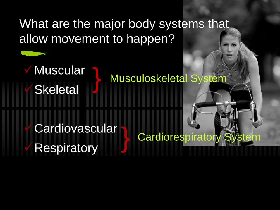
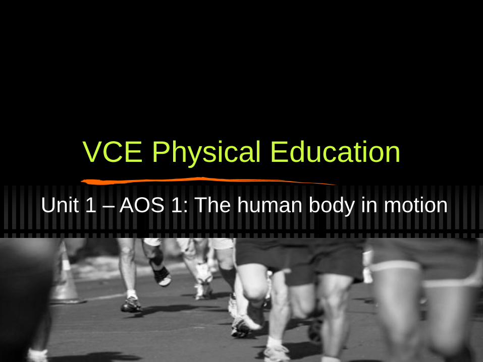
What are the major body systems that
allow movement to happen?
Muscular
Skeletal
Cardiovascular
Respiratory
}
}
Musculoskeletal System
Cardiorespiratory System
Skeletal System
What do you know about it?
Can you name any?
How many bones are there?
Have you broken any bones?
Skeletal System
There are 206 bones in the body!
5 major function
Support
Protection
Movement
Storage
Blood cell production
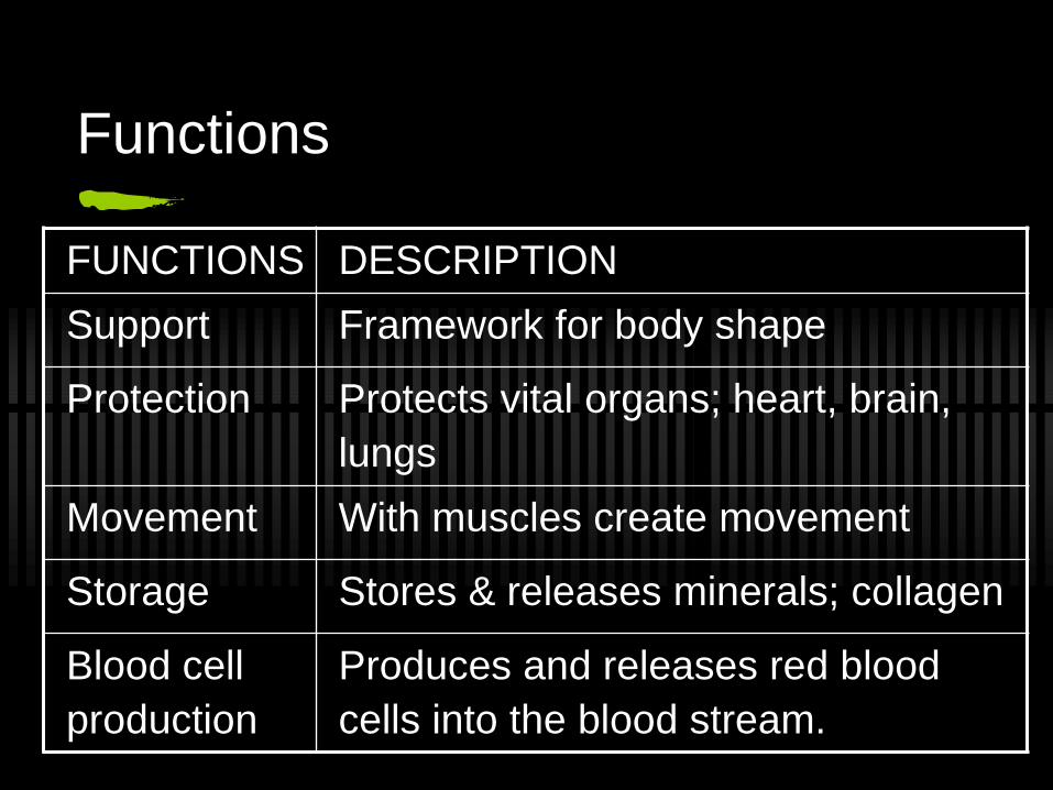
Functions
FUNCTIONS DESCRIPTION
Support Framework for body shape
Protection Protects vital organs; heart, brain,
lungs
Movement With muscles create movement
Storage Stores & releases minerals; collagen
Blood cell
production
Produces and releases red blood
cells into the blood stream.
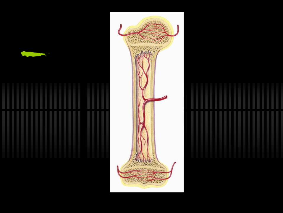
What’s in a bone?
Periosteum
tough membrane around the bones.
Marrow
produces blood cells (white, red and platelets)
Spongy Bone
holey and filled with bone marrow
Compact bone
dense and heavy, provides strength
Blood vessels
Developing bones
As a baby, bones are more flexible. They
contain cartilage which gradually
becomes harder & turns into bone.
You can find cartilage in your nose.
Have you ever wondered how a babies
head can fit through as a slow opening?
Dietary intake
Which foods do you know help to make
bones strong?
Vitamin A
Vitamin C
Vitamin D
Calcium
Exercise and Bones
You can eat all of those things but you MUST combine
it with weight-bearing exercise. Eg. running, jumping,
weights
Consequences of inadequate dietary intake and lack
of exercise can lead to serious health conditions such
as Osteoporosis.
Osteoporosis literally means ‘bones with holes’. It is
where the bones become weak and fragile. It is more
common in elderly females
Weight training and a diet high in calcium and vitamin
D are effective in preventing the onset of
osteoporosis.
Identifying the bones in your body
Label the bones in your body in the workbook
provided.
Axial Skeleton
Provides the main support for the body and
includes the skull, vertebral column and rib cage
Appendicular Skeleton
Made up of the limb bones and their ‘girdles’ that
connect onto the axial skeleton
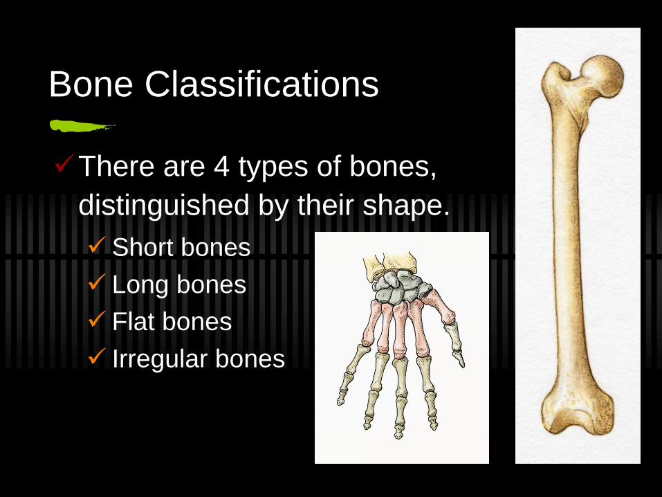
Bone Classifications
There are 4 types of bones,
distinguished by their shape.
Short bones
Long bones
Flat bones
Irregular bones
Vertebral Column
There are 3 curves:
Cervical Curve (7)
Thoracic Curve (12)
Lumbar Curve (5)
Protects the Spinal Cord!
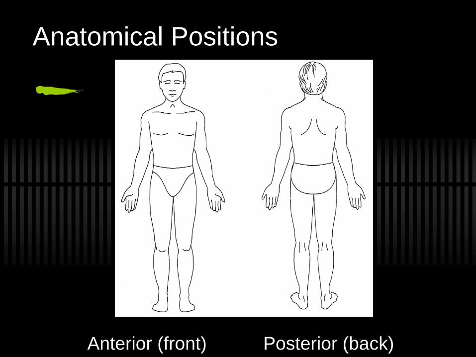
Anatomical Positions
Anterior (front) Posterior (back)
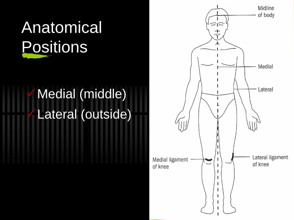
Anatomical
Positions
Medial (middle)
Lateral (outside)
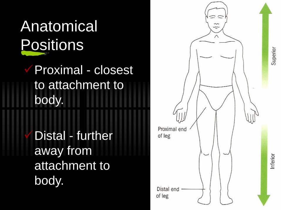
Anatomical
Positions
Proximal - closest
to attachment to
body.
Distal - further
away from
attachment to
body.
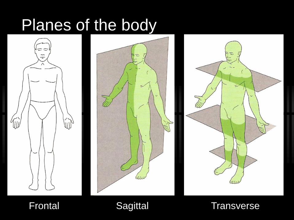
Planes of the body
Frontal Sagittal Transverse
Anatomical positioning
Complete the worksheet
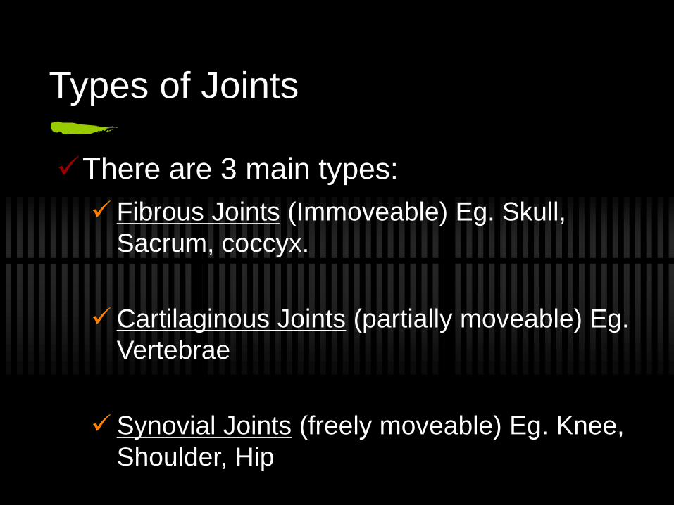
Types of Joints
There are 3 main types:
Fibrous Joints (Immoveable) Eg. Skull,
Sacrum, coccyx.
Cartilaginous Joints (partially moveable) Eg.
Vertebrae
Synovial Joints (freely moveable) Eg. Knee,
Shoulder, Hip
Synovial Joints
Cartilage covers the ends of bones.
Ligaments stabilise joint.
Synovial membrane contains synovial
fluid to allow easy movement.
Joint capsule encases the joint.
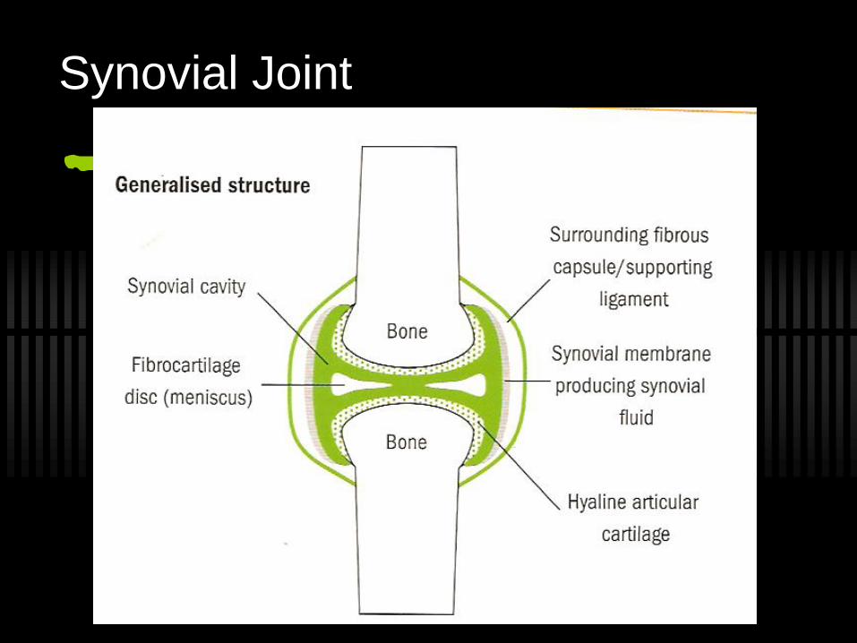
Synovial Joint
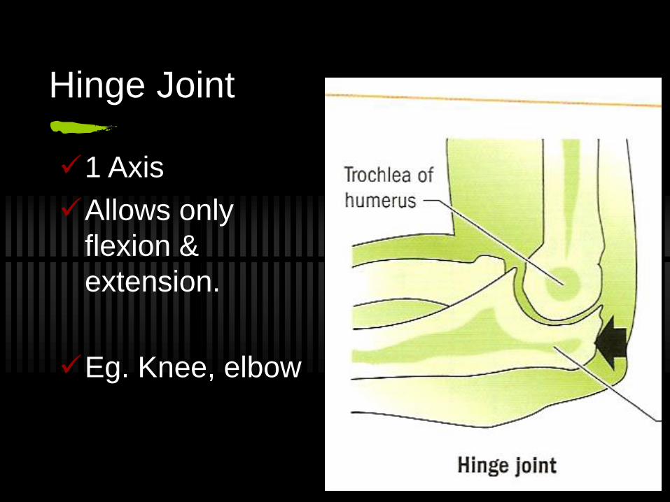
Hinge Joint
1 Axis
Allows only
flexion &
extension.
Eg. Knee, elbow
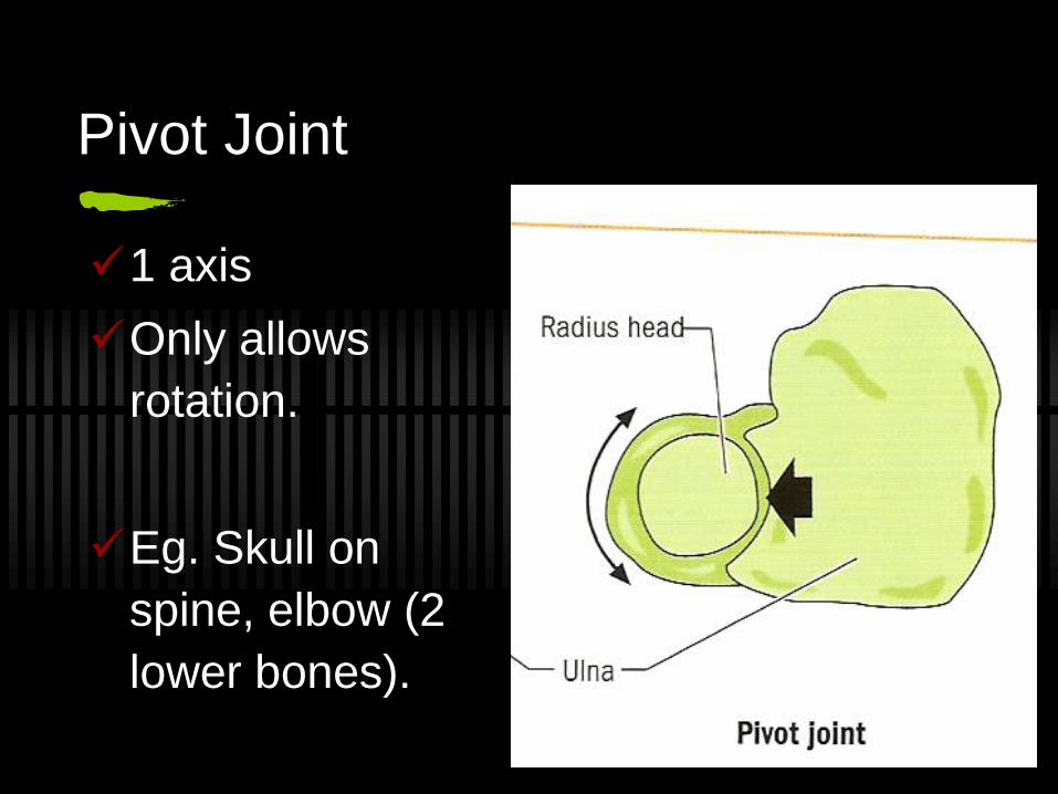
Pivot Joint
1 axis
Only allows
rotation.
Eg. Skull on
spine, elbow (2
lower bones).
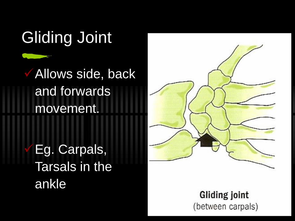
Gliding Joint
Allows side, back
and forwards
movement.
Eg. Carpals,
Tarsals in the
ankle
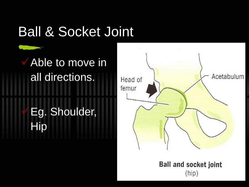
Ball & Socket Joint
Able to move in
all directions.
Eg. Shoulder,
Hip
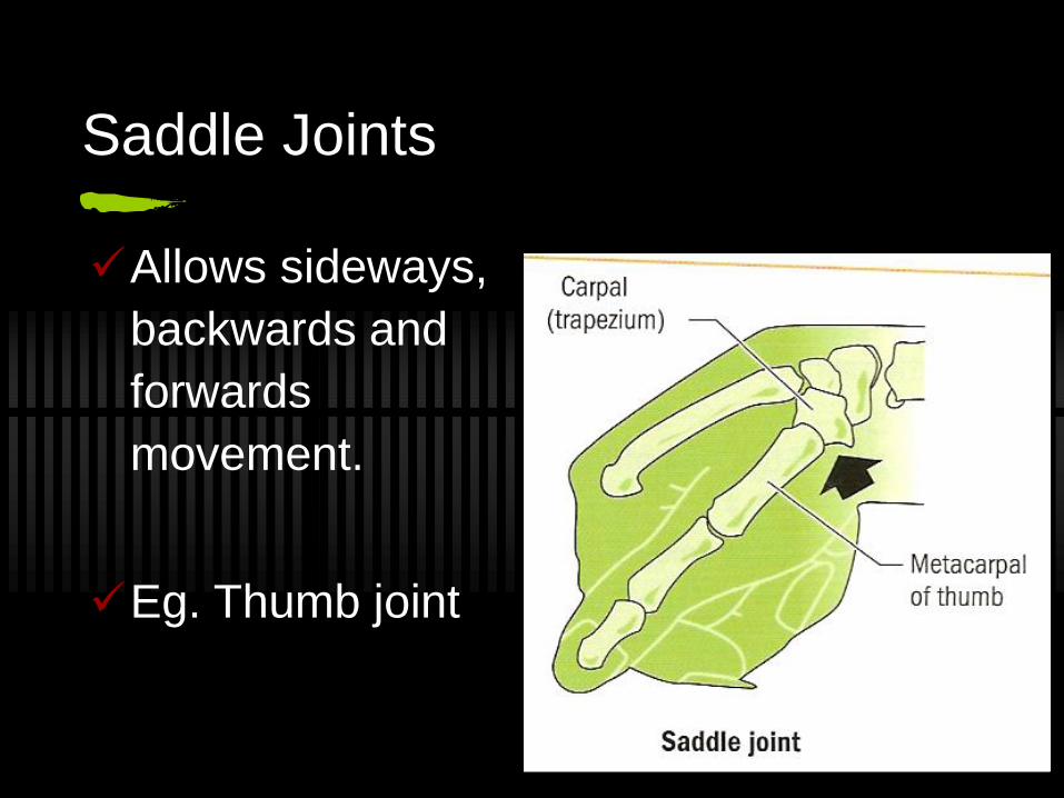
Saddle Joints
Allows sideways,
backwards and
forwards
movement.
Eg. Thumb joint
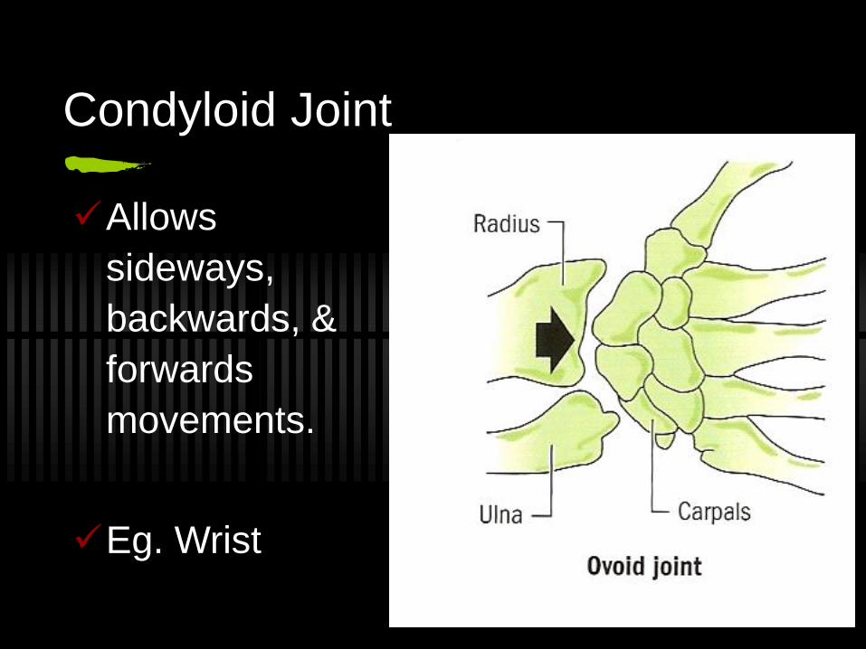
Condyloid Joint
Allows
sideways,
backwards, &
forwards
movements.
Eg. Wrist
Muscular System
Do you know how many muscles there
are in the body?
Over 600
Without muscles our hearts would not
beat, we could not breathe, digest food,
walk, talk or reproduce.
Functions of Muscles
Movement
Pulls on bones.
Posture
Keeps body balanced and aligned correctly.
Body Heat
Contractions produces heat for the body.
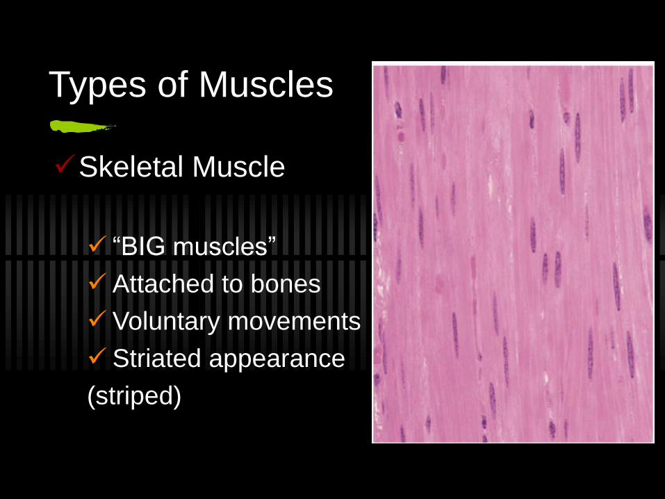
Types of Muscles
Skeletal Muscle
“BIG muscles”
Attached to bones
Voluntary movements
Striated appearance
(striped)
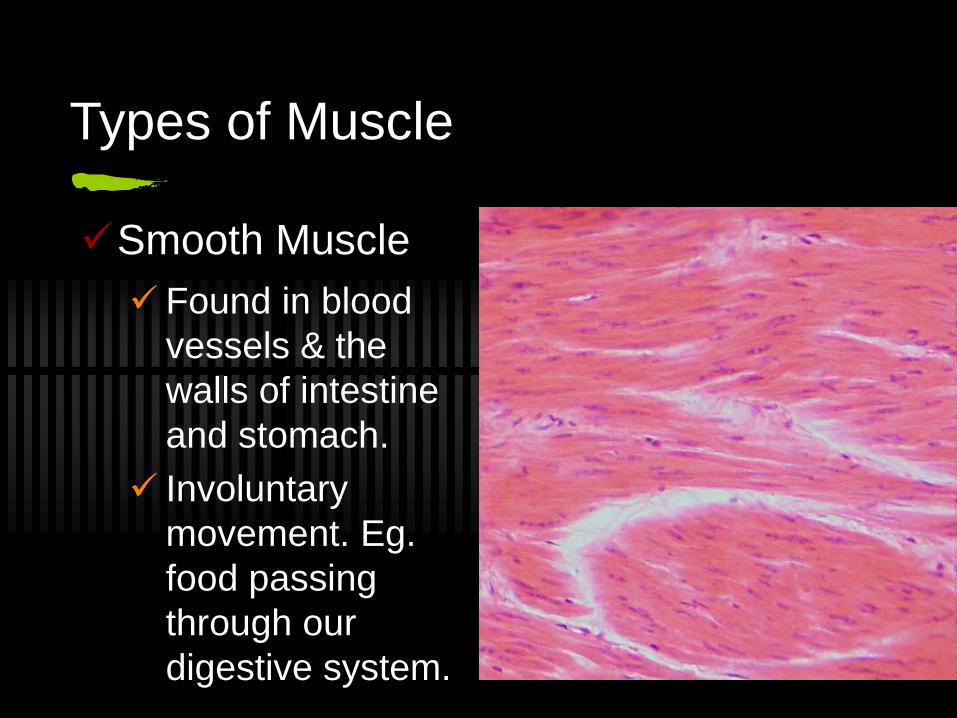
Types of Muscle
Smooth Muscle
Found in blood
vessels & the
walls of intestine
and stomach.
Involuntary
movement. Eg.
food passing
through our
digestive system.
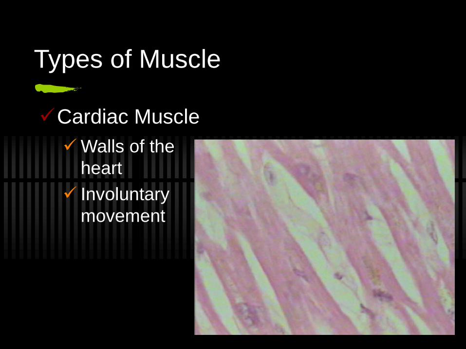
Types of Muscle
Cardiac Muscle
Walls of the
heart
Involuntary
movement
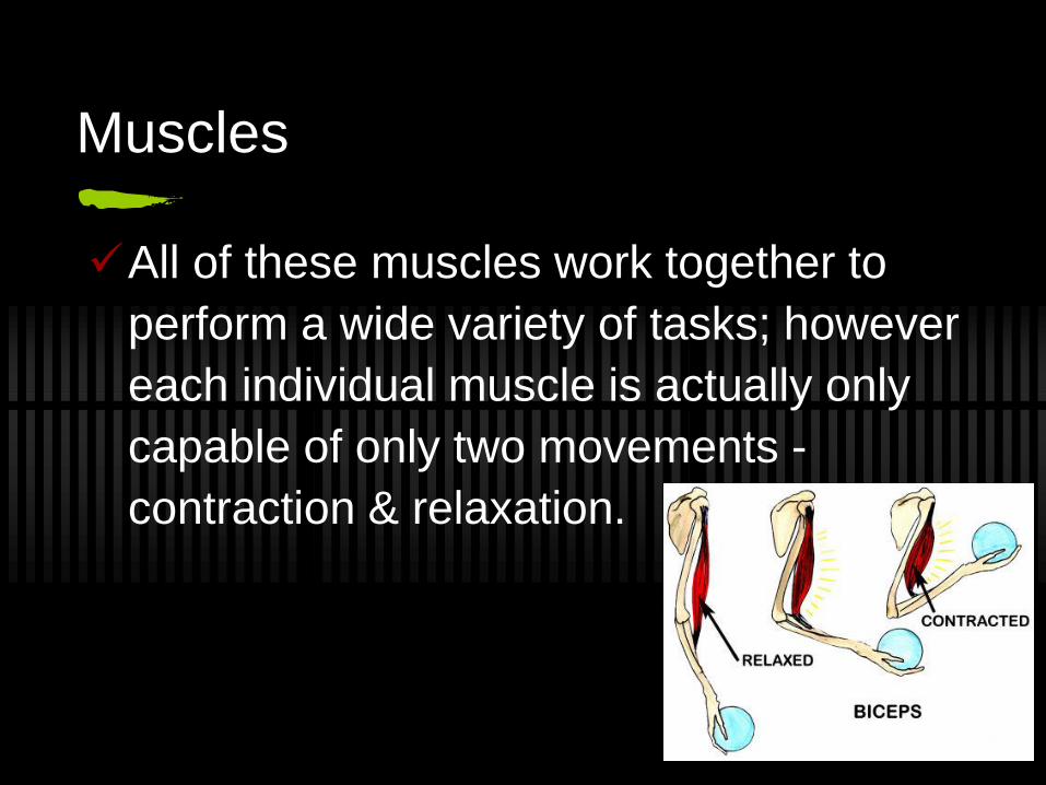
Muscles
All of these muscles work together to
perform a wide variety of tasks; however
each individual muscle is actually only
capable of only two movements -
contraction & relaxation.
Connective Tissue
Cartilage:
Cushions bones. Eg. between ribs and
sternum, b/n vertebrae.
Tendons:
Attaches muscle to bones. Eg. Achilles
Tendon.
Ligaments:
Joins bone to bone. Eg. ACL, PCL.
Origins & Insertions
Skeletal muscle attaches to bones via
tendons.
The one proximal (closest) to the body is
called the origin, the one distal (further
away) from the body is called the
insertion.
Muscles of the Body
Refer to page 28
Note there are 4 Quadriceps Muscles &
the 3 Hamstrings muscles
Identifying the muscles in your body
Label the muscles in your body in the
workbook provided.
Body movements Muscles can only pull. To make a joint move in two
directions, you need two muscles that can pull in opposite directions.
Antagonistic muscles are pairs of muscles that work against each other. One muscle contracts (agonist, or prime mover) while the other one relaxes (antagonist) and vice versa.
Stabilisers are also involved in contractions. These provide stability to the origin e.g. the trapezius during elbow flexion.
Reciprocal Inhibition is the term used to explain how muscles work in ‘teams’. It essentially describes how one muscle contracts and its opposite relaxes to allow ease of movement and reduce muscle tears.
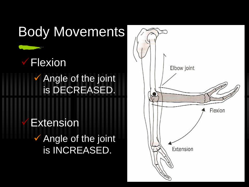
Body Movements
Flexion
Angle of the joint
is DECREASED.
Extension
Angle of the joint
is INCREASED.
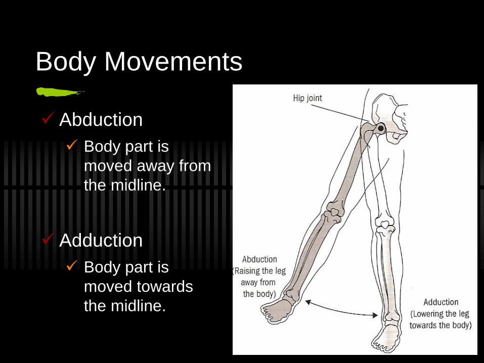
Body Movements
Abduction
Body part is
moved away from
the midline.
Adduction
Body part is
moved towards
the midline.
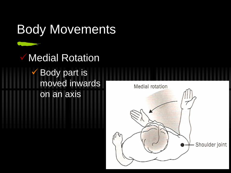
Body Movements
Medial Rotation
Body part is
moved inwards
on an axis
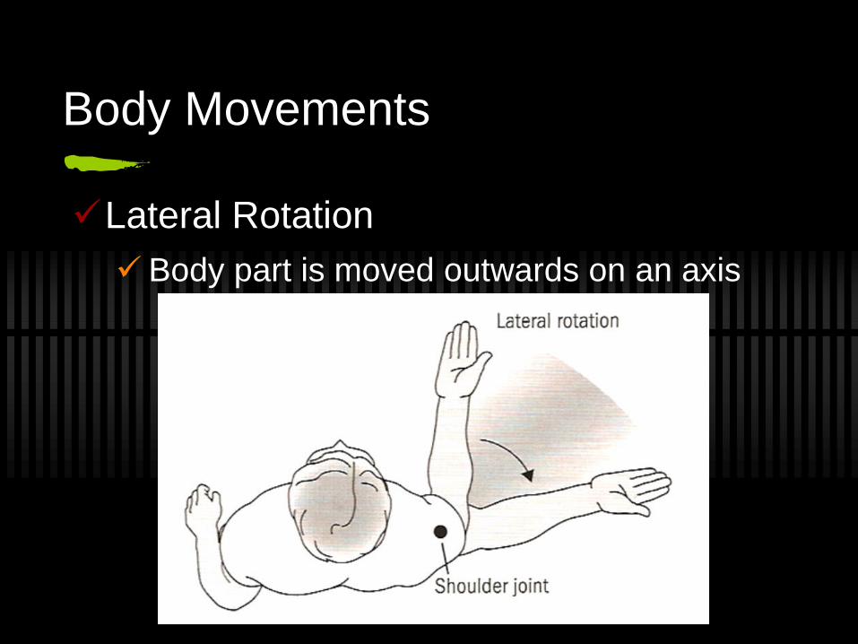
Body Movements
Lateral Rotation
Body part is moved outwards on an axis
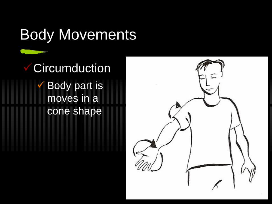
Body Movements
Circumduction
Body part is
moves in a
cone shape
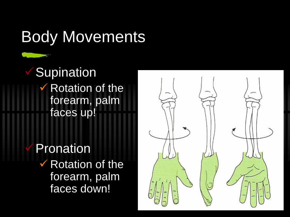
Body Movements
Supination
Rotation of the forearm, palm faces up!
Pronation
Rotation of the forearm, palm faces down!
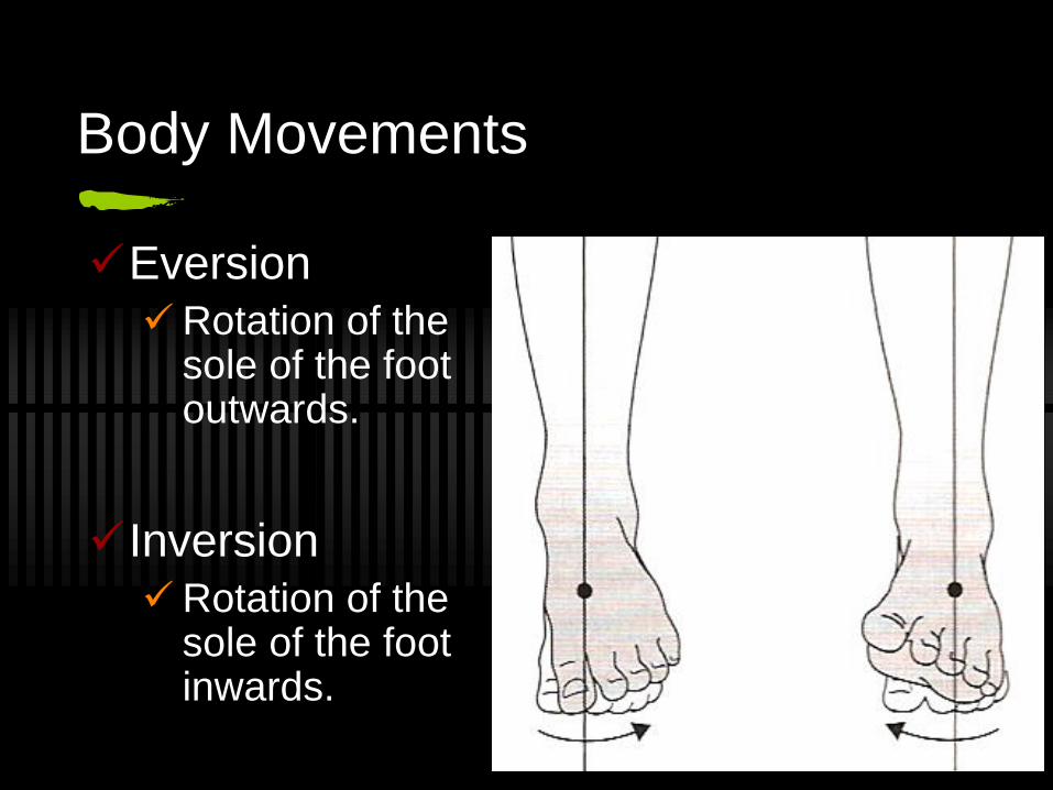
Body Movements
Eversion
Rotation of the sole of the foot outwards.
Inversion
Rotation of the sole of the foot inwards.
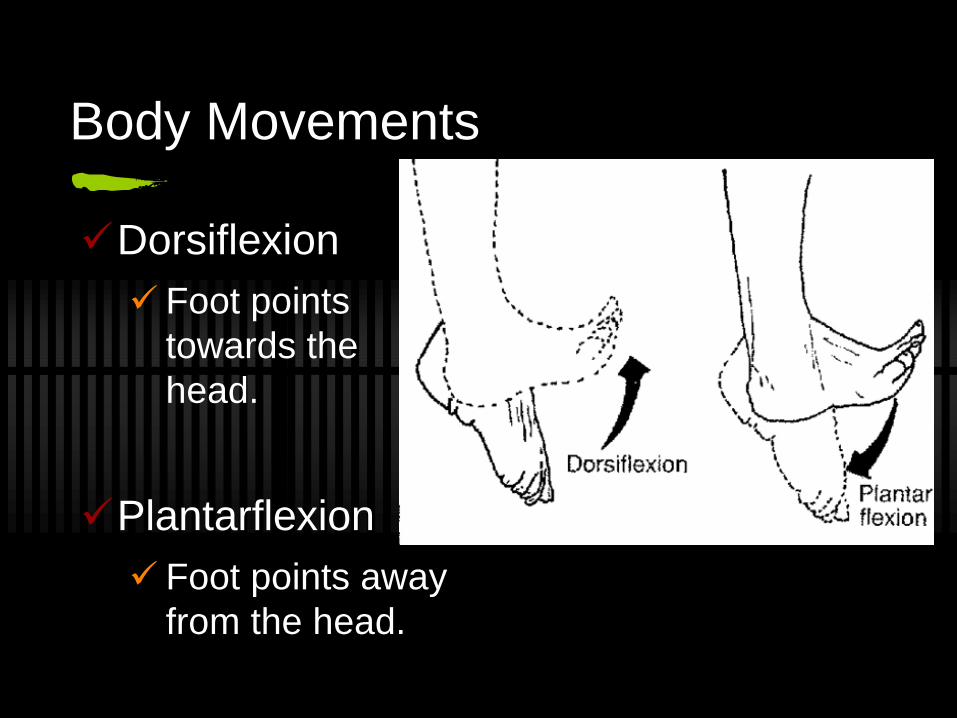
Body Movements
Dorsiflexion
Foot points
towards the
head.
Plantarflexion
Foot points away
from the head.
The types of muscular contraction
There are 3 types of contractions:
Isoinertial
Isometric
Isokinetic
Isoinertial Contraction
Most common
Occurs when muscle length changes while
creating force.
Eg. person picks up a shot put.
There are two types:
Concentric contraction - muscle shortens/contracts
Eccentric contraction - muscle lengthens/extends
Eg. Bicep Curl (Up & Down phase)
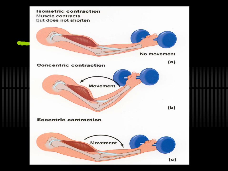
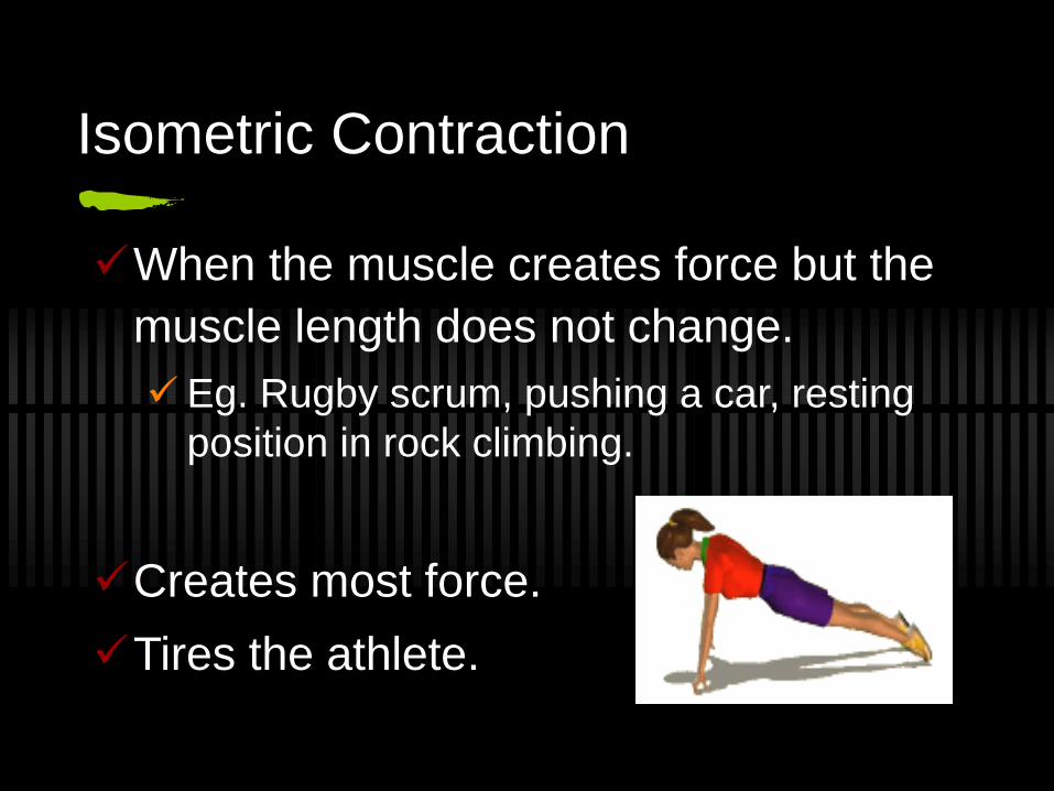
Isometric Contraction
When the muscle creates force but the
muscle length does not change.
Eg. Rugby scrum, pushing a car, resting
position in rock climbing.
Creates most force.
Tires the athlete.
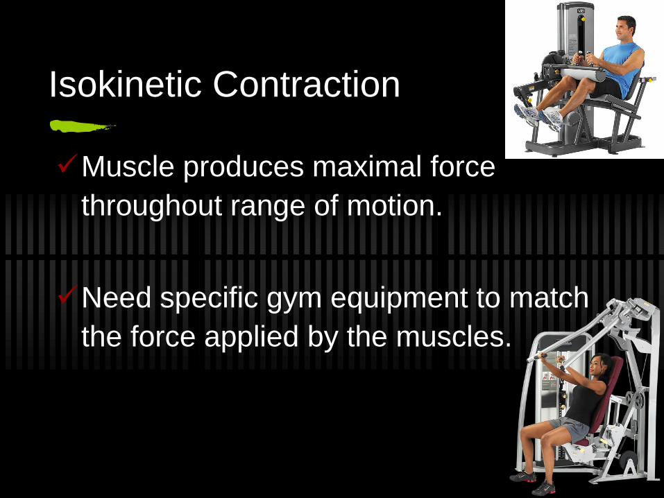
Isokinetic Contraction
Muscle produces maximal force
throughout range of motion.
Need specific gym equipment to match
the force applied by the muscles.
Lab
Sit-up
Planks
Push-ups
Push against the wall
Muscle Fibres
There are two types:
Slow Twitch Fibres
Fast Twitch Fibres
We all have a combination of both. The percentage of
each type varies depending on genetic inheritance and
training undertaken.
They can influence the type of activity an athlete is best
suited to and can be determined through a biopsy.
https://www.youtube.com/watch?v=Uxwh2IIg_Z0
Type I - Slow Twitch Fibres
Red colour
Contract slowly over a period of time
Aerobic & endurance sports
Exert less force for long periods
Athletes that have a high proportion of slow
twitch fibres include: triathletes, marathon
runners and rowers
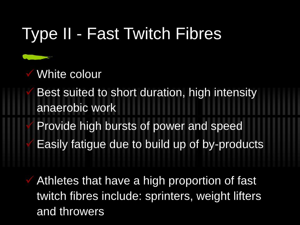
Type II - Fast Twitch Fibres
White colour
Best suited to short duration, high intensity
anaerobic work
Provide high bursts of power and speed
Easily fatigue due to build up of by-products
Athletes that have a high proportion of fast
twitch fibres include: sprinters, weight lifters
and throwers
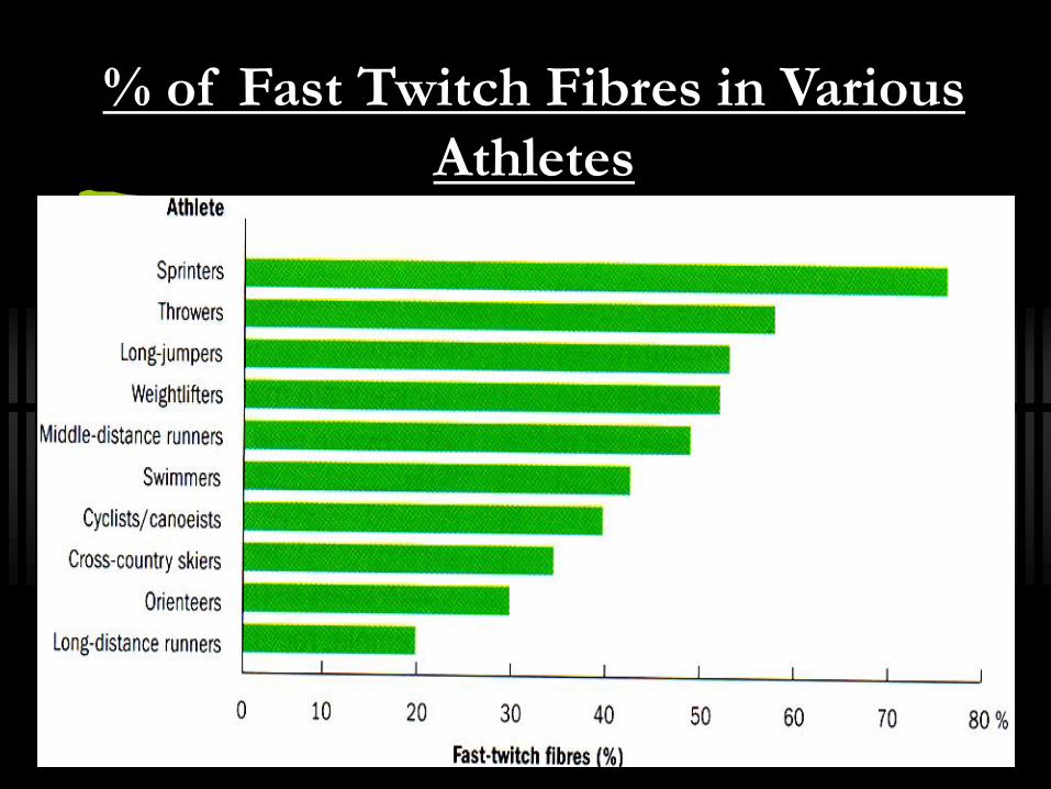
% of Fast Twitch Fibres in Various
Athletes
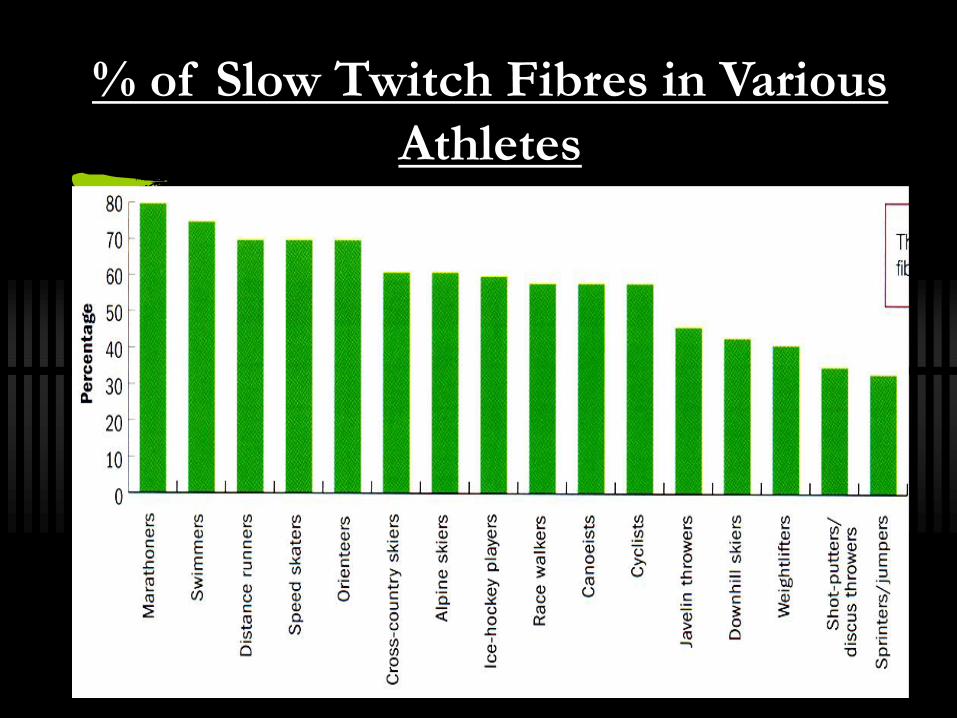
% of Slow Twitch Fibres in Various
Athletes
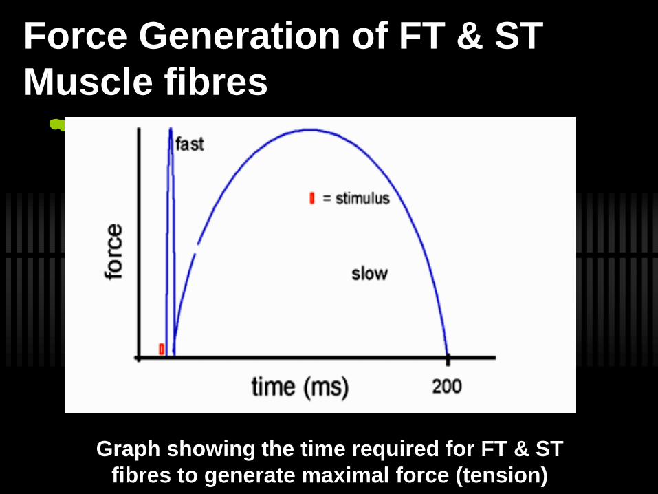
Force Generation of FT & ST
Muscle fibres
Graph showing the time required for FT & ST
fibres to generate maximal force (tension)
Further classification of Type II fibres
Type IIa
Partially aerobic
Also known as intermediate fast-twitch
fibres
Type IIb
Purely anaerobic
Highest rate of contraction for explosive
bursts of energy
Fibre Recruitment Theory
Slow-twitch fibres are recruited before fast twitch units
in most activities, and as muscular forces increase, so
does the pattern of recruitment of fibres.
Eg. A slow swim at a local pool (slow lane) will use
slow-twitch fibres, while an ‘all out’ 100m swim will
recruit fast-twitch A fibres. Depending on how hard the
swim is done (force exerted) the fast-twitch B fibres
may also contribute to the performance.
Gender & Age differences
How do you think they could vary?
Until puberty there isn’t much difference.
Testosterone enables males to develop
a larger muscles to produce greater
power.
Muscles are strongest between 20 - 30.
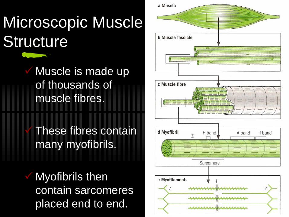
Microscopic Muscle
Structure
Muscle is made up
of thousands of
muscle fibres.
These fibres contain
many myofibrils.
Myofibrils then
contain sarcomeres
placed end to end.
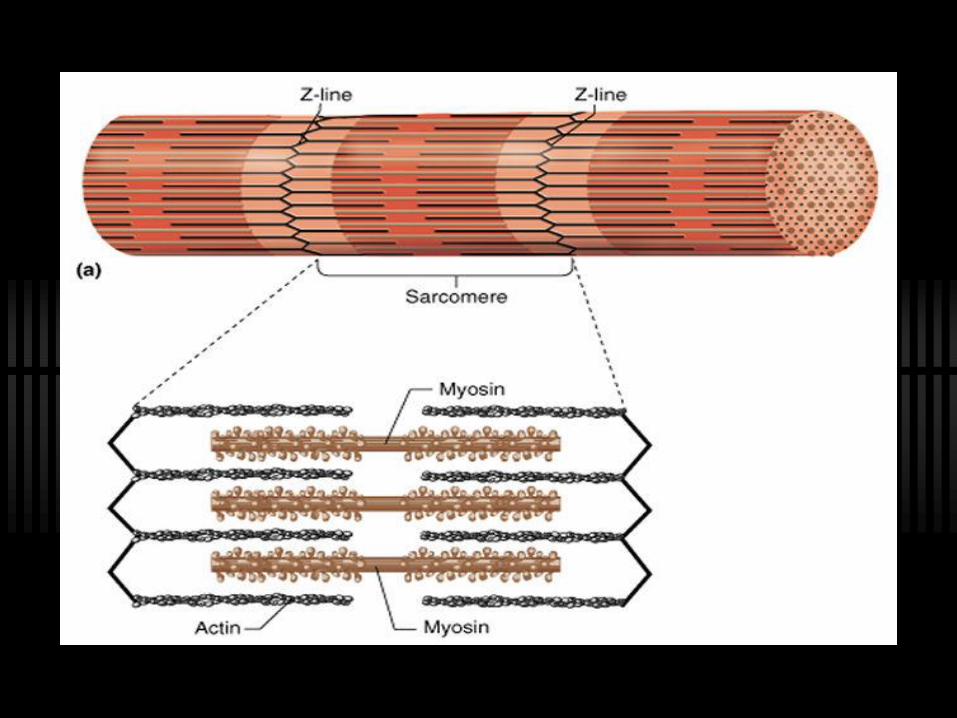
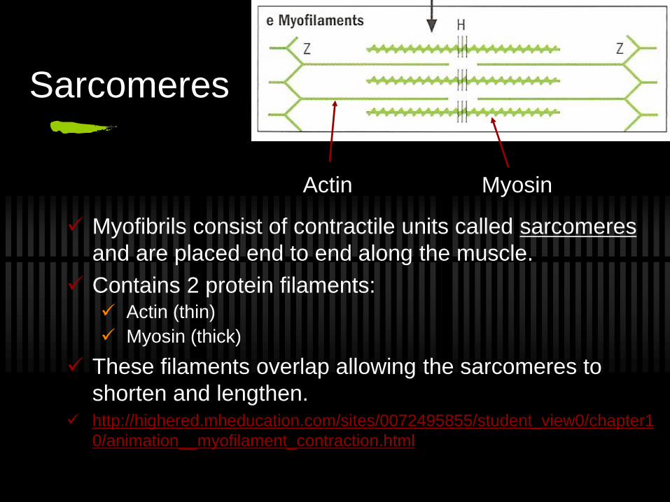
Sarcomeres
Myofibrils consist of contractile units called sarcomeres
and are placed end to end along the muscle.
Contains 2 protein filaments: Actin (thin)
Myosin (thick)
These filaments overlap allowing the sarcomeres to
shorten and lengthen. http://highered.mheducation.com/sites/0072495855/student_view0/chapter1
0/animation__myofilament_contraction.html
MyosinActin
Parts of the sarcomere…
Many sarcomere units run the length of a
myofibril. Z lines are used to indicated where
one sarcomere begins & ends.
Action & myosin occupy different parts along
the length of the sarcomere.
The lighter section contains only the thin actin
filaments. This section is known as the I Band.
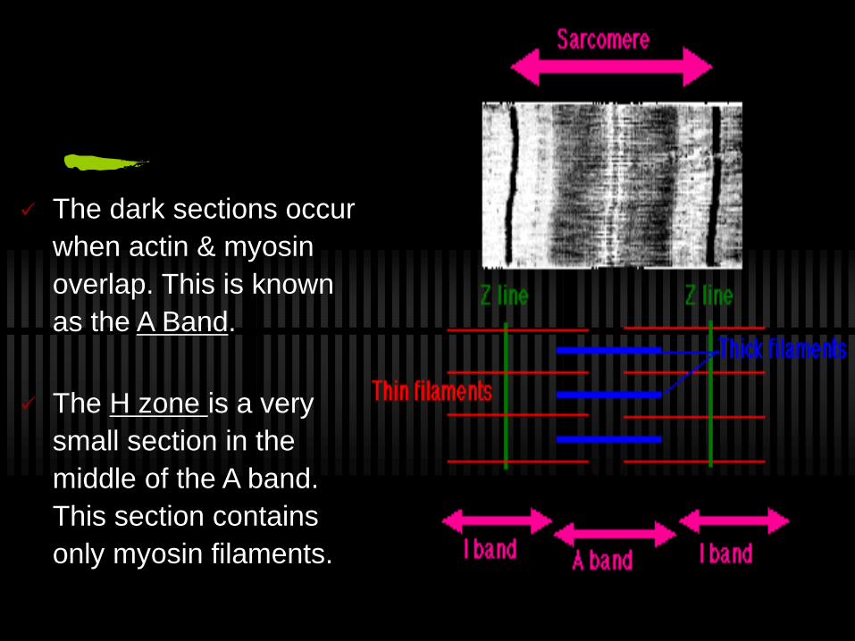
The dark sections occur
when actin & myosin
overlap. This is known
as the A Band.
The H zone is a very
small section in the
middle of the A band.
This section contains
only myosin filaments.
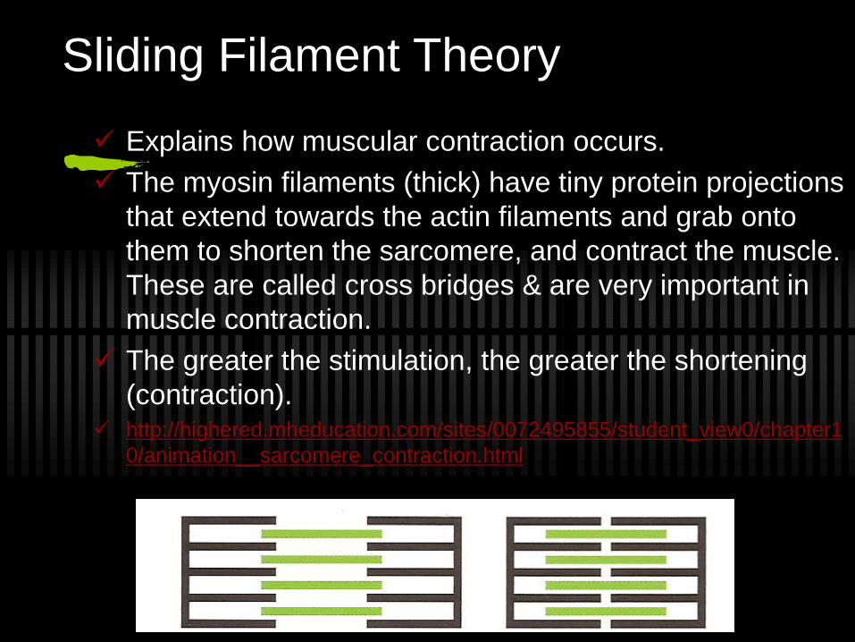
Sliding Filament Theory
Explains how muscular contraction occurs.
The myosin filaments (thick) have tiny protein projections
that extend towards the actin filaments and grab onto
them to shorten the sarcomere, and contract the muscle.
These are called cross bridges & are very important in
muscle contraction.
The greater the stimulation, the greater the shortening
(contraction). http://highered.mheducation.com/sites/0072495855/student_view0/chapter1
0/animation__sarcomere_contraction.html
What occurs in a Sarcomere during
Muscular Contraction
1. H zone disappears because actin filaments slide over myosin filaments.
2. I Band shortens the actin filaments attached to the Z lines on either side of the sarcomere are pulled toward the centre.
3. The A Band does not change in length.
4. Neither the actin nor the myosin filaments change in length, because of the sliding mechanism.
5. http://wps.prenhall.com/wps/media/objects/2688/2752944/Web_Tutorials/25_A01.swf
Nervous control of muscular contractions
Nervous impulses are sent by nerve cells called neurons.
There are two main types of neurons:
Sensory neurons, which pass information about stimuli such as light, heat or chemicals from sensory organs and receptors to your central nervous system.
Motor neurons, which pass instructions from your central nervous system to other parts of your body, such as muscles and glands.
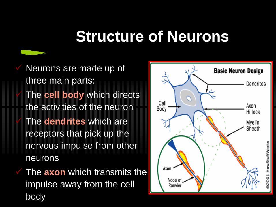
Structure of Neurons
Neurons are made up of
three main parts:
The cell body which directs
the activities of the neuron
The dendrites which are
receptors that pick up the
nervous impulse from other
neurons
The axon which transmits the
impulse away from the cell
body
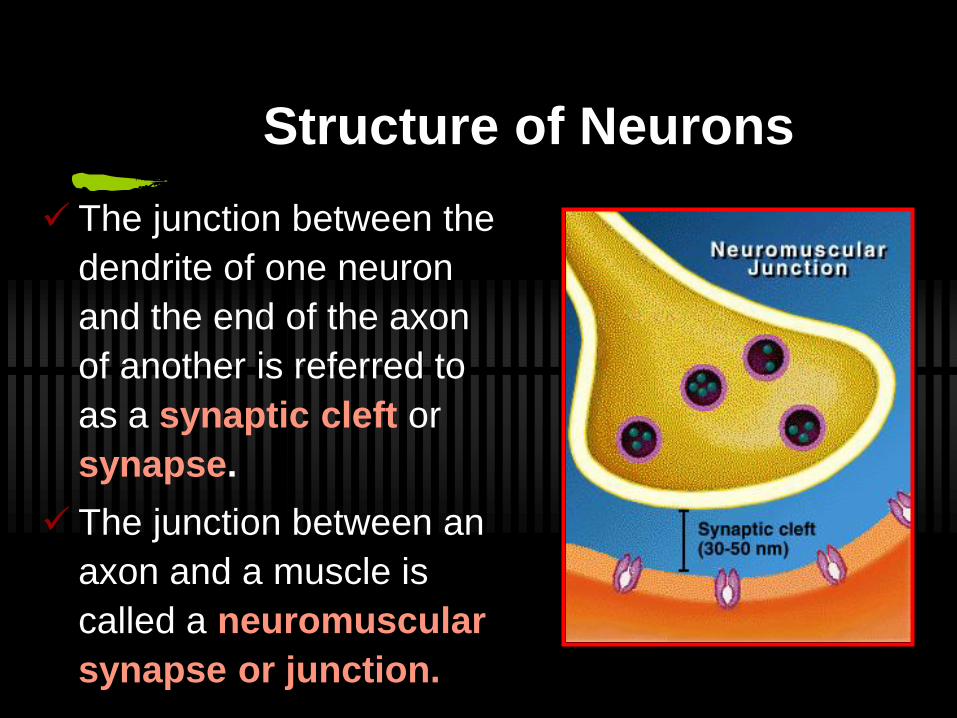
Structure of Neurons
The junction between the
dendrite of one neuron
and the end of the axon
of another is referred to
as a synaptic cleft or
synapse.
The junction between an
axon and a muscle is
called a neuromuscular
synapse or junction.
The Neuromuscular Synapse
The neuromuscular synapse is a specialized excitatory synapse that allows motor neurons to communicate with muscle fibres.
It is a chemical synapse using acetylcholine (ACh)as the neurotransmitter.
As a nerve impulse reaches the end of the axon, it triggers the release of ACh which is stored within the nerve ending.
This transmitter substance travels across the synaptic cleft and stimulates an electrical impulse (action potential) in the muscle fibre, which in turn brings about a muscular contraction.
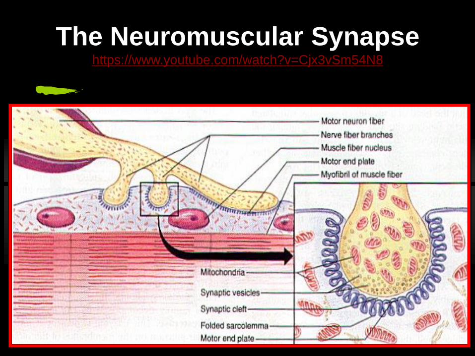
The Neuromuscular Synapsehttps://www.youtube.com/watch?v=Cjx3vSm54N8
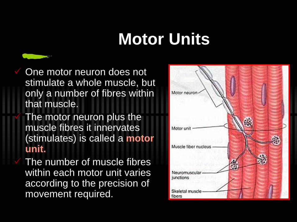
Motor Units
One motor neuron does not stimulate a whole muscle, but only a number of fibres within that muscle.
The motor neuron plus the muscle fibres it innervates (stimulates) is called a motor unit.
The number of muscle fibres within each motor unit varies according to the precision of movement required.
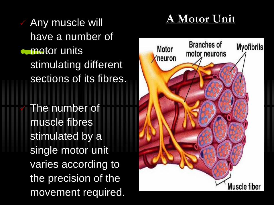
Any muscle will
have a number of
motor units
stimulating different
sections of its fibres.
The number of
muscle fibres
stimulated by a
single motor unit
varies according to
the precision of the
movement required.
A Motor Unit
“ALL OR NOTHING” PRINCIPLE
Once the nerve impulse reaches threshold
level, all of the fibres in a given motor unit will
contract fully.
If the impulse is insufficient no fibres will
contract.
https://www.youtube.com/watch?v=ZpUYOAzBce0
Recommended