7/22/2019 Updates in the Management of the Small Animal Patient with Neurologic Trauma
http://slidepdf.com/reader/full/updates-in-the-management-of-the-small-animal-patient-with-neurologic-trauma 1/26
U p d a t e s i n t h e M a n a g em e n t o ft h e S m a l l A n i m a l P a t i e n t w i t h
Neurologic TraumaJillian DiFazio, DVM
a, Daniel J. Fletcher, PhD, DVMb,*
Neurologic trauma, encompassing traumatic brain injury (TBI) and acute spinal cord
injury (SCI), is a cause of significant morbidity and mortality in veterinary patients. In
one recent retrospective study evaluating blunt trauma in dogs, a diagnosis of TBI
was made in 25% of cases and was associated with increased mortality.1 Acute
SCIs occurring secondary to trauma (including vertebral fracture or luxation [VFL],
traumatic intervertebral disk herniation, spinal cord parenchymal contusions, and
extra-axial hemorrhage) are also common, with an estimated incidence rate of 14%
in cats and 9% in dogs based on the information from single-center retrospective
studies.2,3 The causes of neurologic trauma in dogs and cats include motor vehicular
trauma, falls, crush injuries, bite wounds, missile injuries (eg, gunshot wounds), and
either accidental or purposeful human-inflicted trauma.4–7 Essential to the man-
agement of TBI and SCI is a thorough understanding of the pathophysiology of the pri-
mary and secondary injury that occurs following trauma.8 This article reviews the
a
Section of Emergency and Critical Care, Cornell University Hospital for Animals, Upper TowerRoad, Ithaca, NY 14853, USA; b Department of Clinical Sciences, Cornell University College ofVeterinary Medicine, DCS Box 31, Ithaca, NY 14853, USA* Corresponding author.E-mail address: [email protected]
KEYWORDS
Neurotrauma Traumatic brain injury Head trauma Acute spinal cord injury Resuscitation Dog Cat Cerebral ischemia
KEY POINTS
Neurotrauma, including traumatic brain injury (TBI) and acute spinal cord injury (SCI), is acause of significant morbidity and mortality in veterinary patients.
Damage to neuronal cells can be divided into primary and secondary injury.
Pharmacologic and nonpharmacologic therapies are directed at addressing primary injury
in SCI as well as minimizing the effects of secondary injury in both TBI and SCI.
Prognosis for neurotrauma patients depends on the severity of injury, the site of the lesion,
and the timing and efficacy of the treatment of primary and secondary injury.
Vet Clin Small Anim 43 (2013) 915–940http://dx.doi.org/10.1016/j.cvsm.2013.03.002 vetsmall.theclinics.com
0195-5616/13/$ – see front matter Ó 2013 Elsevier Inc. All rights reserved.

7/22/2019 Updates in the Management of the Small Animal Patient with Neurologic Trauma
http://slidepdf.com/reader/full/updates-in-the-management-of-the-small-animal-patient-with-neurologic-trauma 2/26
pathophysiology of this primary and secondary injury, as well as recommendations
regarding clinical assessment, diagnostics, pharmacologic and nonpharmacologic
therapy, and prognosis.
MANAGEMENT GOALSDamage to nervous tissue can be divided into primary and secondary injury. Primary
injury occurs immediately after trauma and is the direct result of traumatic impact.9–12
Secondary injury is often referred to as delayed injury, but usually begins within mi-
nutes of injury and can last several days to weeks afterward.4,6,7,11,13–15 These cate-
gories may seem artificial at first, but are important when considering management.
Most TBI therapies are aimed at minimizing the effects of secondary injury. Because
instability contributes to exacerbation of primary injury, depending on the type, man-
agement of acute SCI may include surgical therapy directed at stabilization to prevent
further primary injury in addition to therapies directed at minimizing the effects of sec-
ondary injury.15
Primary Injury
Primary injury associated with TBI and SCI involves the physical disruption of intracra-
nial structures (eg, TBI) and the spinal cord, vertebrae, and supporting structures (eg,
SCI) that occurs at the time of impact. Primary injury is broadly classified as focal or
diffuse depending on the extent of injury, and more specifically can be defined based
on the location and type of injury.11 The principal mechanical forces involved in neuro-
logic trauma include concussion (eg, acceleration and deceleration), compression,
shear, laceration, distraction, and contusion.15–17
Primary injuries associated withTBI include epidural hematomas, subdural hematomas, subarachnoid hemorrhage,
cortical contusions/hematomas, and traumatic axonal injury.10 Primary SCI includes
VFL, traumatic intervertebral disc herniation, intraparenchymal contusion, and extra-
axial hemorrhage.
Secondary Injury
Box 1 summarizes the local factors contributing to secondary injury in neurologic
trauma.4,7,13,15–21 In addition, multiple systemic factors can potentiate secondary
injury, most importantly hypoxia and hypotension but also hypercapnia, hypocapnia,
hyperglycemia, hypoglycemia, acid-base disturbances, electrolyte abnormalities, hy-perthermia, and systemic inflammation.17,18 Other intracranial factors can also exacer-
bate secondary injury in TBI, including intracranial hypertension, edema, compromise
of the blood-brain barrier (BBB), vasospasm, hemorrhage, infection, mass effects, and
seizure activity.17
Secondary injury is potentiated by compromise of perfusion. Cerebral perfusion
pressure (CPP) is defined as the net pressure facilitating blood flow to the brain,
and is the difference between the mean arterial blood pressure (MAP) and intracranial
pressure (ICP): CPP 5 MAP À ICP. Similarly, spinal cord perfusion pressure (SCPP)
is the difference between MAP and cerebrospinal fluid pressure (CSFP): SCPP 5
MAP À CSFP.22
The Monroe-Kellie doctrine states that the cranial vault is a rigid, defined space that
has a fixed volume with contributions from the brain parenchyma, cerebrospinal fluid
(CSF), blood, and mass lesions (if present). An increase in the volume of any of these
will result in a compensatory decrease in 1 or more of the others (defined as intracra-
nial compliance; mainly reliant on changes in CSF or blood volumes), without which a
pathologic increase in ICP will occur. With TBI the compensatory capacity of
DiFazio & Fletcher916

7/22/2019 Updates in the Management of the Small Animal Patient with Neurologic Trauma
http://slidepdf.com/reader/full/updates-in-the-management-of-the-small-animal-patient-with-neurologic-trauma 3/26
intracranial compliance can be overwhelmed, and intracranial hypertension may
occur. Increases in ICP combined with decreases in MAP, a finding that is common
in trauma patients, can result in decreases in CPP. In addition, compromise of autor-
egulatory mechanisms (eg, vasodilation/constriction of cerebral arterioles that main-
tain constant cerebral and spinal cord blood flow over a wide range of MAP
([0–150 mm Hg]) results in a more linear association between blood flow and MAP,
leading to a greater risk of hypoperfusion or hyperemia.11
Severe, acute intracranial hypertension may result in the Cushing reflex or central
nervous system (CNS) ischemic response. Decreased cerebral blood flow results in
elevations in carbon dioxide (CO2 ) levels sensed locally at the vasomotor center,
causing a dramatic increase in sympathetic tone, ultimately leading to systemic vaso-
constriction and increased cardiac output.23 Increases in MAP stimulate barorecep-
tors in the aortic and carotid sinuses, resulting in a reflex sinus bradycardia. This
response signifies potentially life-threatening intracranial hypertension and should
be treated immediately.4,7,17
DIAGNOSTIC EVALUATIONSystemic Assessment
Initial triage assessment of the trauma patient should focus on global patient stability
with special emphasis on the respiratory and cardiovascular systems. In patients with
neurotrauma, this is perhaps even more important because hypotension, hypoxemia,
and changes in ventilation contribute to secondary injury and worsen outcome.
Neurologic Assessment
The initial neurologic examination should occur before administration of any analgesic
therapy to allow adequate assessment of the neurologic system. Initial neurologic
examination should include an evaluation of mentation, cranial nerve reflexes, ambu-
latory status, presence of voluntary motor function (assessed only in recumbency in
the nonambulatory patient with potential VFL), presence of superficial pain perception
in the patient who does not demonstrate voluntary motor function, presence of deep
pain sensation in the patient who does not demonstrate intact superficial pain percep-
tion, spinal reflexes, panniculus reflex, anal tone, and perineal reflex. If the patient is
ambulatory and the clinician is not suspicious of a VFL, assessment of gait and pro-
prioceptive function can also occur. Gentle palpation of the spinal column shouldbe done in all patients presenting with possible acute SCI to localize regions of mala-
lignment (eg, “step” fracture), instability, discomfort, or crepitus. When performing a
neurologic assessment, it is important that the patient has been adequately resusci-
tated, as shock can affect neurologic status. In addition, it is important that a thorough
recumbent orthopedic examination also be perf ormed to rule out orthopedic injury as
a potential cause for apparent neurologic signs.13,15
Whenever SCI secondary to VFL is suspected in the nonambulatory patient, minimal
movement of the patient should occur. The patient should be immobilized and
secured to a backboard until definitive assessment for fractures and luxations can
occur.13,15
Patients should be neurolocalized and graded based on severity of signs. The Modi-
fied Glasgow Coma Scale (MGCS) has been validated in dogs, and is useful in the
assessment of TBI patients, as it provides a means of more objectively determining
improvement or progression of clinical signs. It also yields prognostic information
( Box 2 ).24,25 One retrospective study showed that the MGCS correlated well with
the probability of survival in the first 48 hours after TBI in dogs.25 Repeated neurologic
Neurologic Trauma Small Animal Patients 917

7/22/2019 Updates in the Management of the Small Animal Patient with Neurologic Trauma
http://slidepdf.com/reader/full/updates-in-the-management-of-the-small-animal-patient-with-neurologic-trauma 4/26
Box 1
Mechanisms of secondary injury
Secondary Injury
Glutamate accumulation
Occurs secondary to:
Adenosine triphosphate (ATP) depletion
Neuronal cell injury
Positive feedback
Decreased conversion
Potentiated by low interstitial magnesium
Results in:
Loss of ionic gradients
Excitotoxicity
Generation of free radical oxygen species
Influx of sodium into neuronal cells
Occurs secondary to:
Glutamate accumulation
Results in:
Cytotoxic edema
Influx of calcium into neuronal cells Occurs secondary to:
Glutamate accumulation
Primary injury
Results in:
Cytotoxic edema
Neuronal cell destruction through activation of proteases, lipases and endonucleases
Reactive oxygen species production through calpain activation
Inflammatory mediator release
Mitochondrial dysfunction and ATP depletion
Free radical production
Occurs secondary to:
Glutamate accumulation
Inflammatory mediator release
Increased cytosolic calcium concentrations
Ischemia-reperfusion injury
Results in:
Neuronal cell destruction
Inflammatory mediator release
Occurs secondary to:
Primary injury
DiFazio & Fletcher918

7/22/2019 Updates in the Management of the Small Animal Patient with Neurologic Trauma
http://slidepdf.com/reader/full/updates-in-the-management-of-the-small-animal-patient-with-neurologic-trauma 5/26
Neuronal cell destruction with secondary injury
Results in:
Activation of nitric oxide with alterations in blood flow and vascular permeability
Inflammatory cell influx
Coagulation cascade activation and thrombosis
Loss of autoregulation
Occurs secondary to:
Primary injury
Results in:
Ischemia
All mechanisms contribute to neuronal cell death
Box 2
Modified Glasgow Coma Scale
Level of Consciousness6. Occasional periods of alertness and responsive to environment
5. Depression or delirium, capable of responding but response may be inappropriate
4. Semicomatose, responsive to visual stimuli
3. Semicomatose, responsive to auditory stimuli
2. Semicomatose, responsive only to repeated noxious stimuli
1. Comatose, unresponsive to repeated noxious stimuli
Brainstem Reflexes
6. Normal pupillary light reflexes and oculocephalic reflexes
5. Slow pupillary light reflexes and normal to reduced oculocephalic reflexes
4. Bilateral unresponsive miosis with normal to reduced oculocephalic reflexes
3. Pinpoint pupils with reduced to absent oculocephalic reflexes
2. Unilateral, unresponsive mydriasis with reduced to absent oculocephalic reflexes
1. Bilateral, unresponsive mydriasis with reduced to absent oculocephalic reflexes
Motor activity
6. Normal gait, normal spinal reflexes
5. Hemiparesis, tetraparesis, or decerebrate activity
4. Recumbent, intermittent extensor rigidity
3. Recumbent, constant extensor rigidity
2. Recumbent, constant extensor rigidity with opisthotonus
1. Recumbent, hypotonia of muscles, depressed or absent spinal reflexes
Neurologic Trauma Small Animal Patients 919

7/22/2019 Updates in the Management of the Small Animal Patient with Neurologic Trauma
http://slidepdf.com/reader/full/updates-in-the-management-of-the-small-animal-patient-with-neurologic-trauma 6/26
assessment is recommended every 30 to 60 minutes after initial presentation to
assess clinical response to therapy or progression of clinical signs. There are currently
3 validated scoring systems available for assessing the severity of deficits associated
with SCI: the Modified Frankel Score, the 14-Point Motor Score, and the Texas Spinal
Cord Injury Score.26,27
Brachial plexus injuries should be suspected if a patient has decreased reflexes in
only one of the forelimbs, Horner syndrome on the affected side, and decreased pan-
niculus reflex on the affected side.13 The presence of spinal shock may affect neuro-
localization in patients with acute SCI. Spinal shock leads to deficits in segmental
spinal reflexes caudal to a lesion, even though the reflex arcs remain physically intact,
causing flaccid paralysis, due to a sudden interruption in descending supraspinal input
that occurs with acute SCI. Recovery from spinal shock in humans is protracted, but in
dogs and cats occurs much more rapidly, typically within 12 to 24 hours.28
ImagingExtra-CNS assessment
As with any trauma patient, imaging should include thoracic radiographs to rule out
pulmonary contusions, pneumothorax, and other chest or pulmonary trauma, as
well as imaging of the abdomen. Ideally, additional diagnostics such as ultrasono-
graphic imaging via focused assessment with sonography for trauma can also be per-
formed to rule out organ fracture and peritoneal effusion (eg, hemoperitoneum,
uroperitoneum, septic peritonitis).29,30
Intracranial and spinal assessment
Intracranial imaging for the TBI small animal patient is indicated in patients who fail torespond to aggressive medical management, patients who deteriorate after an initial
response to medical therapy, and/or those patients with focal or asymmetric neuro-
logic signs. Computed tomography (CT) is the modality of choice for characterization
of TBI in the acute setting, as it is quick, relatively inexpensive, and has excellent
ability to identify extra-axial hemorrhage (eg, epidural, subdural, and subarachnoid/
intraventricular hemorrhage), intra-axial hemorrhage (eg, cortical contusion, intrapar-
enchymal hematoma, and traumatic axonal injury), cerebral swelling, and cerebral
herniation.9–11 Beyond the acute setting, magnetic resonance imaging (MRI) is rec-
ommended when patients continue to be nonresponsive to medical therapy or dete-
riorate with continued aggressive management despite having normal CT scans.10,11 Although plain radiography can yield important information regarding SCI in small
animal patients, it has been shown to have relatively low sensitivity for detecting verte-
bral fractures (72%) and subluxations (77.5%) in dogs.31 Orthogonal radiographs (ie,
both views obtained in lateral recumbency using the horizontal beam technique)
should be obtained if more advanced imaging is unavailable. The entire spine should
be imaged, as approximately 20% of patients with spinal trauma have multiple VFLs.32
Absence of VFLs on radiographs should not be used to definitively exclude their pres-
ence. Radiographic signs associated with intervertebral disk herniation include nar-
rowing of the disk space, mineralized disk material, narrowing of the articular
facets, and narrowing or increased opacity of the intervertebral foramen.33,34 Thesesigns have relatively low accuracy (51%–61%), sensitivity (64%–69%), and positive
predictive value (63%–71%) in diagnosing disk herniation.33 In addition, other SCIs
can occur with trauma that may not be evident with radiography alone.
CT is the imaging modality of choice for bone, and therefore is recommended in the
patient whose clinical signs are suggestive of an unstable VFL. CT has been docu-
mented to have sensitivity of up to 100% in some human studies for the diagnosis
DiFazio & Fletcher920
7/22/2019 Updates in the Management of the Small Animal Patient with Neurologic Trauma
http://slidepdf.com/reader/full/updates-in-the-management-of-the-small-animal-patient-with-neurologic-trauma 7/26
of VFLs.13 Myelography and CT can be combined, and yields the highest sensitivity for
detecting intervertebral disk herniation sites, though CT alone still maintains relatively
good sensitivity. Although CT requires sedation or general anesthesia, modern CT
scanners are very quick, making this a feasible imaging approach for the polytrauma
patient. Whole-body CT scans can often be obtained in less than a minute and allow
assessment of the skull, brain, spine, thorax, and abdomen.
Myelography involves injecting contrast into the subarachnoid space and identifying
attenuation of the ventral, dorsal, or lateral contrast columns at sites of extradural
compression. Myelography provides more information than plain radiography
regarding the site of intervertebral disk herniation. Studies have shown agreement be-
tween myelographic and surgical findings of approximately 81% to 98%, with accu-
racy for lateralization of the lesion being approximately 53% to 100%.34–37 However,
myelography provides little additional information regarding presence of VFLs or intra-
parenchymal injury. Myelography requires general anesthesia and also carries risks
associated with contrast administration, including postprocedure seizures.38,39
MRI is considered the superior imaging modality for soft tissue including the spinal
cord parenchyma, intervertebral disks, and nerve roots. However, it provides relatively
poor detail of bony structures and, therefore, is not the modality of choice when pur-
suing further imaging for VFLs.40 This modality is more expensive than other tech-
niques and requires longer periods of anesthesia. At the authors’ institution it is
typically used when other techniques (eg, CT) fail to reveal a cause for neurologic
dysfunction in the traumatic SCI patient.
PHARMACOLOGIC STRATEGIES
Systemic Therapy Oxygen therapy
Oxygen should be supplemented if needed to maintain normoxemia (oxygen partial
pressure [PaO2] 5 80–100 mm Hg and pulse oxygen saturation [SpO2] 5 94%–
98%), but should be titrated to avoid hyperoxemia, which could worsen reperfusion
injury.7 Methods for providing oxygen include flow-by mask, nasal or nasopharyngeal
cannulation, oxygen cages or tents, and endotracheal administration.41 Flow-by mask
administration is typically recommended during initial assessment and resuscitation
until oxygenation monitoring can be initiated. Nasal or nasopharyngeal cannulation
has the benefit of ensuring high concentrations of inspired oxygen, but nasal stimula-
tion can induce sneezing, which can lead to increases in ICP. Because of reduced
levels of consciousness, most TBI patients tolerate nasal oxygen quite well. Oxygen
cages also can provide relatively high levels of inspired oxygen, but unfortunately mini-
mize access to the critically ill patient.42 Each patient should be evaluated and the best
modality for oxygen administration determined for the individual. If adequate oxygen-
ation cannot be maintained with high fractional oxygen concentrations (FiO2 ) greater
than 60%, mechanical ventilation is indicated.43
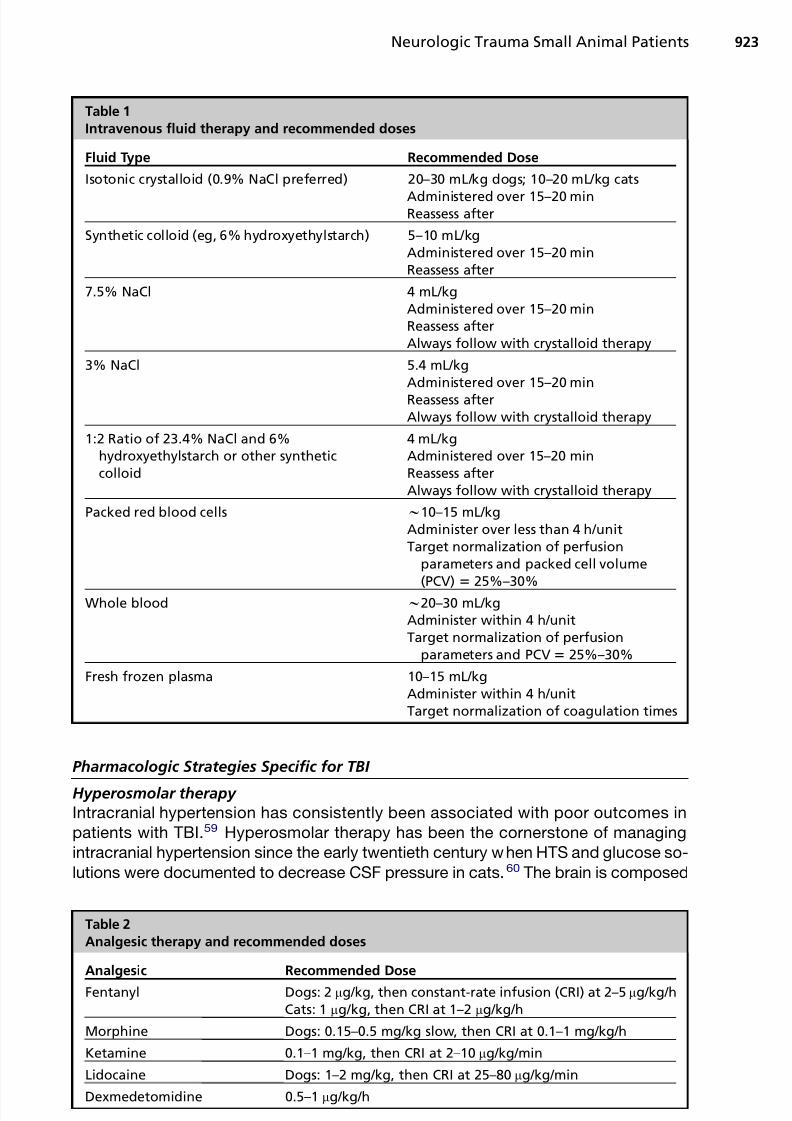
Intravenous fluid therapy
Controversy exists in veterinary medicine regarding the best choice of fluid for resus-
citation in the neurotrauma patient. Options for fluid resuscitation include isotonic crys-talloids, hypertonic solutions, artificial colloidal solutions, and blood products. Concern
exists particularly in TBI management regarding the injured brain’s capacity to protect
against increases in cerebral edema when faced with fluids containing large amounts
of free water caused by disruption of cellular tight junctions and subsequent influxes
of ions and larger, colloid-sized molecules with secondary osmotic pull.4 It is therefore
recommended that isotonic fluids containing the least amount of free water (eg, 0.9%
Neurologic Trauma Small Animal Patients 921

7/22/2019 Updates in the Management of the Small Animal Patient with Neurologic Trauma
http://slidepdf.com/reader/full/updates-in-the-management-of-the-small-animal-patient-with-neurologic-trauma 8/26
NaCl) be administered, barring significant sodium derangements already present on
presentation. Because fluid shifts between the interstitial and intracellular compart-
ments of the brain are predominantly dictated by osmolality as opposed to plasma
oncotic pressure, colloidal solutions have not demonstrated any significant benefit
over crystalloid therapy.44 However, owing to rapid redistribution of crystalloids after
administration, a combination of colloidal therapy with crystalloid therapy (either iso-
tonic or hypertonic) can be considered to provide longer-lasting volume resuscitation.7
Hypertonic saline (HTS) has several potential benefits in the neurologic trauma
patient (particularly the TBI patient), including rapidly increasing intravascular volume,
increasing cardiac output, improving regional cerebral and spinal cord blood flow by
dehydrating cerebrovascular endothelial cells, increasing vessel diameter, decreasing
ICP, and enhancing cerebral oxygen delivery.45–50 It is important that the concentra-
tion of HTS being used is noted, as this will affect the dosing of the solution. 4 HTS
should be used only in euhydrated patients without significant sodium derangements.
In addition, it is imperative that HTS be followed by crystalloid therapy to maintain
adequate tissue hydration.
Anemic patients should be treated with packed red blood cells or whole blood to
maintain adequate arterial oxygen content and oxygen delivery to damaged nervous
tissue. Transfusion goals include normalization of perfusion parameters (including
central venous oxygenation saturation >70%). Fresh frozen plasma should be admin-
istered to the coagulopathic patient.7 Those patients that do not respond to fluid
resuscitation warrant vasopressor support.7 After initial fluid resuscitation, fluid ther-
apy should be continued to account for maintenance needs, deficits, and any ongoing
losses. Recommended initial bolus dosing of intravenous fluids are listed in Table 1.
Pain management
Analgesic therapy is essential in the management of the neurotrauma patient. The de-
gree of analgesia and sedation must be balanced with preservation of blood pressure
and ventilatory status, as depression of each of these parameters can contribute to
secondary injury, and if possible should not impede reassessment of neurologic
status. Adequate analgesia in the TBI patient avoids transient increases in ICP caused
by pain and agitation, which can lead to increased cerebral metabolic rate and, conse-
quently, cerebral blood flow and volume.51
Opioids are the analgesic drugs of choice in critical care medicine, because of their
ease of reversal and relative safety when titrated to effect. Several studies suggest thatbolus infusion of opioids should be avoided, and constant-rate infusions (CRIs) are
preferred.51–55 Because of the ease of reversal, it is recommended that full agonist
opioids be used.56,57
Ketamine noncompetitively inhibits the N -methyl-D-aspartate (NMDA) receptor;
therefore, this agent may have neuroprotective properties against ischemic and
glutamate-induced injury in addition to its cardiovascular and respiratory-sparing
properties. Recent studies have failed to demonstrate that ketamine results in the
ICP increases typically reported in the older literature.51 However, it has been
shown to increase cerebral oxygen consumption, possibly through inhibition of
the g-aminobutyric acid (GABA) receptor. Therefore, administration with a GABA agonist (eg, propofol) potentially could decrease these negative effects.
Medetomidine, an a2-agonist, has been documented to have no effect on ICP in
anesthetized dogs.58 Caution must be exercised in using this class of drugs for their
analgesic or sedative properties, as they can cause clinically significant reductions
in heart rate and cardiac output, thereby affecting CNS perfusion.23 Table 2 lists
the recommended analgesics and their respective doses.
DiFazio & Fletcher922
7/22/2019 Updates in the Management of the Small Animal Patient with Neurologic Trauma
http://slidepdf.com/reader/full/updates-in-the-management-of-the-small-animal-patient-with-neurologic-trauma 9/26
Pharmacologic Strategies Specific for TBI
Hyperosmolar therapy
Intracranial hypertension has consistently been associated with poor outcomes in
patients with TBI.59 Hyperosmolar therapy has been the cornerstone of managing
intracranial hypertension since the early twentieth century when HTS and glucose so-
lutions were documented to decrease CSF pressure in cats.60 The brain is composed
Table 1
Intravenous fluid therapy and recommended doses
Fluid Type Recommended Dose
Isotonic crystalloid (0.9% NaCl preferred) 20–30 mL/kg dogs; 10–20 mL/kg cats
Administered over 15–20 minReassess after
Synthetic colloid (eg, 6% hydroxyethylstarch) 5–10 mL/kgAdministered over 15–20 minReassess after
7.5% NaCl 4 mL/kgAdministered over 15–20 minReassess afterAlways follow with crystalloid therapy
3% NaCl 5.4 mL/kgAdministered over 15–20 minReassess afterAlways follow with crystalloid therapy
1:2 Ratio of 23.4% NaCl and 6%hydroxyethylstarch or other syntheticcolloid
4 mL/kgAdministered over 15–20 minReassess afterAlways follow with crystalloid therapy
Packed red blood cells w10–15 mL/kgAdminister over less than 4 h/unitTarget normalization of perfusion
parameters and packed cell volume(PCV) 5 25%–30%
Whole blood w20–30 mL/kgAdminister within 4 h/unitTarget normalization of perfusion
parameters and PCV 5 25%–30%
Fresh frozen plasma 10–15 mL/kgAdminister within 4 h/unitTarget normalization of coagulation times
Table 2
Analgesic therapy and recommended doses
Analgesic Recommended Dose
Fentanyl Dogs: 2 mg/kg, then constant-rate infusion (CRI) at 2–5 mg/kg/hCats: 1 mg/kg, then CRI at 1–2 mg/kg/h
Morphine Dogs: 0.15–0.5 mg/kg slow, then CRI at 0.1–1 mg/kg/h
Ketamine 0.1–1 mg/kg, then CRI at 2–10 mg/kg/min
Lidocaine Dogs: 1–2 mg/kg, then CRI at 25–80 mg/kg/min
Dexmedetomidine 0.5–1 mg/kg/h
Neurologic Trauma Small Animal Patients 923
7/22/2019 Updates in the Management of the Small Animal Patient with Neurologic Trauma
http://slidepdf.com/reader/full/updates-in-the-management-of-the-small-animal-patient-with-neurologic-trauma 10/26
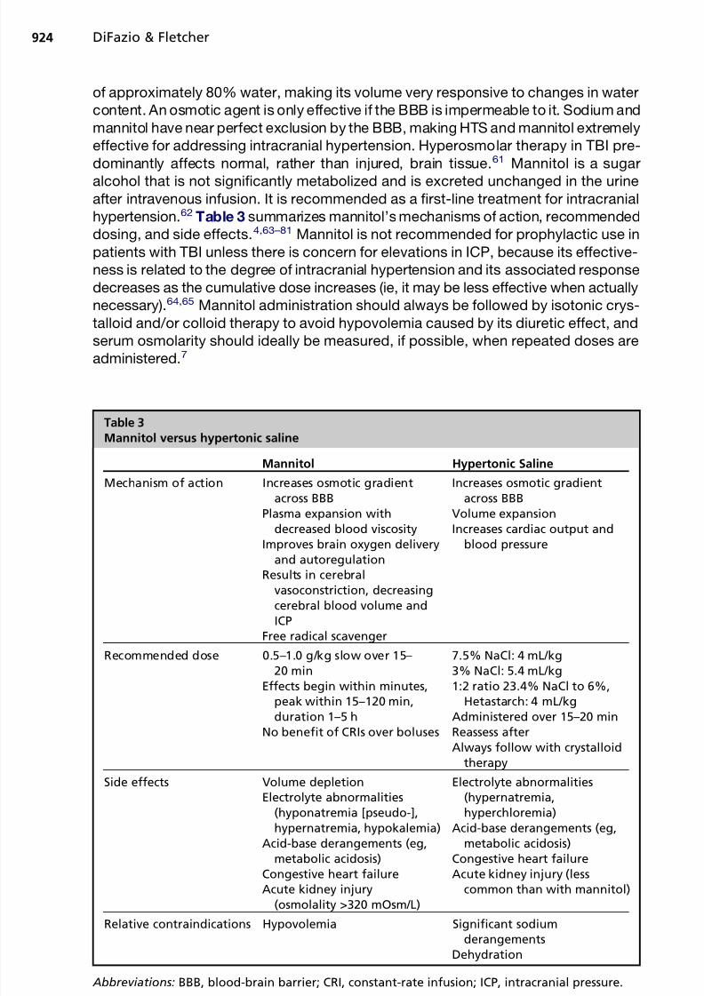
of approximately 80% water, making its volume very responsive to changes in water
content. An osmotic agent is only effective if the BBB is impermeable to it. Sodium and
mannitol have near perfect exclusion by the BBB, making HTS and mannitol extremely
effective for addressing intracranial hypertension. Hyperosmolar therapy in TBI pre-
dominantly affects normal, rather than injured, brain tissue.61 Mannitol is a sugar
alcohol that is not significantly metabolized and is excreted unchanged in the urine
after intravenous infusion. It is recommended as a first-line treatment for intracranial
hypertension.62 Table 3 summarizes mannitol’s mechanisms of action, recommended
dosing, and side effects.4,63–81 Mannitol is not recommended for prophylactic use in
patients with TBI unless there is concern for elevations in ICP, because its effective-
ness is related to the degree of intracranial hypertension and its associated response
decreases as the cumulative dose increases (ie, it may be less effective when actually
necessary).64,65 Mannitol administration should always be followed by isotonic crys-
talloid and/or colloid therapy to avoid hypovolemia caused by its diuretic effect, and
serum osmolarity should ideally be measured, if possible, when repeated doses are
administered.7
Table 3
Mannitol versus hypertonic saline
Mannitol Hypertonic Saline
Mechanism of action Increases osmotic gradientacross BBB
Plasma expansion withdecreased blood viscosity
Improves brain oxygen deliveryand autoregulation
Results in cerebralvasoconstriction, decreasingcerebral blood volume andICP
Free radical scavenger
Increases osmotic gradientacross BBB
Volume expansionIncreases cardiac output and
blood pressure
Recommended dose 0.5–1.0 g/kg slow over 15–20 min
Effects begin within minutes,
peak within 15–120 min,duration 1–5 h
No benefit of CRIs over boluses
7.5% NaCl: 4 mL/kg3% NaCl: 5.4 mL/kg1:2 ratio 23.4% NaCl to 6%,
Hetastarch: 4 mL/kgAdministered over 15–20 minReassess afterAlways follow with crystalloid
therapy
Side effects Volume depletionElectrolyte abnormalities
(hyponatremia [pseudo-],hypernatremia, hypokalemia)
Acid-base derangements (eg,metabolic acidosis)
Congestive heart failureAcute kidney injury
(osmolality >320 mOsm/L)
Electrolyte abnormalities(hypernatremia,hyperchloremia)
Acid-base derangements (eg,metabolic acidosis)
Congestive heart failure
Acute kidney injury (lesscommon than with mannitol)
Relative contraindications Hypovolemia Significant sodiumderangements
Dehydration
Abbreviations: BBB, blood-brain barrier; CRI, constant-rate infusion; ICP, intracranial pressure.
DiFazio & Fletcher924

7/22/2019 Updates in the Management of the Small Animal Patient with Neurologic Trauma
http://slidepdf.com/reader/full/updates-in-the-management-of-the-small-animal-patient-with-neurologic-trauma 11/26
Hypertonic saline therapy affords similar osmotic benefits as mannitol therapy, but
is a less potent diuretic. Table 3 summarizes HTS’s mechanisms of action, recom-
mended dosing, adverse effects, and relative contraindications.63,66 It is generally rec-
ommended that sodium concentrations be maintained at less than 160 mEq/L,
although concentrations of up to 180 mEq/L have been reported in humans treated
with hypertonic saline with no complications.67
There is no evidence that mannitol is superior to HTS in the treatment of intracranial
hypertension or vice versa. The small number of studies available have shown con-
flicting results.68–71 It is likely that HTS is preferable in hypovolemic patients, but in
euvolemic patients, either is reasonable. When patients do not respond to treatment
with one agent, the alternative should be considered.
Administration of furosemide alone or concurrently with mannitol to treat intracranial
hypertension has not been shown to have any additional benefit and increases the risk
of volume depletion.72 Therefore, its use is not recommended.
CorticosteroidsCorticosteroids were previously advocated for in the treatment of TBI patients on the
basis that corticosteroids decreased cerebral edema. Ground-breaking evidence from
the CRASH trials showed that high-dose methylprednisolone was associated with an
increase in mortality at 2 weeks and 6 months after injury.73,74 Corticosteroids are
therefore no longer recommended in the treatment of TBI patients.
Anticonvulsant therapy
Posttraumatic seizures are classified as immediate (occurring within 24 hours of
injury), early (occurring 24 hours to 7 days after injury), or late (occurring more than
7 days after injury).62 Seizures increase secondary brain injury by increasing cerebralmetabolic demands, increasing ICP, and leading to the release of excessive neuro-
transmitters. A recent Cochrane meta-analysis concluded that prophylactic antiepi-
leptic drugs are effective in reducing early seizures, but there is no evidence that
they are effective in preventing late-onset seizures. Therefore, prophylactic antiepi-
leptic drugs are recommended for 7 days post-TBI in humans.75 There are few data
in veterinary medicine, but if seizures develop, aggressive antiepileptic drug (AED)
therapy is indicated to reduce secondary brain injury. The incidence of posttraumatic
seizures in small animals is not well documented, but seizures are known to occur. At
present, there are no clear recommendations for prophylactic AED therapy in veteri-
nary medicine. If risk factors for seizures are present (eg, penetrating head wounds,depressed skull fractures, and so forth), it is reasonable to consider prophylactic
AED therapy for the first 7 days after injury, based on human recommendations.
The duration of AED therapy is debatable. Should seizures occur, benzodiazepines
should be used as a first-line treatment to stop seizure activity.76 A variety of AEDs
for continued seizure control are available for use in dogs and cats, and these are
listed in Table 4. At the authors’ institution, levetiracetam is frequently used for its
rapid onset of action, minimal side effects, and low toxicity potential.
Barbiturate therapy
Barbiturates are considered secondary therapy for the treatment of refractory intracra-nial hypertension in people, as high-dose therapy can control ICP when other medical
and surgical therapies have failed. However, no outcome benefit has been docu-
mented.62 The neuroprotective effects of barbiturates are related to their ability to
cause cerebral vasoconstriction, decrease cerebral metabolism, reduce ICP, decrease
excitotoxicity, and decrease free radical–mediated injury.77 Barbiturates also have
anticonvulsant properties. Complications associated with the use of barbiturates
Neurologic Trauma Small Animal Patients 925

7/22/2019 Updates in the Management of the Small Animal Patient with Neurologic Trauma
http://slidepdf.com/reader/full/updates-in-the-management-of-the-small-animal-patient-with-neurologic-trauma 12/26
include cardiovascular and respiratory depression, with potentially clinically significant
hypotension (and associated decreases in CPP) and hypoventilation. The most widely
used barbiturate for TBI is pentobarbital.4 Barbiturate coma (induced with drugs such
as phenobarbital or other sedatives) was recently described in association with thera-
peutic hypothermia (TH) in a dog with TBI and refractory seizure activity.78 Patients in
which barbiturate therapy is instituted must be monitored closely for hypoventilation,
and may require mechanical ventilation.
Novel therapies
New therapies targeting excitotoxicity and production of reactive oxygen species arecurrently being investigated in human medicine, but none have been examined in vet-
erinary practice to date. A recent randomized controlled trial of amantadine in vegeta-
tive or minimally conscious humans recovering from TBI showed significantly faster
functional recovery over a 4-week period in treated patients.79 In the near future,
more options specifically targeting secondary injury may become available to veteri-
nary practitioners.
Table 4
Antiepileptic drugs and recommended doses
Antiepileptic Drug Recommended Dose
Diazepam 0.5 mg/kg IV, rectally, intranasally
CRI 0.2–1 mg/kg/hPhenobarbital Loading 12–20 mg/kg IV, PO divided q 4–6 h over
24 hDogs: 2.5 mg/kg IV, PO q 12 h initiallyTargeting trough levels of 20–30 mg/mLCats: 1–2 mg/kg IV, PO q 24 h initiallyTargeting trough levels of 10–20 mg/mL
Potassium bromide Loading 400–600 mg/kg KBr PO/rectally dividedq 6–12 h over 24–48 h
Loading 600–1200 mg/kg NaBr IV as CRI over8–24 h
30–50 mg/kg/d as a monotherapy15–30 mg/kg/d as an add-on drugTargeting trough levels of 0.8–3 mg/mLNot recommended in cats (because of high risk
of lower airway signs)
Levetiracetam (Keppra) 20–30 mg/kg IV, PO q 8 hNo load necessary
Zonisamide Loading 10 mg/kg PO q 12 h  3 d5–10 mg/kg q 12 h thereafterTargeting therapeutic range described for
humans (10–40 mg/mL)
Gabapentin Dogs: 30–60 mg/kg q 24 h divided q 8–12 hCats: 5–10 mg/kg q 12–24 h
Propofol 1–4 mg/kg IV bolusCRI 0.05–0.4 mg/kg/min
PentobarbitalAntiepileptic drug used most commonly
for induction of barbiturate coma
2–15 mg/kg bolus over 20 minCRI 0.2–1 mg/kg/h
Abbreviations: CRI, constant-rate infusion; IV, intravenously; PO, by mouth; q, every.
DiFazio & Fletcher926
7/22/2019 Updates in the Management of the Small Animal Patient with Neurologic Trauma
http://slidepdf.com/reader/full/updates-in-the-management-of-the-small-animal-patient-with-neurologic-trauma 13/26
Pharmacologic Strategies Specific for Acute SCI
Corticosteroids
The use of corticosteroids in the treatment of acute SCI in humans and animals
remains controversial despite extensive clinical research. Proposed mechanisms
supporting the use of corticosteroids in SCI include free radical scavenging, anti-inflammatory effects, and improved regional blood flow.13,15 Much of the clinical
and experimental research on corticosteroid therapy in SCI has focused on methyl-
prednisolone sodium succinate (MPSS). The main neuroprotective property of
MPSS appears to be its free radical–scavenging ability. Other corticosteroids (eg,
prednisone and dexamethasone) lack this property, and are unlikely to have any bene-
ficial effect in the treatment of secondary SCI.80 Specific evaluation of dexamethasone
therapy in dogs has also failed to reveal any benefit in SCI, either experimentally or
clinically.81,82 A series of 3 human clinical trials (National Acute Spinal Cord Injury
Studies [NASCIS] I–III) provide the majority of primary evidence relating to the use
of MPSS for the treatment of acute SCI.83–85
None of these studies convincinglydemonstrated a benefit of steroids in improving motor function scores, as most of
the statistically significant results were based on post hoc subgroup analyses.86 An
experimental study in dogs comparing urgent surgical decompression with MPSS
for the treatment of experimentally induced SCI showed that surgical decompression
6 hours after injury (with or without MPSS) resulted in better neurologic outcomes than
treatment with MPSS alone.87 Another experimental dog study showed no improve-
ment in outcome with MPSS administration.88 There are no published clinical
placebo-controlled trials evaluating the efficacy of MPSS in the treatment of SCI in
dogs, although there is currently one under way.16 Given the potential for significant
adverse side effects, such as gastrointestinal ulceration, immunosuppression, andcompromise of renal perfusion in hypovolemic patients, the routine administration of
corticosteroids (including MPSS) is not recommended.13
Box 3 summarizes additional pharmacologic therapies directed at minimizing
secondary injury in acute SCI that have been investigated.89–101
NONPHARMACOLOGIC STRATEGIESSystemic Therapy
Airway management and ventilation
The upper airway should be directly examined and suctioned if necessary when aneurotrauma patient is initially presented. If the airway is deemed nonpatent or if the
patient is unable to control its airway, immediate endotracheal intubation or emergency
tracheostomy (if unable to intubate) is indicated.7 Carbon dioxide has profound effects
on cerebral and spinal cord blood flow and blood volume.102 Both hypoventilation and
hyperventilation should be avoided in neurotrauma patients, and close monitoring of
CO2 using end-tidal CO2 (ETCO2 ) monitors or blood-gas analysis is warranted. Normal
partial pressure of carbon dioxide (PCO2 ) (venous, 40–45 mm Hg; arterial, 35–40 mm Hg)
should be targeted in all cases.62 Titration of analgesic medication, positioning in ster-
nal recumbency, and ensuring that the airway is unobstructed can help address venti-
lation issues, but if these interventions are unsuccessful, endotracheal intubation andmechanical ventilation are indicated. Those patients at risk of cerebral herniation or that
have experienced significant neurologic decompensation can be hyperventilated for
short periods of time. However, the targets of short-term hyperventilation should be
conservative (ETCO25 30–35 mm Hg) to prevent excessive cerebral vasoconstriction
and ischemic brain injury. Studies evaluating prophylactic hyperventilation during initial
resuscitation consistently have shown poor outcomes.103–105
Neurologic Trauma Small Animal Patients 927

7/22/2019 Updates in the Management of the Small Animal Patient with Neurologic Trauma
http://slidepdf.com/reader/full/updates-in-the-management-of-the-small-animal-patient-with-neurologic-trauma 14/26
Box 3
Pharmacologic agents for the treatment of secondary injury in acute SCI
Agents Directed at Free Radical Injury
Vitamin E and selenium
Pretreatment of cats before SCI resulted in improved neurologic outcome and spinal cordblood flow following injury
Tirilazad (21-aminosteroid)
Reduced spinal cord ischemia in cats
Effect not documented in dogs
N -Acetylcysteine
No improvement in outcome in dogs with intervertebral disk herniation
Sulfoxide and ε-aminocaproic acid
No improvement in outcome in dogs
Antioxidants require a prolonged period of administration to achieve therapeutic concentra-tions within the CNS, limiting their use in the acute phase of SCI
Agents Directed at Ionic Disturbances and Excitotoxicity
Verapamil, diltiazem, nifedipine (calcium-channel antagonists)
Improved spinal cord blood flow in cats postinjury with diltiazem and nifedipine; notverapamil
Sodium-channel blockers
Beneficial effects noted in experimental models
NMDA and non-NMDA glutamate receptor antagonists
Delayed administration improved tissue sparing and functional recovery in rodent models
Most have adverse side effects and have largely failed clinical trials
Agents Directed at Inflammation
Minocycline (second-generation tetracycline derivative)
Reduces activation of microglia and macrophages experimentally
Not yet evaluated in a trial
Tacrolimus, cyclosporine, mycophenolate mofetil
Neuroprotective effects in experimental models of injury
Polyethylene Glycol
Hydrophilic polymer that seals damaged neuronal membranes
Evaluated in deep pain perception negative dogs; resulted in restored function in 60% ofdogs
Randomized, controlled trial in dogs currently underway
4-Aminopyridine
Potassium-channel blocker
Improves conduction by blocking the channels that would ordinarily be blocked by intactmyelin
Improves conduction in vitro and vestibulospinal reflexes in cats
Phase I clinical trial with 39 dogs with SCI
DiFazio & Fletcher928

7/22/2019 Updates in the Management of the Small Animal Patient with Neurologic Trauma
http://slidepdf.com/reader/full/updates-in-the-management-of-the-small-animal-patient-with-neurologic-trauma 15/26
Supportive care
Supportive care of the patient with neurologic trauma should include the provision of
dry, clean bedding; frequent turning; passive range of motion exercises; bladder care;
ocular care (eg, frequent lubrication and inspection for corneal ulceration); and nutri-
tion. Hypermetabolic states have been documented in patients with neurotrauma, and
early feeding is recommended.106 The method of feeding should be based on the
assessment of a patient’s ability to protect the airway. Although hyperglycemia has
been associated with worsened mortality rates and neurologic outcome scores in
humans with TBI107 and degree of hyperglycemia has been associated with severity
of TBI in animals,108 intensive insulin therapy is not recommended for the control of
hyperglycemia, based on the available human literature.109–111
Bladder dysfunction is common in SCI patients, and depends on the location andseverity of the lesion. Table 5 summarizes therapies for upper and lower motor neuron
bladder dysfunction.112 Bladder care should include frequent (minimum every
12 hours) manual bladder expression or urinary catheterization. Compared with
indwelling catheterization, intermittent urinary catheterization using a diligent aseptic
technique is associated with a decreased risk of urinary tract infection, and there is no
increased risk of infection with intermittent catheterization in comparison with manual
expression.113,114
Nonpharmacologic Strategies Specific for TBI
Decreasing cerebral blood volumeElevation of the head by 15 to 30 reduces cerebral blood volume, thereby decreasing
ICP and increasing CPP without harmful reductions in cerebral oxygenation.115 A
stiff slant board should be used for head elevation to avoid bending of the neck and
occlusion of the jugular veins. Elevation should not exceed 30, as this can contribute
to a decrease in CPP with associated effects on cerebral oxygenation.7
64% of dogs had transient improvements in neurologic function
Side effects rare, but significant
Additional Therapies
Naloxone (opiate receptor antagonist)
Failed to yield a benefit in human clinical trial
Thyrotropin-receptor antagonist
Failed to yield a benefit in canine clinical trial
Erythropoietin, progesterone and estrogen, magnesium, atorvastatin, melatonin
Table 5
Pharmacologic agents for bladder dysfunction
Upper Motor Neuron Bladder Lower Motor Neuron Bladder
a-Adrenergic antagonistPrazosinPhenoxybenzamine
ParasympathomimeticBethanechol
Skeletal muscle relaxantDiazepam
a-Adrenergic agonistPhenylpropanolamine
Neurologic Trauma Small Animal Patients 929

7/22/2019 Updates in the Management of the Small Animal Patient with Neurologic Trauma
http://slidepdf.com/reader/full/updates-in-the-management-of-the-small-animal-patient-with-neurologic-trauma 16/26
Therapeutic hypothermia
Many of the processes of neurotraumatic secondary injury are temperature depen-
dent.116 TH, targeting temperatures of 32 to 34C (89.6–93.2F), decreases basal
and cerebral metabolism, prevents apoptosis and necrosis and decreases cerebral
edema formation and disruption of the BBB by decreasing release of excitotoxic amino
acids (EAAs), decreases production of proinflammatory cytokines, and decreases
excitatory signaling that can result in seizure activity.117 Therapeutic hypothermia is
indicated as a second-line treatment in humans for intracranial hypertension and
status epilepticus116,117; however, it has not gained widespread acceptance as a
first-line treatment for TBI, likely because it is challenging to implement and has
many potential complications.116,117 Few veterinary practices have the facilities to offer
TH, but its successful use in a dog with protracted seizure activity associated with TBI
has been described.78 As technologies for inducing TH and supporting patients being
treated become more accessible in veterinary medicine, this therapy may gain more
acceptance.
Decompressive craniectomy
Early craniotomy is indicated for evacuation of extra-axial hematomas. The role of
decompressive craniectomy is more controversial in the management of TBI patients.
It may be performed prophylactically at the time of mass evacuation if early crani-
otomy is performed, or later as a second-line, rescue therapy when medical man-
agement has failed.118 The results of the ongoing RESCUEicp trial may provide
additional insight into the role of early decompressive craniectomy in the management
of patients with TBI.119
Nonpharmacologic Strategies Specific for Acute SCI Surgery is usually indicated in patients with moderate to severe deficits, neurologic
deterioration, and/or instability of the vertebral column. Controversy exists regarding
the best timing for surgical intervention in spinal cord trauma. Many traumatic SCI
patients have extraspinal injuries that require initial stabilization. Nevertheless, earlier
surgical treatment of human patients with traumatic SCI has been associated with
improved outcomes.120–122
Management of vertebral fracture and luxation
The stability of spinal fractures in both humans and veterinary patients can be
assessed using a 3-compartment model. This model divides the vertebral columninto dorsal, middle, and ventral compartments, and a fracture is considered unstable
when 2 or more compartments are disrupted. Table 6 lists the structures included in
each of the 3 compartments.123,124
General indications for surgical management include moderate to severe neurologic
deficits (eg, minimal to no motor function), evidence of vertebral column instability on
imaging, and neurologic deterioration despite aggressive conservative manage-
ment.13 Spinal cord decompression, reduction, and fixation are the aims of surgical
management. There are many surgical techniques for stabilization of VFLs after
reduction. The technique chosen depends on the location within the vertebral column,
the type of fracture, and surgeon’s preference.123 Stabilization techniques includebone plates, screws, Steinmann pins, Kirschner wires, and polymethylmethacrylate
(PMMA) cement.13 VFLs should be managed by board-certified orthopedic surgeons
or neurosurgeons. Although surgical management is often indicated, return to function
is generally shorter, and postoperative supportive care is generally less intensive
than that required in conservative management, complications can occur, potentially
leading to worsening of SCI. Worsening of SCI can occur with surgical manipulation,
DiFazio & Fletcher930

7/22/2019 Updates in the Management of the Small Animal Patient with Neurologic Trauma
http://slidepdf.com/reader/full/updates-in-the-management-of-the-small-animal-patient-with-neurologic-trauma 17/26
loosening of implants, and implant failure or infection (particularly with PMMA). Revi-sional surgeries are, therefore, sometimes required.13
Conservative management of primary injury is most appropriate for patients with
minimal deficits, static disease, and good 3-compartment stability. External coapta-
tion has been described, but application of these devices is fraught with complications
including inadequate stabilization, increased mobility of VFL sites, and abrasions and
ulcerations. Conservative management, therefore, usually consists of strict cage rest
for 6 to 8 weeks, analgesia, and nursing care.
Traumatic intervertebral disk herniation
Patients who are nonambulatory, have progressive neurologic deficits, are nonrespon-sive to conservative management, or have cervical lesions causing severe pain should
be considered for surgical management.34,125,126 With Type III (traumatic) interverte-
bral disk disease, surgical therapy is often not indicated unless there is an associated
compressive extra-axial hematoma. Depending on the site, degree of lateralization,
and severity of herniation, dorsal laminectomy, hemilaminectomy or ventral slot proce-
dures may be indicated.13,34,127–132
Conservative management can be used in treating patients with hyperpathia (neuro-
pathic pain) alone or with minimal neurologic deficits.13,34 It is recommended that strict
cage rest be instituted for a minimum of 4 to 6 weeks, although a recent retrospective
evaluation showed that the duration of rest in the management of thoracolumbar diskdisease had no effect on outcome or quality-of-life scores.133 Conservative manage-
ment is inappropriate for patients that are surgical candidates based on imaging and/
or have lost deep pain perception (DPP).34
Spinal cord contusion and extra-axial hemorrhage
A spinal cord contusion is an intraparenchymal hemorrhage that occurs most
commonly secondary to other causes of primary injury including VFLs, intervertebral
disk extrusion, and penetrating injuries.13,134 Therapy for parenchymal contusions is
aimed at treating concurrent primary injury (see earlier discussion).13 Extra-axial hem-
orrhage can occur epidurally or subdurally, and can cause direct compression to the
cord. It has also been reported secondary to intervertebral disk extrusion.135–137
Decompressive surgical techniques are recommended in patients with compressive
extra-axial hematomas.13
Cellular transplantation therapy
Intraspinal olfactory glial cell transplantation was evaluated in a phase I trial in 9 dogs
with thoracolumbar SCI caused by VFLs secondary to vehicular trauma or disk
Table 6
Three-compartment model for assessing stability of VFLs
Dorsal Compartment Middle Compartment Ventral Compartment
Vertebral arch (spinous process, articular
processes, laminae, pedicles)
Dorsal longitudinal
ligament
Ventral portion of the
vertebral bodyDorsal ligamentous complex (facet joint
capsule, interarcuate ligaments,interspinous ligaments, supraspinousligaments)
Dorsal aspect of theannulus fibrosus
Nucleus pulposus
Intertransverse ligaments Dorsal cortex of thevertebral body
Ventral aspect of theannulus fibrosus
Ventral longitudinalligament
Neurologic Trauma Small Animal Patients 931

7/22/2019 Updates in the Management of the Small Animal Patient with Neurologic Trauma
http://slidepdf.com/reader/full/updates-in-the-management-of-the-small-animal-patient-with-neurologic-trauma 18/26
herniation (8 of 9 dogs were DPP negative). Significant functional improvement was
noted in this small population of dogs, with 7 of 8 having improved motor function.138
A recent investigation of the safety of autologous bone marrow stromal cell transplan-
tation in 7 dogs with severe acute SCI caused by VFL found this technique to be
feasible and safe, with no complications noted. Two of 7 dogs were able to walk
without support during follow-up (29–62 months after SCI).139 Although these tech-
niques are currently in the early stages of development, they may represent new alter-
natives for patients with acute SCI.
Prognosis
Prognosis for recovery from neurotrauma depends on the severity of injury, the cause
of injury, the site of the lesion, and the timing and efficacy of the treatment of primary
and secondary injury.13 Small animals with neurologic trauma can demonstrate signif-
icant neurologic improvement and have a tremendous ability to compensate for
neurologic deficits, so serial neurologic reassessments are recommended regardlessof the presentation of the patient.
The MGCS can be used to assess prognosis for recovery in dogs with TBI, as it has
been documented to be linearly associated with 48-hour survival.25 The presence of
DPP has consistently been associated with improved outcome in acute SCI. A retro-
spective study evaluating severe thoracolumbar SCI in dogs showed that only 12%
of dogs with VFL regained the ability to walk, whereas 69% of dogs with injury attribut-
able to intervertebral disk herniation regained motor function.140 Surgical management
of cases with thoracolumbar disk herniation and intact pain perception is excellent,
with expected return of functional motor activity being 80% to 95% (reports range
from 72% to 100%).141
In one study of surgically managed cervical disk herniation,the overall success rate was 99%.126 A retrospective study of dogs with cervical
VFLs found that injuries that could be managed conservatively (ie, nonsurgically) had
good functional outcomes in 90% of cases.142 Those requiring surgical management
had a high risk of perioperative mortality (36%), but in those cases that survived the
perioperative period the prognosis was excellent (100%). This same study also showed
that nonambulatory patients and patients presented at more than 5 days after injury
had worse prognoses.142 Prognostic studies in cats are limited, therefore much of
the relevant information is extrapolated from dogs.143 Finally, neurotrauma rarely oc-
curs independent of other systemic injury. It is therefore crucial to take the entire clinical
picture of the patient into account when assessing prognosis and guiding owners intheir decision-making process.
REFERENCES
1. Simpson S, Syring R, Otto C. Severe blunt trauma in dogs: 235 cases (1997-
2003). J Vet Emerg Crit Care (San Antonio) 2009;19(6):588–602.
2. Marioni-Henry K, Vite CH, Newton AL, et al. Prevalence of diseases of the spinal
cord of cats. J Vet Intern Med 2004;18(6):851–8.
3. Fluehmann G, Doherr MG, Jaggy A. Canine neurological diseases in a referral
hospital population between 1989 and 2000 in Switzerland. J Small Anim Pract2006;47(10):582–7.
4. Sande A, West C. Traumatic brain injury: a review of pathophysiology and man-
agement. J Vet Emerg Crit Care (San Antonio) 2010;20(2):177–90.
5. Dewey C. Emergency management of the head trauma patient. Vet Clin North
Am Small Anim Pract 2000;30:207–25.
6. Hopkins A. Head trauma. Vet Clin North Am Small Anim Pract 1996;26:875–91.
DiFazio & Fletcher932

7/22/2019 Updates in the Management of the Small Animal Patient with Neurologic Trauma
http://slidepdf.com/reader/full/updates-in-the-management-of-the-small-animal-patient-with-neurologic-trauma 19/26
7. Dewey C, Fletcher D. Head trauma management. In: Dewey C, editor. A practical
guide to canine and feline neurology. 2nd edition. Ames (IA): Wiley-Blackwell;
2008. p. 221–35.
8. Aries M, Czrosynka M, Budohoski K, et al. Continuous determination of optimal
cerebral perfusion pressure in traumatic brain injury. Crit Care Med 2012;40(8):
2456–63.
9. Ling G, Marshall S, Moore D. Diagnosis and management of traumatic brain
injury. Continuum 2010;16(6):27–40.
10. Kim J, Gean A. Imaging for the diagnosis and management of traumatic brain
injury. Neurotherapeutics 2011;8(1):39–53.
11. Tsang K, Whitfield P. Traumatic brain injury: review of current management stra-
tegies. Br J Oral Maxillofac Surg 2012;50:298–308.
12. Proulx J, Dhupa N. Severe brain injury: part I: pathophysiology. Compend
Contin Educ Vet 1998;20:897–905.
13. Fletcher DJ, Dewey CW. Spinal trauma management. In: Dewey CW, editor.
A practical guide to canine and feline neurology. 2nd edition. Ames (IA): Wiley-
Blackwell; 2008. p. 405–17.
14. Johnson K, Vite C. Spinal cord injury. In: Silverstein D, Hopper K, editors. Small
animal critical care medicine. 1st edition. St Louis (MO): Elsevier Saunders;
2009. p. 419–23.
15. Park EH, White GA, Tieber LM. Mechanisms of injury and emergency care of
acute spinal cord injury in dogs and cats. J Vet Emerg Crit Care (San Antonio)
2012;22(2):160–78.
16. Olby N. The pathogenesis and treatment of acute spinal cord injuries in dogs.
Vet Clin North Am Small Anim Pract 2010;40(5):791–807.17. Fletcher D, Syring R. Traumatic brain injury. In: Silverstein D, Hopper K, editors.
Small animal critical care medicine. 1st edition. St Louis (MO): Elsevier Saun-
ders; 2009. p. 658–62.
18. Leonard SE, Kirby R. The role of glutamate, calcium and magnesium in sec-
ondary brain injury. J Vet Emerg Crit Care 2002;12(1):17–32.
19. McMichael M, Moore R. Ischemia and reperfusion injury pathophysiology, part I.
J Vet Emerg Crit Care (San Antonio) 2004;14(4):231–41.
20. Oyinbo C. Secondary injury mechanisms in traumatic spinal cord injury: a
nugget of this multiply cascade. Acta Neurobiol Exp 2011;71(2):281–99.
21. Tator C, Fehlings M. Review of the secondary injury theory of acute spinal cordtrauma with emphasis on vascular mechanisms. J Neurosurg 1991;75(1):15–26.
22. Dooney N, Dagal A. Anesthetic considerations in acute spinal cord trauma. Int J
Crit Illn Inj Sci 2011;1(1):36–43.
23. Armitage-Chan E, Wetmore L, Chan D. Anesthetic management of the head
trauma patient. J Vet Emerg Crit Care (San Antonio) 2007;17(1):5–14.
24. Shores A. Craniocerebral trauma. In: Kirk R, editor. Current veterinary therapy X.
X. Philadelphia: WB Saunders; 1983. p. 847–54.
25. Platt S, Radaelli S, McDonnell J. The prognostic value of the Modified Glasgow
Coma Scale in head trauma in gods. J Vet Intern Med 2001;15(6):581–4.
26. Levine GJ, Levine JM, Budke CM, et al. Description and repeatability of anewly developed spinal cord injury scale for dogs. Prev Vet Med 2009;
89(1–2):121–7.
27. Olby N, De Risio L, Munana K, et al. Development of a functional scoring system
in dogs with acute spinal cord injuries. Am J Vet Res 2001;62(10):1624–8.
28. Smith P, Jeffery N. Spinal shock—comparative aspects and clinical relevance.
J Vet Intern Med 2005;19:788–93.
Neurologic Trauma Small Animal Patients 933

7/22/2019 Updates in the Management of the Small Animal Patient with Neurologic Trauma
http://slidepdf.com/reader/full/updates-in-the-management-of-the-small-animal-patient-with-neurologic-trauma 20/26
29. Boysen S, Rozanski E, Tidwell A, et al. Evaluation of a focused assessment with
sonography for trauma protocol to detect free abdominal fluid in dogs involved
in motor vehicle accidents. J Am Vet Med Assoc 2004;225(8):1198–204.
30. Lisciandro GR. Abdominal and thoracic focused assessment with sonography
for trauma, triage, and monitoring in small animals. J Vet Emerg Crit Care
(San Antonio) 2011;21(2):104–22.
31. Kinns J, Mai W, Seiler G, et al. Radiographic sensitivity and negative predictive
value for acute canine spinal trauma. Vet Radiol Ultrasound 2006;47(6):563–70.
32. Vitale C, Coates J. Acute spinal injury. Compendium Standards of Care: Emer-
gency and Critical Care Medicine 2007;9(7):1–11.
33. Lamb CR, Nicholls A, Targett M, et al. Accuracy of survey radiographic diagnosis
of intervertebral disc protrusion in dogs. Vet Radiol Ultrasound 2002;43(3):222–8.
34. Brisson BA. Intervertebral disc disease in dogs. Vet Clin North Am Small Anim
Pract 2010;40(5):829–58.
35. Israel SK, Levine JM, Kerwin SC, et al. The relative sensitivity of computed to-
mography and myelography for identification of thoracolumbar intervertebral
disk herniations in dogs. Vet Radiol Ultrasound 2009;50(3):247–52.
36. Olby N, Dyce J, Houlton J. Correlation of plain radiographic and lumbar myelo-
graphic findings with surgical findings in thoracolumbar disc disease. J Small
Anim Pract 1994;35(7):345–50.
37. Griffin J, Levine J, Kerwin S. Canine thoracolumbar intervertebral disk disease:
pathophysiology, neurologic examination, and emergency medical therapy.
Compend Contin Educ Vet 2009;31(3):E1–13.
38. Lewis D, Hosgood G. Complications associated with the use of iohexol for mye-
lography of the cervical vertebral column in dogs: 66 cases (1988-1990). J AmVet Med Assoc 1992;200(9):1381–4.
39. Barone G, Ziemer L, Shofer F, et al. Risk factors associated with the develop-
ment of seizures after use of iohexol for myelography in dogs: 182 cases
(1998). J Am Vet Med Assoc 2002;220(10):1499–502.
40. Johnson P, Beltran E, Dennis R, et al. Magnetic resonance imaging characteris-
tics of suspected vertebral instability associated with fracture or subluxation in
eleven dogs. Vet Radiol Ultrasound 2012;53(5):552–9.
41. Rozanski EA, Rondeau MP. Respiratory pharmacotherapy in emergency and
critical care medicine. Vet Clin North Am Small Anim Pract 2002;32(5):1073–86.
42. Mazzaferro E. Oxygen therapy. In: Silverstein D, Hopper K, editors. Small animalcritical care medicine. 1st edition. St Louis(MO): Elsevier Saunders; 2009. p. 78–81.
43. Hopper K. Basic mechanical ventilation. In: Silverstein D, Hopper K, editors.
Small animal critical care medicine. 1st edition. St Louis (MO): Elsevier Saun-
ders; 2009. p. 900–4.
44. Zhuang J, Shackford S, Schmoker J, et al. Colloid infusion after brain injury: an
effect on intracranial pressure, cerebral blood flow, and oxygen delivery. Crit
Care Med 1995;23(1):140–8.
45. Wade C, Grady J, Kramer G, et al. Individual patient cohort analysis of the
efficacy of hypertonic saline/dextran in patients with traumatic brain injury and
hypotension. J Trauma 1997;42:561–5.46. Vassar M, Perry C, Gannaway W, et al. Analysis of potential risks associated with
7.5% sodium chloride resuscitation of traumatic shock. Arch Surg 1990;124:
1309–15.
47. Vassar M, Perry C, Gannaway W, et al. 7.5% sodium chloride/dextran for resus-
citation of trauma patients undergoing helicopter transport. Arch Surg 1991;126:
1065–72.
DiFazio & Fletcher934
7/22/2019 Updates in the Management of the Small Animal Patient with Neurologic Trauma
http://slidepdf.com/reader/full/updates-in-the-management-of-the-small-animal-patient-with-neurologic-trauma 21/26
48. Vassar M, Perry C, Holcroft J. Prehospital resuscitation of hypotensive trauma
patients with 7.5% NaCl versus 7.5% NaCl with added dextran: a controlled trial.
J Trauma 1993;34:622–32.
49. Vassar M, Fisher R, O’Brien P, et al. A multicenter trial for resuscitation of injured
patients with 7.5% sodium chloride. Arch Surg 1993;128:1003–11.
50. Cooper J, Myles P, McDermott F, et al. Prehospital hypertonic saline resuscita-
tion of patients with hypotension and severe traumatic brain injury. JAMA
2004;291:1350–7.
51. Roberts D, Hall R, Kramer A. Sedation for critically ill adults with severe trau-
matic brain injury: a systematic review of randomized controlled trials. Crit
Care Med 2012;39(12):2743–51.
52. Sperry R, Bailey P, Reichman M, et al. Fentanyl and sulfentanil increase intracra-
nial pressure in head trauma patients. Anesthesiology 1992;77:416–20.
53. Lauer K, Connolly L, Schmeling W. Opioid sedation does not alter intracranial
pressure in head injured patients. Can J Anaesth 1997;44:929–33.
54. Albanese J, Viviand X, Potie F, et al. Sulfentanil, fentanyl, and alfentanil in head
trauma patients: a study on cerebral hemodynamics. Crit Care Med 1999;27:
407–11.
55. de Nadal M, Munar F, Poca M, et al. Cerebral hemodynamic effects of morphine
and fentanyl in patients with severe head injury: absence of correlation to cere-
bral autoregulation. Anesthesiology 2000;92:11–9.
56. Hansen B. Acute pain management. Vet Clin North Am Small Anim Pract 2000;
30(4):899–916.
57. Pascoe P. Opioid analgesics. Vet Clin North Am Small Anim Pract 2000;30(4):
757–69.58. Keegan R, Green S, Bagley R, et al. Effects of medetomidine administration on
intracranial pressure and cardiovascular variables of isoflurane-anesthetized
dogs. Am J Vet Res 1996;56(2):193–8.
59. Treggiari M, Schultz N, Yanez N, et al. Role of intracranial pressure values and
patterns in predicting outcome of traumatic brain injury: a systematic review.
Neurocrit Care 2007;6:104–12.
60. Weed L, McKibbon P. Pressure changes in the cerebrospinal fluid following
intravenous injection of solutions of various concentrations. Am J Phys 1919;
48:512–30.
61. Ropper A. Hyperosmolar therapy for raised intracranial pressure. N Engl J Med2012;367(8):746–52.
62. Brain Trauma Foundation, American Association of Neurological Surgeons,
Congress of Neurological Surgeons. Guidelines for the management of severe
traumatic brain injury. J Neurotrauma 2007;24(1):S1–106.
63. Fink M. Osmotherapy for intracranial hypertension: mannitol versus hypertonic
saline. Continuum 2012;18(3):640–54.
64. Kaufmann A, Cardoso E. Aggravation of vasogenic cerebral edema by multiple-
dose mannitol. J Neurosurg 1992;77(4):584–9.
65. McManus M, Soriano S. Rebound swelling of astroglial cells exposed to hyper-
tonic mannitol. Anesthesiology 1998;88(6):1586–91.66. Oddo M, Levine J, Frangos S, et al. Effect of mannitol and hypertonic saline
on cerebral oxygenation in patients with severe traumatic brain injury and re-
fractory intracranial hypertension. J Neurol Neurosurg Psychiatr 2009;80(8):
916–20.
67. Aiyagari V, Deibert E, Diringer M. Hypernatremia in the neurologic intensive care
unit: how high is too high? J Crit Care 2006;21(2):163–72.
Neurologic Trauma Small Animal Patients 935

7/22/2019 Updates in the Management of the Small Animal Patient with Neurologic Trauma
http://slidepdf.com/reader/full/updates-in-the-management-of-the-small-animal-patient-with-neurologic-trauma 22/26
68. Battison C, Andrews P, Graham C, et al. Randomized, controlled trial on the
effect of a 20% mannitol solution and a 7.5% saline/6% dextran solution on
increased intracranial pressure after brain injury. Crit Care Med 2005;33(1):
196–202.
69. Francony C, Fauvage B, Falcon D, et al. Equimolar doses of mannitol and hyper-
tonic saline in the treatment of intracranial pressure. Crit Care Med 2008;36(3):
795–800.
70. Zeng H, Wang Q, Deng Y, et al. A comparative study on the efficacy of 10%
hypertonic saline and equal volume of 20% mannitol in the treatment of exper-
imentally induced cerebral edema in adult rats. BMC Neurosci 2010;11:153.
71. da Silva J, de Lima F, Valenca M, et al. Hypertonic saline more efficacious than
mannitol in lethal intracranial hypertension model. Neurol Res 2010;32(2):
139–43.
72. Todd M, Cutkomp J, Brian J. Influence of mannitol and furosemide, alone and in
combination, on brain water content after fluid percussion injury. Anesthesiology
2006;105:1176–80.
73. Edwards P, Arango M, Balica L, et al. Final results of MRC CRASH, a random-
ized placebo-controlled trial of intravenous corticosteroids in adults with head
injury- outcomes at 6 months. Lancet 2005;365(9475):1957–9.
74. Roberts I, Yates D, Sandercock P, et al. Effect of intravenous corticosteroids on
death within 14 days in 10,008 adults with clinically significant head injury (MRC
CRASH trial): randomized placebo-controlled trial. Lancet 2004;364:9442.
75. Schierhout G, Roberts I. Withdrawn: antiepileptic drugs for preventing seizures
following acute traumatic brain injury. Cochrane Database Syst Rev
2012;(6):CD000173.76. Musulin S, Mariani C, Papich M. Diazepam pharmacokinetics after nasal drop
and atomized nasal administration in dogs. J Vet Pharmacol Ther 2011;34(1):
17–24.
77. Kassell N, Hitchon P, Gerk M, et al. Alterations in cerebral blood flow,
oxygen metabolism, and electrical activity produced by high-dose thiopental.
Neurosurgery 1980;7:598–603.
78. Hayes G. Severe seizures associated with traumatic brain injury managed by
controlled hypothermia, pharmacologic coma, and mechanical ventilation in a
dog. J Vet Emerg Crit Care (San Antonio) 2009;19(6):629–34.
79. Giacino J, Whyte J, Bagiella E, et al. Placebo-controlled trial of amantadine forsevere traumatic brain injury. N Engl J Med 2012;366:819–26.
80. Hall E. The neuroprotective pharmacology of methylprednisolone. J Neurosurg
1992;76(1):13–22.
81. Parker A, Smith C. Functional recovery from spinal cord trauma following dexa-
methasone and chlorpromazine therapy in dogs. Res Vet Sci 1976;21(2):246–7.
82. Levine J, Levine G, Boozer L, et al. Adverse effects and outcome associated
with dexamethasone administration in dogs with acute thoracolumbar interverte-
bral disk herniation: 161 cases (2000-2006). J Am Vet Med Assoc 2008;232(3):
411–7.
83. Bracken M, Shepard M, Collins W. A randomized, controlled trial of methylpred-nisolone or naloxone in the treatment of acute spinal-cord injury. Results of the
second national acute spinal cord injury study. N Engl J Med 1990;322(20):
1405–11.
84. Bracken M, Shepard M, Hellenbrand K. Methylprednisolone and neurological
function 1 year after spinal cord injury. Results of the National Acute Spinal
Cord Injury Study. J Neurosurg 1985;63(5):704–13.
DiFazio & Fletcher936

7/22/2019 Updates in the Management of the Small Animal Patient with Neurologic Trauma
http://slidepdf.com/reader/full/updates-in-the-management-of-the-small-animal-patient-with-neurologic-trauma 23/26
85. Bracken M, Shepard M, Holford T. Administration of methylprednisolone for 24 or
48 hours or tirilazad mesylate for 48 hours in the treatment of acute spinal cord
injury. Results of the Third National Acute Spinal Cord Injury Randomized
Controlled Trial. National Acute Spinal Cord Injury. JAMA 1997;277(20):1597–604.
86. Nesathurai S. Steroids and spinal cord injury: revisiting the NASCIS 2 and
NASCIS 3 trials. J Trauma Acute Care Surg 1998;45(6):1088–93.
87. Rabinowitz R, Eck J, Harper C. Urgent surgical decompression compared to
methylprednisolone for the treatment of acute spinal cord injury. Spine 2008;
33(21):2260–8.
88. Coates J, Sorjonen D, Simpson S, et al. Clinicopathologic effects of a 21-
aminosteroid compound (U74389G) and high dose methylprednisolone on
spinal cord function after simulated spinal cord trauma. Vet Surg 1995;24:
128–39.
89. Baltzer W, McMichael MA, Hosgood G. Randomized, blinded, placebo-
controlled clinical trial of N-acetylcysteine in dogs with spinal cord trauma
from acute intervertebral disc disease. Spine 2008;33(13):1397–402.
90. Bains M, Hall E. Antioxidant therapies in traumatic brain injury and spinal cord
injury. Biochim Biophys Acta 2012;1822(5):675–84.
91. Hall E, Wolf D. A pharmacological analysis of the pathophysiological
mechanisms of posttraumatic spinal cord ischemia. J Neurosurg 1986;
64(6):951–61.
92. Ates O, Cayli S, Gurses I. Comparative neuroprotective effect of sodium channel
blockers after experimental spinal cord injury. J Clin Neurosci 2007;14(7):
658–65.
93. Kaptanoglu E, Solaroglu I, Surucu H. Blockade of sodium channels byphenytoin protects ultrastructure and attenuates lipid peroxidation in experi-
mental spinal cord injury. Acta Neurochir 2005;147(4):405–12.
94. Hains B, Saab C, Lo A. Sodium channel blockade with phenytoin protects spinal
cord axons, enhances axonal conduction, and improves functional recovery
after contusion SCI. Exp Neurol 2004;188(2):365–77.
95. Schwartz G, Fehlings M. Secondary injury mechanisms of spinal cord trauma:
novel therapeutic approach for the management of secondary pathophysiology
with the sodium channel blocker riluzole. Prog Brain Res 2002;137:177–90.
96. Park E, Velumian A, Fehlings M. The role of excitotoxicity in secondary mecha-
nisms of spinal cord injury: a review with an emphasis on the implications forwhite matter degeneration. J Neurotrauma 2004;21:754–74.
97. Walters M, Kaste M, Lees K, et al. The AMPA antagonist ZK 200775 in patients
with acute ischaemic stroke: a double-blind, multi-centre, placebo-controlled
safety and tolerability study. Cerebrovasc Dis 2005;20:304–9.
98. Chan P. White matter injury in spinal cord ischemia: protection by AMPA/kainite
glutamate receptor antagonism—Editoral comment. Stroke 2000;31:1952.
99. Chen HS, Lipton S. The chemical biology of clinically tolerated NMDA receptor
antagonists. J Neurochem 2006;97:1611–26.
100. Tadie M, D’Arbigny P, Mathe J, et al. Acute spinal cord injury. Early care and
treatment in a multicenter study with gacylidine. Soc Neurosci 1999;25:1090.101. Kwon B, Okon E, Hillyer J. A systematic review of non-invasive pharmacologic
neuroprotective treatments for acute spinal cord injury. J Neurotrauma 2011;
28(8):1545–88.
102. Sturges B, LeCouteur R. Intracranial hypertension. In: Silverstein D, Hopper K,
editors. Small animal critical care medicine. 1st edition. St Louis (MO): Elsevier
Saunders; 2009. p. 423–9.
Neurologic Trauma Small Animal Patients 937

7/22/2019 Updates in the Management of the Small Animal Patient with Neurologic Trauma
http://slidepdf.com/reader/full/updates-in-the-management-of-the-small-animal-patient-with-neurologic-trauma 24/26
103. Brain Trauma Foundation T, National Association of State EMS Officials T,
National Association of EMTs. Guidelines for prehospital management of trau-
matic brain injury. Prehosp Emerg Care 2007;12(1):1–52.
104. Davis D, Dunford J, Poste J, et al. The impact of hypoxia and hyperventilation on
outcome after paramedic rapid sequence intubation of severely head-injured
patients. J Trauma 2004;57(1):1–8.
105. Muizelaar J, Marmarou A, Ward J, et al. Adverse effects of prolonged hyperven-
tilation in patients with severe head injury; a randomized clinical trial.
J Neurosurg 1991;75(5):731–9.
106. Deutschman C, Konstantinides F, Raup S. Physiological and metabolic
response to isolated closed-head injury. Part i: basal metabolic state: correla-
tions of metabolic and physiological parameters with fasting and stressed con-
trols. J Neurosurg 1986;64:89–98.
107. Jeremitsky E, Omert L, Dunham C, et al. The impact of hyperglycemia on pa-
tients with severe brain injury. J Trauma 2005;58(1):47–50.
108. Syring R, Otto C, Drobatz K. Hyperglycemia in dogs and cats with head trauma:
122 cases (1997-1999). J Am Vet Med Assoc 2001;218:1124–9.
109. Vespa P, McArthur D, Stein N, et al. Tight glycemic control increases metabolic
distress in traumatic brain injury: a randomized controlled within subjects trial.
Crit Care Med 2012;40(6):1923–9.
110. Green D, O’Phelan K, Bassin S, et al. Intensive versus conventional insulin ther-
apy in critically ill neurologic patients. Neurocrit Care 2010;13(3):299–306.
111. Bilotta F, Caramia R, Cernak I, et al. Intensive insulin therapy after severe trau-
matic brain injury: randomized clinical trial. Neurocrit Care 2008;9(2):159–66.
112. Dewey C. Urinary bladder management. In: Dewey CW, editor. A practical guideto canine and feline neurology. St Louis (MO): Elsevier Saunders; 2003. p. 419–26.
113. Bubenik L, Hosgood G. Urinary tract infection in dogs with thoracolumbar inter-
vertebral disc herniation and urinary bladder dysfunction managed by manual
expression, indwelling catheterization or intermittent catheterization. Vet Surg
2008;37:791–800.
114. Smarick S, Haskins S, Aldrich J, et al. Incidence of catheter-associated urinary
tract infection among dogs in a small animal intensive care unit. J Am Vet Med
Assoc 2004;224(12):1936–40.
115. Ng I, Lim J, Wong H. Effects of head posture on cerebral hemodynamics its in-
fluences on intracranial pressure, cerebral perfusion pressure, and cerebraloxygenation. Neurosurgery 2004;54(3):593–7.
116. Sadaka F, Veremakis C. Therapeutic hypothermia for the management of intra-
cranial hypertension in severe traumatic brain injury: a systematic review. Brain
Inj 2012;26(7–8):899–908.
117. McCarthy P, Scott L, Ganta C, et al. Hypothermic protection in traumatic brain
injury. Pathophysiology 2012;735–57.
118. Wijayatilake D, Shepherd S, Sherren P. Updates in the management of intracra-
nial pressure in traumatic brain injury. Curr Opin Anaesthesiol 2012;25(5):540–7.
119. Hutchinson P, Corteen E, Czosnyka M, et al. Decompressive craniectomy in
traumatic brain injury: the randomized multicenter RESCUEicp study. Acta Neu-rochir 2006;96(S):17–20.
120. La Rosa G, Conti A, Cardali S, et al. Does early decompression improve neuro-
logical outcome of spinal cord injured patients? Appraisal of the literature using
a meta-analytical approach. Spinal Cord 2004;42(9):503–12.
121. Croce M, Bee T, Pritchard E, et al. Does optimal timing for spine fracture fixation
exist? Ann Surg 2001;233(6):851–8.
DiFazio & Fletcher938

7/22/2019 Updates in the Management of the Small Animal Patient with Neurologic Trauma
http://slidepdf.com/reader/full/updates-in-the-management-of-the-small-animal-patient-with-neurologic-trauma 25/26
122. Fehlings M, Perrin R. The timing of surgical intervention in the treatment of spinal
cord injury: a systematic review of recent clinical evidence. Spine 2006;31(11):
S28–35.
123. Jeffery ND. Vertebral fracture and luxation in small animals. Vet Clin North Am
Small Anim Pract 2010;40(5):809–28.
124. Denis F. Spinal instability as defined by the three-column spine concept in acute
spinal trauma. Clin Orthop Relat Res 1984;189:65–76.
125. Hillman R, Kengeri S, Waters D. Reevaluation of predictive factors for complete
recovery in dogs with nonambulatory tetraparesis secondary to cervical disk
herniation. J Am Anim Hosp Assoc 2009;45:155–63.
126. Cherrone K, Dewey C, Coates J, et al. A retrospective comparison of cervical
intervertebral disk disease in nonchondrodystrophic large dogs versus small
dogs. J Am Anim Hosp Assoc 2004;40:316–20.
127. Gage E, Hoerlein B. Hemilaminectomy and dorsal laminectomy for relieving
compressions of the spinal cord in the dog. J Am Vet Med Assoc 1968;152:
351–9.
128. Seim H 3rd, Prata R. Ventral decompression for the treatment of cervical disk
disease in the dog: a review of 54 cases. J Am Anim Hosp Assoc 1982;18:
233–40.
129. Gill P, Lippincott C, Anderson S. Dorsal laminectomy in the treatment of cervical
intervertebral disk disease in small dogs: a retrospective study of 30 cases.
J Am Anim Hosp Assoc 1996;32:77–80.
130. Felts J, Prata R. Cervical disk disease in the dog: intraforaminal and lateral ex-
trusions. J Am Anim Hosp Assoc 1983;19:755–60.
131. Tanaka H, Nakayama M, Takase K. Usefulness of hemilaminectomy for cervicalintervertebral disk disease in small dogs. J Vet Med Sci 2005;67:679–83.
132. McCartney W. Comparison of recovery times and complication rates between a
modified slanted slot and the standard ventral slot for the treatment of cervical
disc disease in 20 dogs. J Small Anim Pract 2007;48:498–501.
133. Levine J, Levine G, Johnson S, et al. Evaluation of the success of medical man-
agement for presumptive thoracolumbar intervertebral disk herniation in dogs.
Vet Surg 2007;36:482–91.
134. Kent M. Intraaxial spinal cord hemorrhage secondary to atlantoaxial subluxation
in a dog. J Am Anim Hosp Assoc 2010;46(2):132–7.
135. Cerda-Gonzalez S, Olby NJ. Fecal incontinence associated with epidural spinalhematoma and intervertebral disk extrusion in a dog. J Am Vet Med Assoc 2006;
228(2):230–5.
136. Caswell JL, Nykamp SG. Intradural vasculitis and hemorrhage in full sibling
Welsh springer spaniels. Can Vet J 2003;44(2):137–9.
137. Tidwell AS, Specht A, Blaeser L, et al. Magnetic resonance imaging features of
extradural hematomas associated with intervertebral disc herniation in a dog.
Vet Radiol Ultrasound 2001;43(4):319–24.
138. Jeffery N, Lakatos A, Franklin R. Autologous olfactory glial cell transplantation is
reliable and safe in naturally occurring spinal cord injury. J Neurotrauma 2005;
22(11):1282–93.139. Nishida H, Nakayama M, Tanaka H. Safety of autologous bone marrow stromal
cell transplantation in dogs with acute spinal cord injury. Vet Surg 2012;41:
437–42.
140. Olby N, Levine J, Harris T. Long-term functional outcome of dogs with severe
injuries of the thoracolumbar spinal cord: 87 cases (1996-2001). J Am Vet
Med Assoc 2003;222(6):762–9.
Neurologic Trauma Small Animal Patients 939

7/22/2019 Updates in the Management of the Small Animal Patient with Neurologic Trauma
http://slidepdf.com/reader/full/updates-in-the-management-of-the-small-animal-patient-with-neurologic-trauma 26/26
141. Dewey CW. Myelopathies: disorders of the spinal cord. In: Dewey CW, edi-
tor. A practical guide to canine and feline neurology. 2nd edition. Ames (IA):
Wiley-Blackwell; 2008. p. 323–88.
142. Hawthorne J, Blevins W, Wallace L, et al. Cervical vertebral fractures in 56 dogs:
a retrospective study. J Am Anim Hosp Assoc 1999;35:135–46.
143. Eminaga S, Palus V, Cherubini G. Acute spinal cord injury in the cat: causes,
treatment and prognosis. J Feline Med Surg 2011;13(11):850–62.
DiFazio & Fletcher940
Recommended